Embed Size (px)
Citation preview
A Comparative Evaluation of Fracture Resistance of Endodontically Treated Teeth
Journal of Contemporary Dentistry, May-August 2017;7(2):97-102 97
JCD
A Comparative Evaluation of Fracture Resistance of Endodontically Treated Teeth restored with Composite Resin Core using Two Different Designs of Prefabricated Metal Posts: An in vitro Study1Amit M Gaikwad, 2Naisargi Shah, 3Sabita M Ram
ABSTRACT
Aim: To comparatively evaluate the fracture resistance of endodontically treated teeth restored with light-cured com-posite resin core using two different designs of prefabricated metal posts.
Materials and methods: A total of 30 single-rooted anterior teeth were selected for the study and endodontically treated. Teeth were sectioned 2 mm above the cementoenamel junction and were randomly divided into two groups (n = 15). Teeth in group I were restored with Parallel post—EG post and group II with parallel post with coronal flare—i post. Light-cured com-posite core buildup was done in all samples using a customized core former. Compressive load was applied at a 135° angle to the long axis of the tooth at a cross-head speed of 1 mm/minute until visible signs of fracture were observed. Levene's test and t-test were used to determine the difference of the failure loads between the groups (α = 0.05).
Results: The mean values (standard deviation [SD]) for fracture resistance were 295.55 N and 469.59 N for parallel post—EG post and parallel post with coronal flare—i post respectively. Since the p-value for the t-test is less than 0.05, it indicates that we should reject null hypothesis and conclude that the mean fracture load of parallel post with coronal flare—i post is significantly more than that of mean fracture load of parallel post—EG post.
Conclusion: The study conducted evaluated that the fracture resistance of endodontically treated teeth with parallel post with coronal flare—i post and core buildup had better strength as compared with parallel post—EG post and core buildup.
Clinical significance: The present study will help the clinician to select the appropriate prefabricated metal post that will fit exactly into the coronal flare of the canal improving clinical performance, thus increasing the longevity of the restoration.
Keywords: EG post, Fracture resistance, i post, Post designs.
1Postgraduate Student, 2Professor, 3Dean, Professor, and Head1-3Department of Prosthodontics, Crown and Bridge, Mahatma Gandhi Mission’s Dental College and Hospital, Navi Mumbai Maharashtra, India
Corresponding Author: Amit M Gaikwad, Postgraduate Student Department of Prosthodontics, Crown and Bridge, Mahatma Gandhi Mission’s Dental College and Hospital, Navi Mumbai Maharashtra, India, Phone: +919960813046, e-mail: agaikwad [email protected]
JCD
ORIGINAL RESEARCH10.5005/jp-journals-10031-1193
How to cite this article: Gaikwad AM, Shah N, Ram SM. A Comparative Evaluation of Fracture Resistance of Endodonti-cally Treated Teeth restored with Composite Resin Core using Two Different Designs of Prefabricated Metal Posts: An in vitro Study. J Contemp Dent 2017;7(2):97-102.
Source of support: Nil
Conflict of interest: None
INTRODUCTION
Successful restorations of severely mutilated teeth depend on the endodontic treatment and restorative ability of the remaining tooth structure. Restoring mutilated teeth, maintaining their structural durability, and preventing fracture of the restoration to meet the esthetic and func-tional requirements is a challenge for the clinician. Res-toration of endodontically treated teeth can be achieved by fabricating customized cast post and core or by using prefabricated post with varying core buildup materials. Custom-made post fits exactly into the radicular portion of pulp chamber and root canal of teeth.1 They could be fabricated in metal or zirconia. The metallic custom-made post and core require an increased chair-side time and cumbersome laboratory procedures.2 The zirconia post needs a computer-aided design and computer-aided manufacturing technology for its fabrication, which also requires laboratory support.3 Both the metallic and zirco-nia post and core may be advocated in limited situations.
Post-and-core placement using metal prefabricated posts and composite resin is an everyday procedure in most general practices. Such posts are easy to place, strong, relatively inexpensive, and predictable. The pre-fabricated posts are available in a variety of materials, designs, and sizes to cater to the needs of the situation. The procedure of post placement and composite core buildup can be completed in a single visit reducing the time of fabrication; they have, therefore, gained popula-rity.1 The various designs of metallic prefabricated posts have been made available over the years. However, their fit into the canal may not be as good as the customized post, which may affect the stability of the post.
A finite element study was carried out to evaluate the stress distribution on maxillary central incisors using three different designs of prefabricated metal posts, which
Amit M Gaikwad et al
98
concluded that the adaptation of parallel post with coronal flare was better and stress values in the dentin were the least as compared with model with tapered and parallel post.4 Though the finite element study showed better results for parallel post with coronal flare, further in vitro and in vivo studies need to be carried out to evaluate the effective-ness of posts for restoration of endodontically treated teeth. Therefore, this study was carried out to assess the fracture resistance of endodontically treated teeth using two dif-ferent designs of prefabricated metal posts restored with composite resin cores. This study would be helpful for the clinician to select the appropriate designs of prefabricated post to achieve predictable success while restoring muti-lated endodontically treated teeth with crown or bridges.
MATERIALS AND METHODS
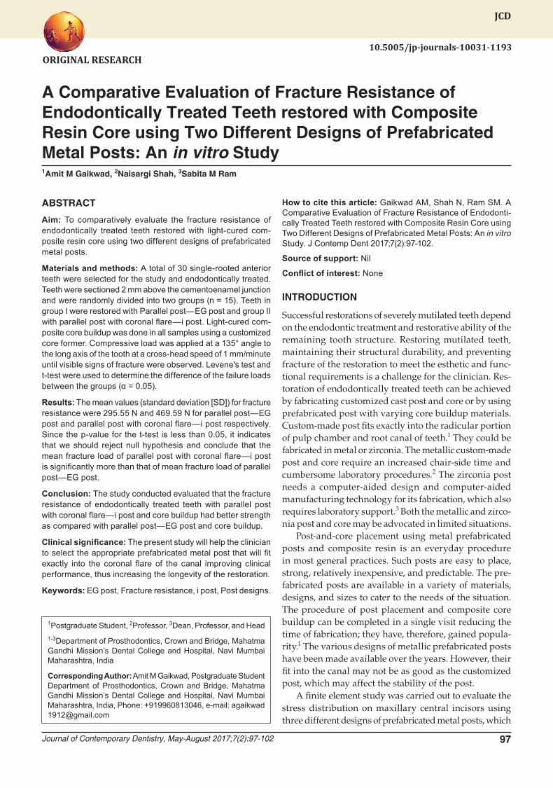
A total of 30 freshly extracted single-rooted anterior human teeth of similar dimension and root form were selected taking into consideration all inclusion and exclu-sion criteria (Fig. 1). All the selected teeth were kept in 0.2% thymol for 7 days for disinfection.5 Selected teeth
were scaled to remove calculus and hard debris with ultrasonic scaler. All the teeth were endodontically treated. The access of canal was sealed with noneugenol temporary restorative material. Radiovisiography (RVG) was taken for verification of proper obturation. All the teeth were stored in normal saline until the post and core were initiated. All the 30 samples were sectioned horizontally 2 mm coronal to cementoenamel junction with diamond disk to achieve desired ferrule effect (Figs 2 and 3).6 Post space preparations of all 30 teeth were done for receiving the post (Fig. 4). The post space was prepared according to the size of the post to be selected.
Peso reamer no. 1 and 2 were used for post space preparation. The post space preparations were carried out to include coronal two-thirds of root length maintaining 3 to 4 mm of apical seal after the post space prepara-tion. All 30 endodontically treated teeth were randomly divided into two groups after the post space preparation in groups I and II—15 teeth in each group. Selection of post was based on the size of last peso reamer, which was introduced into the canal.
Fig. 1: Selected teeth samples Fig. 2: Sectioning of teeth
Fig. 3: Ferrule preparation Fig. 4: Post space preparation
A Comparative Evaluation of Fracture Resistance of Endodontically Treated Teeth
Journal of Contemporary Dentistry, May-August 2017;7(2):97-102 99
JCD
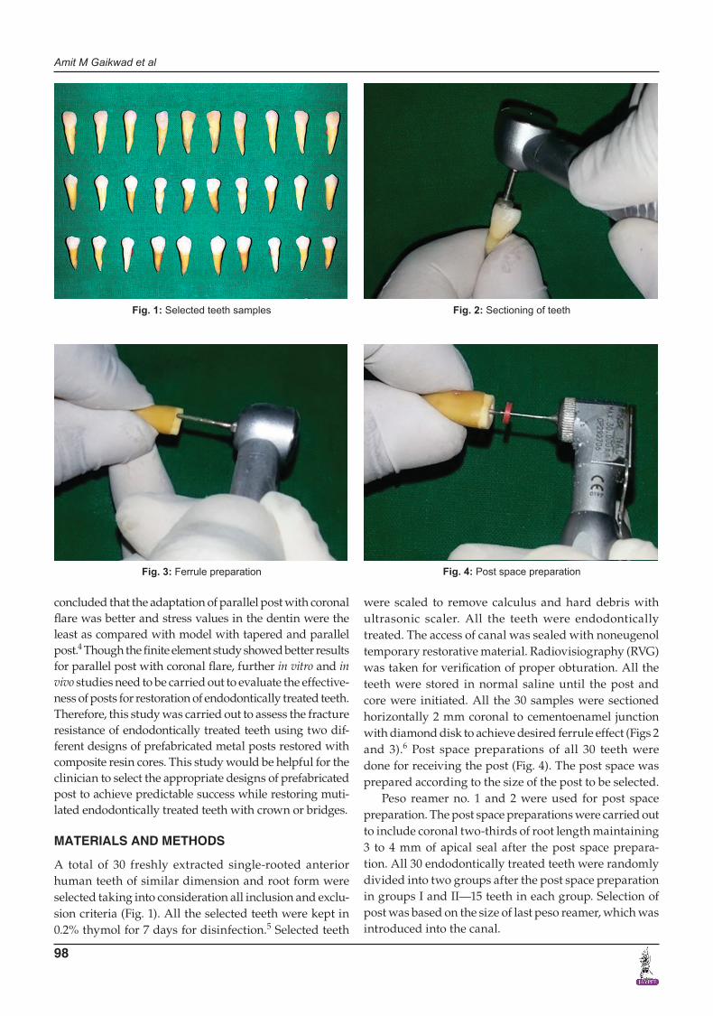
Before cementation, fit of the post was determined. Canal was irrigated with normal saline and dried with absorbent paper point. The wall of the canal was coated with a layer of cement using lentulospiral. Cementation of post was done with zinc phosphate cement for both the groups (Fig. 5). An extracted human tooth of similar dimension was selected and mounted on acrylic resin block. The tooth was prepared with diamond point to simulate desired composite core. The palatal surface was made concave in the area of lingual fossa above the cingulum to prevent the slippage of load applicator tip.7 Vacuum form sheet of 2 mm thickness was selected and core former was made using the Easy Vac vacuum form machine. Excess of material was trimmed with burlew disk and final finishing of core former was done with alpine stone. The core former was used to form the core buildup of all 30 teeth and core buildup was done with light-cured composite resin.
For the mounting of teeth on acrylic resin block, petro-leum jelly was applied on the custom-made aluminum
metal blocks so that resin block could be easily separated from aluminum metal block. Acrylic resin was mixed with desired powder liquid ratio and mix was placed in the aluminum metal block. Tooth was placed at the center of block such that the long axis of tooth was perpendicular to the floor. A surveyor was used to make sure for correct positioning of tooth. Acrylic resin was allowed to set. After final setting of acrylic resin, acrylic blocks along with tooth were removed from aluminum block and were ready for load application (Figs 6 and 7).
RESULTS
The study was carried out to assess the fracture resistance of endodontically treated teeth restored with composite resin core using two different designs of prefabricated metal posts. The 30 samples were loaded on the universal testing machine at an angle of 130° to the long axis of the tooth.8 Load was applied until there was an audible or visible sign of fracture (Fig. 8).
Fig. 5: Cementation of post Fig. 6: Mounted sample for testing group I—EG post
Fig. 7: Mounted sample for testing group II—i post Fig. 8: Application of load on universal testing machine for fracture resistance
Amit M Gaikwad et al
100
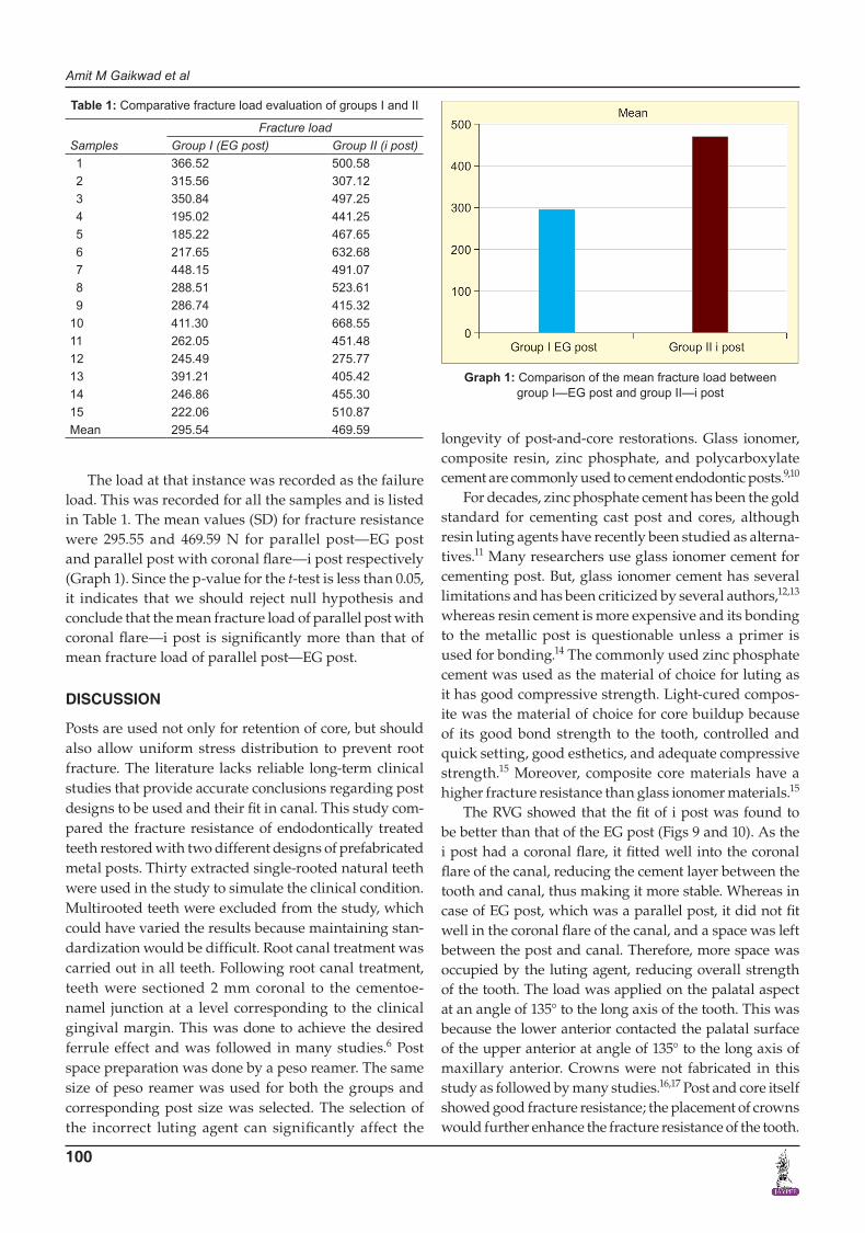
The load at that instance was recorded as the failure load. This was recorded for all the samples and is listed in Table 1. The mean values (SD) for fracture resistance were 295.55 and 469.59 N for parallel post—EG post and parallel post with coronal flare—i post respectively (Graph 1). Since the p-value for the t-test is less than 0.05, it indicates that we should reject null hypothesis and conclude that the mean fracture load of parallel post with coronal flare—i post is significantly more than that of mean fracture load of parallel post—EG post.
DISCUSSION
Posts are used not only for retention of core, but should also allow uniform stress distribution to prevent root fracture. The literature lacks reliable long-term clinical studies that provide accurate conclusions regarding post designs to be used and their fit in canal. This study com-pared the fracture resistance of endodontically treated teeth restored with two different designs of prefabricated metal posts. Thirty extracted single-rooted natural teeth were used in the study to simulate the clinical condition. Multirooted teeth were excluded from the study, which could have varied the results because maintaining stan-dardization would be difficult. Root canal treatment was carried out in all teeth. Following root canal treatment, teeth were sectioned 2 mm coronal to the cementoe-namel junction at a level corresponding to the clinical gingival margin. This was done to achieve the desired ferrule effect and was followed in many studies.6 Post space preparation was done by a peso reamer. The same size of peso reamer was used for both the groups and corresponding post size was selected. The selection of the incorrect luting agent can significantly affect the
longevity of post-and-core restorations. Glass ionomer, composite resin, zinc phosphate, and polycarboxylate cement are commonly used to cement endodontic posts.9,10
For decades, zinc phosphate cement has been the gold standard for cementing cast post and cores, although resin luting agents have recently been studied as alterna-tives.11 Many researchers use glass ionomer cement for cementing post. But, glass ionomer cement has several limitations and has been criticized by several authors,12,13 whereas resin cement is more expensive and its bonding to the metallic post is questionable unless a primer is used for bonding.14 The commonly used zinc phosphate cement was used as the material of choice for luting as it has good compressive strength. Light-cured compos-ite was the material of choice for core buildup because of its good bond strength to the tooth, controlled and quick setting, good esthetics, and adequate compressive strength.15 Moreover, composite core materials have a higher fracture resistance than glass ionomer materials.15
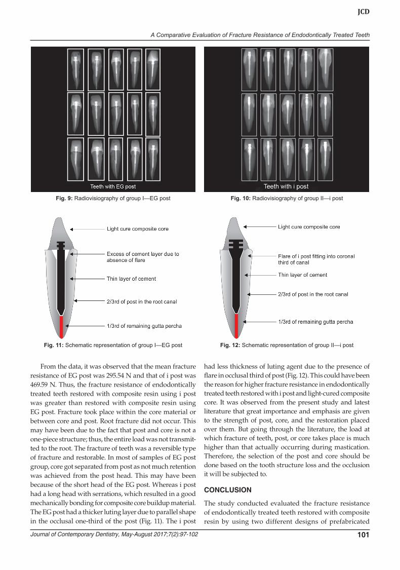
The RVG showed that the fit of i post was found to be better than that of the EG post (Figs 9 and 10). As the i post had a coronal flare, it fitted well into the coronal flare of the canal, reducing the cement layer between the tooth and canal, thus making it more stable. Whereas in case of EG post, which was a parallel post, it did not fit well in the coronal flare of the canal, and a space was left between the post and canal. Therefore, more space was occupied by the luting agent, reducing overall strength of the tooth. The load was applied on the palatal aspect at an angle of 135° to the long axis of the tooth. This was because the lower anterior contacted the palatal surface of the upper anterior at angle of 135° to the long axis of maxillary anterior. Crowns were not fabricated in this study as followed by many studies.16,17 Post and core itself showed good fracture resistance; the placement of crowns would further enhance the fracture resistance of the tooth.
Graph 1: Comparison of the mean fracture load between group I—EG post and group II—i post
Table 1: Comparative fracture load evaluation of groups I and II
SamplesFracture load
Group I (EG post) Group II (i post) 1 366.52 500.58 2 315.56 307.12 3 350.84 497.25 4 195.02 441.25 5 185.22 467.65 6 217.65 632.68 7 448.15 491.07 8 288.51 523.61 9 286.74 415.3210 411.30 668.5511 262.05 451.4812 245.49 275.7713 391.21 405.4214 246.86 455.3015 222.06 510.87Mean 295.54 469.59
A Comparative Evaluation of Fracture Resistance of Endodontically Treated Teeth
Journal of Contemporary Dentistry, May-August 2017;7(2):97-102 101
JCD
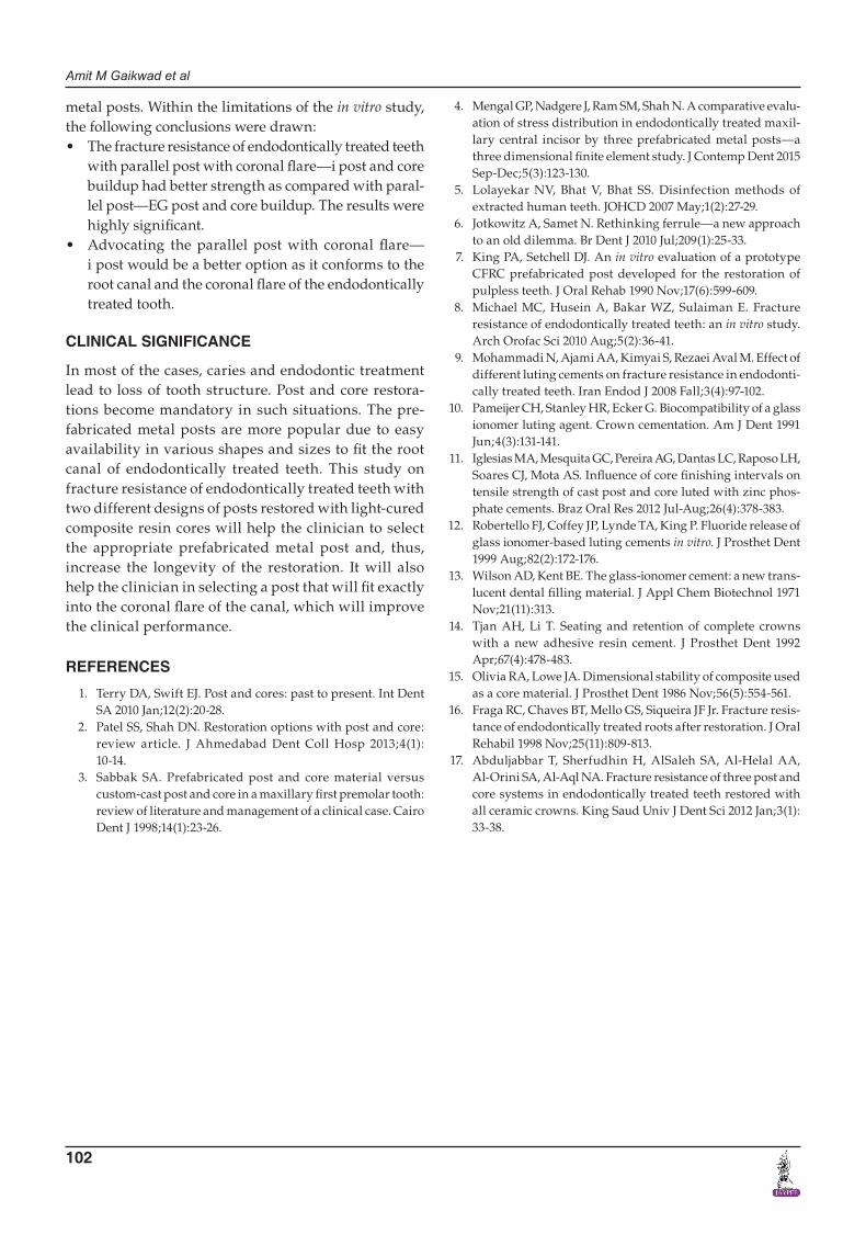
From the data, it was observed that the mean fracture resistance of EG post was 295.54 N and that of i post was 469.59 N. Thus, the fracture resistance of endodontically treated teeth restored with composite resin using i post was greater than restored with composite resin using EG post. Fracture took place within the core material or between core and post. Root fracture did not occur. This may have been due to the fact that post and core is not a one-piece structure; thus, the entire load was not transmit-ted to the root. The fracture of teeth was a reversible type of fracture and restorable. In most of samples of EG post group, core got separated from post as not much retention was achieved from the post head. This may have been because of the short head of the EG post. Whereas i post had a long head with serrations, which resulted in a good mechanically bonding for composite core buildup material. The EG post had a thicker luting layer due to parallel shape in the occlusal one-third of the post (Fig. 11). The i post
had less thickness of luting agent due to the presence of flare in occlusal third of post (Fig. 12). This could have been the reason for higher fracture resistance in endodontically treated teeth restored with i post and light-cured composite core. It was observed from the present study and latest literature that great importance and emphasis are given to the strength of post, core, and the restoration placed over them. But going through the literature, the load at which fracture of teeth, post, or core takes place is much higher than that actually occurring during mastication. Therefore, the selection of the post and core should be done based on the tooth structure loss and the occlusion it will be subjected to.
CONCLUSION
The study conducted evaluated the fracture resistance of endodontically treated teeth restored with composite resin by using two different designs of prefabricated
Fig. 9: Radiovisiography of group I—EG post Fig. 10: Radiovisiography of group II—i post
Fig. 11: Schematic representation of group I—EG post Fig. 12: Schematic representation of group II—i post
Amit M Gaikwad et al
102
metal posts. Within the limitations of the in vitro study, the following conclusions were drawn:• Thefractureresistanceofendodonticallytreatedteeth
with parallel post with coronal flare—i post and core buildup had better strength as compared with paral-lel post—EG post and core buildup. The results were highly significant.
• Advocating the parallel post with coronal flare— i post would be a better option as it conforms to the root canal and the coronal flare of the endodontically treated tooth.
CLINICAL SIGNIFICANCE
In most of the cases, caries and endodontic treatment lead to loss of tooth structure. Post and core restora-tions become mandatory in such situations. The pre-fabricated metal posts are more popular due to easy availability in various shapes and sizes to fit the root canal of endodontically treated teeth. This study on fracture resistance of endodontically treated teeth with two different designs of posts restored with light-cured composite resin cores will help the clinician to select the appropriate prefabricated metal post and, thus, increase the longevity of the restoration. It will also help the clinician in selecting a post that will fit exactly into the coronal flare of the canal, which will improve the clinical performance.
REFERENCES
1. Terry DA, Swift EJ. Post and cores: past to present. Int Dent SA 2010 Jan;12(2):20-28.
2. Patel SS, Shah DN. Restoration options with post and core: review article. J Ahmedabad Dent Coll Hosp 2013;4(1): 10-14.
3. Sabbak SA. Prefabricated post and core material versus custom-cast post and core in a maxillary first premolar tooth: review of literature and management of a clinical case. Cairo Dent J 1998;14(1):23-26.
4. Mengal GP, Nadgere J, Ram SM, Shah N. A comparative evalu-ation of stress distribution in endodontically treated maxil-lary central incisor by three prefabricated metal posts—a three dimensional finite element study. J Contemp Dent 2015 Sep-Dec;5(3):123-130.
5. Lolayekar NV, Bhat V, Bhat SS. Disinfection methods of extracted human teeth. JOHCD 2007 May;1(2):27-29.
6. Jotkowitz A, Samet N. Rethinking ferrule—a new approach to an old dilemma. Br Dent J 2010 Jul;209(1):25-33.
7. King PA, Setchell DJ. An in vitro evaluation of a prototype CFRC prefabricated post developed for the restoration of pulpless teeth. J Oral Rehab 1990 Nov;17(6):599-609.
8. Michael MC, Husein A, Bakar WZ, Sulaiman E. Fracture resistance of endodontically treated teeth: an in vitro study. Arch Orofac Sci 2010 Aug;5(2):36-41.
9. Mohammadi N, Ajami AA, Kimyai S, Rezaei Aval M. Effect of different luting cements on fracture resistance in endodonti-cally treated teeth. Iran Endod J 2008 Fall;3(4):97-102.
10. Pameijer CH, Stanley HR, Ecker G. Biocompatibility of a glass ionomer luting agent. Crown cementation. Am J Dent 1991 Jun;4(3):131-141.
11. Iglesias MA, Mesquita GC, Pereira AG, Dantas LC, Raposo LH, Soares CJ, Mota AS. Influence of core finishing intervals on tensile strength of cast post and core luted with zinc phos-phate cements. Braz Oral Res 2012 Jul-Aug;26(4):378-383.
12. Robertello FJ, Coffey JP, Lynde TA, King P. Fluoride release of glass ionomer-based luting cements in vitro. J Prosthet Dent 1999 Aug;82(2):172-176.
13. Wilson AD, Kent BE. The glass-ionomer cement: a new trans-lucent dental filling material. J Appl Chem Biotechnol 1971 Nov;21(11):313.
14. Tjan AH, Li T. Seating and retention of complete crowns with a new adhesive resin cement. J Prosthet Dent 1992 Apr;67(4):478-483.
15. Olivia RA, Lowe JA. Dimensional stability of composite used as a core material. J Prosthet Dent 1986 Nov;56(5):554-561.
16. Fraga RC, Chaves BT, Mello GS, Siqueira JF Jr. Fracture resis-tance of endodontically treated roots after restoration. J Oral Rehabil 1998 Nov;25(11):809-813.
17. Abduljabbar T, Sherfudhin H, AlSaleh SA, Al-Helal AA, Al-Orini SA, Al-Aql NA. Fracture resistance of three post and core systems in endodontically treated teeth restored with all ceramic crowns. King Saud Univ J Dent Sci 2012 Jan;3(1): 33-38.