Embed Size (px)
Citation preview

A Comparison of Standard Organ Culture
and Standard Transplant Techniques in
the Fusion of the Palatal Processes
of Rat Embryos
JOHN S. KONEGNI, D.D.S., M.S.D.
BYRON C. CHAN, D.D.S., M.S.D.
THOMAS M. MORIARTY, D.D.S., M.S.D.
SAM WEINSTEIN, D.D.S., M.S.D.
R. D. GIBSON, Ph.D.Lincoln, Nebraska
Numerous investigations have been undertaken to discover the etiology
and pathogenesis of cleft palate. Although this anomaly has been pro-
duced by various experimental methods in laboratory animals (5), mor-
phologic, histologic, and biochemical studies are needed to gain insight
into the etiological mechanisms.
In an earlier study, Moriarty, Weinstein, and Gibson (7) used organ
culture and transplantation techniques to study palatal closure and
fusion in normal embryonic rat palates. It was suggested from that in-
vestigation that such techniques could be employed to study the mor-
phologic and histologic effects of teratogenic agents on embryonic palatal
tissues.
Using modifications of the in vivo and in vitro techniques employed
in that earlier study, the current investigation was performed to
standardize the methods and provide estimates for the probability of
fusion of the palatal processes derived from rat embryos. The stand-
ardized methods and probability estimates are needed as guides and
controls for future experiments which utilize teratogenic agents.
Materials and Methods
Sprague-Dawley rats were used exclusively as experimental animals
in both the organ culture and the transplantation studies. The animals
were mated according to the 'weight drop' method described by Hamp-
son (4). An age value for the embryos was arbitrarily assigned by des-
ignating the day of mating as 'day minus one', the following day, 'day
Presented at the annual meeting of the International Somety of CraniofacialBiology, Chicago, Illinois, May 2, 1964.
Drs. Konegni and Chan are in practice in Lakewood, Colorado, and Torrance,California, respectively. Drs. Moriarity and Weinstein are 'affiliated with the Depart-ment of Orthodontics in the College of Dentistry and Dr. Gibson is affiliated withthe Department of Pharmacology, College of Pharmacy, University of Nebraska.
219

220 Konegni and others
zero', etc. All transplanted and explanted tissues were from embryos of
a chronologic age of 16 days and six hours. The embryo age was selected
on the basis of findings by Zeiler (18) which suggested that palatal fu-
sion seldom occurred prior to 16 days and eight hours in the Sprague-
Dawley rats.
The operating, transplantation, and cultivation procedures were simi-
lar to those described by Moriarty and associates (7). A few modifica-
tions were employed, however, for improvement in the overall technique.
The more significant modifications are discussed in the following para-
graphs.
a) The age of the embryos used in the initial study was 15 days plus
14 to 21 hours. In the present investigation the embryos were all 16
days plus six hours of age. This change insured a somewhat closer .
approximation of the palatal processes at the onset of the experimental
procedure. Also, the tongue was not a consideration when using the 16
day six hour embryos since the palatal processes had already assumed
a horizontal position cranial to the tongue. In the initial study the tongue
was described as being between the processes at the time of the removal of
the palatal tissues from the younger embryos and therefore removal of the
tongue was necessary prior to cultivation.
b) In some cases in the present study it was noted that the processes
were actually contacting at the time they were planted (Figure 1). To
be certain that the processes were only in contact and not fused, a fine
glass rod was gently teased between the processes during the operation.
c) In the removal of the palatal tissue in the previous study, four
excisions were employed. The head was removed first and then a second
excision was used to remove the mandible. In this investigation the head
was removed above the body of the mandible by an excision which, in
essence, combined the two cuts just mentioned. The removal of the
excess tissue cranial to the palatal section and the excess distal to the
processes were described previously as the third and fourth exeisions,
respectively, but became the second and third excisions, respectively, for
this study.
d) The most significant change in the in vivo study was from a homo-
transplant method to an isotransplant method. In theory, this change
should have minimized the rejection phenomenon that is inherent with
the homotransplant (12). The fabrication of the components of the
transparent chamber in the technique employed by Sabet, Hidvegi, and
Ray (8) was also modified by the addition of the removable transparent
window.
e) Modifications in the organ culture procedure included the use of
homologous type medium, that is, the embryonic extract and the plasma
were obtained from Sprague-Dawley rats. Also, thrombin was often
added to aid in coagulation of the medium. The moist chamber (watch,
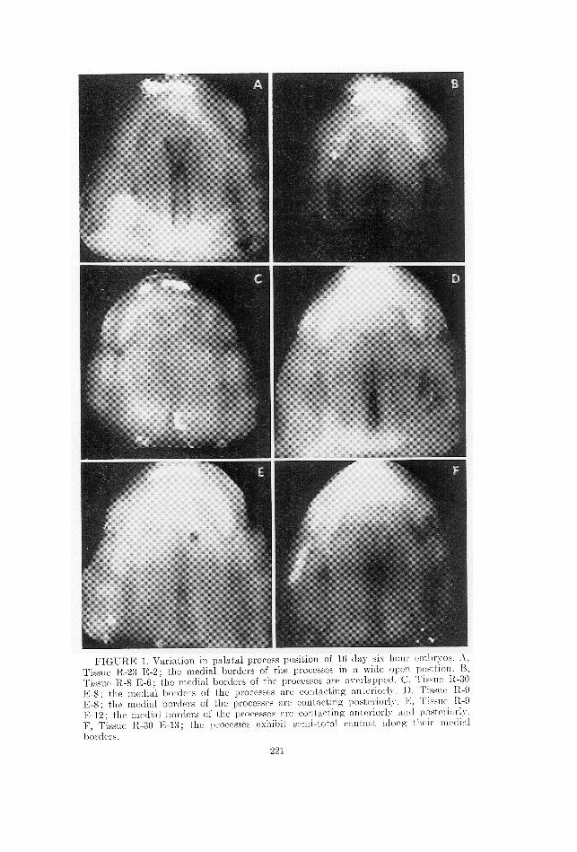
FIGURE 1. Variation in palatal process position of 16 day six hour embryos. A,
Tissue R-23 E-2; the medial borders of the processes in a wide open position. B,
Tissue R-8 E-6; the medial borders of the processes are overlapped. C, Tissue R-30
E-8; the medial borders of the processes are contacting anteriorly. 1, Tissue R-9
E-8; the medial borders of the processes are contacting posteriorly. E, Tissue R-9
E-12; the medial borders of the processes are contacting anteriorly and posteriorly.
F, Tissue R-30 E13; the processes exhibit semi-total contact along their medial
borders.

222 Konegna and others
glass) apparatus used in this experiment was similar to that described
in the earlier study.
The embryonic extract was prepared in five separate lots. Lot number
one was composed of extract from 15 day 12 hour embryos whereas
lot numbers two through five were prepared from 16 day six hour em-
bryos. (Clots in the previous work were composed of equal parts of
9/4-day-old chick embryonic extract and chicken plasma.)
The plasma was prepared from blood obtained by a cardiac puncture
on unanesthetized male rats less than one year of age.
All instruments, equipment, fluids, etc. used in these studies were steri-
lized by appropriate methods. Operating procedures were performed
under sterile conditions.
All experimental tissues were observed, sketched, and photographed
initially and approximately every 12 hours during cultivation.
At the termination of the organ culture and transplant period the
palatal tissues were prepared for histologic verification of process fusion
by using a hematoxylin-eosin stain and standard laboratory procedures.
In accordance with the techniques used in these studies, the data
obtained fitted the statistical methods which treat a binomial population.
The proportion p of successes in the population is estimated unbiasedly
by
estimated p = ¥/n
where Y is the number of success observed and n is the sample size. A
meaningful method of relating the results from these studies to ensuing
studies is by applying confidence intervals for p. Confidence intervals
were obtained from tables appropriate for binomial populations (9).
Results and Discussion
Tur TranspLANTATION ExrpeErimEnNt. A sample of 60 palatal trans-
plants was studied. ‘AThe palatal transplants were maintained on the host animals from 48
to 72 hours. Initially, each transplant was to be maintained on the hostof 72 hours. However, it was noted early in thestudy that there was littlechange in palatal development after the first 24 hours. If no change wasnoted in the tissue on two successive observations, these transplantswere terminated at 48 to 60 hours.
Investigators have cited the role of the extracellular fluids in the sur-vival of the transplants during the early stages following the transplanta-tion (Mir Y Mir, L., 6, Taylor and Lehrfeld, 10, and Conway, Stark,and Joslin, 2). In the present study the transplantation period was shortand was probably terminated before vascularization between the hostand the transplant could be established. The survival of the palataltransplants, then, was dependent upon a metabolic exchange occurringbetween the transplant and extracellular fluid rather than through a direct
ORGAN CULTURE AND TRANSPLANTS 229
vascularization. The value of the extracellular fluid resulted in at
least two noteworthy disadvantages in the transplant chamber method.
First, the amount of time necessary to prepare the host animals re-
quired that the transplantation procedure be performed in two phases.
The loss of extracellular fluid that had accumulated between the first
and second phases proved to be a disadvantage of a two phase procedure
and may have influenced the results. Secondly, the size of the trans-
plant chamber was limited in order to confine the extracellular fluuds to
the immediate vicinity of the transplant. Limiting the size of the cham-
ber resulted in the tissues impinging upon the chamber walls because of
a swelling of the tissues after grafting. How much the impingement af-
fected the growth of the tissues was not discernible.
Tur OrcaN-CuULTURE ExpErimENt. Sixty-five embryonic palatal tis-
sues were explanted for periods up to 73 hours. Sixty-one of these tissues
were included in the statistical evaluation whereas four tissues were
studied separately due to a possible antigen-antibody reaction.
The dynamic age (16 days plus six hours) of each of the cultivated
palatal tissues in this investigation eventually exceeded the relatively
more static age (15 days 12 hours and 16 days six hours) of the em-
bryonic extract present in the culture medium. Under these circumstances,
according to the 'ascending range' theory of Gaillard (3), growth (de-
velopment) of the explant should have been greatly curtailed. This did
not occur in this investigation. Perhaps the occurrence of growth (de-
velopment) could be explained as Walton (11) suggests by the presence
of the embryonic tissue in a relatively older plasma.
One batch of plasma was obtained from the blood of young male rats
which had been used earlier as host animals in the transplantation study.
Four tissues were cultivated in a medium containing this plasma. Due to
the possibility of an antigen-antibody reaction within the host animal as
a result of the original transplantation procedure these four tissues were
not evaluated statistically with the other 61 tissues. However, this ex-
periment revealed no observable evidence of a specific incompatability
and, in fact, all four tissues exhibited closure and fusion of the lateral
palatal processes.
__ Also, no difference was observed in either the growth or fusion of the
tissues cultivated in a medium to which a clot promoting substance
(thrombin) had been added.
The palatal tissues were cultured for periods ranging from 48 to 78
hours. The specific time for the removal of the explant from the medium
was empirically determined by the investigator in relation to a) the
general appearance of the tissue, i.e., distortion, b) the amount of tissue
or clot deterioration, and c) apparent fusion of the palatal processes.
Histromocy or THE TissuErs. Each of the palatal tissues
was studied and evaluated with reference to the following: a) tissue
morphology; b) cellular morphology; c) histological determination of

224 Konegm and others
fusion for the transplanted tissue only; d) the amount of vascular infil-
tration.
The general tissue morphology of the transplanted and cultured tissueswas good in all cases and the slight general distortion was consideredwithin normal limits. Disruption of the normal relationship of the nasal
cavity, nasal septum, and palatal processes, as well as a lack of develop-
ment of the nasal septum, were noted. The disruption of the relationship
of these structures was more severe in the transplant sample when com-
I
FIGURE 2. Comparative histological morphology of transplanted, explanted, andnormally developed tissue. G, Frontal plane section of a 16 day six hour tissue after50 hours in vivo. H, Frontal plane section of a 16 day six hour tissue after 48 hoursin vitro. I, Frontal plane section of a 16 day 16 hour normally developed tissue. Line's' denotes the cartilaginous nasal septum. Line 'c' denotes the nasal cavity. Line'f' denotes the midline fusion area between the palatal processes.

ORGAN CULTURE AND TRANSPLANTS 225
pared with the normal tissue and with the organ culture sample (Figure
2).
When the experimental tissues were examined histologically, they
exhibited various degrecs of histological 'depression' such as pyknosis,
shortening of the stellate processes of the mesenchymal cells, cornification
of the epithelial cells, and presence of chromatin debris in the nucleus
This 'depression' as described by Moriarty and associates (7) may have
been the result of such factors as a lack of adequate nutrition, traumas,
rejection reaction of the host animals in the transplant study, ete. The
most logical of these explanations seems to be the lack of adequate nutri-
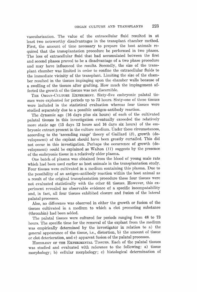
FIGURE 3. J, K, and I are photomicrographs of a frontal plane section of the
midline area of fusion of a tissue which developed fusion in vitro. The lines repre-
sent a) the mitotic figure in the united cpithelium along the midline; b) the oral
cavity; c) the fusion area of the medial borders of the palatine processes; d) the
nasal cavity; e) the mitotic figure in the oral epithelium.
226 Konegna and others
tion due to the size of the experimental tissue and the duration of theexperimental procedure.
Numerous mitotic figures were observed in the midline epithelium(between the palatal processes) of the explanted tissues. A few mitoticfigures were also noted in the oral epithelium (Figure 3). An activelyhigh rate of proliferation of epithelial cells in the midline has beendescribed by Barry (1) as a possible etiologic factor in the production ofa cleft palate. There is little doubt that continued epithelial proliferationin this area could result in the presence of epithelial pearls after fusion
has occurred.
Infiltration of red blood cells and white blood cells (primarily leuco-
cytes) from the host was evident in all the transplanted tissues.
The determination of fusion of the palatal process of the secondary
palate depended upon positive findings from the histological examination
of the experimental tissues. In the sample of 60 transplants that wasexamined, 44 exhibited fusion somewhere along the midline of the second-ary palate. Of the 61 explanted tissues, 54 were found to have fused
in the secondary palate. Seven explants did notexhibit fusion within the secondary palate but did show fusion of the
secondary palate to the primary palate. One explant was destroyed during
histologic preparation and could not be examined.
As in the earlier study (7), the degrees of fusion ranged from early
fusion by epithelial elements (double cell strands of epithelial elements
remaining in the line of fusion) to loss of all epithelial elements and
complete mesenchymal penetration.
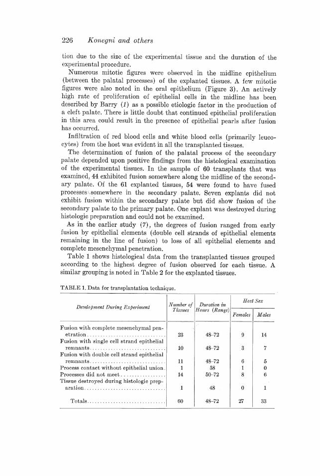
Table 1 shows histological data from the transplanted tissues grouped
according to the highest degree of fusion observed for each tissue. A
similar grouping is noted in Table 2 for the explanted tissues.
TABLE 1. Data for transplantation technique.
f Host SexNumber of Duration inTissues |Hours (Range) Development During Experiment
Females Males
Fusion with complete mesenchymal pen- .
nenee 23 48-72 9 14
Fusion with single cell strand epithelial
.... 10 48-72 3 7Fusion with double cell strand epithelial
...ll} 11 48-72 6 5
Process contact without epithelial union . 1 58 1 0Processes did not meet.. 14 50-72 8 6Tissue destroyed during histologic prep- .
ATAtIONM . ...... ...... ..... lll rre ee 1 48 _ 0 1
..a ls 60 48-72 27 33

ORGAN CULTURE AND TRANSPLANTS 227
TABLE 2. Data for organ culture technique.
X . Numb Duration iDevelopment During Experiment éfggszzsof Hog? 86727223)
Fusion with complete mesenchymal penetration ...... 35 48-73
Fusion with single cell strand epithelial remnants .... 5 48-59
Fusion with double cell strand epithelial remnants ... 14 48-73
Process contact without epithelial union . ............ 5 59-783
Processes did not meet.. ................. ...l. e. es 2 48-59
jiece, 61 48-73
StATISTICAL EvampvaATiON. The data obtained from the histological
determination of fusion of the palatal processes were evaluated statisti-
cally. An estimate for p was calculated for the transplanted tissues as
follows:
estimated p = Y¥/n = 44/66 = 0.73
A 95% two-sided confidence interval for the estimated p was determined
to be:
0.61 < p < 0.84
The estimated p for the explanted tissues was calculated as:
estimated p = Y¥/n = 54/61 = 0.89
and the one-side 95% confidence interval is:
0.80 < p < 1.0
Summary
a) A sample of 60 palatal transplants were evaluated histologically to
determine fusion of the processes of the secondary palate. On the basis of
the number of successes observed in the experimental sample, an esti-
mated p of 0.73 and a 95% confidence interval for p of 0.61 < p < 0.84
was obtained. b) The sample of 61 explanted tissues was evaluated histo-
logically for the occurrence of fusion. The probability p of fusion of the
palatal processes, using the standard organ culture technique, is 0.89
with a one sided 95% confidence interval of 0.80 to 1.0. c) Some histologi-
cal aspects of the tissues were described and it was noted that the trans-
planted tissues exhibited greater disruption of the various tissue struc-
tures when compared with the corresponding explanted tissues and with
normally developed tissue. d) There were no apparent effects on the
growth and/or development of embryonic rat palates cultivated in vitro
for periods of time up to 73 hours in a medium: (1) in which the em-
bryonic extract constituent was of a relatively younger age than that of
the explant and/or (2) in which the plasma constituent has been derived

228 Konegni and others
from an isografted animal and/or (38) to which the clot-promoting sub-
stance, thrombin, has been added. e) Active cell proliferation was observed
in the epithelial remnants of the midline fusion area. This phenomena may
explain the presence of epithelial pearls after complete mesenchymal pene-
tration has occurred.
Sam Weinstein, D.D.8.
Department of Orthodontics
College of Dentistry
University of Nebraska
Inncoln, Nebraska
References
1. Barry, A., Development of the branchial region of human embryos with specialreference to the fate of epithelial. In S8. Pruzansky (Ed.), p. 46-62, CongemialAnomalies of the Face and Associated Structures. Springfield: Charles C Thomas,1961.
2. Conway, H., Starx, R. B., and JostIn, D., Observation on the development ofcirculation in autografts. Plastic reconstr. Surg., 8, 312-319, 1951.
3. P. J., Developmental changes in the composition of the body fluids inrelation to growth and differentiation of tissue cultures. Protoplasma, 28, 145-174,1935. '
4. Hampson, F. F., A morphological study of palates from rat embryos subjected toa transitory maternal pteroyglutamic acid deficiency. Unpublished M.S. thesis,Univ. of Nebraska, 1962.
5. KautEr H., and WARKANY, J., Experimental production of congenital malforma-tion in mammals by metabolic procedure. Physiol. Review, 39, 69-115, 1959.
6. Mir Y Mir, L., Biology of the skin graft. Plastic reconstr. Surg., 8, 378-389, 1951.7. Mortarty, T. M., WreinstEIn, S., and GisBson, R. D., The development in vitro
and in vivo of fusion of the palatal processes of rat embryos. J. embryol. exp.Morph ., 11, 605-619, 1963.
8. Samet, T. Y., E. B., and Ray, R. D., A chamber for in vivo observationsof living organs. Plastic reconstr. Surg., 27, 105-108, 1961.
9. Tables of the Cumulative Binomial Probability Distribution. Cambridge: HarvardUniv. Press, 1955.
10. Taymor, A. C., and KEmrrELD, J. W., Determination of survival time of skinhomografts in the art by observation of vascular changes in the graft. Plasticreconstr. Surg., 12, 423-481, 1953.
11. Warron, A. J., On the variation in the growth of mammalian tissue in vitroaccording to the age of the animal. Proc. Roy. Soc. Lond. Bull., 88, 476-482, 1914.
12. WooprUrFF, M. F. A., The Transplantation of Tissues and Organs. p. 59-64, Spring-field: Charles C Thomas, 1960.
13. K. B., A study of the morphology and the time of closure of the palatein the albino rat. Arch. oral Biol., 9, 545-554, 1964.