Embed Size (px)
Citation preview

A COMPARISON OF THE MAXIMUM DEVIATION MEASURED IN
INTERMITTENT EXOTROPIA USING VARIOUS CLINICAL CONDITIONS
by
Kailee P. Algee
Submitted in partial fulfilment of the requirements
for the degree of Master of Science
at
Dalhousie University
Halifax, Nova Scotia
March 2017
© Copyright by Kailee P. Algee, 2017
ii
TABLE OF CONTENTS
List of Tables v
List of Figures vi
Abstract vii
List of Abbreviations Used viii
Acknowledgments ix
Chapter 1 Introduction 1
1.1 Background 1
1.1.1 Etiology 2
1.1.2 Classification 3
1.1.3 Symptomatology 5
1.1.4 Clinical Assessment 6
1.2 Presentation Of The Problem 9
1.2.1 Purpose Of The Study 9
1.3 Research Questions 9
1.4 Hypothesis 10
Chapter 2 Literature Review 11
2.1 Introduction 11
2.2 Background Of The Clinical Tests (Conditions) 15
2.2.1 Prolonged Monocular Occlusion (PMO) 17
2.2.2 Plus Lenses (+3.00 D) 18
2.2.3 Far Distance Test (20m) 20
2.3 Conclusion 21
Chapter 3 Methodology 22
3.1 Preliminary Chart Review 22
3.1.1 Design 22
3.1.2 Results 23
3.1.3 Predicted Sample Size 23
3.2 Research Design 24
3.2.1 Rational For Chosen Methods 24
3.3 Study Population 24

iii
3.3.1 Inclusion Criteria 25
3.3.2 Exclusion Criteria 25
3.3.3 Sample Size 26
3.3.4 Participants 26
3.3.5 Examiners 26
3.3.6 Risk Analysis 26
3.3.7 Benefit Analysis 27
3.3.8 Ethical Considerations 27
3.3.8.1 Informed Consent And Child Assent 28
3.3.9 Funding And Reimbursement 28
3.4 Experimental Procedures 28
3.4.1 Randomization 28
3.4.2 Clinical Testing Protocol 29
3.5 Data Collection 32
3.5.1 Deviation Measurements 33
3.5.2 Quantifying 33
Chapter 4 Results 35
4.1 Subject Analysis 35
4.2 Deviation Analysis (Strabismus Measurement) 38
4.2.1 Analysis Of Strabismus Measurements By Group (RM MANOVA) 42
4.2.2 Analysis Of Strabismus Measurements (One-Way RM ANOVA) 42
4.2.2.1 Near Measurements 42
4.2.2.2 Distance Measurements 44
4.2.2.3 Summary of Deviation Analysis 47
4.2.3 Analysis Of Sub-Group (Reliability) 49
Chapter 5 Discussion 52
5.1 Summary Of Results 54
5.2 Fixation Distance And Associated Measurement Conditions 54
5.2.1 Near Conditions 56
5.2.1.1 Prolonged Monocular Occlusion (PMO) (0.33m) 57
5.2.1.2 Plus Lenses (+3.00D) 58
5.2.2 Distance Conditions 60
5.2.2.1 PMO (6m) 61

iv
5.2.2.2 Far-Distance Test (20m) 61
5.3 Practical Implications For Orthoptics 64
5.4 Potential Limitations And Future Directions 67
Chapter 6 Conclusion 71
References 72
Appendix A – Information And Consent Form 77
Appendix B – Child Assent 84
Appendix C – Group 1 Data Collection Sheet 86
Appendix D – Group 2 Data Collection Sheet 88
Appendix E – Mahoney/Holmes IXT Control Scale 90
Appendix F – Participants Exodeviations; Including Those In Subgroup 91

v
LIST OF TABLES
Table 3.1 All strabismus measurements to be collected 31
Table 3.2 Participant groups and order of measurements 32
Table 4.1 Descriptive statistics by group and whole sample (means) 37
Table 4.2 Summary of mean strabismus measurements at each fixation 39
distance with each measurement condition.
Table 4.3 All near measurements and the comparison combinations 42
Table 4.4 Pair wise comparisons for the means of all near measurements, 44
showing the mean differences
Table 4.5 All distance measurements and the comparison combinations 45
Table 4.6 Pair wise comparisons for the means of all distance 47
measurements, showing the mean differences
Table 4.7 Summarizes the mean of the strabismus measurements obtained 50
from the sub-group; a comparison of the means of the original
(initial) strabismic measurements and the repeated measurements
Table 4.8 Summarizes the reliability observing Cronbach’s Alpha, for 51
internal consistency, obtained from the sub-group. Below
are the correlations between the original (initial) and the
repeated strabismic measurements.

vi
LIST OF FIGURES
Figure 4.1 Demonstrates all near measurements using each condition 40
by individual participant response
Figure 4.2 Demonstrates all distance measurements using each 41
condition by individual participant response
Figure 4.3 Displays the mean differences from the initial near (0.33m) 48
and distance (6m) measurements with each condition
performed. Near (blue) and distance (red) fixations are separate
analyses, displayed on a single figure to demonstrate the
difference in the magnitude of change of the deviations at
near and distance fixations. The p values for the initial near
measurements and each condition are displayed below the
condition labels; p values between conditions are displayed
above columns.

vii
ABSTRACT
In the Intermittent Exotropia (IXT) population determining the largest exodeviation for
surgical planning has been suggested for desired surgical outcomes (Kushner, 1998; Kim
& Hwang, 2005). In this study the exodeviation of 24 IXT participants were measured at
near and distance fixation, and additionally using +3.00D lenses, an increased fixation
distance (20m), and after prolonged monocular occlusion (PMO), to elicit the largest
exodeviation. The results of this study indicate that all near conditions increase the
exodeviation. Larger deviations were observed with +3.00D lenses and +3.00D after
PMO. There was no statistically significant difference between those two conditions. At
distance, PMO did not produce a statistically significant increase, but 20m and 20m after
PMO did. There was no statistically significant difference between the 20m conditions.
This research indicates that the +3.00D lens measurement and the 20m measurement are
the most clinically efficient measurements for the maximum deviation in IXT patients.
viii
LIST OF ABBREVIATIONS USED
AC/A Ratio of accommodative convergence to accommodation
ANOVA Analysis of variance
APCT Alternate prism cover test
BOFA Base out fusional amplitudes
BSV Binocular single vision
BVA Binocular visual acuity
ETDRS Early treatment of diabetic retinopathy study
D Diopter
DVA Distance visual acuity
IXT Intermittent exotropia
m Meters
M Mean
MD Mean difference
MANOVA Multivariate analysis of variance
NVA Near visual acuity
logMAR Logarithm of minimum angle of resolution
PAT Prism adaptation test
rPAT Rapid prism adaptation test
pd Prism diopter
PMO Prolonged monocular occlusion
RM Repeated measures
SD Standard deviation
TPF Tenacious proximal fusion
VA Visual acuity
X Exophoria
XT Exotropia

ix
ACKNOWLEDGEMENTS
First and foremost I would like to thank my supervisors, Leah Walsh and Erik Hahn for
your support and guidance throughout the entirety of this project, and for the confidence
you have both instilled in me. I am forever thankful to you both for your dedication and
the time you devoted to me throughout each phase of this research. Your expertise and
passion in our field provided such a solid resource for me. I am so grateful to have you as
educators and mentors. Leah, thank you for always pushing me to strive for more as a
student and in my career. I am especially thankful for your ability to always “talk me off
the ledge” and give me the reassurance I needed when I’ve doubted myself. Erik, thank
you for your endless encouragement, and reminding me to always ‘exhibit your
excellence’. Also, for your patience when dealing with “Kaleah”, especially after a long
day and too much coffee, but I think we can all agree it was all worth the “GOLD”!
Next, I would like to thank my supervisory committee members, Dr. Robert LaRoche and
Steve Van Iderstine. I felt confident in my work as you provided a solid support system
for me, and I am thankful for your knowledge and input. I would also like to
acknowledge my external examiner Leah Wood. Thank you, for your time, input and
willingness to be involved.
Thank you to Darren Oystreck, the CVS program, and the rest of the IWK Eye Care
Team for all of your time and dedication towards my education, research and career as an
orthoptist.
I would like to thank my family and friends for all their support. Mom, Dad and my
sisters, your unwavering support and encouragement throughout my entire life, including
this, got me to where I am today. Mom, you always know when to pick up the pieces,
even when I have said nothing, and you always know how to put them back together.
Libby and Martha, for the endless reassurance and support, for always believing in me,
and of course making sure I ‘get it done’. To the ‘collective brain’, thank you for the
constant banter and discussion, and keeping me laughing even on the stressful days.
Finally, Colin, I am so grateful for your ongoing support, no matter where we were,
together or apart. Through the ups and downs, you always managed to be my rock, and
find a way to put a smile on face. My sweet daughter, Dayva, for being the best little girl
and keeping Mommy company while she was busy writing, and always lighting up our
day.

1
CHAPTER 1 INTRODUCTION
Exodeviation refers to the divergent misalignment of the eyes. In western
populations exodeviations are less common than esodeviations and have been described
to affect about 1% of children under the age of 11 years (Govindan, Mohney, Diehl &
Burke, 2005). Exodeviations can be classified based on control of the deviation. An
exophoria (X) is a latent deviation, controlled by fusion; exotropia is a manifest
deviation, whereas intermittent exotropia (IXT) is intermittently controlled. Although the
clinical management of IXT has been discussed extensively in the literature, the timing of
intervention, either surgical or non-surgical, is often dependant on the control of the
exodeviation and/or the patient’s symptoms. This chapter will discuss the etiology,
classification, symptomatology, and clinical assessment of IXT.
1.1 Background
Intermittent Exotropia (IXT) is defined as an outward deviation of an eye that is
intermittently controlled by fusional mechanisms. IXT is the most common type of
childhood onset exodeviation (Mohney & Huffaker, 2003). While little population-based
data exists, one study shows that IXT comprises slightly more than 50% of the
exodeviations in children younger than 19 years of age (Govindan, Mohney, Diehl &
Burke, 2005); and Wright (2003), stated that IXT represented approximately 90% of all
exodeviations.
Generally it has been thought that IXT progresses from an exophoria to IXT and
eventually into a manifest deviation, but this is still debated (von Noorden & Campos,
2002; Jampolsky, 1954). The current literature suggests that some IXT patients remain

2
stable with no deterioration of control over time, and though few, others have been
reported to improve (Romanchuk, Dotchin & Zurevinsky, 2006). During the period of
controlled ocular alignment, binocular single vision is achieved. There has been
suggestion that IXT is actually a large phoria that is controlled by fusional convergence,
some of the time, before spontaneously breaking into a manifest deviation, with or
without dissociative influences (Wright, 2003). The period of manifest exotropia
generally occurs at distance fixation and during periods of fatigue or inattention
(Romanchuck, 2011). When the deviation is manifest, the majority of patients
demonstrate suppression and are often asymptomatic (Wright, 2003).
1.1.1 Etiology
The etiology of intermittent exodeviations remains obscure; various theories
including mechanical, anatomical, and/or innervational imbalances, have been postulated
(Wright, 2003). In 1897, Duane proposed that exodeviations are caused by an
innervational imbalance that upsets the reciprocal relationship between active
convergence and divergence mechanisms (Duane, 1897). Bielschowsky challenged
Duane’s claim that the majority of exodeviations are a results of hyperactive tonic
divergence. He stated that Duane’s theory failed to take into account the anatomical and
mechanical factors that result in an abnormal position of rest associated with
exodeviations (as cited in von Noorden & Campos, 2002). Alternatively, Worth (1929)
stated that defective fusion faculty is responsible for ocular misalignment. The inability
to maintain adequate fusion results in a state of unstable equilibrium that will manifest as
either an inward or outward deviation. Other theories include a possible role of a high
accommodative convergence to accommodation (AC/A) ratio in the etiology of IXT

3
(Cooper & Medow, 1993). Kushner (1988) investigated the link between AC/A ratio and
IXT, reporting that approximately 60 percent of true divergence excess patients as having
a high AC/A ratio. Knapp (1953) and Jampolsky (1954) proposed that patients with IXT
have developed bilateral, bitemporal hemiretinal suppression mechanism, which permits
the eyes to deviate outward. Uncorrected refractive errors have also been suggestive as
playing a role in the development of exodeviations. Patients with uncorrected myopia
require less than the normal amount of accommodation effort at near vision, which in
turn, results in decreased accommodative convergence. This lack of convergence
stimulation could cause the development of an exodeviation (Donders, 1899). Patients
with high amounts of uncorrected hyperopia can similarly make little accommodative
effort, as a clear retinal image is not obtainable. This lack of accommodation and
subsequently accommodative convergence can cause an outward deviation (von Noorden
& Campos, 2002). The unequal clarity of retinal images, secondary to uncorrected
anisometropia, has also been suggested to play a role in the development of an
exodeviation. This retinal image inequality poses as a barrier to fusion, which can result
in ocular misalignment (Jampolsky, Flom, Weymouth & Moster, 1955).
Despite the lack of consensus on the etiology of IXT, current literature coincides
with Burian’s theory supporting a multifactorial etiology: a combination of mechanical
(anatomical) and innervation factors (von Noorden & Campos, 2002).
1.1.2 Classification
Duane initially classified Exodeviations in 1896 based on the near/distance
measurement disparity (Duane, 1897). Duane’s classification is based on the assumption
that divergence is an active process rather than relaxation of convergence with a return of

4
the eyes to parallelism or a divergent position by mechanical or elastic forces. Duane
initially proposed three classifications for exodeviations that include, basic type
(exodeviation at near and distance fixation is within 15 prism diopters (pd)), convergence
insufficiency type (near deviation is 15pd or more than the distance deviation) and
divergence excess type (distance deviation is larger than the near deviation by 15pd or
more). Divergence excess type was further subdivided into simulated and true
divergence excess type. In simulated divergence excess type the deviation is greater at
distance than near however, following monocular occlusion, the near deviation increases
to becomes similar (within a basic angle) to the distance deviation. In true divergence
excess type, the distance deviation remains larger than near, despite monocular occlusion
(Duane, 1897).
Burian proposed that simulated divergence excess could be distinguished in one
of two ways, those who’s near measurements increased using +3.00D lenses at near, and
those who’s measurements were of a basic type range after a period of monocular
occlusion (Santiago, Ing, Kushner & Rosebaum, 1999). In a study by Kushner, he pointed
out that Burian also changed the near/distance disparity in the classification from 15pd to
10pd (Kushner, 1988). While Burian described that there can be differences in the size of
the deviation at near versus distance, Kushner expanded on the mechanism behind the
near/distance disparities; adding the fusional mechanism he coined as tenacious proximal
fusion (TPF). His classification elaborated on the types of near/distance disparities by the
mechanism affecting them with consideration to the accommodative component.
5
1.1.3 Symptomatology
The management of IXT patients relies heavily on whether they are symptomatic.
Visually immature patients typically remain asymptomatic because of cortical
suppression adaptations. Symptomatic IXT patients are usually older children and adults
with asthenopic complaints (headaches, blurred vision and/or diplopia) (Santiago et al.,
1999). Clinicians closely observe the patients for deterioration of their fusional status,
presence of a fixation preference, or the detection of amblyopia, to determine a need for
intervention (Santiago et al., 1999). Symptoms amongst patients with IXT are variable
and often inconsistent with the presence or degree of the symptoms reported (Kushner,
2008). Illness, fatigue or inattention contributes to the variability noted in control and
magnitude of the deviation in IXT (Romanchuck, 2011).
Clinicians use a combination of patient reported symptoms and clinical
observations, both subjective and objective, to determine the need for either non-surgical
or surgical intervention. Binocular blurred vision may occur if patient is utilizing their
accommodative convergence to control the deviation (Walsh, LaRoche, & Tremblay,
1999). Diplopia is typically experienced only in visually mature patients during periods
when the deviation is manifest. Monocular eye closure is a frequently reported finding in
patients with IXT. This phenomenon has been noted to occur in bright illumination or
with fatigue. There is some debate about the relationship of monocular eye closure as a
response to the dissociative nature of bright light or the presence of photosensitivity.
Previous literature describes that monocular eye closure can be seen in all ages, with or
without suppression, and pre and post surgical correction (Kushner, 2008). Monocular
eye closure as a response to the dissociative nature of bright light was thought to be used

6
as a tactic to alleviate binocular diplopia (Wang & Chryssanthou, 1988). It has been
speculated that bright light dazzles the retina and creates a dissociation with consequent
diplopia (Campos & Cipolli, 1992; Wang & Chryssanthou, 1988). Another study found
there to be an association with photalgia, light induced pain of the eyes, resulting in
monocular eye closure in the setting of bright light, without diplopia. Monocular eye
closure in IXT patents’ results in the relief of photalgia by decreasing the summation of
illuminance experienced under binocular conditions (Witschafter & Bourassa, 1966;
Wiggins & von Noorden, 1990). Monocular eye closure has also been documented in
non-strabismic patients (Wiggins & von Noorden, 1990). The etiology of monocular eye
closure as a response to IXT remains obscure in patients who persist with monocular eye
closure post-operatively despite a good surgical result (Santiago et al., 1999).
1.1.4 Clinical Assessment
There are numerous factors that can effect the control of a patient with IXT, in
both the home and the clinical setting such as; fatigue, illness, attention, and
accommodative status (Romanchuck, 2011). Patients with IXT utilize various types of
convergence mechanisms to control the deviation (Wright, 2003). There are five types of
convergence described throughout the literature. These include: fusional,
accommodative, tonic, voluntary, and proximal convergence (Wright, 2003). Fusional
convergence is a binocular state of convergence when there is a blending of the two
images seen by each eye, and can be suspended by occluding one eye. Accommodative
convergence is a physiologic response that occurs with changes in the crystalline lens
thickness when attempting to view an object clearly at near (Wright, 2003). The amount
of convergence in relation to the amount of accommodation exerted is known as the

7
Accommodative Convergence/Accommodation (AC/A) ratio, and can be suspended by
adding plus lenses. Tonic convergence is a form of convergence believed to be a
proprioceptive response that persists even after brief monocular occlusion and the eyes
continue to converge. It is not until a period of prolonged monocular occlusion that this
convergence is suspended (Wright, 2003). Voluntary convergence occurs on demand
when a person chooses to turn both eyes inwards. Proximal convergence is simply the
need to converge the eyes to view an object because of its perceived location at a near
fixation distance and the eyes must converge; it is an awareness of the nearness of the
object of regard, and frequently can be suspended by having the patient fixate on a far
distance target (Wright, 2003).
Patients with IXT often elicit variability in control of the deviation thus most
clinicians utilize both objective as well as subjective assessment tools to gain a
comprehensive evaluation of individual control. These clinical tests are useful for
monitoring deterioration of control over time and can be suggestive of the need for an
intervention (Rosenbaum & Stathacopoulos, 1992). Clinicians can objectively assess
office-based control by observing the fixation, re-fixation, and recovery (from manifest
state to a controlled state) of the patients with the cover/uncover test. Home-based control
describes the patient’s fusional state, relying on parental report, of which the reliability
has been questioned (Mohney & Holmes, 2006). Thus office-based control scales were
established in an effort to standardize these objective observations. Recent literature has
identified a way to quantify control on an ordinal scale. These scales are not universally
used but do offer a clinician another objective means to monitor control (Mohney &
Holmes, 2006). Other objective tests include, convergence amplitudes, near point of

8
convergence (NPC), and stereoacuity testing at both near and distance. Reduction in the
level of stereoacuity at distance had been postulated to be indicative of deterioration of
control (Stathacopoulos et al., 1993; Walsh et al., 2000). Another study suggested that
there is a correlation with reduced binocular visual acuity (BVA) and decreased distance
stereoacuity, suggesting that BVA can be used to monitor deterioration of control of IXT
(Walsh et al., 2000).
Other clinical tests commonly used in the evaluation of IXT include, measuring
the deviation with additional plus lenses (+3.00D) at 0.33m, increasing the fixation
distance to greater than 6 meters, and performing a prolonged monocular occlusion test
(PMO). Measuring a patient’s deviation with additional plus lenses at near fixation,
relaxes accommodative convergence, in theory, eliminating any masked additional
deviation at near being controlled by an accommodative mechanism (Wright, 2003).
Increasing the fixation distance to greater than six meters, in theory, uncovers any
additional deviation by suspending tonic convergence (Wright, 2003). Burian and
Franceschetti (1970) described that a testing distance greater than 20 feet was important
as the divergence mechanism is more effective the greater the fixation distance. Others
have described this increased distanced measurement or far distance test, to suspend
additional proximal convergence (Wright, 2003). PMO disrupts tonic, fusional
convergence, and what has been previously described by Kushner as tenacious proximal
fusion (TPF) (Kushner, 1988; Kushner & Morton, 1998; Wright, 2003).

9
1.2 Presentation Of The Problem
The specific tests (conditions) or combination of tests to obtain the largest
measurements of the exodeviation are unproven. Therefore, there is a need for evidence
as to which condition(s) is the most effective in determining the maximum deviation.
1.2.1 Purpose Of The Study
The aim of this research was to determine if the deviation measurements obtained
by Alternate Prism Cover Test (APCT), using three specific clinical testing conditions
elicit clinically significant differences in the size of deviation amongst IXT subjects, and
if there is any statistical significance between these conditions and measurements they
elicit. These conditions included: the use of additional plus lenses at 0.33m (+3.00D
lenses), a fixation distance beyond the standard 6 meter distance fixation (20m), and a
period of prolonged monocular occlusion (PMO; 45 minutes). The results were analysed
to determine if these methods yield the largest deviation, of clinical significance, and
detect any statistically significant difference between the conditions. To our knowledge,
there has not been any investigation performed to determine if any significant differences
exist between the use of +3.00D lenses, an increased distance test and a prolonged
occlusion test.
1.3 Research Questions
1. Are there clinically significant differences between the deviations measured at
the standard distances of 0.33m (near) and 6m (distance) and the deviation assessed
by the following methods:
1.1 At near fixation (0.33m):

10
a) with additional+3.00D lenses
b) after a period of PMO
c) after a period of PMO with additional +3.00D lenses
1.2 At distance fixation (6m):
a) with increased fixation distance (20m)
b) after a period of PMO
c) after a period of PMO with increased fixation distance (20m)
2. Are there statistically significant differences between deviations elicited by
these specific testing conditions?
1.4 Hypothesis
I hypothesize that there is no significant difference in the size of the deviation at
near and at distance fixation using prolonged monocular occlusion versus using plus
lenses at near, and a distance fixation greater than 6m (20m).

11
CHAPTER 2 LITERATURE REVIEW
2.1 Introduction
Intermittent exotropia is the most common type of exodeviation and comprises
approximately 90% of all exodeviations (Wright, 2003). It has been proposed by many
that IXT is a progressive deviation, starting as a well-controlled phoria, progressing into
and an intermittent deviation, and eventually resulting in a manifest deviation without
intervention (Jampolsky, 1954). Von Noorden and Campos (2002) reported on 51
patients with intermittent exotropia over a 3.5-year period. They reported 9% of patients
to show no change in the deviation, 16% demonstrated improvement, and in 75% of
patients they reported one or more signs of progression. As pointed out by Romanchuk,
Dotchin and Zurevinsky (2006), this study was the only research to report progression
over time. Von Noorden and Campos (2002) emphasized that not all exodeviations are
progressive in nature and that some even remain unchanged or even improve over time.
While the natural course of intermittent exotropia remains obscure, recent literature has
been published in attempt to better define this course.
In a retrospective study by, Romanchuk, Dotchin and Zurevinsky (2006), the
authors reviewed the charts of 2664 patients with exodeviations and reported findings on
109 patients with intermittent exotropia that fit their criteria. These patients were
followed for a mean of 9 years over a 17-year time frame (from 1982 to 1998). They
reported on the change of the distance exodeviation at the initial visit versus the final, and
that when using 10pd as the criterion for change 19% decreased, 58% remained stable,
and 23% increased. The authors concluded that in the majority of the patients the size of
the deviation does not change or progress over time in the majority of patients, nor do

12
they always experience deterioration of control over time (Romanchuk, Dotchin &
Zurevinsky, 2006).
Patients that experience an increase in the size and/or control of an IXT deviation
may be candidates for strabismus surgery. Kushner (1998), stated surgical
undercorrections in the IXT population are typically more common than overcorrections.
Kushner describes that the distance angle may increase after 1-hour of monocular
occlusion or when measured to an outdoor far-distance target. He postulated that this
increase in the measured angle, using these two techniques, could be due to different
mechanisms. When the deviation increases after 1-hour of monocular occlusion he
describes the increase as ‘vergence aftereffect at distance’. The increased deviation with
an outdoor target, he suggests could be due to an ‘outdoor sensitivity’. From his previous
work, patients that had demonstrated either phenomenon preoperatively resulted in
postoperative undercorrections. Surgery in those patients was targeted for the initial 6m
measurement. In the patients that showed an ‘outdoor sensitivity’ preoperatively, 40%
were undercorrected. In those that demonstrated ‘vergence aftereffect at distance’
preoperatively, 35% were undercorrected. From this, Kushner postulated that surgery
should be performed on the largest angle measured (Kushner, 1998). From his study in
1998, he concluded that when surgery was targeted for the maximum deviation 86% had
a ‘satisfactory’ outcome 1-year postoperatively compared to 62.5% with ‘satisfactory’
outcome when surgery was based on the initial 6m measurements. Kushner concluded
that targeting the maximum deviation gave better surgical outcomes (Kushner, 1998).
Pineles, Ela-Dalma, Zvansky and Rosenbaum (2010) investigated the long-term
surgical success rate of IXT patients from their clinical population. They attempted to

13
contact all patients who underwent IXT surgery over a 28-year period (between 1970-
1998), only including those with a minimum of 10-year follow-up. Out of 197 patients
contacted, 50 returned for a follow-up sensory/motor evaluation. The authors analyzed
sensory and motor status separately to determine surgical success. They found that the
majority of patients (64%) had an ‘excellent’ motor outcome postoperatively. The
remainder of patients had either a fair (18%) or poor (18%) motor outcome. They also
reported that during their 10-year follow-up period 30 (60%) patients required at least
one reoperation. In 24 (80%) of these patients additional surgery was performed for either
residual or recurrent IXT.
Previous studies have suggested augmenting the original surgical dose in an
attempt to reduce recurrent exodeviations postoperatively (Lee, Kim & Thacker, 2007;
Arda, Atalay, & Orge, 2014; Yuksel, Spiritus, & Vandekannoitte, 1998). The majority of
these studies included basic type exodeviations. In a more recent study, Kim, Yang and
Hwang (2016) compared the surgical outcomes of patients when surgery was based on
‘original’ dosage tables versus their augmented table. They augmented surgery for an
additional 1.0-1.5mm of bilateral lateral rectus recession compared to what they refer to
as the ‘original’ methods. They found that in the group where surgery was performed on
the ‘original’ dosage table 49% had recurrence, and in their augmented group, 37% had
recurrence. Overcorrection occurred in 4% of both groups, and successful alignment was
reported in 48% of the ‘original’ group and 59% in the augmented group.
A number of factors causing these undercorrections or recurrences of the
exodeviation postoperatively have been postulated. The exact cause of these
reoccurrences and undercorrections remains unclear (Kim & Hwang, 2005). The

14
intermittency and variability of IXTs has been described by von Noorden and Campos
(2002), and could be due to a variety of reasons; whether it is the alertness of the patient,
inattention, or general fatigue (von Noorden & Campos, 2002). Pritchard (1993)
suggested that an obvious explanation for surgical undercorrections could be that the
surgeon did not operate on the full angle of deviation.
In the study by Kim and Hwang (2005) the authors investigated whether surgery
should be targeted for the largest angle or the ‘more common or stable angle’ measured.
The authors used three clinical tests to obtain the largest angle, these included: an outdoor
far distance target, after 1-hour of monocular occlusion, and with +3.00D lenses before
allowing the patient to regain binocular fusion (Kim & Hwang, 2005). The mean age of
the patients was 6.4 years (range 2.8 – 11 years). Patients with A and V patterns were not
excluded. Short-term surgical outcomes were assessed at 1-week post-operatively and
long-term outcomes were assessed between 6-9 months and a final assessment between
12-36 months. The average follow-up period was 13.8 (range 6-36 months). All patients
were treated with bilateral lateral rectus recessions by the same surgeon. They reported
that 22 out of 33 patients demonstrated an ‘excellent’ or ‘good’ result based on their
criteria. Overcorrections of no more than 9pd of esodeviation at near or distance was
reported by any patient’s final visit, and only two patients had as much as 9pd of
esodeviation (one intermittent deviation and one phoria). No patients experienced the
development of amblyopia or loss of binocularity as a consequence from an
overcorrection. They suggest that the largest angle measured can be used for surgical
planning, when using bilateral lateral rectus recessions, without much fear of persistent
overcorrection. The authors also reported that only two of their patients had their largest

15
angle measured at the outdoor far-distance target, and that 13 patients had largest
measurements to accommodative target at 1/3m and 6 meters. From this they suggest that
multiple measurements at various times might be more useful than just using the outdoor
far-distance measurement. The authors suggest that more extensive comparative studies
need to done to address which, an outdoor far-distance target, or the most commonly
measured angle (angle measured most frequently visit to visit), is best for reducing
undercorrections. However, from their work they did conclude that surgery based on the
largest angle was both safe and did not result in overcorrections (Kim & Hwang, 2005).
The rate of exodeviation recurrences also vary. One potential factor for this
variability in the recurrence rate could be a result of the range of postoperative follow-up
assessments. One study reported the postoperative alignment at 6 months and 5 years
with 27.6% and 77.9% recurrence rate, respectively (Lim, Hong, & Kim, 2011).
There is a need to determine the ideal target measurement for IXT. Thus, there is
a need to determine which clinical tests elicit the maximum deviation at near and distance
fixation, as there is currently no consensus throughout the literature.
2.2 Background Of The Clinical Tests (Conditions)
Patients with IXT utilize fusion to control their deviation to maintain binocular
single vision (BSV). This control requires constant effort on the patient’s behalf to
maintain binocular alignment. As intermittent exotropia varies between a controlled and a
manifest state there can be variability in the deviation measured (Hatt, Leske,
Liebermann, Mohney & Holmes, 2012). Clinicians have established a variety of tests in
attempt to obtain the maximum deviation in these patients. The literature heavily

16
investigates the effects of these tests for classification purposes and their roles in patient
management. Von Noorden (1969), studied intermittent exotropia, divergence excess
types, true versus simulated. He used both the occlusion test and +3.00D lenses for
classifying and planning surgery for IXT, emphasizing the importance of adequate
dissociation of the eyes. The mechanisms these tests target are well defined, but there
does not seem to be a direct comparison of the deviation measurements obtained using
these tests, nor a consensus as to which, if any, elicit the largest deviation.
In an abstract by Lin, Li and Wang (2013), the authors investigated 50
participants and compared 4 methods of measurement at distance fixation, and 3
measurements at near fixation. The full text copy of this article was only available in
Chinese; the abstract however was available in English translation. This study measured
distance deviations of IXT patients at 6m, after a 1-hour monocular occlusion, fixating
far distance outdoor target, and after a prism adaptation test (PAT). The near deviation
measurements they obtained included 0.33m, after 1-hour monocular occlusion, and after
a PAT. They found that deviations at distance, when compared to the initial 6m
measurements, had a positive rate of increased angle of deviation of 8% using 1-hour of
monocular occlusion, 16% while fixating on an outdoor far distance target, and 44% after
a PAT. At near, when compared to the initial 0.33m measurements, they found a positive
rate of increased angle of deviation of 38% after 1-hour of monocular occlusion and 66%
after a PAT. They concluded that using a 1-hour monocular occlusion test and the PAT
both elicit larger near deviations, and that the prolonged monocular occlusion and the
PAT elicit larger distance deviation, but the maximal deviations for both near and
distance were elicited with the PAT (Lin, Li & Wang, 2013). From this abstract we do

17
not know whether this was a rapid PAT or the original PAT. In this research study I did
not investigate the rapid PAT, as Kushner and Morton found that the prolonged
monocular occlusion test elicited greater near deviations when compared to the rapid
PAT (Kushner & Morton, 1998). This will be discussed further in the next section.
2.2.1 Prolonged Monocular Occlusion (PMO)
Marlow (1932) first described using a prolonged occlusion test to elicit small
heterophorias while using unilateral occlusion of the dominant eye for 1-2 weeks. From
there it has evolved to a commonly used clinical test of shorter duration in the
classification of IXTs.
As previously stated, PMO disrupts fusional and tonic convergence. Following
Marlow, Scobee originally recommended that 24-hours of occlusion was necessary
(Scobee, 1952). Later, von Noorden suggested that simply 1-hour was adequate (1969),
and Burian & Franceschetti went on to conclude that 30-45 minutes was comparable and
adequate for full dissociation (Burian & Franceschetti, 1970). A range from 30 minutes to
1-hour continues to be used at the discretion of the clinician (Wright, 2003; Gürlü &
Erda, 2008).
Prism adaptation has also been used throughout the literature to elicit a greater
deviation for the purpose of classification and distance/near disparities. Kushner and
Morton (1998), compared the results of the rapid PAT and prolonged monocular
occlusion. Their results showed that the two tests were not equivalent and were in fact
statistically significantly different, concluding that the prolonged monocular occlusion
test elicits a greater near deviation than the rapid PAT (Kushner & Morton, 1998). In the

18
same study they described the use of the monocular occlusion test for the purposes of
classification, in conjunction with plus lenses, to assess AC/A ratio. In this study we will
use +3.00D lenses in an attempt to obtain the maximum angle of deviation at near.
In the study by Kushner (1999), he suggests that prolonged occlusion followed by
a near measurement of +3.00D lenses is required for proper assessment of AC/A ratio.
While this is important information to consider, this study is looking at the effects these
tests have on the maximum deviation, not AC/A ratio classification. However, a +3.00D
lens measurement after a PMO was included as part the investigation for maximal
deviation in the current study.
2.2.2 Plus Lenses (+3.00D)
It was initially suggested by Brown (1971) that plus lenses increase the near
deviation, though he suggested that plus lenses and/or a period of monocular occlusion
could both differentiate patients with true versus simulated divergence excess type IXT
(Brown, 1971). However as stated by Kushner and Morton, it was Helveston (1974) that
acknowledged that there were two different convergence mechanisms being targeted by
using plus lenses and prolonged monocular occlusion, and recommended that the two
tests not be used interchangeably (Kushner & Morton, 1998). They suggested that plus
lenses were affecting accommodative convergence and that prolonged monocular
occlusion was affecting fusional convergence (Kushner & Morton, 1998; Helveston,
1974). As Kushner and Morton (1998) pointed out, Brown recognized that these two tests
effect two different mechanisms, but he argued that they could be used interchangeably to
diagnose simulated versus true divergence excess type IXT (Kushner & Morton, 1998).

19
Measuring a patient’s deviation with additional plus lenses relaxes
accommodative convergence, and in theory, eliminates any masked additional deviation
at near being controlled by the accommodative mechanism (Wright, 2003; Burian &
Franceschetti, 1970). Using plus lenses at near may produce an increase in the deviation
obtained at near comparable to that after PMO. To the best of my knowledge there is no
current research that uses +3.00D lenses to elicit a greater near deviation for the purposes
of obtaining the maximum deviation.
As previously mentioned, a study by Kushner and Morton (1998) investigated the
role of +3.00D lenses after a period of monocular occlusion to obtain accurate AC/A
ratios, and its use in classifying the type of deviation with consideration to the AC/A
ratio. Kushner identifies that for the purpose of diagnosis and classification, AC/A ratio
must be calculated after a post-occlusion measurement with the addition of +3.00D lenses
to avoid reporting falsely high AC/A ratios, or what he describes as pseudo high AC/A
ratio. This study acknowledges this work by Kushner and Morton, but seeks to utilize
+3.00D lenses at near to determine the maximal deviation for a surgical target.
Patients with IXT have been reported to use accommodative convergence to help
control IXT when fusional convergence is insufficient (von Noorden & Campos, 2002;
Walsh et al., 2000). It is believed that the patient may sacrifice clear vision to maintain
binocular single vision when utilizing their accommodative convergence to control the
deviation (Walsh et al, 2002). Thus, blurred vision may be the result of the increased
demand on accommodation via accommodative convergence (Walsh et al, 2002). When a
patient is asked to read an accommodative target through +3.00D lenses, which typically
result in a larger exodeviation, this may result in a manifest deviation if they do not have

20
sufficient fusional convergence amplitudes to maintain their control. For this reason we
postulate that plus lenses are a useful clinical test that may elicit the maximum deviation
at near.
2.2.3 Far Distance Test (20m)
Burian and Franceschetti (1970) described the use of an increased distance
measurement or testing beyond 20 feet. They stated the importance of a far distance
measurement, as the divergence mechanism controlling the deviation is more effective
the greater the fixation distance, thus producing a larger deviation. Others have described
this increased distanced measurement, or far distance test, as an effort to suspend
additional proximal convergence (Wright, 2003).
A clinical trial by Kushner (1998) suggests that surgical results are improved
when the greatest distance angle of deviation is established preoperatively and used for
surgical planning. He performed bilateral lateral rectus recessions on 90 subjects that fit
their criteria, out of 118 subjects. Ages ranged from 3-18 years, with an outcome follow
up at 1-year follow up post surgery. In addition to routine strabismus measurements,
Kushner added a post-occlusion (after 1-hour of monocular occlusion) measurement at
6m and an outdoor far-distance measurement (0.25 mile away (402 m)). He defined a
clinically significant change for the post-occlusion measurement or the far-distance
measurement as 3pd, as this would change his surgical dose. There was an experimental
and control group of 50 patients and 40 patients, respectively. The experimental group
had strabismus surgery for the largest angle and 86% had a satisfactory outcome
compared to 62.5% with satisfactory outcomes in the control group, for which the initial
6m measurement was the targeted surgical angle. Kushner suggested, from the results of

21
his study, to target the largest distance angle for optimal surgical outcomes. However, he
stated that the largest angle must be determined by one of two tests, either 6m post-
occlusion or an outdoor far-distance target, and they should not replace one another.
The current study used an indoor far-distance accommodative target at 20m. This
was done to maintain transferable measurements directly to clinic practice, as some
clinics do not have windows. Also, an indoor target in the same hallway permits a
controlled lighting environment for each patient.
2.3 Conclusion
The literature has described the roles of these tests and how they effect deviation
measurements by the mechanisms they target. Using the maximum angle of deviation for
planning the surgical dose to obtain optimal post-operative outcomes is suggested. There
does not appear to be a consensus for any one individual test, or combinations of tests, to
yield the maximum deviations. Thus the need for this investigation into how effective and
comparable these tests are at obtaining the maximum deviation.

22
CHAPTER 3 METHODOLOGY
3.1 Preliminary Chart Review
A preliminary chart review was performed prior to the larger current study to
determine the normal distribution of data. Ethical approval was obtained through the
IWK Health Centre Research Ethics Board for this retrospective chart review. The review
was titled, Pre-Study: A Comparison of the Maximum Deviation in People with
Intermittent Exotropia Using Three Common Clinical Techniques (File number
1015771).
The purpose of this chart review was to collect a sample of data and
measurements obtained during routine orthoptic assessments. This data allowed for an
evaluation of the variability (standard deviation) in these measurements and to test for
normality in the distribution of the data. Power and sample size calculations were also
obtained to ensure that the larger current study would be appropriately powered.
Potential participants for the chart review were identified from a list of current
patients with IXT that were followed in the Eye Clinic at the IWK Health Centre,
Halifax, Nova Scotia, Canada. All participants with IXT were 5 years of age and older.
All participants did not have any previous strabismus, refractive, or intraocular surgery.
As well, patients must not have had any mental health or neurological conditions. These
criteria were set to best represent the desired population for the current study.
3.1.1 Design
The retrospective chart review of known IXT patients was completed and data
collected by the Primary Investigator (PI). The charts from which the data was collected

23
were only of those patients with IXT, whom had previously completed a PMO test. From
these charts the data recorded included their age and strabismic measurements at 0.33m,
0.33m with +3.00D lenses, at 6m, at a fixation distance greater than 6m, and all
strabismus measurements repeated after the PMO test. Charts were reviewed via the IWK
Health Centre's electronic charting system, MediTECH. This data was recorded into a
Microsoft Excel worksheet, from which data analysis was completed to aid in the
determination of the feasibility of the current study.
3.1.2 Results
In this chart review of 212 charts of patients with an IXT, 17 patients met the
inclusion/exclusion criteria and had all the desired Alternate Prism Cover Test (APCT)
measurements recorded on previous orthoptic reports. The data set was analyzed at the
advice of a consulting scientist for interdisciplinary research, in the IWK Health Centre.
With power set at .80 and alpha set at .05, in order to detect a medium effect size, a
sample size of 20 was determined. Statistically significant mean differences were found
with this data set of 17.
3.1.3 Predicted Sample Size
This study determined a minimum sample size of 20 participants to adequately
power the proposed research project. The current study planned to obtain an additional 4
participants (at the advice of the consulting scientist). These additional participants will
be enrolled to account for any unexpected variability.

24
3.2 Research Design
The current study used a non-interventional, observational, cross-sectional, cohort
design to measure and analyze the ocular alignment of twenty-four IXT participants
under various clinical conditions.
3.2.1 Rational for Chosen Methods
A cross-sectional, observational study permits a practical method of studying the
various measurements in conditions where a prospective randomized control study is
impractical.
A subgroup was included, involving 50% of participants to be measured
additionally at both 0.33m and 6m, as well as with +3.00D and at 20m, to investigate the
test-retest reliability of the strabismus measurements obtained by the examiners, as well
as examiner reliability.
3.3 Study Population
All participants were established patients of the IWK Health Centre, Eye Care
Team. A master list of all known IXT patients was screened by the PI. As potential
participants were identified, the parents/guardians were contacted by the PI via telephone.
The PI provided a detailed explanation of the study, discussed the purpose and how the
appointment would proceed; this would be part of their regularly scheduled orthoptic
assessment should they consent to participate in this research. It was explained to the
parent/guardian during this telephone conversation that they would have a chance to
review this information again (in written form) prior to any enrollment in this study on
the day of their scheduled appointment.

25
Upon verbal consent to participate, their regularly scheduled orthoptic assessment
was booked as a potential one –time study appointment. On the day of the examination,
the PI carefully reviewed the details of this research with both the parent and the child. If
written consent/assent was obtained, the study examination protocol proceeded.
In the event that a verbally consented participant declined written consent on the
day of the exam, then the PI in routine fashion would have conducted the patient’s
regular orthoptic evaluation. No potential participants declined enrollment.
3.3.1 Inclusion Criteria
Participants were required to have an IXT at either near and/or distance fixation,
with a minimum exodeviation at distance of 10pd. Only participants of 5 years of age and
older, and had the ability to cooperate for the duration of the exam, were enrolled in the
study. All participants were required to have at least 400 seconds of arc on stereo-acuity
testing, and have had a cycloplegic refraction performed within the past 12 months.
Participants were required to have refractive correction for myopic correction >1.50D,
hyperopia >3.50D, astigmatic correction >1.50D, and anisometropia >1.50D (Donahue,
2007). Participants were required to have a minimum best-corrected visual acuity of 6/9
(0.3 logMAR) in each eye.
3.3.2 Exclusion Criteria
Excluded were those whom had any history of strabismus surgery, Botox
injections, intra-ocular surgery or refractive surgery. Participants were also excluded if
they had any other ocular or neurological disease /abnormalities. Patients were required
to understand the English language.

26
3.3.3 Sample Size
As previously discussed, the results of the preliminary study determined a
minimum sample size of 20 participant to be adequate, and it was recommenced, by the
consulting scientist of the IWK research department, that this research obtain an
additional 4 participants. These additional participants were enrolled to account for any
unexpected variability.
3.3.4 Participants
All participants had an IXT with no prior strabismus surgery. The age range of
participants was from 5-14 years old.
3.3.5 Examiners
Two experienced orthoptists (each with 20 years or more clinical practice
experience) were used to obtain the strabismus measurements. The purpose of using
experienced orthoptists was to ensure reliability and consistency of the strabismus
measurements. Using two orthoptist made obtaining the measurements more feasible,
while limiting it to two orthoptists was intended to maintain internal consistency. All
measurements per participant were obtained by the same orthoptist.
3.3.6 Risk Analysis
There were no identified potential harms associated in the participation of this
study other than the potential for a breach of confidentiality. Assigning study ID numbers
and labeling study documents with the ID number instead of unique identifiers helped

27
protect confidentiality. A master list, linking IDs to identifiers, was created and stored
separately and securely.
3.3.7 Benefit Analysis
There was no intervention prescribed during this one time appointment other than
what would be received from a standard orthoptic assessment. All orthoptic reports were
forwarded to the referring ophthalmologist as per IWK Health Centre Eye Care Team
policy and procedures.
There were no guarantees that participants would personally experience any
benefits from participating in this study. However, the knowledge gained from this study
may help decide which clinical methods are most efficient and effective in measuring
IXT deviations. This information could in turn potentially provide new knowledge for
the management of patients with IXT.
3.3.8 Ethical Considerations
Ethical approval was obtained from the IWK Health Centre Research Ethics
Board on September 11, 2014. For the current research titled, A Comparison of the
Maximum Deviation Measured in Intermittent Exotropia Using Various Clinical
Conditions, (File # 1017428). This observational study involved no medical intervention.
The examinations did not affect the standard of care given to each participant. The PI
monitored each examination and ensured protocol was followed to maintain consistency
amongst all participant examinations. There were no apparent conflicts of interest.

28
3.3.8.1 Informed Consent And Child Assent
Informed consent was obtained and signed by the participant’s parent or
legal guardian on the day of the exam (Appendix A). A child assent was given
and read with each participant to ensure they understood and could ask any
additional questions (Appendix B). Both documents were submitted and
approved through the IWK Health Centre Research Ethics Board.
3.3.9 Funding And Reimbursements
Funding was obtained through a Category A grant from the IWK Health Centre
Research Department. The funding was used for reimbursement for the cost ($13.00) of
parking for all participants. Participants that took part in the sub-study, requiring them to
stay for an additional set of measurements, were given additional monetary compensation
($10.00).
3.4 Experimental Procedures
The routine orthoptic examination for each participant was completed by the PI,
followed by the examination of ocular measurements by the examiners. All data was
recorded on the study examination sheet that corresponded to the participants group by
the PI (Appendix C and D).
3.4.1 Randomization
There were two groups (1 and 2; PMO second and PMO first respectively), and
upon enrollment participants were alternately assigned into groups 1 or 2. This allowed
29
for an unbiased placement into groups and it also maintained equal numbers of
participants in each group as data collection proceeded.
3.4.2 Clinical Testing Protocol
All patients had a cycloplegic refraction within the past 12 months to ensure they
were wearing their best correction. Lensometry was performed for all participants with
spectacles. All testing was completed with the participants prescribed correction in place.
Each participant’s near and distant control scale, visual acuity (VA), stereoacuity,
convergence amplitudes, and binocular visual acuity (BVA) were collected in the
standard fashion. Control of the exodeviation was measured at near and distance using
the office control scale (Mohney & Holmes, 2006). See details on control scale in
Appendix E. VA was obtained at distance using the Vector Vision CSV 1000, Vector
Vision, Greenville, OH, USA, and near acuity was obtained using the Sloan Letter Near
Card ® (catalog number: 72500), Good-Lite Co. Elgin IL USA. Stereoacuity was tested
with the Adult Vectorgraphic Projector Slide 9100, Stereo Optical Company Inc.,
Chicago, IL, at distance and Original Stereo Fly Stereotest® Stereotest, Stereo Optical,
Chicago, IL, USA, at near.
Convergence amplitudes were obtained while fixating the smallest visible letter at
distance and near. BVA was obtained using the letter acuity portion from the Adult
Vectorgraphic Projector Slide 9100, Stereo Optical Inc., Chicago, IL, for distance, and
the Clement Clarke Children’s Fixation Bar (Catalog number: 7004001), Haag-Streit,
Essex, UK, at near. Each patient’s near point of convergence (NPC) was measured and
recorded in centimeters (cm). Ocular motility was performed in standard fashion using a

30
scale of 0 to +/- 4. Pupils were checked for relative afferent pupillary defect with the
swinging flash light test. Following these assessments the strabismus measurements were
obtained.
Upon completion of the detailed orthoptic exam, patients in Group 1 would have a
10-minute break to regain BSV. For this group, deviation control was retested following
the break using the Mohoney/ Holmes control scale (Appendix E). Once control was re-
established, the first four-strabismic measurements were completed (0.33m, 0.33m with
+3.00, 6m, and 20m). Following the deviation measurements, the patch was placed on the
participant for 45-minutes. The patch for all patients was placed over the non-dominant
eye where applicable. However if no obvious dominancy was noted, the patch was placed
over the left eye. Hash marks were made on the patch to ensure no tampering occurred
throughout the 45-minute period. The four-strabismic measurements were repeated as
soon as the patch was removed, without permitting any binocular viewing until all
measurements were obtained (0.33m after PMO, 0.33m with +3.00D after PMO, 6m after
PMO, and 20m after PMO).
In Group 2 the patch was put on immediately following the completion of the
orthoptic examination. Forty-five minutes later, the four post-PMO measurements were
obtained, with no binocular viewing until all measurements were completed. These
participants then had 10-minutes of uninterrupted binocular viewing followed by the
control scale measurement, and the final four measurements were obtained once the PI
established that control was regained.
Participant’s included in the subgroup were given another 10-minute break of
uninterrupted binocular viewing, control scale was tested again, and the final (3rd set) of

31
four strabismus measurements were obtained once the PI established that control was
regained.
All measurements of ocular alignment were completed while looking in primary
position at both 0.33m (near) and 6m (distance). The examiners used the APCT
technique, while the participant was reading the smallest accommodative target
discernible. These procedures were implemented for all additional measurement
conditions including, the +3.00D lenses at 0.33m, at a fixation distance greater than 6m
(20m), and again after a PMO. Thus, there were three additional near measurements and
three additional distance measurements for each patient, for a total of four measurements
at near and four at distance.
Near Distance
Set A
0.33m 6m
0.33m with +3.00D 20m
Set B
0.33m after PMO 6m after PMO
0.33m with +3.00D
after PMO
20m after PMO
Table 3.1 All strabismus measurements to be collected
Each participant underwent two sets of strabismus measurements, for a total of 8
strabismus measurements, as shown above in Table 3.1. Set A included a measurement at
near fixation (0.33m), at near with +3.00D lenses, at distance (6m), and at an indoor far-
distance target (20m). Set B included all four of the previously described conditions after
a PMO. These strabismus measurements were completed in one of two orders shown in
Table 3.2. Group 1 had a 10-minute break to reestablish BSV following their initial
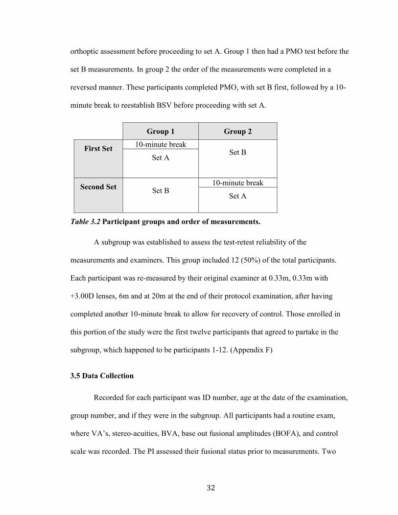
32
orthoptic assessment before proceeding to set A. Group 1 then had a PMO test before the
set B measurements. In group 2 the order of the measurements were completed in a
reversed manner. These participants completed PMO, with set B first, followed by a 10-
minute break to reestablish BSV before proceeding with set A.
Group 1 Group 2
First Set 10-minute break
Set B Set A
Second Set Set B
10-minute break
Set A
Table 3.2 Participant groups and order of measurements.
A subgroup was established to assess the test-retest reliability of the
measurements and examiners. This group included 12 (50%) of the total participants.
Each participant was re-measured by their original examiner at 0.33m, 0.33m with
+3.00D lenses, 6m and at 20m at the end of their protocol examination, after having
completed another 10-minute break to allow for recovery of control. Those enrolled in
this portion of the study were the first twelve participants that agreed to partake in the
subgroup, which happened to be participants 1-12. (Appendix F)
3.5 Data Collection
Recorded for each participant was ID number, age at the date of the examination,
group number, and if they were in the subgroup. All participants had a routine exam,
where VA’s, stereo-acuities, BVA, base out fusional amplitudes (BOFA), and control
scale was recorded. The PI assessed their fusional status prior to measurements. Two

33
experienced orthoptists (each with 20 years or more clinical practice experience) were
used to obtain the strabismus measurements. The purpose of using experienced
orthoptists was to ensure reliability and consistency of the strabismus measurements.
Using two orthoptist made obtaining the measurements more feasible, while limiting it to
two orthoptists maintained consistency. See Appendix C and D.
3.5.1 Deviation Measurements
Strabismus was evaluated using the APCT. The PI assessed the initial alignment
of the eyes performing a cover-uncover test of each eye, the examiner then performed the
APCT; at no point allowing the eyes to simultaneously view the target. The same
examiner performed all measurements on the participant. Each participant underwent two
sets (set A and B) of strabismus measurements. All measurements were conducted while
the patient fixated on an accommodative target. This ensured that the participant was
maintaining a clear image throughout the measurement as well as constant
accommodative effort.
3.5.2 Quantifying
For the purposes of this study we have defined a clinically significant change in
deviation measurements as 5pd, as this would have an affect on the surgical dose chosen
by the surgeon (Santiago & Rosebaum, 1999). For this research we used the Luneau
Prism Bar Set (catalog number: TE1LU161239) and the Prism Set Luneau Loose (22
Prisms) (catalog number: TE1LU120014), from Innova, Toronto, ON, Canada, which
increments increase from 1pd up to 2pd, and from then on continue to increase by 2pd
increments until reaching 20pd. After 20pd on the prism bar, and in loose prisms, the

34
increments increase to that of 5pd increments. For the purpose of this study loose prisms
of 2.5pd increments were used after measuring a deviation greater then 20pd. These
prisms were commission to be manufactured for the use of PEDIG researchers by Gulden
Ophthalmics ®, Elkins Park, PA, USA. The smaller increments allowed for more
definitive and accurate measurements.

35
CHAPTER 4 RESULTS
4.1 Subject Analysis (Descriptive Statistics)
The data collected from all 24 participants was analyzed using version 20 of
SPSS. Descriptive statistics (Table 4.1) were analyzed by group (1 and 2) and by the
entire population. Gender was almost equally distributed, as there were 13 (54%) males
and 11 females (46%). Participants were not selected based on the classification of their
IXT. Analyzing the measurements of the participants for this research, there were 20
participants with basic type IXT and 4 participants with simulated divergence excess
(SDE) type IXT. All participants were healthy and no participants were on any
medications at the time of the study examination. The mean age of participants’ was 9
years, and ranged from 5-14 years old. At 0.33m participants had either an X or an IXT
with a mean of 18.10pd, and a range of from 8-40pd (SD = 7.74pd). At 6m only one
participant had an XT, the rest of the participants had either an X or an IXT with a mean
deviation of 22.21pd, ranging from 12-40pd (SD= 6.99pd). All participants tested were
wearing their best correction. The best-corrected distance visual acuity (DVA) amongst
all participants ranged from 6/7.5 (0.1 logMAR) to 6/4.8 (-0.1 logMAR), with a mean of
6/6-2 (0.03 logMAR) in either eye. The near visual acuity (NVA) for all participants
ranged from 6/4.8 (-0.1 logMAR) to 6/7.5 (0.1 logMAR), with a mean of 6/6 RE (0.00
logMAR) and 6/6-1 LE (0.01 logMAR). Binocular Visual Acuity (BVA) was also
recorded for near and distance fixation. At near BVA ranged from 6/6 (0.0 logMAR) to
6/30 (0.7 logMAR), with a mean of 6/7.5 (0.1 logMAR). Distance BVA ranged from 6/6
to >6/60, with a mean of 6/12 (0.3 logMAR), and only 4 participants lost control when
attempting to read (>6/60). All participants demonstrated at least 140 seconds of arc on

36
the Original Stereo Fly Stereotest® (range 40”-140”; M= 50”); Vectorgraph at 6m scores
ranged from 60” to 120” with a mean of 87.5”; only 3 participants were unable to
appreciate any stereo-acuity at that distance. The group mean for base out fusional
amplitudes (BOFA) at near was 23.1pd and 6.6pd at distance. The mean near point of
convergence (NPC) for the group was 2.3 cm to the nose.

37
Table 4.1 Descriptive statistics by group and whole sample (means)
Group 1 Group 2 Total
Age 9 years
(stdv 2.3)
9 years
(stdv 2.8)
9 years
(stdv 2.5)
Sex 6 Males
6 Females
7 Males
5 Females
13 Males
11 Females
Classification 11 Basic type
1 SDE type
9 Basic type
3 SDE type
20 Basic type
4 SDE type
DVA RE (LogMAR)
DVA LE (LogMAR)
0.02
0.02
0.03
0.03
0.03
0.03
NVA RE (LogMAR)
NVA LE (LogMAR)
0.00
0.01
-0.01
0.01
0.00
0.01
Near (0.4m) Stereo-
acuity
(seconds of arc)
46” 53” 50”
Dist. (6m) Stereo-
acuity
(seconds of arc)
80” 95” 87.5”
Near BVA
(LogMAR)
0.0 0.1 0.1
Distance BVA
(LogMAR)
0.1 0.4 0.3
Near BOFA (pd)
Dist. BOFA (pd)
25.8
9.5
20.4
3.8
23.1
6.6
NPC (cm to nose) 1.9 2.8 2.4
Participant Total 12 12 24

38
4.2 Deviation Analysis (Strabismus Measurements)
Data was normally distributed, thus parametric analyses were used to detect
statistically significant mean differences (MD) amongst the data set. More specifically,
the data was analyzed using a repeated measures multivariate analysis of variance (RM
MANOVA) for the between group comparison and a one-way RM ANOVA was used for
analysis of the strabismic measurements for the whole sample.
Each near condition had a greater mean deviation measurement compared to the
mean at 0.33m alone, and each distance condition had a greater mean when compared to
the mean at 6m alone. Table 4.2 displays the mean deviation measurements obtained by
group and whole sample, as well as each fixation distance and condition.

39
Condition Group # Mean (pd) Std. Deviation
(pd)
N
1 0.33m
1
2
Total
21.08
15.13
18.10
8.22
6.19
7.74
12
12
24
2 +3.00D lenses
1
2
Total
34.79
36.17
35.48
7.03
11.85
9.55
12
12
24
3 PMO 0.33m
1
2
Total
26.67
23.42
25.04
6.15
7.79
7.06
12
12
24
4 PMO +3.00D
lenses
1
2
Total
35.00
37.42
36.21
6.91
9.37
8.15
12
12
24
5 6m
1
2
Total
22.00
22.42
22.21
7.06
7.22
6.99
12
12
24
6 20m
1
2
Total
23.54
24.50
24.02
6.62
7.40
6.88
12
12
24
7 PMO 6m
1
2
Total
22.50
23.29
22.90
6.47
6.86
6.53
12
12
24
8 PMO 20m
1
2
Total
23.88
23.92
23.90
6.19
7.12
6.52
12
12
24
Table 4.2 Summary of mean strabismus measurements at each fixation distance with
each measurement condition.
The largest deviation that was measured for each participant, with the near
conditions, produced a clinically significant change (increase 5pd or greater). The

40
maximum near angle increased as little as 7pd up to 42pd greater than the initial 0.33m
measurement.
At near (0.33m) fixation, 2 (8%) participants had the largest deviation with all
three conditions (+3.00D, 0.33m after PMO, +3.00D after PMO). Three (13%)
participants had the largest deviation measurements after the +3.00D condition. A total of
7 (29%) participants had the largest deviations with +3.00D lenses after PMO. Finally, 12
(50%) participants had the largest deviation with +3.00D lenses and after PMO with
+3.00D lenses. Figure 4.1 graphically demonstrates all near measurements for all
participants under all conditions.
Figure 4.1 Demonstrates all near measurements using each condition by individual
participant response
The largest deviation that was measured for each participant, with the distance
conditions, did not always produce a clinically significant change (increase of 5pd or
0
5
10
15
20
25
30
35
40
45
50
55
60
0 1 2 3 4 5 6 7 8 9 10 11 12 13 14 15 16 17 18 19 20 21 22 23 24
Dev
iati
on
(p
d)
Participant
Near Measurements
0.33m plus 3 PMO 0.33m PMO plus 3

41
greater). The maximum distance angle achieved, increased by 2pd up to 6.5pd greater
than the initial 6m measurement.
A clinically significant change occurred in 4 (17%) participants. Of those 4
participants, only 1 (4%; percentages of whole sample) participant had the largest
deviation with all three conditions (6m after PMO, 20m, and 20m after PMO). At 6m
after PMO and 20m after PMO, 1 (4%) participant had their largest deviation. The largest
deviation in 2 (8%) participants was obtained using both the 20m and 20m after PMO. Of
the 20 remaining participants, 10 (42%) had no change in deviation measurements with
any of the experimental conditions, and the other 10 (42%) were not clinical significant.
The distance strabismic measurements for all participants using each condition are
graphically represented in Figure 4.2.
Figure 4.2 Demonstrates all distance measurements using each condition by
individual participant response.
0
5
10
15
20
25
30
35
40
45
0 1 2 3 4 5 6 7 8 9 10 11 12 13 14 15 16 17 18 19 20 21 22 23 24
Dev
iati
on
(p
d)
Participant
Distance Measurements
6m 20m PMO 6m PMO 20m

42
4.2.1 Analysis of Strabismus Measurements By Group (RM MANOVA)
A between group comparison was conducted to investigate any significant
differences between the two study groups. A RM MANOVA showed no significant
differences between groups (p= .251). Thus no post hoc analyses were completed.
4.2.2 Analysis of Strabismus Measurements Of All Participants (one-way RM ANOVA)
A one-way RM ANOVA was conducted to investigate any statistically significant
differences of the strabismic measurements using each measurement condition, by
observing changes of the mean difference. There were statistically significant differences
in the data using the additional experimental testing conditions, F(3.02, 69.43) = 55.62,
p= < .001, partial n2 = .71, which were further investigated with post hoc analyses.
4.2.2.1 Near Measurements
Fixation for all near measurements was at 0.33m alone, 0.33m with
+3.00D lenses, 0.33m after PMO, and 0.33m with +3.00D lenses after PMO.
Thus there were a total of 6 possible comparisons for all near measurements,
without repetition, as displayed in Table 4.3.
Conditions Comparisons
1 0.33m alone 1-2, 1-3, 1-4
2 0.33m with +3.00D lenses 2-3, 2-4
3 0.33m after PMO 3-4
4 0.33m with +3.00D lenses after PMO -
Table 4.3 All near measurements and the comparison combinations

43
The group mean of the strabismus measurements obtained at 0.33m (1)
was 18.10pd (SD = 7.74pd). The greatest mean, 36.21pd, was observed after
PMO with +3.00D lenses (4) (SD = 8.14pd), followed by a mean of 35.48pd,
which was observed using +3.00D lenses (2) alone (SD = 9.60pd), and finally
the smallest mean, 25.04pd, was observed after PMO (3) (SD = 7.06pd).
Post hoc analysis with pair wise comparisons revealed that the mean
difference of the measurements were statistically significantly different from the
initial 0.33m measurements when treated with each measurement condition as
follows.
The first comparison to 0.33m (1) alone, was using +3.00D lenses (2)
alone, this yielded a statistically significant mean difference of 17.38pd
(comparison 1-2: MD = -17.38pd, 95% CI [-21.24, -13.51], p < .001). The
second comparison was after PMO (3) with a statistically significant mean
difference of 6.94pd (comparison 1-3: MD = -6.94pd, 95% CI [-10.00, -3.87], p
< .001). Next was with +3.00D lenses after PMO (4) with another statistically
significant mean difference of 18.10pd (comparison 1-4: MD = -18.10pd, 95%
CI [-21.58, -14.63], p < .001).
The mean of the +3.00D lens (2) condition was greater than the mean of
the PMO (3) condition, with a statistically significant mean difference
(comparison 2-3: MD = 10.44pd, 95% CI [6.94, 13.94], p <.001). The mean of
the PMO with +3.00D lenses (4) was greater than the PMO (3) condition, with a
statistically significant difference (comparison 3-4: MD = -11.17pd, 95% CI [-
14.23, -8.11], p <.001).

44
The mean difference of the initial mean at 0.33m (1) to the mean using
+3.00D lenses after PMO (4) did yield a greater mean difference than +3.00D
lenses (2), there was not a statistically significant difference between those two
conditions (comparison 2-4: MD = 0.73pd, 95% CI [-.057, 2.03], p = .258).
Table 4.4 shows the mean differences for all near measurements combinations
without repeated comparisons.
Conditions Condition
Comparisons
Mean
Difference
(pd)
p-
value
95% CI
(Upper)
95% CI
(Lower)
1 (0.33m)
1-2 (0.33m – +3.00D)
1-3 (0.33m – PMO) 1-4 (0.33m – PMO
+3.00D)
-17.38
-6.94
-18.10
<.001
<.001
<.001
-21.24
-10.00
-21.58
-13.51
-3.87
-14.63
2 (+3.00D) 2-3 (+3.00D –PMO)
2-4 (+3.00D – PMO
+3.00D)
10.44
-.73
<.001
.258
6.94
-2.03
13.94
.57
3 (PMO) 3-4 (PMO – PMO
+3.00D)
-11.17 <.001
-14.23 -8.11
4 (+3.00D
PMO)
- - - - -
Table 4.4 Pair wise comparisons for the means of all near measurements,
showing mean differences
4.2.2.2 Distance Measurements
The distance measurements were obtained at the standard fixation distance
of 6m, an increased fixation distance of 20m, 6m after PMO, and 20m after

45
PMO. Thus there were a total of 6 possible comparisons for all distance
measurements, without repetition, as displayed in Table 4.5.
Conditions Comparisons
5 6m alone 5-6, 5-7, 5-8
6 20m alone 6-7, 6-8
7 6m after PMO 7-8
8 20m after PMO -
Table 4.5 Demonstrates all distance measurements and the comparison
combinations
The mean of strabismus measurements obtained at 6m (5) was 22.21pd
(SD = 6.99pd). The condition yielding the greatest mean was obtained at 20m
(6) with a mean of 24.02pd (SD = 6.88pd). The next largest mean was 20m after
PMO (8) 23.90pd (SD = 6.52pd). The smallest mean of the strabismus
measurements was 6m after PMO (7) (MD = 22.90pd, SD = 6.53pd).
Post hoc analysis with pair wise comparisons revealed that the mean
difference of strabismus measurements were greater than the mean of the initial
6m measurements when treated with each measurement conditions. The mean
difference was not statistically significant with all conditions.
The first comparison to the mean at 6m (5) alone was with the mean at
20m (6), there was a statistically significant mean difference of 1.81pd
(comparison 5-6: MD = -1.81pd, 95% CI [-2.65, -0.97], p < .001). The mean
difference at 6m (5) compared to the mean after PMO (7) was not statistically
significantly different (comparison 5-7: MD = -0.69pd, 95% CI [-1.51, 0.41], p=

46
.097). The mean difference at 20m after PMO (8) (comparison 5-8: MD = -
1.69pd, 95% CI [-2.61, -0.77], p = .001). The mean at 20m (6) was greater than
the mean 6m after PMO (7), and the mean difference was statistically
significantly different (comparison 6-7: MD = 1.13pd, 95% CI [0.240, 2.010], p
= 0.015). The mean difference when comparing 6m after PMO (7) and 20m after
PMO (8) was statistically significant (7-8: MD = -1.00pd, 95% CI [-1.65, -0.35],
p = .004).
The mean differences of the strabismus measurements 20m and 20m after
PMO were both statistically significantly greater than the mean at 6m. The
largest mean distance measurement obtained was at 20m (6), the next largest
measurement obtained was at 20m after PMO (8), however, the difference was
not statistical significant (comparison 6-8: MD = 0.13 pd, 95% CI [-0.433, .683],
p = .647). Table 4.6 shows the mean differences for all distance measurement
combinations without repeated comparisons.

47
Conditions Condition
Comparisons
Mean
Difference
(pd)
p-
value
95% CI
(Upper)
95% CI
(Lower)
5 (6m) 5-6 (6m – 20m)
5-7 (6m – 6m PMO)
5-8 (6m – 20m
PMO)
-1.81
-.69
-1.69
<.001
.097
.001
-2.65
-1.51
-2.61
-.97
.14
-.77
6 (20m) 6-7 (20m –6m PMO)
6-8 (20m – 20m
PMO)
1.13
.13
.015
.647
.24
-.43
2.01
.68
7 (6m PMO) 7-8 (6m PMO – 20m
PMO)
-1.00 .004 -1.65 -.35
8 (20m PMO) - - - - -
Table 4.6 Pair wise comparisons for the means of all the distance
measurements, showing mean differences
4.2.2.3 Summary of Deviation Analysis
The mean differences and associated p values, as described throughout
this section are represented in Figure 4.3. Although near (0.33m) and distance
(6m) fixation are separate analyses, both fixation distances and their associated
conditions are included on the same figure. These were included on the same
figure to better represent the difference in the magnitude of change at near versus
distance fixation.

48
Figure 4.3 Displays the mean differences from the initial near (0.33m) and distance
(6m) measurements with each condition performed. Near (blue) and distance (red)
fixations are separate analyses, displayed on a single figure to demonstrate the
difference in the magnitude of change of the deviations at near and distance
fixations. The p values for the initial near measurements and each condition are
displayed below the condition labels; p values between conditions are displayed
above columns.

49
4.2.3 Analysis Of Sub-Group (Reliability)
Of the total study population, 50% of participants were later re-measured after a
period of uninterrupted binocular viewing conditions. The purpose of the subgroup was
to assess the test-retest reliability with a sample of repeated measurements. The means of
the deviation measurements from the 12 participants with each repeated condition were
calculated. The paired samples were then analyzed using Cronbach’s Alpha statistic to
further investigate the correlation between the pairs, and are represented below in Table
4.7.
The subgroup mean of the initial strabismus measurements at 0.33m was 18.25pd
(SD = 6.74pd). The subgroup mean of the repeated deviation measurements at 0.33m was
19.17pd (SD = 6.73pd). The subgroup mean at 0.33m with +3.00D lenses was 36.88pd
(SD = 6.04pd), and the subgroup mean of the repeated deviation measurements at 0.33m
with +3.00D lenses was 36.46pd (SD = 7.94pd).
The subgroup mean of the initial measurements at 6m was 21.79pd (SD= 6.14pd),
which measured similarly to the mean of the repeated measurements at 6m, with a mean
of 21.75pd (SD= 6.14pd). The mean of the subgroup’s strabismus measurements at 20m
was 23.46pd (SD= 5.50pd), and the mean of the repeated measurements at 20m was
23.67pd (SD= 5.51pd).

50
Mean (pd) Std. Deviation (pd)
Pair 1
1/3m (original deviation)
1/3m (re-test deviation)
19.17
18.25
6.73
6.74
Pair 2
1/3m with +3.00D lenses
(original deviation)
1/3m with +3.00D lenses
(re-test deviation)
36.46
36.88
7.94
6.04
Pair 3
6m (original deviation)
6m (re-test deviation)
21.79
21.75
6.14
6.14
Pair 4
20m (original deviation)
20m (re-test deviation)
23.46
23.67
5.50
5.51
Table 4.7 Summarizes the mean of the strabismus measurements obtained from the
sub-group; a comparison of the means of the original (initial) strabismic
measurements and the repeated measurements
Cronbach’s alpha statistic was determined for each condition. Cronbach’s alpha
score range from 0-1, 1 being highly correlated; these correlations are displayed in Table
4.8.
Repeated near conditions included deviation measurements at 0.33m and 0.33m
with +3.00D lenses. Each analysis assessed the correlation of the two deviation
measurements, the initial measurement and the repeated measurement. The initial
measurements at 0.33m and 0.33m with +3.00D lenses were highly correlated with the
repeated measurements, as determined by a Cronbach’s alpha .976, and .986,
respectively.

51
Repeated distance conditions included deviation measurements at 6m and 20m.
Each analysis consisted of two deviation measurements, the initial measurement and the
repeated measurement. The 6m and 20m measurements were highly correlated, as
determined by a Cronbach’s alpha .986, and .996, respectively.
Correlation N of items
Pair 1
0.33m .976 2
Pair 2
0.33m with +3.00D lenses .986 2
Pair 3
6m .986 2
Pair 4
20m .996 2
Table 4.8 Summarizes the test-retest reliability, using Cronbach’s Alpha statistic,
for internal consistency obtained from the sub-group. Displaying the correlations
between the original and the repeated strabismic measurements.

52
CHAPTER 5 DISCUSSION
Although IXT is a common type of childhood strabismus, the most effective
method of eliciting the maximum deviation in these patients remains obscure. Previous
studies have attempted to identify an isolated clinical assessment tool that consistently
yields the maximum deviation in this population. A definitive method to elicit the
maximum deviation has not been clearly established. Knowledge of the maximum
deviation has important clinical implications, not just in terms of therapeutic
management, but to gain a more compressive understanding of the various mechanisms
used by these patients to control their misalignment.
Scobee (1952) acknowledged that a 24-hour monocular occlusion was needed
preoperatively on IXT patients to obtain the full deviation for surgical planning. Burian
and Franceschetti found 30-45 minutes to be full dissociation (Burian & Franceschetti,
1970). Gürlü and Erda (2008) showed that a 1-hour period was sufficient. Brown (1962),
demonstrated the use of plus lenses to increase the near deviation, and Burian and
Franceschetti (1970) suggested that a measurement greater than 20 feet could detect a
larger distance deviation. Kushner (1998) thoroughly investigated the fusional
mechanisms controlling the deviation and what testing order is required to properly
assess AC/A ratio; however he did not state which, if any, clinical test would yield the
maximum deviation.
The prevalence of undercorrection and recurrence of IXT postoperatively has
been established throughout the literature (Arda, Atalay, & Orge, 2014; Kushner 1998;
Lee, Kim & Thacker, 2007; Pineles et al., 2010; Yuksel, Spiritus, & Vandekannoitte,
1998). Targeting IXT surgery for the largest, or maximal deviation has been identified in

53
an attempt to reduce these undercorrections and/or recurrences. Kushner (1998)
suggested that the largest angle be targeted for surgical correction. Kim and Hwang
(2005) investigated the largest deviation to target for surgery in IXTs as well. All
measurements were performed in primary position. They used an outdoor far distance
target, a 0.33m measurement after 1-hour of monocular occlusion and another 0.33m
measurement after 1-hour of monocular occlusion with additional +3.00D lenses to
obtain the maximum deviation for their surgical planning. Kim and Hwang, also report
targeting the largest angle for surgical correction; and from their surgical outcomes,
targeting the largest angle is an effective approach and safe from the potential
consequences of minor overcorrections (<9pd of esodeviation). The lack of consensus in
the literature lead to this investigation of the previously mentioned clinical tests, to
determine if one consistently resulted in the largest measured deviation.
Twenty-four participants with IXT, aged 5-14 (9 years) were included in this
research. In addition to a primary position alignment measurement at 0.33m and 6m, all
subjects underwent three additional measures both at near and at distance fixation (total
of six). All of the six experimental variables were included for data analysis. The
additional experimental near fixation measurements include; +3.00 lenses, PMO and
+3.00’s after PMO. At Distance fixation, subjects underwent measurements at a far-
distance (20m) target, 6m following a PMO, and at 20m after PMO.
Knowing if these clinical tests (conditions) alter the original primary position alignment
is important for the therapeutic management planning. We acknowledged that some of
our experimental testing conditions are more time efficient, while others require more
clinical time and patient cooperation. Ideally less time consuming techniques would

54
promote exam efficiency. This research sought to determine if the less time consuming
techniques were as effective as others. The PMO test requires an additional 45 minutes
(30-60 min depending on the clinician and various clinical protocols) to complete, and
additional time is needed to obtain the measurements afterwards. Whereas techniques like
measuring the deviation at an increased fixation distance or using +3.00D lenses at near
take approximately 5 minutes to do both, from start to finish.
5.1 Summary Of Results
This research study was completed to determine which of these specific
experimental testing conditions or combination of testing conditions elicited the largest
measurements of exodeviation. Providing evidence-based research as to which
condition(s) is the most effective in determining the maximum deviation will enhance our
clinical knowledge transfer.
By hypothesis I predicted that there was no significant difference in the size of the
deviation at near fixation using PMO versus using +3.00D lenses, and at distance fixation
using PMO versus an increased fixation distance (20m). The results of this study revealed
that there was a statistically significant difference between PMO at 0.33m versus +3.00D
alone, and PMO at 6m compared to 20m alone. Interestingly, the greatest mean deviation
was found after PMO with +3.00D lenses at near and with an increased fixation distance
of 20m at distance.
5.2 Fixation Distance And Associated Measurement Conditions
Throughout the literature there are gaps in the methodology, as studies investigate
various fixation distances and/or methods of eliciting the largest deviation. For example,
55
Kushner’s study in 1998, looked at distance fixation only. Measurements were obtained
at 6m and at a 0.25 mile outdoor target. Prolonged monocular occlusion test was done at
6m only, and not at the outdoor fixation distance. Kim and Hwang (2005) and Lin, Li &
Wang’s work in 2013 include both near and distance measurements. However, in the
study by Kim and Hwang (2005), they do not report specifically which test(s) elicit the
largest deviation. They only report the numerical data of the maximum deviation and the
correlation to postoperative outcomes. They were not consistent with the conditions
performed for maximal deviation at individual fixation distances. For example, they did
include a +3.00D lens measurement after 1-hour monocular occlusion but not with
+3.00D lenses alone. In the study by Lin, Li and Wang (2013), only their abstract was
available, therefore the methodology was not clearly defined and could not be
reproduced.
This research study included a comprehensive methodology for obtaining the
maximum deviation at near and distance fixations. Deviation measurements were done
using all possible combinations for the chosen conditions and fixation distances. It looked
directly at the individual measurements from the cohort for clinically significant changes,
as well as the mean difference, of the deviation measurements obtained with each
condition. By calculating the mean difference between each condition any statistically
significant differences between the experimental testing conditions could be detected.
This indicates which conditions elicit the largest change in deviation, and any presence of
statistical significance between the experimental testing conditions.
Investigations of the measurements in this research were completed by comparing
those taken at near, and those taken at distance, separately. A p-value of 0.05 or less was

56
considered significant. Clinical significance was considered as any change in the
strabismus measurements of 5pd or more. The clinical significance was based on surgical
dose tables, because a change in strabismus measurements of as little as 5pd can alter the
surgical dose (Santiago & Rosenbaum, 1999).
5.2.1 Near Conditions
In the literature there are gaps in the research for obtaining the maximum
deviation at near. While studies have investigated maximum deviation, often the
methodology is unclear and their results are not transparent. One of the purposes of this
research was to determine, and therefore, clarify which of these chosen near conditions
elicit the maximum deviation. These findings would provide a foundation for obtaining
the maximum deviation at near which can be readily transferred to the clinical setting.
At near fixation, we compared the primary position measurement, to the +3.00D
lenses measurement, measurements after PMO, and measurements after PMO with
+3.00D lenses. The maximum deviation that was elicited for each participant was a
clinically significant change (increase 5pd or greater). At near 2 (8%) participants had the
largest deviation measured using all three conditions. Three (13%) participants had the
largest deviation measured using the +3.00D lens condition. A total of 7 (29%)
participants had the largest deviation measured using +3.00D lenses after PMO. Finally,
12 (50%) participants had the largest deviations after +3.00D lenses and +3.00D lenses
after PMO. Using either +3.00D lenses and/or +3.00D lenses after a PMO test elicited the
largest deviation for all participants.
If we look at the mean difference in the measurements from the initial 0.33m
measurement, the condition that produced the greatest mean difference was +3.00D

57
lenses after PMO (M= -18.10pd, p < .001). Using +3.00D lenses yielded the next greatest
mean difference (M = -17.38pd, p < .001). The smallest mean difference was after PMO
(M = -6.94pd, p < .001). Statistical significance, and clinical significance, of the mean
difference of measurements was observed for the mean of each of the experimental
testing conditions (PMO (p < .001), +3.00D (p < .001), and PMO +3.00D (p < .001)), and
was compared to the mean of the strabismic measurements at 0.33m.
5.2.1.1 Prolonged Monocular Occlusion (PMO) (0.33m)
PMO, that can easily be disrupted if a patient has even a moment of
binocular viewing, is a cumbersome and time-consuming test. However, this test
is widely accepted and utilized in many institutions.
Although Kim and Hwang (2005) did include a near measurement after
occlusion, they did not report specifically on how often this condition elicited the
largest angle. They do state the largest deviation at near and the mean difference,
however, they do not identify which testing condition produced those maximal
measurements. This ambiguity in Kim and Hwang’s (2005) testing protocols
make it impossible to directly compare their findings to the current research.
In this research, there were two participants where the largest deviation
was obtained at 0.33m after PMO. However, the PMO test at 0.33m, alone, did
not produce the largest deviation with any individual participant. Although, the
mean deviation at 0.33m when compared to the mean at 0.33m after PMO had a
statistically significant mean difference (p <.001), that same size deviation was
also obtained, on the same participants, with at least one other condition. More
specifically these 2 (8%) participants had the same, largest, deviation

58
measurement after all three conditions (+3.00D, 0.33m after PMO, +3.00D after
PMO). While, PMO did produce the largest deviation in those 2 participants, the
other two experimental conditions at near, produced the same (largest) angle as
well. With consideration to how well these tests elicit the largest angle of
deviation, PMO at near, failed to do so in 22 participants. For those 22
participants the largest deviation was obtained using the other 2 conditions,
+3.00D lenses and +3.00D after PMO; either individually or in some cases both
elicited the maximal deviation.
5.2.1.2 Plus Lenses (+3.00D)
Measuring a patient’s deviation with additional +3.00D lenses at 0.33m in
theory relaxes accommodation and therefore accommodative convergence. In
theory, this would eliminate any masked additional deviation at near being
controlled by the accommodative convergence mechanism (Burian &
Franceschetti, 1970; Wright, 2003). This research investigated the maximal
deviation for a surgical target by using +3.00D lenses to suspend accommodation
and relax accommodative convergence at near. It was found that when
participants had strabismus measurements with +3.00D lenses in place or after
PMO with +3.00D in place their deviations increased. These deviation increases
were clinically significant changes for all participants. Similar to this research,
Kim and Hwang (2005) did include near measurements with +3.00’s lenses.
However, they failed to include the +3.00D lens measurement in isolation, only
performing this measurement following monocular occlusion. This current
investigation included the +3.00D lens data in isolation.

59
In this research, 13% of participants had their largest near deviation
measured using only the +3.00D lenses. In 29% of participants their largest angle
of deviation was measured using the +3.00D lenses after PMO, and 50% of
participants increased to their largest angle of the collected measurements using
+3.00D lenses and +3.00D after PMO. The remaining 8% also had their
deviations largest measured with +3.00D lenses and +3.00D lenses after PMO,
but also had the same angle measured after PMO as well. As stated previously, no
patients had their largest near angle of deviation measured using only the PMO
test.
These tests were compared using the mean difference to find any
significant differences. The greatest mean difference was 18.10pd, and was
observed when comparing 0.33m and +3.00D after PMO. The second greatest
mean difference occurred when comparing 0.33m to +3.00D lens condition, a
mean difference of -17.38pd was determined. These both demonstrated a
statistically significant difference with a p<.001 for comparison.
From this data it is clear the near measurements with +3.00D lenses and
+3.00D lenses after PMO bring out the largest angle in this data set. While the
+3.00D lenses after PMO had a larger mean difference than +3.00D lenses, no
statistical significance was found in the mean difference of the +3.00D and
+3.00D after PMO conditions (p= .258). These results confirm that there is no
statistically significant difference between these two experimental testing
conditions. Since there was no statistical significance between these two
experimental testing conditions, either test could be used for maximal deviation

60
measurements at near. However, the test with +3.00D lenses alone is much
quicker and therefore more clinically acceptable for both the patient and the
practitioner.
5.2.2 Distance Conditions
At distance fixation, comparisons were made between the 6m primary position
measurement to the measurements at 20m, measurements at 6m after PMO, and
measurements at 20m after PMO.
In only 4 (17%) of the participants did the experimental testing conditions used
elicit larger distance deviations that were clinically significant compared to the 6m
measure alone. Only 1 (4%) of the participants had all three conditions elicit the same,
largest deviation. One (4%) participant had the largest deviation for both PMO
conditions, 6m after PMO and 20m after PMO. The largest deviations in 2 (8%)
participants were obtained using both the 20m and 20m after PMO. Of the 20 remaining
participants, 10 (42%) had no change in deviation measurements with any of the
experimental conditions, and the other 10 (42%) had changes in measurements that were
not clinical significant (<5pd).
Even though clinically significant changes only occurred in a few participants, we
still had statistical significance amongst the experimental testing conditions. The mean
difference was statistically significant when the mean at 6m was compared to the means
at 20m after PMO (p= .001) and at 20m (p<.001). Comparing the mean at 6m to the mean
at 6m after PMO did not produce a statistically significant difference (p = .097).

61
5.2.2.1 PMO (6m)
Kushner (1998) suggested the standard distance (6m) measurement post-
occlusion and an outdoor measurement should be done for each patient, and that
neither test can replace the other. The results from Kushner’s study in 1998
indicated that the measurements at 6m after monocular occlusion were
significantly greater than those at 6m alone. In contrast to Kushner’s findings the
results of this study found there was no significant difference between the two
conditions. In fact, the distance PMO test at 6m, alone, did not elicit the maximal
deviation in any one participant. Of the 2 participants that the PMO test elicited
the maximum deviation at 6m, at least one of the other two conditions elicited the
same, maximum deviation. One participant had the same deviation elicited at 20m
after PMO, and the other had the same deviations at 20m and at 20m after PMO.
Of the remaining 22 participants, 2 of them had clinically significant increases at
20m and 20m after PMO. Of the remaining 20 participants, 10 of them had no
change and 10 of them had non-clinically significant changes. The results of this
research show that even when the PMO test at 6m did elicit the largest deviation,
the other conditions at 20m did as well.
5.2.2.2 Far Distance Test (20m)
In the current study, the largest deviation, of clinical significance, was not
obtained solely by the 20m condition in any of the participants. The largest
deviation, of clinical significance, was elicited at both 20m, and 20m after PMO
in, 2 (8%) participants. For one other participant the largest deviation was elicited
at 20m, but was already discussed above as having had the same, clinically
62
significant, increase in deviation measurements with all conditions (6m after
PMO, at 20m and at 20m after PMO).
The study by Kushner (1998), measured exodeviations at 6m, using an
outdoor target (through a window at 0.25 miles), and 6m after 1-hour PMO. The
study did not include a 24m post occlusion measurement or an outdoor post-
occlusion measurement. In this research, an indoor 20m pre and post occlusion
measurement was conducted. Since Kushner (1998) did not do a far distance test
post-occlusion measurement, we cannot compare our far-distance results to
Kushner’s far-distance (outdoor) measurement. From this data, when a clinically
significant increase in the deviation was measured at far distance fixation, either
20m, 20m after PMO or both were involved. The results of this study
demonstrated that there was no significant mean difference between 20m and 20m
after PMO (P = .647). Either the 20m, or 20m after PMO, will elicit clinically
significant changes in the deviation, if present, thus suggesting that the
measurements at 20m alone could be sufficient in eliciting the largest deviation.
The measurements at 20m with and without PMO are clinically equal, and again,
without PMO are more acceptable to both the patient and the practitioner.
The prospective study by Kushner (1998), investigating the surgical
outcomes when surgery is based on the largest distance deviation, found that the
largest distance angle measured should be targeted for surgery. He reported that
the experimental group had strabismus surgery for the largest angle, and 86% had
a satisfactory outcome compared to 62.5% with satisfactory outcomes in the
control group, where surgery was based on the 6m measurements. Kushner also

63
suggested that the outside far-distance measurement and the standard distance
measurement post-occlusion should be done for each patient, and that neither test
can replace the other. The results of this research indicate that the 20m
measurement can replace the 6m after PMO and the 20m after PMO for clinical
significance.
Kim and Hwang (2005) did not report on the performance of all tests, but
they did state that 2 out of 11 (18%) patients having had an outdoor far-distance
measurement yield the largest deviation, of an unspecified distance. Similarly, the
results of this research found that 4 (17%) of the participants had their largest
distance angle, though not all clinically significant, at 20m. In 22 (92%)
participants the 20m measurement elicited the largest distance deviation (not all
clinically significant), if we consider the situations when the 20m condition was
one of any combination of conditions to elicit the maximum deviation.
The current study used an indoor far-distance accommodative target for
the 20m condition. The Sloan letters used were calibrated for 6/15 (20/50), 6/12
(20/40), and 6/9 (20/30). I acknowledge that this is different from that of the
quarter mile (402m) outdoor distance target used by Kushner (1998), and the
unknown outdoor target used by Kim and Hwang (2005). This was done to
maintain transferable measurements directly to our clinical practice, as some
clinics do not have windows. An indoor target in the same hallway permitted a
controlled distance and lighting environment for each participant, also ensuring
that their accommodation was controlled with letter recognition.

64
For the participants of this research, the 20m measurement provided the
greatest distance deviation if the deviation was to increase at all. As stated by
Kushner (1998), the largest distance angle is best to target for surgery. This
research demonstrated that this can be performed with a measurement at 20m to a
calibrated letter target.
An increased distance measurement, beyond the standard 20 feet (6m),
was described in 1970 by Burian and Franceschetti. The mechanism that is being
utilized to control the deviation at this distance is debated. Regardless, the
effectiveness of this increased testing distance for producing larger distance
measurements is supported in the literature, as well as by the findings in this
research.
5.3 Practical Implications For Orthoptics
This research was designed to answer clinical questions using scientific
methodology that would be clinically transferable, in order to apply the findings as
directly as possible to everyday clinical practice. Maintaining transferability was
important as the findings could directly impact clinical procedures. This research
contributes to the literature and direct clinical practice on the outcomes of commonly
used clinical tools (conditions) to obtain the maximal deviation. Clarifying how these
conditions perform relates directly back to what we do with our patient examination.
Typically, strabismic deviations that are greater than 20pd are measured in
increments of 5pd because of the commercially available prisms. As previously
mentioned, this study used smaller increments, 2.5pd, for deviations greater than 20pd.

65
This was done to obtain more precise measurements and determine if this provided useful
information. Out of all the near the measurements, the 2.5pd increments were used and
recorded on the 12 (50%) participants with deviations > 20pd. Out of those 12
participants, for 4 of them the 2.5pd increment detected an increase, that may not have
been detected if using 5pd increments, in the strabismus measurements. Of all the
distance measurements, in another 12(50%) participants the 2.5pd increments were used
for deviations > 20pd. Clinicians often record a misalignment as a range (i.e.: 30-35pd)
when an obvious neutrality is not reached, and/or note when the reversal (overcorrection)
occurs (i.e.: reversal at 35pd) giving the surgeon the most detailed information they can.
We defined a significant change as 5pd, as this amount can change the surgical dose. For
patients with deviations > 20pd by 5pd increments, there is a risk of planning for too
much, or too little, surgery. As shown in the results of this study, 2.5pd increments could
be useful in obtaining more precise measurements. This research found this increment
useful on 11 participants, providing more definitive and accurate measurements in those
participants. Having the ability to record our measurements in smaller increments than
what is considered clinically significant seems both logical and useful.
In 1998, Kushner reported a 3pd change at distance as clinically significant to
change his surgical dose. Anecdotally, had this research criterion considered 3pd of
change as significant for distance measurements, this would add 2 more participants from
this population as potentially having the surgical dose altered. However, this criteria was
not considered further as we do not have Kushner’s surgical dose table for 3pd and he did
not state how he measures a 3pd change for deviations > 20pd.

66
Cronbach’s Alpha statistic was used to observe the test-retest reliability of the
examiners and the measurement techniques. Clinically, it is important to know that as
clinicians we are consistent and reliable in our evaluations of strabismic measurements.
The repeated measurement from this study were all highly correlated, at near and
distance, with all conditions (Table 4.8). The paired measurements using each condition
were highly correlated. Not only did this analysis confirm that the method of
measurement was reliable and the examiners themselves were reliable and internally
consistent, it also indicated that measurements were not affected by fatigue for the
duration of time the participants were involved per the current protocol (i.e.: measuring
clinically significantly larger by the end of the examination).
As stated by Pritchard (1993), an obvious explanation for IXT and surgical
undercorrections, could be directly related to not having operated on the full angle of
deviation. These findings may serve to improve surgical outcomes, by potentially
reducing post-operative surgical undercorrections, by more effectively determining the
maximum deviation. In addition the results indicate the use of PMO did not obtain a
clinically significant (5pd) difference in the angle of the maximum distance deviation.
The elimination of PMO could eliminate many patient and clinician hours. This
research reviewed 212 active IXT patients, at a minimum each patient is examined
annually and that would require a minimum of 212 extra hours for patients and their
families. In this clinic it would also require a minimum of 106 hours of clinician time.
Which would equal 212, 30-minute patient examination periods.

67
5.4 Potential Limitations And Future Directions
In this study we used specific tests (conditions) to elicit an increase in the
deviation measurements of our cohort. I acknowledge that there are other tests used to
maximize the deviation, but the tests included were commonly used techniques at the
IWK Health Centre where the research took place, and would be common in many
orthoptic clinics. Additional tests used include measuring the deviation while fixating on
a light at both near and distance, and the prism adaptation test (PAT). We excluded
measuring to a light, as examiners are unable to control the accommodative effort that
each patient exerts while looking at a light, and thus the measurements could be variable.
Prism adaption was shown by Kushner and Morton (1998) to also increase the measured
deviation, but the mean value of exodeviation was 4.1pd greater with PMO than PAT.
Also, when prism adaption has been described in the literature for maximum deviation
measurements, the adaption time is poorly defined. Since, it is often unclear how long the
adaption time was in these studies, it would be difficult to reproduce their results because
we are unable to mimic their methodology.
Kushner (1998) found that exodeviation measurements with PMO at 6m were
significantly greater than at 6m. As previously mentioned this investigation did not find a
significant difference between the 6m measurement and the 6m PMO measurement (p =
.097). He also found that the 6m measurement after PMO was the same as the outdoor
far-distance measurement. This investigation found that an indoor accommodative target
at 20m elicited the greatest deviations from this population, but did not explore the
outdoor measurement condition for reasons previously mentioned, but perhaps this can be
a direction for future research in this area.

68
All patients in this research were not wearing or measured with their full
hyperopic correction. This could be viewed as a limitation. However, the inclusion
criteria only allowed for minimal undercorrections and their prescription was the clinical
decision of the ophthalmologist. Participants included in this study had good VA, both at
near (RE M: 0.00; LE M: 0.01 LogMAR) and distance (RE M: 0.03; LE M: 0.03
LogMAR). This was an important factor, obtaining clear NVA ensured that any
uncorrected hyperopia was within their ability to compensate with their own
accommodative effort. One might argue that in some cases the +3.00D lens measurement
was only correcting some uncorrected hyperopia and thus not a full +3.00D effect.
Placing participant in their full cycloplegic hyperopic correction in trial frames would not
have permitted adaptation. Patients often do not accept their full hyperopic correction
immediately, and would potentially require weeks of adjustment (PEDIG, 2006). Also, it
is common practice to undercorrect hyperopic patients with IXT, without compromising
VA, to ease the effort of maintaining BSV. Seven out of the 24 participants were in their
full refractive correction, and the other 17 participants were within the criteria for not
wearing correction. It should be noted, that there were no participants in this research that
had more than +1.00D of uncorrected hyperopia. In this research there were no
participants that had a clinically significant difference at near with PMO alone versus
+3.00D lenses alone. Four of the participants had a clinically significant increase in the
deviations after PMO with +3.00D lenses versus +3.00D lenses alone. However, 3 of
those 4 participants were undercorrected in their hyperopic correction. One participant
had a clinically significant increase with +3.00D lenses alone, compared to +3.00D lenses
69
after PMO. A future direction of this research could consider investigating the difference
in measurements if participants were in their full cycloplegic correction.
The results of this research cannot be interpreted for populations outside of the set
inclusion criteria, and are not necessarily applicable to the excluded population. If these
results were to hold true to a larger population, it may increase our understanding of the
mechanisms for control of IXT and the need for PMO.
The results of this study indicate which of these specific tests elicit the maximum
deviation in this population, but does not claim that another test, not included in this
study, could not obtain an even greater deviation. For that reason future directions
include comparing other tests to those used in this research and investigating how they
compare. This study was a starting point, and obtained baseline information for this
population.
From a clinical vision science perspective, these results indicate that there was no
statistically significant difference in the mean measurement values between the maximum
measurements obtained for near (+3.00s alone and +3.00s after PMO) and distance (20m
alone and 20m after PMO).
From a clinical perspective the results of this study indicate that the 20m measure
alone could replace all other distance measures, used in this research, with this
population, to obtain the maximum clinically significant deviation (within 5pd).
However, despite the evidence of the results at near indicating that there was no mean
difference between +3.00D lenses alone and +3.00D lenses after PMO, there were
individual cases of clinical significance. Near measurements are more likely to be

70
influenced by accommodative factors, which may need fully adapted cycloplegic
correction (hence the future direction, previously mentioned).

71
CHAPTER 6 CONCLUSION
The question of surgical dose to be performed on an IXT patient is the decision of
the surgical ophthalmologist. The results of this research indicate that the +3.00D lens
measurement at near and the 20m measurement at distance are the most clinically
efficient measurements for maximum deviation in IXT patients. Adopting these measures
as routine clinical practice would reduce the need for PMO. This would save many
patient and practitioner hours and permit more patients to be seen, lower waiting lists,
increase efficacy, etc. Based on this research, PMO did not provide any additional
information that would alter the surgical plan. In the four participants that demonstrated
the maximum deviation with the +3.00D lenses following PMO, it would be interesting
to see if this finding still persists if they had been adapted in their full hyperopic
correction.
PMO also remains as a useful tool for children when +3.00D lens measurement is
unobtainable, or with the +3.00D lens measurement the exodeviation would still be
classified as true divergence excess type, creating a surgical conundrum where full
correction of the exodeviation at distance could possibly results in an esotropic deviation
at near.
72
REFERENCES
Arda, H., Atalay, H.T., & Orge, F.H., (2014). Augmented surgical amounts for
intermittent exotropia to prevent recurrence. Indian Journal of Ophthalmology;
62(11): 1056-1059.
Brown, H.W., (1962). Exodeviations: their classification, diagnosis, and treatment. In:
Haik G.M. (ed). Strabismus Symposium of the New Orleans Academy of
Ophthalmology. St. Louis, Mo: CV Mosby Co; pp. 238.
Brown, H.W., (1971). Accommodative convergence in exodeviations. International
ophthalmology clinics; 11:39-56.
Burian, H. M., & Franceschetti, A. T. (1970). Evaluation of diagnostic methods for the
classification of exodeviations. Transactions of the American Ophthalmological
Society; 68: 56-71.
Campos, E.C., & Cipolli, C. (1992). Binocularity and photophobia in intermittent
exotropia. Perceptual and Motor Skills. 74:1168-1170.
Cooper, J. & Medow, N. (1993). Intermittent Exotropia, basic and divergence excess
type. Binocular Vision & Eye Muscle Surgery; 8:185-216.
Donahue, S.P. (2007). Prescribing spectacles in children: A pediatric ophthalmologist’s
approach. Optometry and Vision Science. 84(2), 110:114.
Donders, F.C. (1899). An essay on the nature and consequences of anomalies of
refraction. Oliver, CA. Philadelphia. Blakiston’s Son and Co. pp. 59.
Duane, A. (1897). A new classification of the motor anomalies of the eyes based upon
physiological principles. New York, NY: JH Vail; 6: 84-122; 5: 84, 247.
Govindan, M., Mohney, B.G., Diehl, N.N., & Burke, J.P. (2005).Incidence and types of
childhood exotropia. American Academy of Ophthalmology. 112(1), 104-108.
Gürlü, V. P., & Erda, N. (2008). Diagnostic occlusion test in intermittent exotropia.
Journal of American Association for Pediatric Ophthalmology and Strabismus,
12(5): 504-506.

73
Helveston, E.M. (1974). The use and abuse of +3.00 D lenses. Journal of pediatric
ophthalmology and strabismus. 11:175-176.
Hatt, S.R., Leske, D.A., Liebermann, L., Mohney, B.G., and Holmes, J.M. (2012).
Variability of angle of deviation measurements in children with intermittent
exotropia. Journal of American Association for Pediatric Ophthalmology and
Strabismus. 16(2): 120.124.
Jampolsky, A. (1954). Differential diagnostic characteristics of intermittent exotropia and
true exophoria. American orthoptic journal. 4: 48-55.
Jampolsky, A., Flom, B.C., Weymouth, F.S., and Moster, L.E. (1955). Unequal corrected
visual acuity as related to anisometropia. Arch Ophthalmology; 54: 893.
Kim, C., and Hwang, J-M. (2005). ‘Largest angle to target’ in surgery for intermittent
exotropia. Eye. 19: 637-42.
Kim, H., Yang, H.K., & Hwang, J.M. (2016). Long-term surgical outcomes of augmented
bilateral lateral rectus recession in children with intermittent exotropia. American
Journal of Ophthalmology; 167: 96.
Kushner, B. J. (1988). In symposium. Exotropic Deviations: A functional classification
and approach to treatment. American Orthoptic Journal. 38: 81-93.
Kushner, B. J. (1998). The distance angle to target surgery for intermittent exotropia.
Archives of Ophthalmology. 116, 189-194.
Kushner, B. J. (1999). Diagnosis and treatment of exotropia with high accommodation
convergence-accommodation ratio. Archives of Ophthalmology; 117(2): 221-224.
Kushner, B. J. (2008). In Wilson, E.D., Saunders, R., Rupal, T., (Eds). Paediatric
Ophthalmology: current thought and a practical guide. Springer Science &
Business Media. Charleston, SC. Pg 97-110.
Kushner, B. J. & Morton, G.V. (1998). Distance/near differences in intermittent
exotropia. Archives of Ophthalmology. 116, 478-486.

74
Knapp, P. (1953). Intermittent Exotropia: Evaluation and therapy. American Orthoptic
Journal. 3: 27-33.
Lee, S.Y., Kim, J.H., & Thacker, N.M. (2007). Augmented bilateral lateral rectus
recessions in basic type intermittent exotropia. Journal of Pediatric Ophthalmology
and Strabismus; 11(3): 226-268.
Lin, S., Li, F.J, & Wang, L.H. (2013). Comparative measures of exodeviation in
intermittent exotropia. Abstract only. [Article in Chinese]; 49(7): 609-14.
Lim, S.H, Hong, J.S, & Kim, M.M. (2011). Prognostic factors for recurrence with
unilateral recess-resect procedure in patients with intermittent exotropia. Eye; 25:
449-54.
Marlow, F. W. (1932). The technique of the prolonged occlusion test. The Australian
Journal of Optometry, 15(4): 11-13.
Mohney, B. G., & Holmes, J. M. (2006). An Office-Based Scale for Assessing Control in
Intermittent Exotropia. Strabismus, 14(3), 147-150.
Mohney, B.G., & Huffaker, R.K. (2003). Common forms of childhood exotropia.
American Academy of Ophthalmology. 110(11), 2093-2096.
PEDIG, (2006). Treatment of anisometropic amblyopia in children with refractive
correction. Ophthalmology; 113(6): 895-903.
Pineles, S.L., Ela-Dalma, N., Zvansky, A. G., & Rosenbaum, A.L. (2010). Long-term
results of the surgical management of intermittent exotropia. Journal of American
Association for Pediatric Ophthalmology and Strabismus; 14(4): 298-304.
Pritchard, C. (1993). Intermittent exotropia: how do the ‘turn out’? American Orthoptic
Journal; 43: 60-66.
Romanchuk, K.G. (2011). Intermittent Exotropia: facts, opinions and unknowns.
American Orthoptic Journal. 61: 71-87.
Romanchuk, K.G., Dotchin, S.A., & Zurevinsky, J. (2006). The natural history of
surgically untreated intermittent exotropia –Looking into the distant future. Journal
of the American Association for Pediatric Ophthalmology. 10(3), 225-231.

75
Rosenbaum, A. L., & Stathacopoulos, R. A. (1992). Subjective and objective criteria for
recommending surgery in intermittent exotropia. American Orthoptic Journal. 42:
46-51.
Santiago, A.P., Ing, M.R., Kushner, B.J., & Rosebaum, A.L. (1999). Intermittent
Exotropia. In A.L. Rosenbaum & A.P. Santiago (Eds.). Clinical Strabismus
Management: Principals and Surgical Techniques; Philadelphia, PA: W.B.
Saunders Company; 163-175.
Santiago, A.P. & Rosebaum, A.L. (1999). Appendix: Surgical dose tables. Surgery for
Exotropia. In A.L. Rosenbaum & A.P. Santiago (Eds.). Clinical Strabismus
Management: Principals and Surgical Techniques; Philadelphia, PA: W.B.
Saunders Company; 554.
Scobee, R.G. (1952). Exophoria. In: The Oculorotary Muscles. St. Louis, Mo: CV Mosby
Co; pp 171.
Stathacopoulos, R. A., Rosenbaum, A. L., Zanoni, D., Stager, D. R., McCall, L. C.,
Ziffer, A. J., & Everett, M. (1993). Distance stereoacuity: assessing control in
intermittent exotropia. Ophthalmology, 100(4): 495-500.
von Noorden, G.K. (1969). Divergence excess and simulated divergence excess:
Diagnosis and surgical management. Ophthalmologica: 26:719-28.
von Noorden, G. K., Campos, E.C. (2002). Exodeviations. In: Binocular vision and
ocular motility. Theory and Management of Strabismus (6th ed.), St. Louis: Mosby;
356-367.
Walsh, L. A., Laroche G. R., & Tremblay F. (2000). The use of binocular
visual acuity in the assessment of intermittent exotropia. Journal of American
Association for Pediatric Ophthalmology and Strabismus; 4(3): 154 –157.
Worth, C. (1929). Squint, its causes, pathology and treatment. 6th Ed. London, Bailiere,
Tyndall and Cox.
Wiggins, R.E., & von Noorden, G.K. (1990). Monocular eye closure in sunlight. Journal
of Pediactric Ophthalmology and Strabismus. 27(1), 16-20.

76
Witschafter, J., & Bourassa, C.M. (1966). Binocular facilitation of discomfort with high
luminances. Archives of Ophthalmology. 75, 683-688.
Wright, K.W. (2003). Exotropia. In Wright, K.W. & Spiegel P.H. (Eds). Pediatric
ophthalmology and strabismus (2nd ed.). New York: Springer-Verlag. Pg 154-6,
224-30.
Yuksel, D., Spiritus, M., & Vandekannoitte, S. (1998). Symmetric or asymmetric surgery
for basic type intermittent exotropia. Bulletin of the Belgian Society of
Ophthalmology; 268: 195-199.
77
APPENDIX A
Information and Consent Form
Study Title: A comparison of the maximum deviation measured in
intermittent exotropia using various clinical conditions
Investigator (s): Kailee Algee, BSc, OC(C)
Department of Orthoptics and Ophthalmology, IWK
Health Centre.
Dalhousie University Student: Master of Science in
Clinical Vision Science Candidate
Leah Walsh, BSc, MSc, OC(C), COMT
Eye Care Team, IWK Health Centre.
Dalhousie University Adjunct Associate Professor:
Master of Science in Clinical Vision Science
Erik Hahn, BPE, MSc, OC(C), COMT, DO, CCLF
Eye Care Team, IWK Health Centre.
Dalhousie University Adjunct Associate Professor:
Master of Science in Clinical Vision Science
Dalhousie University Lecturer: Department of
Ophthalmology
Funding: Category A operating grant from the IWK Health Centre
Introduction
You are being invited to take part in the research study named above. It is
important that you understand the purpose of the study, how it may affect you, the
risks and benefits of taking part and what you will be asked to do, before you
decide if you want to take part. This information and consent form is to help you
decide if it is in your best interest to take part in this study. You do not have to
take part in this study. Taking part is entirely voluntary (your choice). If you have

78
any questions that this form does not answer, the principal investigator (Kailee
Algee) will be happy to give you further information.
Purpose of the Study
This study is going to measure eye-turn using multiple clinical methods that bring
out a larger eye-turn. The purpose of the study is to determine if any single one of these
measuring condition brings out the largest amount of eye-turn.
Intermittent Exotropia (one eye turning outward some of the time) is one
of the most common types of childhood eye turns. People with his type of eye
turn can control their outward eye-turn keeping their eyes straight at times.
During the time the outward eye-turn is not present (straight eyes), binocular
single vision (3D vision) can be achieved.
Clinically, we routinely use multiple methods to break this control (the
ability to keep eyes straight) to measure a larger eye-turn. It is unclear in current
research studies, which, if any methods, most effectively break their control of the
eye turn and uncover the larger eye-turn. The clinical time requirements for such
tests can also vary significantly from mere minutes to close to an hour.
Study Design
Person’s ages 5 and older that are current patients of the IWK Health Centre, Eye
Care Team, with Intermittent Exotropia (IXT), who are being followed for their outward
eye-turn and symptom management, or awaiting strabismus surgery, will be invited to
participate. Twenty-four (24) patients will be enrolled in this study. Potential
participants will be identified at the time of their routine orthoptic follow-up appointment
or through referral from colleagues. Next the identified the information and consent form
will be explained and signed if you agree to participate. The principal investigator
(Kailee Algee) will be the person who will verbally invite you participate in this study.
You will be given an opportunity to ask any questions concerning this study at this time.
Once you are enrolled in the study you will be assigned to one of two groups.
Each participant will under go their routine follow-up orthoptic exam, followed
by two sets of eye-turn measurements. All eye-turn measurements will be completed
using the Alternate Prism Cover Test (APCT) technique at near (0.33m) and distance
(6m). These measurements will be done using multiple conditions to determine the
largest eye-turn. These conditions will include measuring the eye-turn at near, at near
with plus lenses, at distance, at greater than distance (20m), and also all four
measurements will be done after a period of occlusion of one eye (patch test). In total
there will be two sets of the four eye-turn measurements.
The order of the measurements will depend on which group you are assigned to.
Group 1 will have their first set of measurements completed before the patch, and then
the same measurements repeated after the patch test. The other group (Group 2) will first
have the patch test with eye-turn measurements and then, after a ten minute rest, the same
series of eye turn of measurements will be repeated.
Two Study Groups

79
In order to determine if any one these conditions is the best at revealing a larger
eye-turn then we need to test all three measuring conditions on each participant. A patch
test is often completed on patients with IXT to increase the size of the eye-turn at near
and distance, and typically the patch test is completed at the end of the exam. However,
in order to ensure that no one condition measures the largest eye-turn because it is always
performed last and revealing a larger eye-turn due to fatigue (tiredness), we will alternate
whether participants do the patch test first or second.
Subgroup
The subgroup involves repeating the four eye-turn measurements an additional
(third) time, in the same day, to determine how reliable the measurements are. This
portion of the exam does require an additional 15 minutes. Subgroup participants will be
randomly selected and compensated for their time.
What Participation Involves
Taking part in this study will involve a one-time assessment in conjunction with
your regularly scheduled orthoptic follow-up appointments, typically scheduled every 3-6
months. The time it takes to examine you with a patch test routinely requires a total 120
minutes. This research project requires an additional 10 minutes (total 130 minutes);
unless you are in the sub group in which an additional 25 minutes is required (total of 145
minutes). The number of visits to the eye clinic will not change due to taking part in this
study.
Each participant will have his or her routine orthoptic examination. Once this
portion of the appointment is completed then the participant will undergo their eye-turn
measurements.
What takes place if you are is in Group 1?
If you are in group 1, then this means that you do not wear the patch first; you/
will have the four eye-turn measurements first. After these measures there will be a 10-
minute break to allow your eyes to rest and return to being used together again. Next an
adhesive eye patch will be placed over one eye for 45 minutes. Following the patch test
the same four eye-turn measurements will be repeated.
What takes place if you or your child is in Group 2?
If you are in group 2, then this means that you will have the patch first. The
examiner will immediately put an adhesive eye patch over one eye. The patch will remain
on for a total of 45 minutes, after this time has passes the four eye-turn measurements
will be completed. After these measures there will be a 10-minute break to allow your
eyes to rest and return to being used together again. Following the break, the same four
eye turn measurements will be repeated.
What takes place if you or your child is selected for the Subgroup?

80
If you are randomly (by-chance) selected to part take in the subgroup, you will
have the same four eye-turn measurements repeated a third time. These measurements
will be completed after a second 10-minute period of rest with both eyes open. The
purpose of this subgroup is to determine how reliable the measurements are and if
repeated do they result in the same measurement.
Group 1 Group 2
At the routine orthoptic exam At the routine orthoptic exam
Eye-turn measures
(Measures at 1/3m, 1/3m with
additional +3.00 D lenses, 6m, and
20m)
45-Minute Patch-test
(Measures at 1/3m, 1/3m with
additional +3.00 D lenses, 6m, and
20m)
10 minutes of Binocularity (both
eyes open)
10 minutes of Binocularity (both eyes
open)
45-Minute Patch-test
(Measures at 1/3m, 1/3m with
additional +3.00 D lenses, 6m, and
>6m)
Eye-turn measures
(Measures at 1/3m, 1/3m with
additional +3.00 D lenses, 6m, and
20m)
Subgroup (If applicable): Eye- turn
measures
(Measures at 1/3m, 1/3m with
additional +3.00 D lenses, 6m, and
20m)
Subgroup (If applicable): Eye-turn
measures
(Measures at 1/3m, 1/3m with
additional +3.00 D lenses, 6m, and
20m)
Potential Harms
There is the potential that someone finds out that you are in this study that should
not know. However to avoid this all participant information will be kept locked and
securely stored in the PI’s office.
Potential Benefits
There is no guarantee that you will personally experience any benefits from
participating in this study. There is no intervention prescribed during this one time
appointment other than what you may receive at any standard follow-up appointment.
The results will be forwarded to your Ophthalmologist (eye doctor). However, the
knowledge gained from this study will help decide which clinical methods of measuring
the largest eye-turn are the most efficient and accurate in eye-turns like yours. This
information will provide us, and possibly others, with important information about
managing patients with IXT.

81
Alternatives to the Study
Before deciding to participate in this study, you should know that you do not have
to take part in the study. If you do not participate in the study, you will receive the current
standard of care, with regular Orthoptic follow-up examinations at your Orthoptists or
Ophthalmologists recommended time.
Withdrawal from Participation
Participation in the study is entirely voluntary (your choice). You may decide not
to enroll yourself, or you may withdraw yourself from the study at any time. This will
not affect your eye care at the IWK Health Centre in any way. If the study is changed in
any way that could affect your decision to continue to have yourself participate, you will
be told about the changes and you may be asked to sign a new consent form. If you
decide to withdrawal from the study you will be scheduled appropriately for your routine
orthoptic follow-up appointment.
Conflicts of Interest
The PI of this research is a Certified Orthoptist and an active part of the IWK
Health Centre’s Eye Care Team. The PI is also a student and Masters of Science
candidate in the joint IWK/Dalhousie University Clinical Vision Science program. This
research is part of the requirements for graduation in the program.
Confidentiality
Any information that is learned about you will be kept private. Research study
staff will have access to the study records. The records may be shown to that of the
Research Services of the IWK Health Centre and regulatory authorities to make sure the
research is being done properly. If the results of the study are published in a medical
journal it will not have any information that would identify you. Study records will be
stored in a locked area for 5 years past the age of majority as required by the IWK
Research Ethics Board.
Costs and Reimbursement
Participation in this study will not result in any expenses to you. You will be
compensated for parking at the IWK Health Centre Parkade. If you are selected to take
part in the sub group, an additional $10.00 will offered as reimbursement for the
additional time (15 minutes) required in this part of the study, beyond the standard
follow-up examination.
Research Rights
Your signature on this form will show that you have understood, to your
satisfaction, the information about the research study. By signing this document you are
not waiving any of yours legal rights, nor are you releasing the investigator(s),
institution(s) and/or sponsor(s) from their legal and professional responsibilities.

82
If you have any questions at any time during or after the study about these legal
rights or about research in general and you would like an independent opinion, you may
contact the Research Office of the IWK Health Centre at 470-8765, Monday to Friday
between 9 am to 5 pm.
Contact Person
The principal investigator (Kailee Algee) will be available to answer any questions or
concerns that you have from Monday to Friday between 7:30 am to 4 pm at 470-6831 OR
e-mail – [email protected]
Communication of Results
Research results will be available at the completion of the study. If you wish to have a
copy of the results please print your address here:
________________________________________________________________________
________________________________________________________________________
________________________________________________________________________
________________________________________________________________________

83
Study title: A comparison of the maximum deviation measured in people with
intermittent exotropia using three common clinical techniques.
Participant ID: _ Participant INITALS: __________________
Personal Authorization- I have read or had read to me this information and
authorization form and have had the chance to ask questions which have been answered
to my satisfaction before signing my name. I understand the nature of the study and I
understand the potential risks of reactions. I understand that I have the right to withdraw
from the study at any time without affecting my care in any way. I have received a copy
of the Information and Authorization Form for future reference. I freely agree to
participate in this research study.
Name of Participant (Print) Signature of Participant
Date: Time: ___________
STATEMENT BY PERSON PROVIDING INFORMATION ON STUDY
I have explained the nature and demands of the research study and judge that the
Parent/Guardian named above understands the nature and demands of the study.
Name (Print): Position: ______________________
Signature: Date: ________________Time_____
STATEMENT BY PERSON OBTAINING CONSENT
I have explained the nature and demands of the research study and judge that they
understand that participation is voluntary and that they/their child may withdraw at any
time from participating.
Name (Print): Position: ______________________
Signature: Date: ________________Time_____
Other people present at time of signing:
Name (Print): Position: ______________________
Signature: Date: ________________Time_____

84
APPENDIX B
A comparison of the maximum deviation measured in intermittent exotropia using
various conditions
Or
How to measure the largest eye-turn in people with outward eye-turns
Information for Children
Researcher
Kailee Algee, Orthoptist, IWK Eye Care Team
Why are we doing this study?
You have a problem with your eyes called intermittent exotropia. This means that your
eyes turn out some of the time and are straight at other times. We are doing a study to
find out how to measure the largest amount of eye-turn in the shortest amount of time in
people with your type of eye-turn.
What will happen during this study?
You will have your normal eye appointments that you regularly have at the eye clinic. At
these appointments the eye care professional will check how straight your eyes are, how
well your eyes work together, and your vision as they normally do, only this visit you
will have a few extra eye-turn measurements.
There are four eye-turn measurements that we will do at two different times, once on
their own and a second time after wearing a patch over one eye for 45 minutes. For each
of the measurements all we ask you to do is read the letters we show you while we
measure the size of your eye-turn. Some children will wear the patch first and others will
wear it second, but everyone at one point will wear the patch before the appointment is
over.
Some children will be asked to stay a little longer for a third set of eye-turn
measurements. A computer program will decide whether or not you will stay for this
extra set. This is like flipping a coin to decide if you will.
Are there any good or bad things about this study?
Being in the study may not help your eyes. We hope that we will learn things in the study
that will help us take better care of other children with same eye problem in the future,
and maybe even make your visits shorter.
If you are chosen to do the extra measurements you will have to stay a little longer the
others (15 minutes). This part is extra but we will make it up to you by giving you $10 as
a thank you.

85
Who will know about what I did in this study?
No one except the researchers will know you are taking part in this study unless you want
to tell them. Your name, your study forms and your chart will only be seen by people
involved in the study.
Do I have to be in this study?
You do not have to be in this study. Being in this study is totally up to you. If you don’t
want to be in this study, tell us. It will not affect how your doctor will look after you if
you decide not to be in the study. Even if you say yes now, you can change your mind
later. Being in this study is totally up to you.
What if I have any questions?
You can ask questions about the study any time, now or later. You can talk to your
parents about things in the study you don’t understand. You can also ask Kailee, Leah or
Erik about the study. You can call them or email them:
Ms. Kailee Algee, 902-470-6831 or
Ms. Leah Walsh, 902-470-8958 or
Mr. Erik Hahn, 902-470-8978 or

86
APPENDIX C
Patient ID#: Gender: M F Group #: 1 Exam Date:
Sub-Group: yes no Consent obtained: yes no Examiner: 1 2
ORTHOPTIC EXAM
Cyclo Refraction: ____________Lensometry IXT Control Scale 1/3m: 6m:
RE:_________________________RE:_____________________
LE:_________________________LE:_____________________
Distance
VA
Full
Chart
Singles PH ______ Near VA Full
Chart
______
RE RE
LE LE
Stereoacuity BO Amplitudes BVA NPC
Titmus 1/3m
Vectograph 6m
STRABISMUS MEASUREMENTS
1. Non-PMO Test Measurements FRE FLE
1/3m 1/3m +3.00’s 6m >6m
10 minutes BSV: yes no
Stereoacuity Control Scale
1/3m: 6m: 1/3m: 6m:
2. PMO Test Measurements FRE FLE
1/3m 1/3m +3.00’s 6m >6m

87
SUB-GROUP ONLY
10 minutes BSV: yes no
Stereoacuity Control Scale
1/3m: 6m: 1/3m: 6m:
Repeat: Non-PMO Test Measurements FRE FLE
1/3m 1/3m +3.00’s 6m >6m

88
APPENDIX D
Patient ID#: Gender: M F Group #: 2 Exam Date:
Sub-Group: yes no Consent obtained: yes no Examiner: 1 2
ORTHOPTIC EXAM
Cyclo Refraction: ____________ Lensometry IXT Control Scale 1/3m: 6m:
RE:______________________________RE:____________________________
LE:______________________________LE:_____________________________
Distance
VA
Full
Chart
Singles PH ______ Near VA Full
Chart
_____
RE RE
LE LE
Stereoacuity BO Amplitudes BVA NPC
Titmus 1/3m
Vectograph 6m
STRABISMUS MEASUREMENTS
1. PMO Test Measurements FRE FLE
1/3m 1/3m +3.00’s 6m >6m
10 minutes BSV: yes no
Stereoacuity Control Scale
1/3m: 6m: 1/3m: 6m:
2. Non-PMO Test Measurements FRE FLE
1/3m 1/3m +3.00’s 6m >6m

89
SUB-GROUP ONLY
10 minutes BSV: yes no
Stereoacuity Control Scale
1/3m: 6m: 1/3m: 6m:
Repeat: Non-PMO Test Measurements FRE FLE
1/3m 1/3m +3.00’s 6m >6m

90
APPENDIX E
IXT Control Scale
5= Constant exotropia
4=Exotropia >50% of the exam before dissociation
3= Exotropia <50% of the exam before dissociation
2= No exotropia unless dissociated, recovers in >5seond
1= No exotropia unless dissociated, recovers <5seconds
0= No exotropia unless dissociated, recovers in <1 second (phoria)
Used at both near and distance fixation.
Levels 5-3 are assessed during an initial 30-second period of observation at distance, then
at near for another 30-second period.
Levels 2-0 are then graded as the worst of three rapidly successive trials; An occluder is
placed over the RIGHT eye for a 10-second period and then removed, measuring the
length of time for recovery (re-establish fusion), and then the LEFT eye is occluded for
another 10-second period, and the time for recovery is measured again. The third trial is
done occluding the eye that took the longest to regain fusion from the first two trials. The
worst level of control is then recorded.

91
APPENDIX F
ID# G# Age 0.33m
PMO
0.33m
0.33m
+3D
PMO
0.33m
+3D 6m
PMO
6m 20m
PMO
20m
1 1 7 12 20 35 35 22.5 22.5 22.5 22.5
2 2 6 10 10 25 30 18 18 18 18
3 1 9 25 25 40 40 22.5 22.5 25 25
4 2 11 18 18 27.5 27.5 16 18 16 18
5 1 12 25 25 42.5 40 27.5 27.5 27.5 27.5
6 2 9 25 35 45 45 37.5 37.5 37.5 37.5
7 1 11 25 25 40 40 20 20 22.5 22.5
8 2 10 12 20 20 30 16 22.5 22.5 22.5
9 1 10 25 35 35 35 16 16 20 20
10 2 8 8 22.5 50 45 25 22.5 27.5 25
11 1 12 14 30 35 32.5 20 20 22.5 22.5
12 2 14 20 22.5 45 42.5 20 20 22.5 22.5
13 1 9 16 20 27.5 32.5 12 18 14 18
14 2 5 10 18 27.5 30 20 20 20 20
15 1 9 10 20 20 20 20 20 20 20
16 2 6 12 37.5 47.5 47.5 30 32.5 35 35
17 1 10 16 25 30 30 16 16 16 16
18 2 9 16 30 27.5 32.5 18 18 20 18
19 1 5 40 40 45 47.5 40 40 40 40
20 2 6 18 20 25 25 16 18 20 18
21 1 9 20 25 30 30 25 25 25 25
22 2 11 22.5 27.5 54 54 32.5 32.5 35 32.5
23 1 5 25 30 37.5 37.5 22.5 22.5 27.5 27.5
24 2 12 20 20 40 40 20 20 20 20

92
ID#
Subgroup
0.33m
Subgroup
0.33m +3D
Subgroup
6m
Subgroup
20m
1 12 35 22.5 22.5
2 10 25 18 18
3 25 40 22.5 22.5
4 16 27.5 14 16
5 25 40 25 27.5
6 27.5 45 37.5 37.5
7 25 40 22.5 22.5
8 18 25 16 22.5
9 25 35 16 20
10 8 50 25 27.5
11 16 32.5 20 22.5
12 22.5 42.5 22.5 22.5


![fileALI RIZWAN.MUHAMMAD AZIM CH. WI vcre PAPER NO.554 or 12] X N s Logarithm of maximum annual flow. Mean Of logarithm of maximum annual floods, X -X, the deviation of a …](https://img.pdfslide.net/doc/110x75/5e0b1c0e8b22ea412c56d499/rizwanmuhammad-azim-ch-wi-vcre-paper-no554-or-12-x-n-s-logarithm-of-maximum.jpg)