Embed Size (px)
Citation preview
202
RADIATION RESEARCH 164, 202–211 (2005)0033-7587/05 $15.00q 2005 by Radiation Research Society.All rights of reproduction in any form reserved.
A Complete Dielectric Response Model for Liquid Water: A Solution ofthe Bethe Ridge Problem
Dimitris Emfietzoglou,a Francis A Cucinottab and Hooshang Nikjooc,1
a Medical Physics Laboratory, University of Ioannina Medical School, 451 10 Ioannina, Greece; b NASA Johnson Space Center,Houston, Texas 77058; and c USRA, NASA Johnson Space Center, Houston, Texas 77058
Emfietzoglou, D., Cucinotta, F. A. and Nikjoo, H. A Com-plete Dielectric Response Model for Liquid Water: A Solutionof the Bethe Ridge Problem. Radiat. Res. 164, 202–211 (2005).
We present a complete yet computationally simple modelfor the dielectric response function of liquid water over theenergy-momentum plane, which, in contrast to earlier models,is consistent with the recent inelastic X-ray scattering spec-troscopy data at both zero and finite momentum transfer val-ues. The model follows Ritchie’s extended-Drude algorithmand is particularly effective at the region of the Bethe ridge,substantially improving previous models. The present devel-opment allows for a more accurate simulation of the inelasticscattering and energy deposition process of low-energy elec-trons in liquid water and other biomaterials. As an example,we calculate the stopping power of liquid water for electronsover the 0.1–10 keV range where direct experimental mea-surements are still impractical and the Bethe stopping for-mula is inaccurate. The new stopping power values are up to30–40% lower than previous calculations. Within the rangeof validity of the first Born approximation, the new values areaccurate to within the experimental uncertainties (a few per-cent). At the low end, the introduction of Born correctionsraises the uncertainty to perhaps ;10%. Thus the presentmodel helps extend the ICRU electron stopping power data-base for liquid water down to about two orders of magnitudewith a comparable level of uncertainty. q 2005 by Radiation Research
Society
INTRODUCTION
The complex dielectric response function is the importantmaterial property for describing the inelastic scattering ofenergetic charged particles in condensed matter (i.e. liquidsor solids) (1). Its value depends on the atomic compositionand state of condensation of the system and is a functionof the energy and momentum transfer in the collision. With-in the Born theoretical framework, it has been used widelyfor calculating differential cross sections, mean free paths,stopping powers and other transport parameters for liquid
1 Address for correspondence: USRA, Center for Advanced SpaceStudies, NASA Johnson Space Center, NASA Road 1, Houston, TX77058; e-mail: [email protected].
and solid water and other biomaterials; recent contributionsare reported in refs. (2–6). The dielectric response functionis also a basic input for Monte Carlo codes simulating ra-diation transport in biological matter in condensed phase(7–13).
No rigorous theory exists for the dielectric responsefunction of liquids or amorphous solids. Therefore, in prac-tice, semi-empirical schemes have been developed. Themost consistent approach appears to be the use of experi-mental optical data (i.e. at zero momentum transfer) and asimple method to extrapolate to finite momentum transfers(i.e. the dispersion algorithm) (8, 14). The emphasis is oncomputational simplicity and applicability while meetingthe accuracy requirements of radiation transport studies.Moreover, the use of experimental data at the optical limitautomatically accounts, for the most part, for many-bodyeffects in electron-electron interactions (e.g. polarization,correlation, collective excitations). These effects, which aremost pronounced at the optical limit, are still computation-ally intractable for condensed media.
Until recently, the optical limit of the dielectric functionsof liquid water had been studied only by VUV (vacuumultraviolet) reflection measurements (15, 16). In particular,the data of Heller and coworkers (16), which cover theimportant—though limited—range from threshold up to 26eV, form the basis of almost all optical models developedso far for liquid water. The momentum dependence is sub-sequently introduced into the models by means of physi-cally plausible though, so far, experimentally untested dis-persion algorithms (17). The limited energy range coveredby the optical data as well as the lack of experimental dataat finite momentum transfer have traditionally been themain points of criticism of the application of the dielectricmethodology to liquid water. Thus accurate inelastic cal-culations were limited above the Bethe cutoff (;10 keV)where Bethe’s stopping power theory—essentially an op-tical-limit theory—is accurate (18–20). Calculations at low-er energies where the spectrum of momentum transfers isalso important are generally viewed as tentative associatedwith a largely unknown uncertainty margin (21).
The nearly complete optical spectrum of liquid water (upto ;160 eV) has been obtained recently by inelastic X-ray
203DIELECTRIC RESPONSE MODEL FOR LIQUID WATER
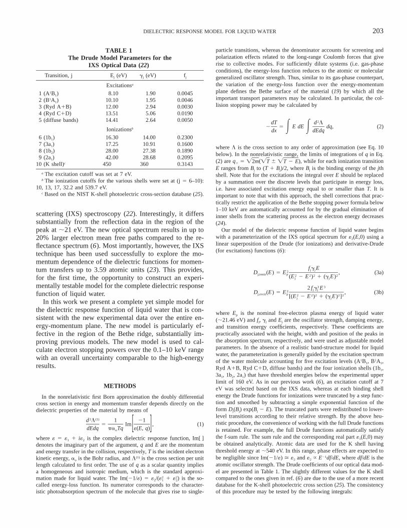
TABLE 1The Drude Model Parameters for the
IXS Optical Data (22)
Transition, j Ej (eV) gj (eV) fj
Excitationsa
1 (A1B1) 8.10 1.90 0.00452 (B1A1) 10.10 1.95 0.00463 (Ryd A1B) 12.00 2.94 0.00304 (Ryd C1D) 13.51 5.06 0.01905 (diffuse bands) 14.41 2.64 0.0050
Ionizationsb
6 (1b1) 16.30 14.00 0.23007 (3a1) 17.25 10.91 0.16008 (1b2) 28.00 27.38 0.18909 (2a1) 42.00 28.68 0.2095
10 (K shell)c 450 360 0.3143
a The excitation cutoff was set at 7 eV.b The ionization cutoffs for the various shells were set at (j 5 6–10):
10, 13, 17, 32.2 and 539.7 eV.c Based on the NIST K-shell photoelectric cross-section database (25).
scattering (IXS) spectroscopy (22). Interestingly, it differssubstantially from the reflection data in the region of thepeak at ;21 eV. The new optical spectrum results in up to20% larger electron mean free paths compared to the re-flectance spectrum (6). Most importantly, however, the IXStechnique has been used successfully to explore the mo-mentum dependence of the dielectric functions for momen-tum transfers up to 3.59 atomic units (23). This provides,for the first time, the opportunity to construct an experi-mentally testable model for the complete dielectric responsefunction of liquid water.
In this work we present a complete yet simple model forthe dielectric response function of liquid water that is con-sistent with the new experimental data over the entire en-ergy-momentum plane. The new model is particularly ef-fective in the region of the Bethe ridge, substantially im-proving previous models. The new model is used to cal-culate electron stopping powers over the 0.1–10 keV rangewith an overall uncertainty comparable to the high-energyresults.
METHODS
In the nonrelativistic first Born approximation the doubly differentialcross section in energy and momentum transfer depends directly on thedielectric properties of the material by means of
2 (1)d L 1 215 Im , (1)[ ]dEdq pa Tq «(E, q)o
where « 5 «1 1 i«2 is the complex dielectric response function, Im[ ]denotes the imaginary part of the argument, q and E are the momentumand energy transfer in the collision, respectively, T is the incident electronkinetic energy, ao is the Bohr radius, and L(1) is the cross section per unitlength calculated to first order. The use of q as a scalar quantity impliesa homogeneous and isotropic medium, which is the standard approxi-mation made for liquid water. The Im(21/«) 5 «2/( 1 ) is the so-2 2« «1 2
called energy-loss function. Its numerator corresponds to the character-istic photoabsorption spectrum of the molecule that gives rise to single-
particle transitions, whereas the denominator accounts for screening andpolarization effects related to the long-range Coulomb forces that giverise to collective modes. For sufficiently dilute systems (i.e. gas-phaseconditions), the energy-loss function reduces to the atomic or moleculargeneralized oscillator strength. Thus, similar to its gas-phase counterpart,the variation of the energy-loss function over the energy-momentumplane defines the Bethe surface of the material (19) by which all theimportant transport parameters may be calculated. In particular, the col-lision stopping power may be calculated by
2dT d L2 5 E dE dq, (2)E Edx dEdq
where L is the cross section to any order of approximation (see Eq. 10below). In the nonrelativistic range, the limits of integrations of q in Eq.(2) are q6 5 ( 6 ), while for each ionization transitionÏ2m ÏT ÏT 2 EE ranges from Bj to (T 1 Bj)/2, where Bj is the binding energy of the jthshell. Note that for the excitations the integral over E should be replacedby a summation over the discrete levels that participate in energy loss,i.e. have associated excitation energy equal to or smaller than T. It isimportant to note that with this approach, the shell corrections that prac-tically restrict the application of the Bethe stopping power formula below1–10 keV are automatically accounted for by the gradual elimination ofinner shells from the scattering process as the electron energy decreases(24).
Our model of the dielectric response function of liquid water beginswith a parameterization of the IXS optical spectrum for «2(E,0) using alinear superposition of the Drude (for ionizations) and derivative-Drude(for excitations) functions (6):
f g Ej j2D (E) 5 E , (3a)j,ioniz p 2 2 2 2(E 2 E ) 1 (g E)j j
3 32 f g Ej j2D (E) 5 E , (3b)j,excit p 2 2 2 2 2[(E 2 E ) 1 (g E) ]j j
where Ep is the nominal free-electron plasma energy of liquid water(;21.46 eV) and fj, gj and Ej are the oscillator strength, damping energy,and transition energy coefficients, respectively. These coefficients arepractically associated with the height, width and position of the peaks inthe absorption spectrum, respectively, and were used as adjustable modelparameters. In the absence of a realistic band-structure model for liquidwater, the parameterization is generally guided by the excitation spectrumof the water molecule accounting for five excitation levels (A1B1, B1A1,Ryd A1B, Ryd C1D, diffuse bands) and the four ionization shells (1b1,3a1, 1b2, 2a1) that have threshold energies below the experimental upperlimit of 160 eV. As in our previous work (6), an excitation cutoff at 7eV was selected based on the IXS data, whereas at each binding shellenergy the Drude functions for ionizations were truncated by a step func-tion and smoothed by subtracting a simple exponential function of theform Dj(Bj) exp(Bj 2 E). The truncated parts were redistributed to lower-level transitions according to their relative strength. By the above heu-ristic procedure, the convenience of working with the full Drude functionsis retained. For example, the full Drude functions automatically satisfythe f-sum rule. The sum rule and the corresponding real part «1(E,0) maybe obtained analytically. Atomic data are used for the K shell havingthreshold energy at ;540 eV. In this range, phase effects are expected tobe negligible since Im(21/«) ù «2 and «2 } E21df/dE, where df/dE is theatomic oscillator strength. The Drude coefficients of our optical data mod-el are presented in Table 1. The slightly different values for the K shellcompared to the ones given in ref. (6) are due to the use of a more recentdatabase for the K-shell photoelectric cross section (25). The consistencyof this procedure may be tested by the following integrals:
204 EMFIETZOGLOU, CUCINOTTA AND NIKJOO
`2E« (E, q) dE 5 1, (4a)E 22pEp 0
`2 21E Im dE 5 1, (4b)E2 [ ]pE «(E, q)p 0
` 21E ln(E)Im dEE [ ]«(E, 0)0
ln(I) 5 , (4c)` 21
E Im dEE [ ]«(E, 0)0
Note that Eqs. (4a, 4b) are generalizations of the f-sum rule and must besatisfied for any value of q, whereas Eq. (4c) defines the I value of thestopping-power theory, which depends on the optical limit of the energy-loss function of the medium.
The momentum dependence of the dielectric functions may be intro-duced by the so-called extended-Drude or d-oscillator models. They arecomputationally simpler and conceptually more appropriate for the inter-band transitions of an insulator like water than Lindhard’s function of thefree electron gas model. An intercomparison for the case of liquid wateris provided in refs. (6, 17).
In the present work we follow the extended-Drude algorithm first sug-gested by Ritchie and Howie (26). In this context the use of Drude-typefunctions is particularly convenient since the momentum dependence maybe introduced directly into the Drude coefficients by a suitable general-ization of the triad {fj, gj, Ej} to {fj(q), gj(q), Ej(q)}. Two forms of theextended-Drude model are currently available in the literature. In thesimplest form (herein named the D1 model), only one Drude coefficientis allowed to depend on the momentum transfer, namely, the transitionenergy coefficient. Along the lines of the impulse approximation, a first-order expansion in q2 gives Ej(q) 5 Ej 1 q2/2m. This approximationasymptotically leads to the free electron limit and is also consistent withthe obvious requirement that at q 5 0 the dielectric function coincideswith its optical limit. Although the use of the D1 model seems reasonablefor the continuum, it is obviously unrealistic for the discrete spectrum.Therefore, for track structure studies on liquid water, an improved schemeis used whereby two Drude coefficients are now allowed to disperse(herein named the D2 model). In this scheme, the dispersion of the D1model is now restricted to the ionization transitions and the momentumdependence of the oscillator strength coefficient for the discrete transi-tions, i.e. fj(q), is taken by an empirical formula obtained from gas-phasedata. The corresponding dispersion for the oscillator strength coefficientof the continuum may then be obtained simply by the application of thef-sum rule. By this development, the asymptotic limit at high momentumtransfers is still retained while a more reasonable approximation for thediscrete transitions is provided. As we show here, the predictions of theD1 and D2 models do not compare well with the IXS data at finitemomentum transfers. Specifically, they do not accurately reproduce theposition and width of the Bethe ridge. As already pointed out in ref. (27)and based on our knowledge on atomic oscillator strength distributions(19), it is expected that medium effects should bring about a broadeningof the Bethe ridge.
To improve upon the earlier models at finite momentum transfers, twofurther modifications are introduced: (1) We also allow the damping en-ergy coefficient (for both ionizations and excitations) to disperse alongwith the other two coefficients. A simple formula of the form
2g (q) 5 g 1 aq 1 bq ,j j (5)
where a 5 10 and b 5 6 (q in atomic units), resulted in a sufficientlybroad Bethe ridge in accordance with the IXS findings. (2) We introducea ‘‘retarding’’ function to the q2 term of the (ionization) transition energydispersion, Ej(q), so that the free electron limit is asymptotically ap-proached at a ‘‘slower’’ rate, that is, at larger q values. A simple modi-fying function of the form
dg(q) 5 1 2 exp(2cq ), (6)
where c 5 1.5 and d 5 0.4 (q in atomic units), provided the necessary
fine-tuning of the position of the peak. The new extended-Drude functionnow becomes
f (q)g (q)Ej j2D (E, q) 5 E , (7a)j,ioniz p 222q2 2E 1 g(q) 2 E 1 [g (q)E]j j5 6[ ]2m
3 32 f (q)g (q)Ej j2D (E, q) 5 E . (7b)j,excit p 2 2 2 2 2{(E 2 E ) 1 [g (q)E] }j j
The new algorithm will hereafter be called the D3 model since all threeDrude coefficients are now allowed to disperse.
The first Born calculations based on Eq. (1) are gradually inaccuratefor incident electron energies below a few hundred eV (19). Higher-orderand exchange terms need to be supplemented at low energies. No exacttheory exists, however, to obtain these corrections for many electron sys-tems, and various heuristic approaches are therefore in use. As a second-order term we have used a formula that has proven useful as a higher-order Z 3 correction to the stopping-power theory (24, 28):
(2)dL 2 215 2 Im L(E; j), (8)[ ]dE pa T «(E, 0)o
where L(E;j) 5 (h/ao)[E/(2T )] 3 I(j) represents a distant collisionÏ2Tmcorrection function weighed by the distribution of energy losses as de-termined by the energy-loss function. The parameter j is associated inthe model with the cutoff distance of glancing collisions which are as-sumed to be the only ones contributing to this correction term. A com-putationally convenient parameterization of I(j) for various intervals ofj is given in ref. (24). It was found in ref. (6) that the cutoff valuesuggested in ref. (29), which gives j 5 1/2 , appears more appro-ÏE/Tpriate for liquid water. Despite its shortcomings, which are well under-stood (18, 30, 31), an important advantage of using Eq. (8) is that itdepends on the optical limit of the energy-loss function and therefore isboth phase-specific and readily available by our optical model. As notedelsewhere (6, 12, 32), the use of Eq. (8) gradually fails below 100 eVelectron impact, and alternative schemes should therefore be used at verylow energies (6). The range below 100 eV, however, is most uncertainsince the discrepancies between the various models increase rapidly inthis range and even the use of the Born theoretical framework may bequestioned (6). For an exchange correction, we follow the standard prac-tice (2, 8) of applying Mott-like modifications to the direct Born term (ofsecond order here),
ioniz dL (T 2 E 1 B , T)dL (E, T) j jex 5 O 5dE dEj
1/2dL (E, T) dL (T 2 E 1 B , T)j j j2 3 , (9)6[ ]dE dE
where Lj 5 1 is the cross section of the jth shell calculated to(1) (2)L Lj j
second order by the use of Eqs. (1) and (8). In the spirit of the Mott crosssection, which is associated with transitions to the continuum (i.e. ioni-zations), no such correction was applied to the discrete transitions (i.e.excitations). Equation (9), which is exact in the binary limit, is obviouslyan oversimplification for a many-electron system. It has the advantage,however, that the dielectric functions of the material (through Lj) arebeing used. Our final formula of second order and exchange-correctedbecomes
˜dL dL dLex5 1 , (10)dE dE dE
where L 5 L(1) 1 L(2). The methodology beyond the first Born approx-imation provides an overall consistent scheme for extrapolating to lowerenergies using, at all steps, the characteristic dielectric properties of thematerial.
205DIELECTRIC RESPONSE MODEL FOR LIQUID WATER
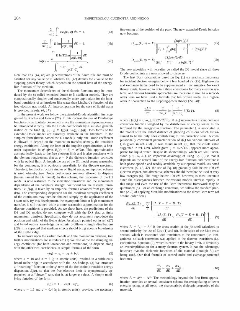
FIG. 1. The optical energy-loss function of liquid water: filled boxes,experimental data based on inelastic X-ray scattering (IXS) measurements(22); open boxes, values obtained from the NIST photoelectric cross-section database for the water molecule (25); solid line, our Drude modelbased on the parameters of Table 1.
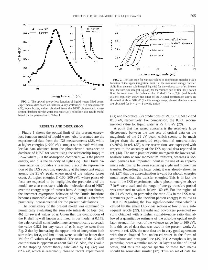
FIG. 2. The sum rule for various values of momentum transfer q as afunction of the upper integration limit, i.e. the maximum energy transfer.Solid line, the sum rule integral Eq. (4a) for the valence part of «2; brokenline, the sum rule integral Eq. (4b) for the valence part of Im(–1/«); dottedline, the total sum rule (valence plus K shell) for «2(E,0) [and Im(–1/«(E,0)] explicitly shown the onset of the K-shell contribution above itsthreshold at about 540 eV (for this energy range, almost identical curvesare obtained for 0 # q # 3 atomic units).
RESULTS AND DISCUSSION
Figure 1 shows the optical limit of the present energy-loss function model of liquid water. Also presented are theexperimental data from the IXS measurements (22), whileat higher energies (.200 eV) comparison is made with mo-lecular data obtained from the photoelectric cross-sectiondatabase of NIST for water using the relationship Im(«) 5mc/v, where m is the absorption coefficient, v is the photonenergy, and c is the velocity of light (25). Our Drude pa-rameterization provides a reasonably accurate representa-tion of the IXS spectrum, especially at the important regionaround the 21 eV peak, where most of the valence lossesoccur. At higher energies (.100–200 eV), where phase ef-fects are expected to be negligible, the predictions of themodel are also consistent with the molecular data of NISTover the energy range of interest here. Although not shown,the incorrect asymptotic behavior of the Drude functionsbecomes noticeable above several keV, and it is thereforepractically inconsequential for the present calculations.
The consistency of the present model was examined bynumerically evaluating the sum rule integrals of Eqs. (4a,4b) for several values of q. Given that the contribution ofthe K shell is well known and fixed in our model at 0.179,the valence shell contribution must asymptotically approachthe value 0.821 for any value of q. It may be seen fromFig. 2 that by increasing the upper limit of integration bothsum rules, for «2 and Im(21/«), were satisfied to better than1% for all values of q examined. The onset of the K-shellcontribution is apparent at about 540 eV. Also, the I valueof the stopping power theory calculated by Eq. (4c) was82.4 eV, which is reasonably close to recent experimental
(33) and theoretical (2) predictions of 79.75 6 0.50 eV and81.8 eV, respectively. For comparison, the ICRU recom-mended value for liquid water is 75 6 3 eV (20).
A point that has raised concerns is the relatively largediscrepancy between the two sets of optical data on themagnitude of the 21 eV peak, which seems to be muchlarger than the associated experimental uncertainties(,30%). In ref. (27), some reservations are expressed withrespect to the accuracy of the IXS optical data reported inref. (34). The main point of criticism regards the low signal-to-noise ratio at low momentum transfers, whereas a sec-ond, perhaps less important, point is the use of an approx-imate relationship between scattering angle and momentumtransfer. Regarding the latter point, it was already shown inref. (27) that the approximation is valid for photon energiesmuch larger than the transfer energies. This is in fact thecase in the IXS experiments, where photon energies above7 keV were used and the range of energy transfers probedwas restricted to values below 160 eV. For the region ofthe 21 eV peak, in particular, the ratio E/v in the IXS mea-surements (with v the incident photon energy) is as low as;0.003. Regarding the low signal-to-noise ratio which iscaused by the small IXS cross section at low q, in a sub-sequent article (22), Hayashi and coworkers presented re-sults obtained with a higher signal-to-noise ratio that al-lowed a quantitative estimate of the absolute optical oscil-lator strength for most of the valence range (up to 160 eV).It is this set of data that was used in the present work. Asshown in ref. (22), the new data are in very good agreementwith those obtained for condensed water in the form ofamorphous and hexagonal ice (35, 36). Amorphous ice, inparticular, bears a similar molecular layout to that of liquidwater, and thus the optical spectra of these two mediashould be somewhat similar (37). Thus no set of data for

206 EMFIETZOGLOU, CUCINOTTA AND NIKJOO
FIG. 3. The energy-loss function of liquid water at (panel a) momen-tum transfer, q 5 1.50 atomic units and (panel b) momentum transfer, q5 3.59 atomic units: filled boxes, experimental data based on inelasticX-ray scattering (IXS) spectroscopy (23); solid line, the D3 extended-Drude model developed in the present work; dotted line, the D2 extended-Drude model; long broken line, the D1 extended-Drude model; shortbroken line, using Ashley’s (A) d-oscillator dispersion (38); double dottedline, using Liljequist’s (L) d-oscillator dispersion (39). Note that for ameaningful comparison all the above dispersion schemes have been ap-plied to the same (IXS-based) optical model.
liquid or solid water seems to support the intense peak ob-tained by the reflectance measurements, which, as ex-plained in ref. (22), may be an experimental artifact.
Figure 3 shows the energy-loss function of liquid waterfor two values of the momentum transfer. The 3.59 atomicunits is the highest momentum probed by the IXS experi-ments (23). Along with the IXS data, results obtained usingthe three forms of the extended-Drude model as well as
Ashley’s (38) and Liljequist’s (39) d-oscillator models arepresented. Evidently, the new model provides a significantimprovement over the previous models in reproducing theexperimental spectrum. For example, at 3.59 atomic units,the predictions of the earlier models differ by at least afactor of 3 from the experimental peak. In contrast, the newmodel, D3, provides a very good overall representation ofthe spectrum, especially in the peak region, where it coin-cides almost exactly with the experimental data. Minor dis-crepancies with the data are unimportant in the present con-text since for energy-loss calculations one needs to inte-grate over the momentum variable. The above improvementconcerning the broadening of the peak is brought aboutmainly by the dispersion of the damping coefficient, gj(q)(Eq. 5). A second observation concerns the position of thepeak. By a careful examination it becomes apparent thatthe position of the peak predicted by the earlier modelsdoes not match the experimental one; the effect is morepronounced at intermediate values of momentum transfers(e.g. at 1.5 atomic units). The fine-tuning of the peak po-sition in the new model is accomplished by the modifyingfunction g(q) in the dispersion of the transition energy co-efficient (Eq. 6). In essence, the g(q) modification causesthe Drude function (Eq. 7a) to peak at smaller energy trans-fers for q values that are not too large. The effect asymp-totically (q .. 0) vanishes in accordance with the free-electron limit. Note also that the peak predicted by the D2model at small energy transfers (;10 eV) in Fig. 3b (whichis not visible in Fig. 3a due to the scale on the y axis) iscaused by the dispersion relationship of the oscillatorstrength coefficient for excitations, fexc(q). Although thesame formula for fexc(q) is being used in D3 as well, thislow-energy excitation peak has been smeared out in thenew model due to the dispersion of the damping energycoefficient, gexc(q).
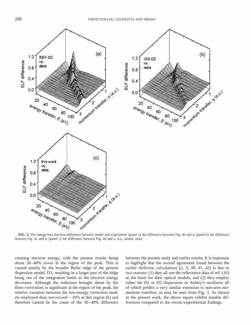
In Fig. 4a–d, the Bethe surface of liquid water as deter-mined by the IXS data (panel a) and model calculations(panels b–d) is presented. Panels b and c are obtained bythe application of the D2 dispersion to the REF- and IXS-based optical models, respectively. Panel d uses the newD3 dispersion to the IXS-based optical model. The devel-opment of a sharply peaked Bethe ridge along the binaryline (E 5 q2/2m) by the D2 model is evident in panels band c (40). In contrast, the predictions of the D3 model(panel d) seem to closely match the experimental broad-ening of the Bethe ridge with increasing energy and mo-mentum transfer. Thus binding effects appear to have beenunderestimated in the D2 model. The effect of the D3 mod-el is perhaps best visualized in Fig. 5, which presents thedifference between the model and the experimental Bethesurfaces. Specifically, the improvement observed in panelb compared to panel a is due to the optical model (IXS-based in panel b and REF-based in panel a), whereas thefurther improvement all along the Berthe ridge in panel cis solely due to the new dispersion model (D3 in panel c
207DIELECTRIC RESPONSE MODEL FOR LIQUID WATER
FIG. 4. The energy-loss function of liquid water over the energy-momentum plane (i.e. the Bethe surface): (panel a) inelastic X-ray scattering (IXS)spectroscopy data (22, 23); (panel b) the REF-based optical model with the D2 dispersion (40); (panel c) the IXS-based optical model with the D2dispersion (40); (panel d) the IXS-based optical model with the D3 dispersion developed in the present work. a.u,, atomic units.
and D2 in panel b) since the same optical models have beenused in panels b and c.
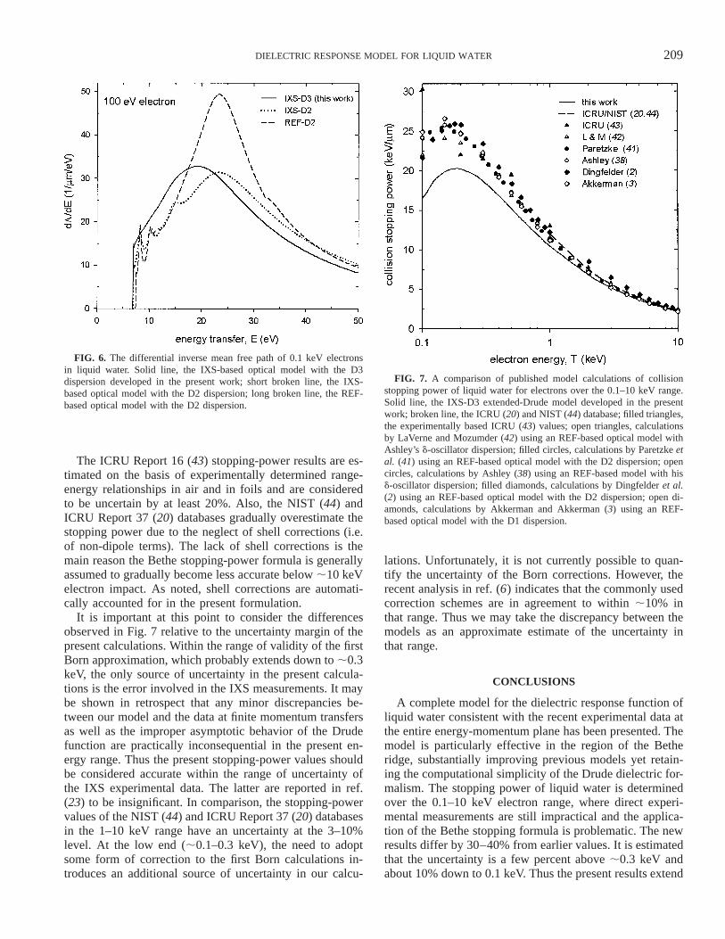
An example of the effect of the new developments onelectron inelastic calculations is presented in the subsequentfigures. Figure 6 shows the differential inverse mean freepath at the low-energy end examined (impact energy 100eV) calculated by the extended-Drude models using theREF and IXS data. The model developed in the presentwork (IXS-D3) seems to have a notable effect on the dis-tribution. This effect is most pronounced at low electronenergies due the increased importance of the spectrum ofmomentum transfers. In particular, there is a shift toward
smaller energy transfers and a smoothing of the discreteexcitation peaks. The former is caused mainly by the ‘‘re-tarded’’ impulse approximation, i.e. the g(q) in Eq. (7a),whereas the latter is caused by the broadening of the en-ergy-loss distribution brought about by the dispersion of thedamping energy, i.e. the gexc(q) in Eq. (7b). In contrast, thediscrepancies observed between the calculations that usethe D2 dispersion are solely the result of the differences inthe optical data employed (REF and IXS).
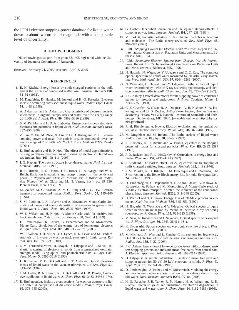
Figure 7 presents the collision stopping power of liquidwater over the 0.1–10 keV electron range as obtained byvarious studies. Differences increase gradually with de-
208 EMFIETZOGLOU, CUCINOTTA AND NIKJOO
FIG. 5. The energy-loss function difference between model and experiment: (panel a) the difference between Fig. 4b and a; (panel b) the differencebetween Fig. 4c and a; (panel c) the difference between Fig. 4d and a. a.u,, atomic units.
creasing electron energy, with the present results beingabout 30–40% lower at the region of the peak. This iscaused mainly by the broader Bethe ridge of the presentdispersion model, D3, resulting in a larger part of the ridgebeing out of the integration limits as the electron energydecreases. Although the reduction brought about by theBorn corrections is significant at the region of the peak, therelative variation between the low-energy correction mod-els employed does not exceed ;10% at this region (6) andtherefore cannot be the cause of the 30–40% difference
between the present study and earlier results. It is importantto highlight that the overall agreement found between theearlier dielectric calculations (2, 3, 38, 41, 42) is due totwo reasons: (1) they all use the reflectance data of ref. (16)as the basis for their optical models, and (2) they employeither the D1 or D2 dispersions or Ashley’s oscillator, allof which predict a very similar extension to non-zero mo-mentum transfers, as may be seen from Fig. 3. As shownin the present work, the above inputs exhibit notable dif-ferences compared to the recent experimental findings.
209DIELECTRIC RESPONSE MODEL FOR LIQUID WATER
FIG. 6. The differential inverse mean free path of 0.1 keV electronsin liquid water. Solid line, the IXS-based optical model with the D3dispersion developed in the present work; short broken line, the IXS-based optical model with the D2 dispersion; long broken line, the REF-based optical model with the D2 dispersion.
FIG. 7. A comparison of published model calculations of collisionstopping power of liquid water for electrons over the 0.1–10 keV range.Solid line, the IXS-D3 extended-Drude model developed in the presentwork; broken line, the ICRU (20) and NIST (44) database; filled triangles,the experimentally based ICRU (43) values; open triangles, calculationsby LaVerne and Mozumder (42) using an REF-based optical model withAshley’s d-oscillator dispersion; filled circles, calculations by Paretzke etal. (41) using an REF-based optical model with the D2 dispersion; opencircles, calculations by Ashley (38) using an REF-based model with hisd-oscillator dispersion; filled diamonds, calculations by Dingfelder et al.(2) using an REF-based optical model with the D2 dispersion; open di-amonds, calculations by Akkerman and Akkerman (3) using an REF-based optical model with the D1 dispersion.
The ICRU Report 16 (43) stopping-power results are es-timated on the basis of experimentally determined range-energy relationships in air and in foils and are consideredto be uncertain by at least 20%. Also, the NIST (44) andICRU Report 37 (20) databases gradually overestimate thestopping power due to the neglect of shell corrections (i.e.of non-dipole terms). The lack of shell corrections is themain reason the Bethe stopping-power formula is generallyassumed to gradually become less accurate below ;10 keVelectron impact. As noted, shell corrections are automati-cally accounted for in the present formulation.
It is important at this point to consider the differencesobserved in Fig. 7 relative to the uncertainty margin of thepresent calculations. Within the range of validity of the firstBorn approximation, which probably extends down to ;0.3keV, the only source of uncertainty in the present calcula-tions is the error involved in the IXS measurements. It maybe shown in retrospect that any minor discrepancies be-tween our model and the data at finite momentum transfersas well as the improper asymptotic behavior of the Drudefunction are practically inconsequential in the present en-ergy range. Thus the present stopping-power values shouldbe considered accurate within the range of uncertainty ofthe IXS experimental data. The latter are reported in ref.(23) to be insignificant. In comparison, the stopping-powervalues of the NIST (44) and ICRU Report 37 (20) databasesin the 1–10 keV range have an uncertainty at the 3–10%level. At the low end (;0.1–0.3 keV), the need to adoptsome form of correction to the first Born calculations in-troduces an additional source of uncertainty in our calcu-
lations. Unfortunately, it is not currently possible to quan-tify the uncertainty of the Born corrections. However, therecent analysis in ref. (6) indicates that the commonly usedcorrection schemes are in agreement to within ;10% inthat range. Thus we may take the discrepancy between themodels as an approximate estimate of the uncertainty inthat range.
CONCLUSIONS
A complete model for the dielectric response function ofliquid water consistent with the recent experimental data atthe entire energy-momentum plane has been presented. Themodel is particularly effective in the region of the Betheridge, substantially improving previous models yet retain-ing the computational simplicity of the Drude dielectric for-malism. The stopping power of liquid water is determinedover the 0.1–10 keV electron range, where direct experi-mental measurements are still impractical and the applica-tion of the Bethe stopping formula is problematic. The newresults differ by 30–40% from earlier values. It is estimatedthat the uncertainty is a few percent above ;0.3 keV andabout 10% down to 0.1 keV. Thus the present results extend
210 EMFIETZOGLOU, CUCINOTTA AND NIKJOO
the ICRU electron stopping-power database for liquid waterdown to about two orders of magnitude with a comparablelevel of uncertainty.
ACKNOWLEDGMENT
DE acknowledges support from grant 62/1405 registered with the Uni-versity of Ioannina Committee of Research.
Received: February 24, 2005; accepted: April 6, 2005
REFERENCES
1. R. H. Ritchie, Energy losses by swift charged particles in the bulkand at the surface of condensed matter. Nucl. Instrum. Methods 198,81–91 (1982).
2. M. Dingfelder, D. Hantke, M. Inokuti and H. G. Paretzke, Electroninelastic-scattering cross sections in liquid water. Radiat. Phys. Chem.53, 1–18 (1998).
3. A. Akkerman and E. Akkerman, Characteristics of electron inelasticinteractions in organic compounds and water over the energy range20–1000 eV. J. Appl. Phys. 86, 5809–5816 (1999).
4. S. M. Pimblott and L. D. A. Siebbeles, Energy loss by non-relativisticelectrons and positrons in liquid water. Nucl. Instrum. Methods B194,237–250 (2002).
5. Z. Tan, Y. Xia, M. Zhao, X. Liu, F. Li, B. Huang and Y. Ji, Electronstopping power and mean free path in organic compounds over theenergy range of 20–10,000 eV. Nucl. Instrum. Methods B222, 27–43(2004).
6. D. Emfietzoglou and H. Nikjoo, The effect of model approximationson single-collision distributions of low-energy electrons in liquid wa-ter. Radiat. Res. 163, 98–111 (2005).
7. I. G. Kaplan, The track structure in condensed matter. Nucl. Instrum.Methods B105, 8–13 (1995).
8. R. H. Ritchie, R. N. Hamm, J. E. Turner, H. A. Wright and W. E.Bolch, Radiation interactions and energy transport in the condensedphase. In Physical and Chemical Mechanisms in Molecular Radia-tion Biology (W. A. Glass and M. N. Varma, Eds.), pp. 99–136.Plenum Press, New York, 1991.
9. M. Zaider, M. G. Vracko, A. Y. C. Fung and J. L. Fry, Electrontransport in condensed water. Radiat. Prot. Dosim. 52, 139–146(1994).
10. S. M. Pimblott, J. A. LaVerne and A. Mozumder, Monte Carlo sim-ulation of range and energy deposition by electrons in gaseous andliquid water. J. Phys. Chem. 100, 8595–8606 (1996).
11. W. E. Wilson and H. Nikjoo, A Monte Carlo code for positive iontrack simulation. Radiat. Environ. Biophys. 38, 97–104 (1999).
12. D. Emfietzoglou, K. Karava, G. Papamichael and M. Moscovitch,Monte Carlo simulation of the energy loss of low-energy electronsin liquid water. Phys. Med. Biol. 48, 2355–2371 (2003).
13. W. E. Wilson, J. H. Miller, D. J. Lynch, R. R. Lewis and M. Batdorf,Analysis of low-energy electron track structure in liquid water. Ra-diat. Res. 161, 591–596 (2004).
14. J. M. Fernandez-Varea, R. Mayol, D. Liljequist and F. Salvat, In-elastic scattering of electrons in solids from a generalized oscillatorstrength model using optical and photoelectric data. J. Phys. Con-dens. Matter 5, 3593–3610 (1993).
15. L. R. Painter, R. D. Birkhoff and E. T. Arakawa, Optical measure-ments of liquid water in the vacuum ultraviolet. J. Chem. Phys. 51,243–251 (1969).
16. J. M. Heller, R. N. Hamm, R. D. Birkhoff and L. R. Painter, Collec-tive oscillation in liquid water. J. Chem. Phys. 60, 3483–3486 (1974).
17. D. Emfietzoglou, Inelastic cross-sections for electron transport in liq-uid water: A comparison of dielectric models. Radiat. Phys. Chem.66, 373–385 (2003).
18. G. Basbas, Inner-shell ionization and the and Barkas effects in3Z1
stopping power. Nucl. Instrum. Methods B4, 277–238 (1984).
19. M. Inokuti, Inelastic collisions of fast charged particles with atomsand molecules—The Bethe theory revisited. Rev. Mod. Phys. 43,297–347 (1971).
20. ICRU, Stopping Powers for Electrons and Positrons. Report No. 37,International Commission on Radiation Units and Measurements, Be-thesda, MD, 1984.
21. ICRU, Secondary Electron Spectra from Charged Particle Interac-tions. Report No. 55, International Commission on Radiation Unitsand Measurements, Bethesda, MD, 1996.
22. H. Hayashi, N. Watanabe, Y. Udagawa and C. C. Kao, The completeoptical spectrum of liquid water measured by inelastic x-ray scatter-ing. Proc. Natl. Acad. Sci. USA 97, 6264–6266 (2000).
23. N. Watanabe, H. Hayashi and Y. Udagawa, Bethe surface of liquidwater determined by inelastic X-ray scattering spectroscopy and elec-tron correlation effects. Bull. Chem. Soc. Jpn. 70, 719–726 (1997).
24. J. C. Ashley, Optical-data model for the stopping power of condensedmatter for protons and antiprotons. J. Phys. Condens. Matter 3,2741–2753 (1991).
25. C. T. Chantler, K. Olsen, R. A. Dragoset, A. R. Kishore, S. A. Ko-tochigova and D. S. Zucker, X-Ray Form Factor, Attenuation andScattering Tables, Ver. 2.1. National Institute of Standards and Tech-nology, Gaithersburg, MD, 2005. [available online at http://physics.nist.gov/ffast]
26. R. H. Ritchie and A. Howie, Electron excitation and the optical po-tential in electron microscopy. Philos. Mag. 36, 463–481 (1977).
27. M. Dingfelder and M. Inokuti, The Bethe surface of liquid water.Radiat. Environ. Biophys. 38, 93–96 (1999).
28. J. C. Ashley, R. H. Ritchie and W. Brandt, effect in the stopping3Z1
power of matter for charged particles. Phys. Rev. B5, 2393–2397(1972).
29. J. D. Jackson and R. L. McCarthy, z3 Corrections to energy loss andrange. Phys. Rev. B6, 4131–4141 (1972).
30. J. Lindhard, The Barkas effect—or , -corrections to stopping of3 4Z Z1 1
swift charged particles. Nucl. Instrum. Methods 132, 1–5 (1976).
31. J. M. Pitarke, R. H. Ritchie, P. M. Echenique and E. Zaremba, Thecorrection to the Bethe-Bloch energy-loss formula. Europhys. Lett.3Z1
24, 613–619 (1993).
32. D. Emfietzoglou, G. Papamichael, I. Androulidakis, K. Karava, K.Kostarelos, A. Pathak and M. Moscovitch, A Monte-Carlo study ofsub-keV electron transport in water: the influence of the condensed-phase. Nucl. Instrum. Methods B228, 341–348 (2005).
33. H. Bichsel and T. Hiraoka, Energy loss of 70 MeV protons in ele-ments. Nucl. Instrum. Methods B66, 345–351 (1992).
34. H. Hayashi, N. Watanabe and Y. Udagawa, Optical spectra of liquidwater in vacuum uv region by means of inelastic X-ray scatteringspectroscopy. J. Chem. Phys. 108, 823–825 (1998).
35. M. Seki, K. Kobayashi and J. Nakahara, Optical spectra of hexagonalice. J. Phys. Soc. Jpn. 50, 2643–2648 (1981).
36. K. Kobayashi, Optical spectra and electronic structure of ice. J. Phys.Chem. 87, 4317–4321 (1983).
37. M. Michaud, A. Wen and L. Sanche, Cross sections for low-energy(1–100 eV) electron elastic and inelastic scattering in amorphous ice.Radiat. Res. 159, 3–22 (2003).
38. J. C. Ashley, Interaction of low-energy electrons with condensed mat-ter: Stopping powers and inelastic mean free paths from optical data.J. Electron Spectrosc. Relat. Phenom. 46, 199–214 (1988).
39. D. Liljequist, A simple calculation of inelastic mean free path andstopping power for 50 eV–50 keV electrons in solids. J. Phys. D:Appl. Phys. 16, 1567–1582 (1983).
40. D. Emfietzoglou, A. Pathak and M. Moscovitch, Modeling the energyand momentum dependent loss function of the valence shells of liq-uid water. Nucl. Instrum. Methods B230, 77–84 (2005).
41. H. G. Paretzke, J. E. Turner, R. N. Hamm, H. A. Wright and R. H.Ritchie, Calculated yields and fluctuations for electron degradation inliquid water and water vapor. J. Chem. Phys. 84, 3182–3188 (1986).

211DIELECTRIC RESPONSE MODEL FOR LIQUID WATER
42. J. A. LaVerne and A. Mozumder, Effect of phase on the stoppingand range distribution of low-energy electrons in water. J. Phys.Chem. 90, 3242–3247 (1986).
43. ICRU, Linear Energy Transfer. Report No. 16, International Com-mission on Radiation Units and Measurements, Bethesda, MD, 1970.
44. M. J. Berger, J. S. Coursey, M. A. Zucker, ESTAR, PSTAR, andASTAR: Computer Programs for Calculating Stopping-Power andRange Tables for Electrons, Protons, and Helium Ions (version1.2.2). National Institute of Standards and Technology, Gaithersburg,MD, 2000. [available online at http://physics.nist.gov/Star]