Embed Size (px)
Citation preview
Earn
2 CE creditsThis course was
written for dentists, dental hygienists,
and assistants.
A Comprehensive Review of Vascular Disease: Part 1- Pathophysiology and Early DetectionA Peer-Reviewed Publication Written by Charles C. Whitney, M.D.
Publication date: June 2012Expiration date: May 2015
AbstractEvidence shows an association between oral disease and systemic vascular disease. Physicians need our dental colleagues’ help if we strive to optimally reduce our patients’ risk of suffering a heart attack or stroke. This four-part series will give dental professionals an understanding of the pathology of cardiovascular disease and describe how you can intervene to reduce risk in your personal life and your patients’ lives.Incorporating a cardiovascular health program in your practice will elevate your credibility as a true health professional, improve your ability to cure dental disease, and drive the much-needed collaboration between physicians and dentists. Part 1 of the series describes the epidemiology of cardiovascular disease and the anatomy, physiology, and pathology of plaque formation. You will also learn the best ways to detect disease at its earliest, most treatable stage.
Learning Objectives:At the conclusion of this course the attendees will be able to understand:1. Epidemiology of vascular disease2. Anatomy and physiology of plaque
development3. Testing available to detect early disease
Author ProfileCharles C. Whitney, M.D. is founder of Revolutionary Health Services, www.revolutionaryhealthservices.com, a practice established by the University of Pennsylvania as the second concierge medical practice in the state of Pennsylvania. He currently serves as Vice President of the American Academy of Private Physicians, www.AAPP.org, and has been a member of the Board of Directors since 2007. Dr. Whitney graduated from Jefferson Medical College in Philadelphia in 1990. He completed his residency at David Grant USAF Medical Center and served as a Physician in the United States Air Force before joining the University of Pennsylvania Health System.
Author DisclosureThe author is not compensated by any of the companies referenced in this course.
This course has been made possible through an unrestricted educational grant.
Supplement to PennWell Publications This course was written for dentists, dental hygienists and assistants, from novice to skilled. Educational Methods: This course is a self-instructional journal and web activity. Provider Disclosure: PennWell does not have a leadership position or a commercial interest in any products or services discussed or shared in this educational activity nor with the commercial supporter. No manufacturer or third party has had any input into the development of course content.Requirements for Successful Completion: To obtain 2 CE credits for this educational activity you must pay the required fee, review the material, complete the course evaluation and obtain a score of at least 70%.
CE Planner Disclosure: Michelle Fox, CE Coordinator does not have a leadership or commercial interest with products or services discussed in this educational activity.
Educational Disclaimer: Completing a single continuing education course does not provide enough information to result in the participant being an expert in the field related to the course topic. It is a combination of many educational courses and clinical experience that allows the participant to develop skills and expertise.
Registration: The cost of this CE course is $49.00 for 2 CE credits. Cancellation/Refund Policy: Any participant who is not 100% satisfied with this course can request a full refund by contacting PennWell in writing.
Go Green, Go Online to take your coursePennWell designates this activity for 2 Continuing Educational Credits
2 www.ineedce.com
Educational ObjectivesAt the conclusion of this course the attendees will be able to understand:1. Epidemiology of vascular disease2. Anatomy and physiology of plaque development3. Testing available to detect early disease
In the United States, a heart attack occurs every 25 seconds and a stroke every 40 seconds. In about 25% of people who suffer sudden death from a heart attack, sudden death is their first symptom, usually caused by an arrhythmia.
Advancing medical science has significantly improved the treatment of known vascular disease, reducing mortality and allowing people to live longer following an event. However, we are ineffectively preventing the first event. The incidence of heart attacks was estimated to be 1.5 million in 1986. That only modestly reduced to 1.2 million by 2006.1
The CDC reports that heart disease and stroke are the #1 and #3 causes of death in the United States, accounting for 31% of all deaths.2 A 2010 United Nations report only ranks the United States as 36th in life expectancy.³ Life expectancy in the United States is 78 years, equivalent to citizens of Cuba, Chile, and Czechoslovakia. We would have a longer life ex-pectancy if we lived in Slovenia, Portugal, Malta, or Iceland.4
If we are to improve, pathology must be detected earlier in the course of disease development, before an event occurs or a procedure is needed to open obstructed arteries. Necessary lifestyle changes, medications, and supplements can then be implemented to heal and stabilize existing plaque to halt and even reverse the disease process.
Dentists are well positioned to participate in the war against heart attacks and strokes. Many middle-aged Americans rarely visit their Physician, but regularly see their Dentist. By understanding the pathophysiology of vascular disease and red flags for disease, Dentists can iden-tify patients at risk, intelligently discuss needed screenings,
and even do some of the testing and health promotion in their office.
Dentists and physicians need to collaborate. Sleep ap-nea causes heart attacks and Dentists will have a hard time optimally treating their sleep apnea patients without weight loss. Periodontal breakdown may not heal until insulin resistance is identified and treated. Physicians often can’t optimally improve vascular inflammation without identify-ing and treating existing high risk oral bacteria.
All Health Professionals who care about their patients need to be involved in promoting total health. It’s about the person, not the body part! During this 4 part series you will learn the pathophysiology of vascular disease, state-of-the-art testing available to detect early treatable disease, direct causes of disease, root causes of disease, genetic testing available, and most importantly, what dentists can do to help stabilize or reverse disease in your patients, and perhaps yourself.
Anatomy of Disease- making of a volcanoThe heart is a muscle. It receives oxygenated blood from the lungs and delivers it to be used for cellular respiration at the tissue level. After the oxygen is extracted by a tissue, veins transport the blood back to the heart to be reoxygenated.
It is estimated that there are about 7000 miles of arteries in the human body ranging in size from the aorta to tiny arte-rioles that feed capillary beds. The coronary arteries exit im-mediately after oxygenated blood leaves the heart. Coronary arteries supply oxygen to the heart’s myocardium.
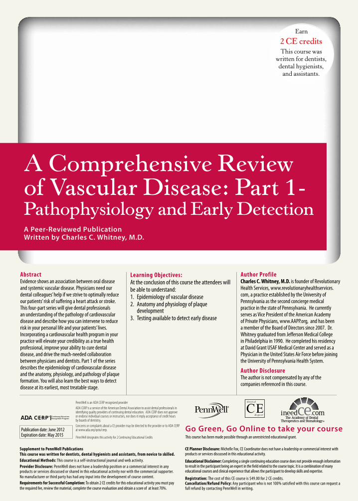
Figure 1 depicts the anatomy of an artery. The section at the far left is a normal arterial wall. Think of it as a garden hose. Its anatomy includes:1. Endothelium-the endothelium is the thin layer of cells
that separates flowing blood from the rest of the wall.2. Intima layer- The intima layer within the arterial wall is
composed primarily of smooth muscle cells.3. Outer capsule
In�ammation Myeloperoxidase Thickening Plaque Calciumbuild-up
Vulnerable plaque/erosion/�ssure
Rupture Sti�eningwall
Blood clot
Oxidation Plaquedeposition
Plaque growth/vulnerable plaque
Plaque rupture Acute coronary syndrome
Figure 1. Anatomy of an artery
Source: Cleveland Heart Lab
www.ineedce.com 3
Within the blood, red blood cells carry oxygen and white blood cells modulate inflammatory reactions intended to fight pathologic processes such as infection, toxic exposures, and malignant transformation of cells. Cholesterol products, lip-ids, are delivered to tissues for cell membrane production. A normal arterial wall does not accumulate a significant amount of lipids. The endothelial lining is intended to block the flow of lipids from the blood into the wall.
Arterial plaque forms from the buildup of lipids within the intima layer of the arterial wall. Plaque does not simply stick to the inner surface of the endothelial lining, it actually penetrates the endothelial barrier to accumulate within the arterial wall. That is why a cardiologist is not able to just “scrape out” the plaque.
There is actually a bidirectional flow of lipid particles across the endothelial lining. Think of it as a military battle. Enemy troops (pathogenic lipids) are trying to work their way behind enemy lines (the endothelial barrier) in an effort to kill their target, us! There are also friendly troops (High Density Lipoproteins, HDL) that are trying to protect the military theater. They bind pathogenic lipids and transport these enemy troops out of the wall, back into the bloodstream where they are washed away. This elimination of pathogenic lipids from the wall of the artery by HDL particles is called reverse cholesterol transport.
In a healthy artery, the endothelium is impervious to the influx of pathogenic lipids. Any excess is effectively eliminated by reverse cholesterol transport. Unfortunately, in most Americans, the enemy troops are winning the war, leading to a slow buildup of lipids within the intima layer of our arteries.
Inflammation and endothelial dysfunction drive the pathologic flow of lipids into the arterial wall. Inflammation is actually a normal process designed to eliminate invading organisms and toxins, heal damaged tissue, and repair mu-tated cells that could lead to cancer. Inflammation is neces-sary for survival. When a developing problem is recognized by our immune system, macrophages migrate to the injured site and release cytokines. Cytokines are immune system chemicals that kill the invading infection, repair the injured tissue, and eliminate a cancerous cell. Cytokines do so by producing inflammation.
The problem in vascular disease is that the inflamma-tory processes are never allowed to turn off, which leads to chronic inflammation. Chronic inflammation causes an increased flow of pathogenic lipids across the endothelium, and leaves it fragile, prone to rupture.
Early plaque formation grows deep into the arterial wall, like an iceberg (figure 1, panels 2-4). Eventually, the endo-thelial lining weakens and small volcanoes begin to bubble up from the surface of the artery. These volcanoes will con-tinue to grow into large plaques that eventually block the flow of blood within the artery as shown on the far right of the diagram.
There are two pathologic outcomes that result from this disease process. In the first, a slow steady build of enemy troops in the arterial wall continues until we can no longer deliver adequate oxygen past the obstruction to adequately supply the distal tissues. At this point, symptoms will be-gin. Symptoms usually begin during activity since exercis-ing muscles require more oxygen.
Symptoms experienced depend on the location of the diseased artery. If the obstruction occurs in the artery of our legs as occurs with peripheral artery disease, we get cramp-ing in our calves called claudication. If the blocked artery is in our heart we get a symptom called angina, typically experienced as chest pressure, left arm discomfort, nausea, and breaking out in a cold sweat. Think of this as the Bill Clinton version of heart disease. President Clinton began to experience symptoms of angina. Testing showed that three arteries in his heart were 90% obstructed, and bypass surgery was required.
The other pathologic outcome that occurs within the arterial wall is much more dangerous. It’s called plaque rupture as is shown in panel 5 of figure 1. White blood cells from the circulating blood penetrate the arterial wall. Oxi-dation occurs within the plaque in the arterial wall, which activates the inflammatory cascade to soften the plaque. The reactions weaken the endothelial barrier leaving it prone to crack. The lining may break open, causing the contents from within the plaque to erupt like a volcano into the bloodstream.
This rupture activates the clotting cascade. If the event occurs in a high pressure artery like the carotid artery in our neck, the clot breaks loose, flows to the brain, and causes a stroke. If rupture occurs in a low pressure vessel like the coronary arteries in our heart, the clot continues to grow un-til it completely blocks blood flow through the artery. If the clot is not recanalized to allow oxygenation of distal tissue, all tissue beyond the event will die. This is a heart attack.
Asymptomatic events can occur in the tiny arteries of our kidneys and brain. These events lead to chronic con-ditions such as chronic kidney disease and “multi infarct” dementia. Multi-infarct dementia is the second leading cause of dementia, accounting for up to 20% of the cases. It is second to Alzheimer’s dementia.
Rupture can occur in very small plaques. It is estimated that up to 85% of coronary plaque ruptures that cause heart attacks occur in plaques not large enough to make a stress test abnormal.5 Think of this as the Tim Russert type of vascular disease. TV anchorman Tim Russert had a normal stress test April 2008 and died of a heart attack two months later. He suffered a plaque rupture in a small plaque not picked up on the stress test. Sudden death occurs when the event occurs in a coronary artery that supplies the electri-cal center of the heart. It’s like tripping a circuit breaker in your house. All electrical activity ceases when this artery thromboses.
4 www.ineedce.com
Early detection of vascular diseaseDiscovered early, vascular disease can be stabilized and even reversed. Vascular risk factors like blood pressure, cholesterol, smoking, etc. simply predict the likelihood a person has dis-ease. More important is to determine whether disease actually exists, and if so, how bad it is. Knowledge is power. An indi-vidual who discovers they have disease often has a stronger motivation to make the changes necessary to improve their health.
Discovered plaque needs to be protected. Even a very small plaque can rupture to cause a heart attack, stroke, or transient ischemic attack (TIA). Ruptures within tiny ar-teries in the brain may not cause symptoms, but when they occur hundreds or thousands of times, it can lead to vascular dementia. On a brain MRI the radiologist will sometimes make a side comment of, “volume loss due to chronic isch-emic changes.” Translated, that says: “volume loss”= death of brain. “Chronic ischemic changes”= arterial disease. The brain is dying from multiple small imperceptible events! Fre-quently, these patients have no history of heart attack, stroke, or other obvious vascular event.
We don’t want to wait until there is death of brain or some other event occurs to start treating existing disease. Early detection of disease allows aggressive treatment with lifestyle changes, medications, and supplements to halt, or even reverse the process. Most importantly, we want to prevent a major event.
Sometimes evidence of disease is found in testing already done. A chest x-ray report may comment, “coronary calcifica-tions present” or “calcifications in the aortic arch.” Calcifica-tions are not seen in healthy vessels, they are only seen along with arterial plaque. Similarly, vascular calcifications may be seen in CT scans, Panorex, or mammograms.
Historically, it has been difficult to identify disease early, and impossible to track whether therapeutic interventions have been effective. However, advancing technology has changed that. The following is a review of cardiovascular testing currently available.
EKGContrary to popular belief, an electrocardiogram is NOT a screening test for vascular disease. All an EKG does is mea-sure the flow of electricity through your heart. If the flow of electricity is disrupted because of death of heart muscle as oc-curs in a heart attack, changes will be seen on the EKG. That is how we often find “silent heart attacks.”
Occasionally we will find evidence of cardiomyopathy (heart muscle thickening), electrical disturbances, electrolyte abnormalities, and drug toxicities, but that is the exception rather than the rule.
A routine EKG is most valuable as a baseline study. The flow of electricity can vary between individuals because of anatomic variations. Having it as a comparison when they present with symptoms can be invaluable.
Stress testStress tests have historically been the standard of care in screening for vascular disease. Unfortunately, they still are. As many as 70-86 percent of heart attacks occur in people who would have had a normal stress test as occurred with Tim Russert. Why?
The heart is a large muscle whose job is to receive oxy-genated blood from the lungs, then contract to deliver oxy-genated blood to distant tissue. Just like every muscle, the heart needs oxygen. The coronary arteries branch off just as the aorta exits the heart. Blood flows through these coronary arteries to supply oxygenated blood to the heart muscle.
When a slow buildup of plaque reaches a critical size, the obstruction prevents an adequate supply of oxygen. The person begins to experience symptoms of angina, which include pressure in their chest, shortness of breath, pain in their left arm, diaphoresis (cold sweat), and nausea. This is what happened to Bill Clinton. Symptoms usually first oc-cur with exertion because of the higher oxygen demand.
A stress test is an extension of this concept. It is a func-tional study, not an anatomical study. A plaque must block at least 60-70 percent of the blood flow before a stress test becomes abnormal.
There are two components to every stress test. The first is exercise to force an increased oxygen requirement. The second is to watch for changes that suggest poor blood flow through the coronary arteries.
The most common method to increase the heart’s oxygen requirement is to follow one of several protocols that use a treadmill. The patient starts by walking slowly on the treadmill with minimal elevation. Every 2-3 minutes the speed and tilt are increased slightly to force a greater workload. The patient’s heart rate, rhythm, and EKG are monitored throughout the test. The workload continues to increase until the patient fatigues, or reaches a target heart rate to qualify as an adequate test. This is called an exercise stress test.
If a person has physical limitations that do not allow them to walk on a treadmill, a pharmacologic stress test is performed. A medication, either Adenosine or Dobutamine are infused through an IV to mimic the effect of exercise.
There are three ways to look for inadequate blood flow to the coronary arteries. The first is to look for EKG changes that occur during exercise. However, false negative tests are common.
Sensitivity to detect disease improves with a stress echo-cardiogram. An echocardiogram is performed immediately after peak exercise. The cardiologist looks for areas of the heart that squeeze weakly during exercise. A localized area of weak squeeze suggests an obstruction at that site.
The third method is called a nuclear stress test. A scan is performed of the heart at rest prior to exercise. Shortly before peak exercise, a nuclear isotope is injected through an IV. Thallium, Cardiolyte, and Myoview are common ones

www.ineedce.com 5
used. A second scan is performed shortly after the treadmill is finished to measure uptake of the tracer in the heart. Poor tracer flow suggests inadequate oxygen supply.
The use of the stress echocardiogram and nuclear stress test has significantly improved the sensitivity of picking up coronary artery blockages. However, our heart only needs about 60% of normal blood flow to function normally, so any smaller blockage will not be detected.
Therefore, stress tests are an inadequate method of find-ing arterial disease at a reasonably early stage. They are very helpful in evaluating a patient who is having atypical chest pain that we don’t think is cardiac in origin. A normal test in that circumstance confirms that their symptoms are from a different source. It is also a good tool to evaluate arrhyth-mias, estimate exercise capacity, and to screen a patient who is sedentary, but wants to initiate a vigorous exercise program.
Carotid artery DopplerCarotid Doppler testing has been used for decades. It is one component of Lifeline screening frequently available in the community. A small ultrasound probe is placed on your neck and the flow of blood through the carotid artery is observed.
As blood flows past a significant blockage, it speeds up, similar to water flowing from a garden hose as you place your thumb over the opening. This change in speed of blood flow is observed as color changes on the ultrasound monitor sug-gesting a partial blockage in that section of the artery. The size can be estimated based on the speed of flow as indicated by the color.
Like a stress test, this is a functional study looking for flow obstruction. It does not look at the specific anatomy of the arterial wall.
Coronary artery calcium score (CACS)In the 1990s a new technology was developed to detect ar-terial disease at a much earlier stage. It was called Electron Beam Computed Tomography (EBCT). It was able to take very rapid pictures of the heart through the use of a CT scan. Like a fast shutter speed on a camera, it was able to take stop action photos of the heart. CT scans are able to detect cal-cium within tissue. Normal coronary arteries do not contain any calcium, but diseased ones often do. EBCT was able to measure the total amount of calcium contained within the arteries of the heart and provide a score. A higher score sug-gested more severe disease, but any disease is significant.
Now most radiology facilities are utilizing ultrafast CT scanners, not EBCT, to do their coronary artery calcium testing. Technology has improved and allows for 64 pictures per second, so the quality of image rivals that of EBCT. Coronary calcium score by ultrafast CT scan is typically more readily available and is usually less expensive.
Although CACS is a significant improvement to screen for early vascular disease, it still has its limitations. A plaque
must be calcified to be detected by CACS. Early plaque and soft plaque, the most dangerous type, are not typically calci-fied. CACS will miss them.
Carotid Intima-Media Thickness ultrasound (CIMT)CIMT ultrasound is the best method to detect early dis-ease. It is much different than carotid Doppler ultrasound screening described above. CIMT uses modern B-mode ultrasound technology together with advanced software capability to measure the thickness of arterial walls. It actually looks at the anatomy of the arterial wall.
An ultrasound probe is placed on your neck and many pictures are taken of the carotid arteries. It is inexpensive and many insurance companies, including Medicare, cover its use.
A task force of the American College of Cardiology/American Heart Association presented a report in Novem-ber 2010 reviewing 25 preventive cardiology tests.6 They gave CIMT ultrasound a very favorable Class IIa recom-mendation based on level B evidence. It was one of their most highly recommended screenings. Their only caution was, “Published recommendations on required equipment, technical approach, and operator training and experience for performance of the test must be carefully followed to achieve high-quality results.”
Their caution is completely valid. Although CIMT ultrasound has been used in research for about 15 years, it has only recently been available to the public. National standards ensuring adequate equipment, technician cer-tification, and quality assurance of results have not been established.
Often, Physicians purchase equipment and perform testing in their office. Another option is thru a company that specializes in CIMT ultrasound. In either case, quality assurance is critical.
There are 2 measurements taken during analysis. The first is the mean arterial thickness. The distance between the outer edge of the intima, and the inner edge of the media is measured. An average of multiple images at the posterior wall of the common carotid artery is calculated to give the mean thickness. This intima-media thickness is approximately 0.50 mm in a disease free artery. As enemy troops penetrate the lining and build up within the intima, the artery wall begins to thicken. There is a normal rate that the carotid artery wall thickens. However, this process is anything but normal, it is developing disease!
The mean thickness is a very precise and consistent measurement to within 0.05mm accuracy. It is quantita-tive and trackable. The test can be a repeated over time to look for worsening or improvement. CIMT is the tech-nology used in many studies over the past 15 years which have shown that plaque regression occurs with adequate treatment.

6 www.ineedce.com
Figure 2. Sample CIMT report
Figure 2 shows a graph the median thickness progression from 16 to 85 years old. The pink curve is women and blue is men. A person’s vascular age is the age at which their thick-ness falls on the curve. A person with a mean arterial thick-ness of 0.81 mm has a three-fold increased risk of stroke.7 Therefore, in a 67 year old man and a 74 year old woman, it’s normal to be abnormal!
The heart attack risk is similar to stroke risk, suggesting that the presence of carotid artery disease predicts the exis-
tence of coronary artery disease.8 This is not surprising since the disease process will likely be similar regardless of where the artery is located.
A vascular age more than five years greater than the ac-tual age is considered advanced plaque deposition. Plaque is accumulating faster than it should. The causes should be identified and treated.
Lastly, a mean thickness measurement is also very help-ful to assess people with known disease, including those who
www.ineedce.com 7
have undergone stents or bypass surgery. Some of the best mean arterial thickness measurements are in people who have had a heart attack, stroke, or undergone a cardiac procedure. It shows that their lifestyle changes, medication, and supple-ments are working!
CIMT ultrasound also measures individual plaques, the volcanoes bubbling up from the surface of your arterial wall. Once a volcano of plaque forms, the endpoints of measurement get fuzzy, so this is a qualitative measurement not a quantita-tive measurement as mean thickness is. We cannot compare measurements between tests looking for improvement.
However, it can be observed whether the plaque is soft, heterogeneous, or calcified. Soft plaque is very dangerous plaque. Think of soft plaque like a blister ready to burst. Its liquefied center is often inflamed. An actively inflamed plaque that cools off and is given a chance to heal becomes fibrous and often calcified. Calcified plaque is the least dangerous type of plaque. It is like an aged blister that has wrinkled and become hard. Heterogeneous plaque is between these two.
An individual plaque larger than 1.9 mm seen on CIMT ultrasound would never be seen on carotid Doppler ultra-sound. However, there is up to a 50% increased risk of event.9 A plaque this size needs to be protected from rupture by ex-tinguishing any inflammation, and optimally controlling all other vascular risk factors.
CIMT ultrasound is able to identify disease earlier than coronary artery calcium score. A study published in the Mayo Clinic Proceedings in 2009 performed CIMT ultrasound on 36-59 year olds with one traditional cardiovascular risk factor but no known disease. 34% were found to have plaque. CACS were also performed in those tested and half with an abnormal CIMT ultrasound had a calcium score of zero!10 Their plaque was not calcified.
An approach to discover diseaseWhat is a reasonable approach to determine whether a per-son is at risk of suffering an event, or has advanced plaque deposition?
Start by reviewing the results of previous testing per-formed like a Panorex, a chest x-ray, CT scans, mammogram, lifeline screening, and cardiac catheterization. If plaque is seen on one of these, the search is over. Arteries are diseased and need to be protected.
The next step is to have CIMT ultrasound performed. CIMT ultrasound may be done even if a patient is known to have existing disease. The mean thickness measurement will tell whether current risk factor treatment is working.
If plaque is seen on CIMT ultrasound, the search is over. If no plaque is seen on CIMT, a coronary artery calcium score should be done to look for evidence of disease in the heart. If no plaque is seen on any of the above tests and their mean arterial thickness is favorable, congratulations! It is time for that person to create optimal health in their life to prevent plaque from ever forming.
What processes allow the flow of lipid particles behind enemy lines to cause disease? There is not one simple an-swer to that very important question. It is not just about cholesterol level and blood pressure. There are many other contributors to the disease process, all of which must be assessed if developing disease is to be reversed, or at least stabilized.
The next course will begin to answer this question regarding the origin of disease. It will describe the causes of disease that directly attack the arterial wall, including dyslipidemia, hypertension, vascular inflammation, endo-thelial dysfunction, and oxidative stress.
References1. Lloyd-Jones et al. Heart Disease and Stroke Statisitics-
2010 Update. Circulation 2009; DOI: 10.1161/CIRCULATIONAHA.109.192667
2. Center for Disease Control – Causes of Death Natural vital statistics report, volume 58 number 19, 5-20-2010
3. United Nations World Population Prospects (2005-2010):2006 revision
4. World Health Organization – World Health Statistics – 2010 report
5. Falk E, Shah PK, Jusler V. Circulation 1995;92:657-716. ACCF/AHA Task Force. J Am Coll Cardiol 2010;
56:2182-2199 7. Li C, et al. Cerebrobasc Dis 2008; 26(3):297-3038. Lorenz MW, et al. Circulation 2007; 115(4):459-4679. Rundek T, et al. Neurology 2008; 70:1200-120710. Lester S, et al. Mayo Clin Proc 2009; 84(3):229-233
Author ProfileCharles C. Whitney, M.D. is founder of Revolution-ary Health Services, www.revolutionaryhealthservices.com, a practice established by the University of Penn-sylvania as the second concierge medical practice in the state of Pennsylvania. He currently serves as Vice Presi-dent of the American Academy of Private Physicians, www.AAPP.org , and has been a member of the Board of Directors since 2007. Dr. Whitney graduated from Jefferson Medical College in Philadelphia in 1990. He completed his residency at David Grant USAF Medical Center and served as a Physician in the United States Air Force before joining the University of Pennsylvania Health System.
DisclaimerThe author is not compensated by any of the companies referenced in this course.
Reader FeedbackWe encourage your comments on this or any PennWell course. For your convenience, an online feedback form is available at www.ineedce.com.

8 www.ineedce.com
Questions
Online CompletionUse this page to review the questions and answers. Return to www.ineedce.com and sign in. If you have not previously purchased the program select it from the “Online Courses” listing and complete the online purchase. Once purchased the exam will be added to your Archives page where a Take Exam link will be provided. Click on the “Take Exam” link, complete all the program questions and submit your answers. An immediate grade report will be provided and upon receiving a passing grade your “Verification Form” will be provided immediately for viewing and/or printing. Verification Forms can be viewed and/or printed anytime in the future by returning to the site, sign in and return to your Archives Page.
1. Which of the following countries has a longer life expectancy than the United States?a. Sloveniab. Maltac. Portugal.d. Icelande. All of the above
2. Which of the following is NOT currently a component of optimal cardiovascular assessment and treatment.a. Identify and treat sleep apnea.b. Identify and treat sinusitis.c. Identify and treat high risk oral bacteria.d. Assess and individual’s genetic makeup.
3. Which of the following statements is true about reverse cholesterol (RCT) transporta. It is the process of cholesterol deposition in arterial
wallsb. It is driven by LDL cholesterolc. High levels of Myelperoxidase (MPO) amplify
RCTd. It is the process of removal of cholesterol buildup in
arterial walls4. Arterial plaque
a. Sticks to the lining of arteriesb. Accumulates within the endothelial liningc. Collects within the intimad. Collect between the intima and outer capsule
5. Reverse cholesterol transporta. Is the process by which lipids enter the arterial wallb. Is accomplished by HDL cholesterolc. Accelerates with endothelial dysfunctiond. Accelerates with inflammation.e. All of the above
6. Acute Inflammationa. Is a process where macrophages release cytokines.b. Is a normal physiologic processc. Leaves vulnerable plaque prone to rupture.d. Is used to kill and invading infection, heal damaged
tissue, and repair mutated cells.e. All of the above
7. All of the following are potential outcomes from systemic vascular disease EXCEPTa. Dementiab. Renal diseasec. Multiple sclerosisd. Heart attacke. Stroke
8. All of the following are true statements about arterial plaque rupture EXCEPTa. Most events occur in non-obstructing plaque.b. Sudden death may occur from plaque rupture in a
coronary arteryc. Small asymptomatic events in the brain are the
second leading cause of dementiad. Most people who suffer plaque rupture have
warning signs prior to the evente. All statements are true
9. Which of the following is NOT a potential resource for discovering vascular diseasea. EKGb. Mammogramc. Panorex.d. Chest x-raye. All are potential resources
10. Which of the following screening tests can detect abnormalities before blood flow is obstructed?a. CIMT ultrasoundb. Coronary artery calcium scorec. Carotid Doppler screeningd. Both A and Be. All of the above
11. Which of the following is NOT a reason to detect vascular disease at an early stage?a. Early detection motivates patients to make neces-
sary lifestyle changes.b. Early detection helps a physician determine how
aggressively to treat risk factors.c. Existing plaques can be protected from plaque
rupture.d. Early disease may be reversed.e. All are reasons for early detection
12. An electrocardiogram a. Is a valuable tool to screen for vascular disease.b. Cannot detect existing vascular diseasec. May detect cardiomyopathyd. May detect electrolyte imbalancee. Is most valuable as a baseline comparison because
of the individual anatomic variations13. Which of the following statements is true
about stress tests?a. Stress tests usually detect asymptomatic vascular
disease.b. The patient must be able to walk on a treadmill to
be tested.c. In a pharmacologic stress test, amiodarone is used
to mimic exercise.d. In a nuclear stress test, the patient is exposed to
ionizing radiation for a CT scan e. A stress echocardiogram is more sensitive to pick
up disease than a regular stress test14. Valuable uses of a cardiac stress test
include all of the following EXCEPTa. Detect early vascular disease.b. Evaluate atypical chest pain.c. Estimate exercise capacity.d. Evaluate certain arrhythmias.e. Screen a patient prior to beginning an exercise
program.15. Which of the following is NOT just flow
study that only detects plaques that are obstructing flow, so have a high false nega-tive rate in finding early vascular disease?a. Stress echocardiogramb. Nuclear stress testc. Carotid Doppler ultrasoundd. Coronary artery calcium scoree. C and D
16. All of the following are true statements about coronary artery calcium scoring EXCEPTa. Detects early, nonobstructing, plaqueb. Helps guide how aggressive to treat existing
vascular risk factors.c. 64 slice CT scanners rival the quality of EBCT.d. Plaque must be calcified to be detected with
calcium scoringe. All statements are true
17. All of the following are true statements about carotid intima-media thickness (CIMT) ultrasound EXCEPTa. It is the most sensitive method for detecting early
vascular disease.b. Mean thickness is quantitative and trackablec. It can detect soft plaques.d. Standards have been established to ensure quality
control of resultse. All statements are true
18. Which of the following is a true statement about carotid intima-media thickness (CIMT) ultrasound?a. The American Heart Association does not
recommend it for screening.b. It is an invasive test.c. It is only valuable to assess stroke risk.d. Physicians may perform the test in their office.e. National standards for quality control have been
established.
19. Vascular disease regression or progres-sion is best seen through repeating which of the following tests a. Carotid intima-media (CIMT) thickness
ultrasoundb. Coronary artery calcium scorec. Carotid Doppler ultrasoundd. Nuclear stress teste. Stress echocardiogram
20. Which of the following is a true statement about finding early vascular disease?a. Disease can be found on chest x-ray, panorex, or
mammogramb. A thorough assessment for early vascular disease
requires both coronary artery calcium scoring and carotid intima-media thickness (CIMT) ultrasound testing
c. Exercise stress tests have a high false negative rated. A and Ce. All of the above
1VASDIS612
If not taking online, mail completed answer sheet to
Academy of Dental Therapeutics and Stomatology,A Division of PennWell Corp.
P.O. Box 116, Chesterland, OH 44026 or fax to: (440) 845-3447
For iMMEDiATE results, go to www.ineedce.com to take tests online.
Answer sheets can be faxed with credit card payment to (440) 845-3447, (216) 398-7922, or (216) 255-6619.
Payment of $49.00 is enclosed. (Checks and credit cards are accepted.)
If paying by credit card, please complete the following: MC Visa AmEx Discover
Acct. Number: _____________________________
Exp. Date: _____________________
Charges on your statement will show up as PennWell
AGD Code 018
ANSWER SHEET
A Comprehensive Review of Vascular Disease: Part 1 - Pathophysiology and Early Detection
Name: Title: Specialty:
Address: E-mail:
City: State: ZIP: Country:
Telephone: Home ( ) Office ( ) Lic. Renewal Date:
Requirements for successful completion of the course and to obtain dental continuing education credits: 1) Read the entire course. 2) Complete all information above. 3) Complete answer sheets in either pen or pencil. 4) Mark only one answer for each question. 5) A score of 70% on this test will earn you 2 CE credits. 6) Complete the Course Evaluation below. 7) Make check payable to PennWell Corp. For Questions Call 216.398.7822
Educational Objectives1. Epidemiology of vascular disease
2. Anatomy and physiology of plaque development
3. Testing available to detect early disease
Course Evaluation1. Were the individual course objectives met? Objective #1: Yes No Objective #3: Yes No
Objective #2: Yes No
Please evaluate this course by responding to the following statements, using a scale of Excellent = 5 to Poor = 0.
2. To what extent were the course objectives accomplished overall? 5 4 3 2 1 0
3. Please rate your personal mastery of the course objectives. 5 4 3 2 1 0
4. How would you rate the objectives and educational methods? 5 4 3 2 1 0
5. How do you rate the author’s grasp of the topic? 5 4 3 2 1 0
6. Please rate the instructor’s effectiveness. 5 4 3 2 1 0
7. Was the overall administration of the course effective? 5 4 3 2 1 0
8. Please rate the usefulness and clinical applicability of this course. 5 4 3 2 1 0
9. Please rate the usefulness of the supplemental webliography. 5 4 3 2 1 0
10. Do you feel that the references were adequate? Yes No
11. Would you participate in a similar program on a different topic? Yes No
12. If any of the continuing education questions were unclear or ambiguous, please list them. ___________________________________________________________________
13. Was there any subject matter you found confusing? Please describe. ___________________________________________________________________ ___________________________________________________________________
14. How long did it take you to complete this course? ___________________________________________________________________ ___________________________________________________________________
15. What additional continuing dental education topics would you like to see? ___________________________________________________________________ ___________________________________________________________________
PLEASE PHOTOCOPY ANSWER SHEET FOR ADDITIONAL PARTICIPANTS.COURSE EVALUATION and PARTICIPANT FEEDBACK
We encourage participant feedback pertaining to all courses. Please be sure to complete the survey included with the course. Please e-mail all questions to: [email protected].
INSTRUCTIONSAll questions should have only one answer. Grading of this examination is done manually. Participants will receive confirmation of passing by receipt of a verification form. Verification of Participation forms will be mailed within two weeks after taking an examination.
COURSE CREDITS/COSTAll participants scoring at least 70% on the examination will receive a verification form verifying 2 CE credits. The formal continuing education program of this sponsor is accepted by the AGD for Fellowship/Mastership credit. Please contact PennWell for current term of acceptance. Participants are urged to contact their state dental boards for continuing education requirements. PennWell is a California Provider. The California Provider number is 4527. The cost for courses ranges from $29.00 to $110.00.
PROVIDER INFORMATIONPennWell is an ADA CERP Recognized Provider. ADA CEROP is a service of the American Dental association to assist dental professionals in identifying quality providers of continuing dental education. ADA CERP does not approve or endorse individual courses or instructors, not does it imply acceptance of credit hours by boards of dentistry.
Concerns or complaints about a CE Provider may be directed to the provider or to ADA CERP ar www.ada.org/cotocerp/
The PennWell Corporation is designated as an Approved PACE Program Provider by the Academy of General Dentistry. The formal continuing dental education programs of this program provider are accepted by the AGD for Fellowship, Mastership and membership maintenance credit. Approval does not imply acceptance by a state or provincial board of dentistry or AGD endorsement. The current term of approval extends from (11/1/2011) to (10/31/2015) Provider ID# 320452
RECORD KEEPINGPennWell maintains records of your successful completion of any exam for a minimum of six years. Please contact our offices for a copy of your continuing education credits report. This report, which will list all credits earned to date, will be generated and mailed to you within five business days of receipt.
Completing a single continuing education course does not provide enough information to give the participant the feeling that s/he is an expert in the field related to the course topic. It is a combination of many educational courses and clinical experience that allows the participant to develop skills and expertise.
CANCELLATION/REFUND POLICYAny participant who is not 100% satisfied with this course can request a full refund by contacting PennWell in writing.
© 2012 by the Academy of Dental Therapeutics and Stomatology, a division of PennWell
www.ineedce.com Customer Service 216.398.7822 9
1.2.3.4.5.6.7.8.9.10.
11.12.13.14.15.16.17.18.19.20.