Embed Size (px)
Citation preview
A csontvaacutezrendszer eacutes a vaacutezizomzat fejlődeacutese
Főbb fejlődeacutesi rendellenesseacutegek
Molnaacuter Gergely
SZTE AacuteOK Anatoacutemiai Szoumlvet- eacutes Fejlődeacutestani Inteacutezete
2015
Korai fejlődeacutes
1) Paraxiaacutelis mesoderma
bull Somitomerek (7 db) + somitaacutek sum 42 db ndash occipitalis somitaacutek 4x
ndash cervicalis somitaacutek 8x
ndash thoracalis somitaacutek 12x
ndash lumbalis somitaacutek 5x
ndash sacralis somitaacutek 5x
ndash coccygealis somitaacutek 5-8x
bull Sclerotom + dermomyotom (dermatomyotom)
Somitaacutek
bull hasznaacuteljaacutek az embrioacute koraacutera is
bull nem egeacuteszen pontos ndash
statisztikailag OK nem
rosszabb mint napokban
megadni
nap somitaacutek szaacutema
20 1-4
21 4-7
22 7-10
23 10-13
24 13-17
25 17-20
26 20-23
27 23-26
28 26-29
30 34-35
28 napos embrioacute
oticus placod
veacutegtagbimboacute
ductus vitellinus
koumlldoumlkzsinoacuter
lencseplacod
allantois
sziacutevdomb
garatiacutevek
Korai fejlődeacutes
2) Oldallemez mesoderma (Mesoderma laminae lateralis) ndash Parietalis mesoderma (Somatopleura)
3) Ganglionleacutec (Crista neuralis)
MESENCHYMA=embryonalis koumltőszoumlvet
a mesenchyma sejtekből ① fibroblastok koumltőszoumlvet desmalis csontosodaacutes
② chondroblastok hyalinporc előtelep chondralis csontosodaacutes
+ osteoblastok primer csontosodaacutes
Ectoderm
Endoderm
Somite sclerotome
Chorda
Intermediate mesoderm
Body cavity
Neural tube Neural crest
Somite dermomyotome
Lateral mesoderm
6
SOMITA
DERMOMYOTOM SCLEROTOM
DERMOMYOTOM SCLEROTOM A SEJTEK MESENCHYMA-
SZERŰVEacute VAacuteLNAK CHORDA DORSALIS IRAacuteNYAacuteBA VAacuteNDOROLNAK (4 HEacuteT) A KOumlZEacutePVONALBAN OumlSSZE-
OLVADNAK
A SOMITA UumlREGE (SOMITOCOEL)
7
RESEGMENTATIO - egy- egy cranialis eacutes caudalis sclerotomfeacutel
oumlsszenő
- emiatt az intersegmentalis arteacuteria koumlzeacutepre
keruumll
- a Chorda dorsalis eltűnik a csigolyatest
magassaacutegaacuteban
- a Discus intervertebralis teruumlleteacuten a Chorda
dorsalis Nucleus pulposust alakiacutetja ki
- egy Myotom keacutet szomszeacutedos
szegmentumot hidal aacutet
1
2
3
1
2
3
FUSION OF TWO SCLEROTOMES TO BUILD A VERTEBRA
11
STAGES OF FURTHER VERTEBRAL DEVELOPMENT A week 5 B week 6 (neural arch is the primordium of the vertebral arch) C week 7 D at birth EF puberty
Mesenchymal vertebra Chondrification of vertebra Ossification of vertebra begins
12
INDUKCIOacute 1) A chorda dorsalis indukaacutelja a sclerotomsejtek koumlzeacutepvonali vaacutendorlaacutesaacutet eacutes fuacutezioacutejaacutet Corpus vertebrae 2) A velőcső indukaacutelja a paacuteros neuraacutelis iacutevek noumlvekedeacuteseacutet eacutes dorsalisan a koumlzeacutepvonalbeli fuacutezioacutejaacutet Arcus vertebrae
A bordaacutek fejlődeacutese
CARTILAGE MODEL OF DEVELOPING BONE
DIAPHYSIAL (PRIMARY) OSSIFICATION DURING
FETAL LIFE
EPIPHYSIAL (SECONDARY) OSSIFICATION AFTER
BIRTH 15
A csigolyaacutek fejlődeacutesi rendellenesseacutegei
bull Hemivertebra (eacutek- vagy feacutelcsigolya)
csigolyatest inkomplett ossificatioja kongenitaacutelis ScoliosisKyphosisLordosis (ritkaacuten)
bull bdquoButterfly vertebrardquo
bull Blokkcsigolya
(Sacrum normaacutelisan)
bull Tranziens (aacutetmeneti) csigolya ndash lumbalizaacutecioacute
ndash szakralizaacutecioacute
bull Spina bifida
Costa cervicalis Costa lumbalis
ABNORMAL DEVELOPMENT OF VERTEBRAE AND RIBS 19
Spina bifida
a sclerotom sejtjeinek koumlzeacutepvonalhoz vaacutendorlaacutesaacutet a chorda dorsalis indukaacutelja Corpus vertebrae a csigolyaiacutevek noumlvekedeacuteseacutehez eacutes zaacuteroacutedaacutesaacutehoz normaacutelisan fejlődő velőcső szuumlkseacuteges ha ez nincs akkor spina bifida alakul ki
SPINA BIFIDA IS A DEFECT OF THE VERTEBRAE COMBINED WITH DEFECT OF THE SPINAL CORD
21
Bifid spine the neural arches of several vertebrae are missing the spinal cord is
exposed to skin
22
Case courtesy of Dr Ahmed Almuslim Radiopaediaorg
Hemivertebra
Case courtesy of Radiopaediaorg
Butterfly vertebra
Block vertebrae
Case courtesy of Dr Roberto Schubert Radiopaediaorg
Case courtesy of Dr M Osama Yonso Radiopaediaorg
Cervical ribs
Case courtesy of Dr Nafisa Shakir Batta Radiopaediaorg
Occipital vertebrae (Prebasioccipital arch)
Cleidocranial dysostosis Dr Matt A Morgan and Dr Basab Bhattacharya et al
Cleidocranial dysostosis (CCD) is a skeletal dysplasia with predominantly membranous bone involvement It carries an autosomal dominant inheritance 4
Pathology
It is characterised by incomplete ossification of skeletal structures inclusive of the clavicle as well as defective development of the pubic bones vertebral column and long bones 5
Clinical features
bull large head with large fontenelles with delayed closure
bull broad mandible
bull supernumerary teeth
bull high arched palate
bull neonatal distress due to thorax being narrowed and bell shaped
bull excessively mobile shoulders
bull may have genu valgum and short fingers
Radiographic features
Plain film
Skull
bull wormian bones
bull widened sutures andor fontanelles
bull premature fusion of the coronal suture (brachycephaly)
bull frontal andor parietal bossing
bull basilar invagination (atlantoaxial impaction)
bull persistent metopic suture
Chest
bull hypoplasiaaplasia of lateral clavicle (absent clavicles) may have two separate hypoplastic segments 5
bull supernumerary ribs
bull hemivertebrae with spondylosis
bull small and high scapulae
Pelvis
bull hypoplasia of iliac bones
bull absentdelayed ossification of the pubic bone (pseudo-widening of the symphysis pubis)
Limbs
bull short absent fibula
bull short absent radius
bull coxa vara
bull hypoplastic terminal phalanges
Radiopaediaorg Case courtesy of Dr Sharifah Intan Radiopaediaorg
Klippel-Feil Syndrome
Case courtesy of Dr Mohammed ElBeialy Radiopaediaorg
Fusion of C2 C3 and C4 vertebral bodies suggestive of type II Klippel-Feil syndrome (full spinal imaging is required to confirm the type) In addition to AP diameter at the level of the affected discovertebral joint is smaller than the diameter at the superior and inferior limits of the vertebrae adjacent to the uninvolved discs which is called the wasp-waist sign
bdquoKlippelldquo von Noble Frawley - TPNoble JMFrawley The Klippel-Feil syndrome Ann Surg 82(5) 728ndash734 (1925)[1] Lizenziert unter Gemeinfrei uumlber Wikimedia Commons - httpcommonswikimediaorgwikiFileKlippelJPGmediaviewerFileKlippelJPG
Atlas amp Axis
Lig apicis dentis
peacuteldaClavicula
Corpus desmalis csontosodaacutes ndash elsőkeacutent kezdődik Extremitasok chondralis csontosodaacutes ndash 20-24 eacuteves korban veacutegződik
peacutelda Sternum
Greyrsquos Anatomy
9 eacutev
2 hoacutenap
Sternum punctio bull voumlroumls csontvelő leveacutetel
bull Foramen sternale 4-5
bull bordakoumlz magassaacutegaacuteban
Case courtesy of Dr Chris ODonnell Radiopaediaorg
Koponya fejlődeacutese
bull Neurocranium
ndash Desmocranium
ndash Chondrocranium
bull Viscerocranium
A mesenchyma eredete
Crista neuralis
Paraxialis mesoderma (SomitaacutekSomitomerek)
Lateralis mesoderma
Desmocranium (haacutertyaacutes neurocranium)
Mesenchyma
- Crista neuralis
- Paraxialis mesoderma (SomitaacutekSomitomerek)
a mesenchyma beboriacutetja az agyat
desmalis csontosodaacutes
- csontspiculumok
terjednek a primer csontosodaacutesi magokboacutel
- noumlvekedeacutes kiacutevuumll appoziacutecioacute + beluumll reszorpcioacute
(pre- eacutes postnatalisan)
3 hoacutenap
Uacutejszuumlloumltt koponya
bull Suturae koumltőszoumlvet (Syndesmosis)
ndash ahogy a csontok uacutegy ezek is keacutefeacutele eredetűek
ndash ganglionleacutec sagittalis oumlsszekoumltteteacutesek hellipfelnőttkorig
ndash paraxialis mesoderma cronoalis struktuacuteraacutek
bull Fonticuli kiszeacutelesedett hellip3 eacutev
MOLDING a koponya deformaacuteloacutedik ideiglenesen szuumlleteacuteskor
Chondrocranium (porcos neurocranium)
bull kuumlloumlnaacutelloacute porcok chondralis csontosodaacutes + oumlsszenőnek egyseacuteges koponyaalap
(pl Synchondrosis sphenoocipitalis)
bull prechordalis chondrocranium ndash ganglionleacutec eredet
bull chordalis chondrocranium ndash occipitalis sclerotomokboacutel paraxialis
mesoderma
40
az arc fejlődeacutese keacutesőbbi előadaacutes teacutemaacuteja
Viscerocranium főkeacutent 1 eacutes 2 garatiacutevből fejlődik
Craniosynostosis (12500)
bull brachycephaly bicoronal andor bilamboid sutures bull scaphocephalydolichocephaly sagittal suture bull plagiocephaly unilateral coronal and lambdoid sutures bull frontal plagiocephaly unilateral coronal suture bull occipital plagiocephaly unilateral lambdoid suture bull trigonocephaly metopic suture bull pachycephaly lamdoid suture bull oxycephalyturricephaly sagittal coronal and lambdoid
sutures (Tower like skull) bull cloverleaf skullKleeblattschadel intrauterine sagittal
coronal lambdoid sutures (most severe) bull harlequin eye ipsilateral coronal suture
httpwwwcraniokidscozaimagesfaqlrgimgjpg
brachycephaly
scaphocephaly
plagiocephaly
trigonocephaly
Scaphocephalia Sutura sagittalis korai synostosisa hosszuacute magas koponya
BrachycephaliaTurricephalia Sutura coronalis eacutesvagy lambdoidea korai synostosisa
roumlvid magas koponya
Khagendra Thapa Magar Nepal 67 cm
Robert Pershing Wadlow
273 cm Pigmeusok IGF-I hiaacuteny (Ituri nők 135 cm feacuterfiak 143 cm)
Akromegaacutelia
csak az acrak nőnekhellip
IZOMRENDSZER
bull vaacutezizomzat paraxialis mesoderma (somitaacutek-somitomerek)
bull simaizom
ndash splanchnicus mesoderma (visceralis lateralis mesoderma) belek eacutes szaacutermazeacutekai falaacuteban
ndash ectoderma() pupilla (neuroectoderma) emlő- eacutes verejteacutekmirigyek (haacutem szrmazeacutekai) izmai
bull sziacutevizom sziacutevcső koumlruumlli splanchnicus mesoderma
Haraacutentcsiacutekolt vaacutezizomzat
bull somitaacutek SCLEROTOM kivaacutendorol + DERMOMYOTOM marad
bull az ectoderma feleacute eső DERMOTOM epitheloid bull az entoderma feleacute eső MYOTOM sejtjei a
dermomyotom keacutet eacuteleacuten (bdquoajakrdquo) talaacutelhatoacutek amik egymaacutes feleacute vaacutendorolnak eacutes bezaacuteroacutednak 1) ventrolateralis ajak (VLL) keacutesőbbi HYPOMER hypaxialis izomzat (testfal veacutegtagok) 1) dorsomedialis ajak (DML) keacutesőbbi EPIMER epaxialis izomzat (haacutetizomzat)
bull occipitalis somitomerek laza szerkezetű keacutepletek nem
kuumlloumlnuumll el bennuumlk sclerotom eacutes demomyotom de az izmok fejlődeacutese hasonloacutean zajlik
bull inak myotommal hataacuteros sclerotomboacutel szaacutermaznak
Haraacutentcsiacutekolt izomrostok
bull myoblastok fuacutezioacuteja sokmagvuacute hosszuacute izomrostokkaacute
bull myofilamentumok keacutepzeacutese
bull a 3 hoacutenapra megjelenik a haraacutentcsiacutekolat
Myoblast fusion in cultured muscle cells
Susan M Abmayr and Grace K Pavlath Development
2012139641-656
A model for the role of myostatin during muscle growth and differentiation
Brett Langley et al J Biol Chem 200227749831-49840
copy2002 by American Society for Biochemistry and Molecular Biology
Izmok fejlődeacutese bull a myoblastok vaacutendorlaacutesaacutet a koumlrnyező
(mesenchyma eredetű) koumltőszoumlvet iraacutenyiacutetja izmok mintaacutezata
bull EPIMERltHYPOMER
bull eredetileg szegmentaacutelt + szegmentaacutelt beidegzeacutes Rr posterioresanteriores
bull Epimer M erector spinae
bull Hypomer testfal + veacutegtagok izmai
FIGYELEM 1) AZ ADOTT SOMITAacuteBOacuteL SZAacuteRMAZOacute IZMOK ELVAacuteNDOROLHATNAK
DE MEGTARTJAacuteK AZ EREDETI SZEGMENTAacuteLIS (SPINALIS) BEIDEGZEacuteSUumlKET (Pl feluumlletes haacutetizmok)
2) EGY SZEGMENTAacuteLIS SOMITAacuteBOacuteL TOumlBB IZOM IS SZAacuteRMAZHAT (Pl M intercostalis externus et internus)
3) EGY IZOM TOumlBB SZEGMENTAacuteLIS SZOMITAacuteBOacuteL SZAacuteRMAZOacute REacuteSZEK OumlSSZEOLVADAacuteSAacuteVAL IS LEacuteTREJOumlHET EKKOR MINDIG AZ EREDETI SZEGMENSEKBŐL KAPJAacuteK A REacuteSZEI A BEIDEGZEacuteST (Pl veacutegtagok izmai ndash az idegeik szegmentaacutelis idegrostok kombinaacutecioacutejaacuteval joumlttek leacutetre)
Testfal izmai bull cervicalis szegmentumok Mm scaleni M geniohyoideus
et Mm prevertebrales
bull thoracalis szegmentumok 3 reacuteteg ndash Mm intercostalis externus + internus + intimusM transversus
thoracis
ndash Mm obliquus externus + internus + M transversus abdominis
mellkason szegmentaacuteltak de a hasfalon oumlsszeolvadnak
bull lumbalis szegmentumok M quadratus lumborum
bull sacraliscoccygealis szegmentumok Diaphragma pelvis M sphincter ani externus
+ ventralis longitudinalis oszlop
Mm infrahyoidei ndash (M sternalis) ndash M rectus abdominis
bdquoszilvahas-szindroacutema kivaacuteltoacute ok lehet a kivaacutelasztoacute rendszer zavara ascites hasfal atroacutefia
Veacutegtagok izomzata ld keacutesőbb
Fej izomzata
bull vaacutezizmok paraxialis mesoderma (somitomer + somita) eredetűek
bull kiveacutetel
ndash iris (szemserleg neuroectoderma)
ndash garatiacutevek izmai
Sziacutevizom bull sziacutevcső koumlruumlli splanchnicus mesodermaacuteboacutel
bull myoblastok kapcsoloacuteszerkezetekkel koumltődnek ( keacutesőbbi Discus intercalaris)
bull myofibrillumok a vaacutezizmokra jellemző elrendeződeacutesben (haraacutentcsiacutekolat) de nem fuacutezionaacutelnak a sejtek
(kiveacuteve ingeruumlletvezető rendszer rendezetlen)
httpswwwyoutubecomwatchv=Y5uKMM8Od9g
Simaizom
bull erek lateralis mesoderma + crista neuralis
bull beacutel eacutes fuumlggeleacutekek splanchnicus mesoderma
bull pupilla izmai neuroectoderma
bull emlő- eacutes verejteacutekmirigy ectoderma
bull SRF myocardinhellip indiacutetjaacutek be az utat
Felhasznaacutelt irodalom
bull T W Sadler Orvosi embrioloacutegia
Medicina Koumlnyvkiadoacute Zrt 10 kiadaacutes
bull Benninghoff-Drenckhahn Anatomie
Urban amp Fischer Verlag 17 kiadaacutes

Korai fejlődeacutes
1) Paraxiaacutelis mesoderma
bull Somitomerek (7 db) + somitaacutek sum 42 db ndash occipitalis somitaacutek 4x
ndash cervicalis somitaacutek 8x
ndash thoracalis somitaacutek 12x
ndash lumbalis somitaacutek 5x
ndash sacralis somitaacutek 5x
ndash coccygealis somitaacutek 5-8x
bull Sclerotom + dermomyotom (dermatomyotom)
Somitaacutek
bull hasznaacuteljaacutek az embrioacute koraacutera is
bull nem egeacuteszen pontos ndash
statisztikailag OK nem
rosszabb mint napokban
megadni
nap somitaacutek szaacutema
20 1-4
21 4-7
22 7-10
23 10-13
24 13-17
25 17-20
26 20-23
27 23-26
28 26-29
30 34-35
28 napos embrioacute
oticus placod
veacutegtagbimboacute
ductus vitellinus
koumlldoumlkzsinoacuter
lencseplacod
allantois
sziacutevdomb
garatiacutevek
Korai fejlődeacutes
2) Oldallemez mesoderma (Mesoderma laminae lateralis) ndash Parietalis mesoderma (Somatopleura)
3) Ganglionleacutec (Crista neuralis)
MESENCHYMA=embryonalis koumltőszoumlvet
a mesenchyma sejtekből ① fibroblastok koumltőszoumlvet desmalis csontosodaacutes
② chondroblastok hyalinporc előtelep chondralis csontosodaacutes
+ osteoblastok primer csontosodaacutes
Ectoderm
Endoderm
Somite sclerotome
Chorda
Intermediate mesoderm
Body cavity
Neural tube Neural crest
Somite dermomyotome
Lateral mesoderm
6
SOMITA
DERMOMYOTOM SCLEROTOM
DERMOMYOTOM SCLEROTOM A SEJTEK MESENCHYMA-
SZERŰVEacute VAacuteLNAK CHORDA DORSALIS IRAacuteNYAacuteBA VAacuteNDOROLNAK (4 HEacuteT) A KOumlZEacutePVONALBAN OumlSSZE-
OLVADNAK
A SOMITA UumlREGE (SOMITOCOEL)
7
RESEGMENTATIO - egy- egy cranialis eacutes caudalis sclerotomfeacutel
oumlsszenő
- emiatt az intersegmentalis arteacuteria koumlzeacutepre
keruumll
- a Chorda dorsalis eltűnik a csigolyatest
magassaacutegaacuteban
- a Discus intervertebralis teruumlleteacuten a Chorda
dorsalis Nucleus pulposust alakiacutetja ki
- egy Myotom keacutet szomszeacutedos
szegmentumot hidal aacutet
1
2
3
1
2
3
FUSION OF TWO SCLEROTOMES TO BUILD A VERTEBRA
11
STAGES OF FURTHER VERTEBRAL DEVELOPMENT A week 5 B week 6 (neural arch is the primordium of the vertebral arch) C week 7 D at birth EF puberty
Mesenchymal vertebra Chondrification of vertebra Ossification of vertebra begins
12
INDUKCIOacute 1) A chorda dorsalis indukaacutelja a sclerotomsejtek koumlzeacutepvonali vaacutendorlaacutesaacutet eacutes fuacutezioacutejaacutet Corpus vertebrae 2) A velőcső indukaacutelja a paacuteros neuraacutelis iacutevek noumlvekedeacuteseacutet eacutes dorsalisan a koumlzeacutepvonalbeli fuacutezioacutejaacutet Arcus vertebrae
A bordaacutek fejlődeacutese
CARTILAGE MODEL OF DEVELOPING BONE
DIAPHYSIAL (PRIMARY) OSSIFICATION DURING
FETAL LIFE
EPIPHYSIAL (SECONDARY) OSSIFICATION AFTER
BIRTH 15
A csigolyaacutek fejlődeacutesi rendellenesseacutegei
bull Hemivertebra (eacutek- vagy feacutelcsigolya)
csigolyatest inkomplett ossificatioja kongenitaacutelis ScoliosisKyphosisLordosis (ritkaacuten)
bull bdquoButterfly vertebrardquo
bull Blokkcsigolya
(Sacrum normaacutelisan)
bull Tranziens (aacutetmeneti) csigolya ndash lumbalizaacutecioacute
ndash szakralizaacutecioacute
bull Spina bifida
Costa cervicalis Costa lumbalis
ABNORMAL DEVELOPMENT OF VERTEBRAE AND RIBS 19
Spina bifida
a sclerotom sejtjeinek koumlzeacutepvonalhoz vaacutendorlaacutesaacutet a chorda dorsalis indukaacutelja Corpus vertebrae a csigolyaiacutevek noumlvekedeacuteseacutehez eacutes zaacuteroacutedaacutesaacutehoz normaacutelisan fejlődő velőcső szuumlkseacuteges ha ez nincs akkor spina bifida alakul ki
SPINA BIFIDA IS A DEFECT OF THE VERTEBRAE COMBINED WITH DEFECT OF THE SPINAL CORD
21
Bifid spine the neural arches of several vertebrae are missing the spinal cord is
exposed to skin
22
Case courtesy of Dr Ahmed Almuslim Radiopaediaorg
Hemivertebra
Case courtesy of Radiopaediaorg
Butterfly vertebra
Block vertebrae
Case courtesy of Dr Roberto Schubert Radiopaediaorg
Case courtesy of Dr M Osama Yonso Radiopaediaorg
Cervical ribs
Case courtesy of Dr Nafisa Shakir Batta Radiopaediaorg
Occipital vertebrae (Prebasioccipital arch)
Cleidocranial dysostosis Dr Matt A Morgan and Dr Basab Bhattacharya et al
Cleidocranial dysostosis (CCD) is a skeletal dysplasia with predominantly membranous bone involvement It carries an autosomal dominant inheritance 4
Pathology
It is characterised by incomplete ossification of skeletal structures inclusive of the clavicle as well as defective development of the pubic bones vertebral column and long bones 5
Clinical features
bull large head with large fontenelles with delayed closure
bull broad mandible
bull supernumerary teeth
bull high arched palate
bull neonatal distress due to thorax being narrowed and bell shaped
bull excessively mobile shoulders
bull may have genu valgum and short fingers
Radiographic features
Plain film
Skull
bull wormian bones
bull widened sutures andor fontanelles
bull premature fusion of the coronal suture (brachycephaly)
bull frontal andor parietal bossing
bull basilar invagination (atlantoaxial impaction)
bull persistent metopic suture
Chest
bull hypoplasiaaplasia of lateral clavicle (absent clavicles) may have two separate hypoplastic segments 5
bull supernumerary ribs
bull hemivertebrae with spondylosis
bull small and high scapulae
Pelvis
bull hypoplasia of iliac bones
bull absentdelayed ossification of the pubic bone (pseudo-widening of the symphysis pubis)
Limbs
bull short absent fibula
bull short absent radius
bull coxa vara
bull hypoplastic terminal phalanges
Radiopaediaorg Case courtesy of Dr Sharifah Intan Radiopaediaorg
Klippel-Feil Syndrome
Case courtesy of Dr Mohammed ElBeialy Radiopaediaorg
Fusion of C2 C3 and C4 vertebral bodies suggestive of type II Klippel-Feil syndrome (full spinal imaging is required to confirm the type) In addition to AP diameter at the level of the affected discovertebral joint is smaller than the diameter at the superior and inferior limits of the vertebrae adjacent to the uninvolved discs which is called the wasp-waist sign
bdquoKlippelldquo von Noble Frawley - TPNoble JMFrawley The Klippel-Feil syndrome Ann Surg 82(5) 728ndash734 (1925)[1] Lizenziert unter Gemeinfrei uumlber Wikimedia Commons - httpcommonswikimediaorgwikiFileKlippelJPGmediaviewerFileKlippelJPG
Atlas amp Axis
Lig apicis dentis
peacuteldaClavicula
Corpus desmalis csontosodaacutes ndash elsőkeacutent kezdődik Extremitasok chondralis csontosodaacutes ndash 20-24 eacuteves korban veacutegződik
peacutelda Sternum
Greyrsquos Anatomy
9 eacutev
2 hoacutenap
Sternum punctio bull voumlroumls csontvelő leveacutetel
bull Foramen sternale 4-5
bull bordakoumlz magassaacutegaacuteban
Case courtesy of Dr Chris ODonnell Radiopaediaorg
Koponya fejlődeacutese
bull Neurocranium
ndash Desmocranium
ndash Chondrocranium
bull Viscerocranium
A mesenchyma eredete
Crista neuralis
Paraxialis mesoderma (SomitaacutekSomitomerek)
Lateralis mesoderma
Desmocranium (haacutertyaacutes neurocranium)
Mesenchyma
- Crista neuralis
- Paraxialis mesoderma (SomitaacutekSomitomerek)
a mesenchyma beboriacutetja az agyat
desmalis csontosodaacutes
- csontspiculumok
terjednek a primer csontosodaacutesi magokboacutel
- noumlvekedeacutes kiacutevuumll appoziacutecioacute + beluumll reszorpcioacute
(pre- eacutes postnatalisan)
3 hoacutenap
Uacutejszuumlloumltt koponya
bull Suturae koumltőszoumlvet (Syndesmosis)
ndash ahogy a csontok uacutegy ezek is keacutefeacutele eredetűek
ndash ganglionleacutec sagittalis oumlsszekoumltteteacutesek hellipfelnőttkorig
ndash paraxialis mesoderma cronoalis struktuacuteraacutek
bull Fonticuli kiszeacutelesedett hellip3 eacutev
MOLDING a koponya deformaacuteloacutedik ideiglenesen szuumlleteacuteskor
Chondrocranium (porcos neurocranium)
bull kuumlloumlnaacutelloacute porcok chondralis csontosodaacutes + oumlsszenőnek egyseacuteges koponyaalap
(pl Synchondrosis sphenoocipitalis)
bull prechordalis chondrocranium ndash ganglionleacutec eredet
bull chordalis chondrocranium ndash occipitalis sclerotomokboacutel paraxialis
mesoderma
40
az arc fejlődeacutese keacutesőbbi előadaacutes teacutemaacuteja
Viscerocranium főkeacutent 1 eacutes 2 garatiacutevből fejlődik
Craniosynostosis (12500)
bull brachycephaly bicoronal andor bilamboid sutures bull scaphocephalydolichocephaly sagittal suture bull plagiocephaly unilateral coronal and lambdoid sutures bull frontal plagiocephaly unilateral coronal suture bull occipital plagiocephaly unilateral lambdoid suture bull trigonocephaly metopic suture bull pachycephaly lamdoid suture bull oxycephalyturricephaly sagittal coronal and lambdoid
sutures (Tower like skull) bull cloverleaf skullKleeblattschadel intrauterine sagittal
coronal lambdoid sutures (most severe) bull harlequin eye ipsilateral coronal suture
httpwwwcraniokidscozaimagesfaqlrgimgjpg
brachycephaly
scaphocephaly
plagiocephaly
trigonocephaly
Scaphocephalia Sutura sagittalis korai synostosisa hosszuacute magas koponya
BrachycephaliaTurricephalia Sutura coronalis eacutesvagy lambdoidea korai synostosisa
roumlvid magas koponya
Khagendra Thapa Magar Nepal 67 cm
Robert Pershing Wadlow
273 cm Pigmeusok IGF-I hiaacuteny (Ituri nők 135 cm feacuterfiak 143 cm)
Akromegaacutelia
csak az acrak nőnekhellip
IZOMRENDSZER
bull vaacutezizomzat paraxialis mesoderma (somitaacutek-somitomerek)
bull simaizom
ndash splanchnicus mesoderma (visceralis lateralis mesoderma) belek eacutes szaacutermazeacutekai falaacuteban
ndash ectoderma() pupilla (neuroectoderma) emlő- eacutes verejteacutekmirigyek (haacutem szrmazeacutekai) izmai
bull sziacutevizom sziacutevcső koumlruumlli splanchnicus mesoderma
Haraacutentcsiacutekolt vaacutezizomzat
bull somitaacutek SCLEROTOM kivaacutendorol + DERMOMYOTOM marad
bull az ectoderma feleacute eső DERMOTOM epitheloid bull az entoderma feleacute eső MYOTOM sejtjei a
dermomyotom keacutet eacuteleacuten (bdquoajakrdquo) talaacutelhatoacutek amik egymaacutes feleacute vaacutendorolnak eacutes bezaacuteroacutednak 1) ventrolateralis ajak (VLL) keacutesőbbi HYPOMER hypaxialis izomzat (testfal veacutegtagok) 1) dorsomedialis ajak (DML) keacutesőbbi EPIMER epaxialis izomzat (haacutetizomzat)
bull occipitalis somitomerek laza szerkezetű keacutepletek nem
kuumlloumlnuumll el bennuumlk sclerotom eacutes demomyotom de az izmok fejlődeacutese hasonloacutean zajlik
bull inak myotommal hataacuteros sclerotomboacutel szaacutermaznak
Haraacutentcsiacutekolt izomrostok
bull myoblastok fuacutezioacuteja sokmagvuacute hosszuacute izomrostokkaacute
bull myofilamentumok keacutepzeacutese
bull a 3 hoacutenapra megjelenik a haraacutentcsiacutekolat
Myoblast fusion in cultured muscle cells
Susan M Abmayr and Grace K Pavlath Development
2012139641-656
A model for the role of myostatin during muscle growth and differentiation
Brett Langley et al J Biol Chem 200227749831-49840
copy2002 by American Society for Biochemistry and Molecular Biology
Izmok fejlődeacutese bull a myoblastok vaacutendorlaacutesaacutet a koumlrnyező
(mesenchyma eredetű) koumltőszoumlvet iraacutenyiacutetja izmok mintaacutezata
bull EPIMERltHYPOMER
bull eredetileg szegmentaacutelt + szegmentaacutelt beidegzeacutes Rr posterioresanteriores
bull Epimer M erector spinae
bull Hypomer testfal + veacutegtagok izmai
FIGYELEM 1) AZ ADOTT SOMITAacuteBOacuteL SZAacuteRMAZOacute IZMOK ELVAacuteNDOROLHATNAK
DE MEGTARTJAacuteK AZ EREDETI SZEGMENTAacuteLIS (SPINALIS) BEIDEGZEacuteSUumlKET (Pl feluumlletes haacutetizmok)
2) EGY SZEGMENTAacuteLIS SOMITAacuteBOacuteL TOumlBB IZOM IS SZAacuteRMAZHAT (Pl M intercostalis externus et internus)
3) EGY IZOM TOumlBB SZEGMENTAacuteLIS SZOMITAacuteBOacuteL SZAacuteRMAZOacute REacuteSZEK OumlSSZEOLVADAacuteSAacuteVAL IS LEacuteTREJOumlHET EKKOR MINDIG AZ EREDETI SZEGMENSEKBŐL KAPJAacuteK A REacuteSZEI A BEIDEGZEacuteST (Pl veacutegtagok izmai ndash az idegeik szegmentaacutelis idegrostok kombinaacutecioacutejaacuteval joumlttek leacutetre)
Testfal izmai bull cervicalis szegmentumok Mm scaleni M geniohyoideus
et Mm prevertebrales
bull thoracalis szegmentumok 3 reacuteteg ndash Mm intercostalis externus + internus + intimusM transversus
thoracis
ndash Mm obliquus externus + internus + M transversus abdominis
mellkason szegmentaacuteltak de a hasfalon oumlsszeolvadnak
bull lumbalis szegmentumok M quadratus lumborum
bull sacraliscoccygealis szegmentumok Diaphragma pelvis M sphincter ani externus
+ ventralis longitudinalis oszlop
Mm infrahyoidei ndash (M sternalis) ndash M rectus abdominis
bdquoszilvahas-szindroacutema kivaacuteltoacute ok lehet a kivaacutelasztoacute rendszer zavara ascites hasfal atroacutefia
Veacutegtagok izomzata ld keacutesőbb
Fej izomzata
bull vaacutezizmok paraxialis mesoderma (somitomer + somita) eredetűek
bull kiveacutetel
ndash iris (szemserleg neuroectoderma)
ndash garatiacutevek izmai
Sziacutevizom bull sziacutevcső koumlruumlli splanchnicus mesodermaacuteboacutel
bull myoblastok kapcsoloacuteszerkezetekkel koumltődnek ( keacutesőbbi Discus intercalaris)
bull myofibrillumok a vaacutezizmokra jellemző elrendeződeacutesben (haraacutentcsiacutekolat) de nem fuacutezionaacutelnak a sejtek
(kiveacuteve ingeruumlletvezető rendszer rendezetlen)
httpswwwyoutubecomwatchv=Y5uKMM8Od9g
Simaizom
bull erek lateralis mesoderma + crista neuralis
bull beacutel eacutes fuumlggeleacutekek splanchnicus mesoderma
bull pupilla izmai neuroectoderma
bull emlő- eacutes verejteacutekmirigy ectoderma
bull SRF myocardinhellip indiacutetjaacutek be az utat
Felhasznaacutelt irodalom
bull T W Sadler Orvosi embrioloacutegia
Medicina Koumlnyvkiadoacute Zrt 10 kiadaacutes
bull Benninghoff-Drenckhahn Anatomie
Urban amp Fischer Verlag 17 kiadaacutes
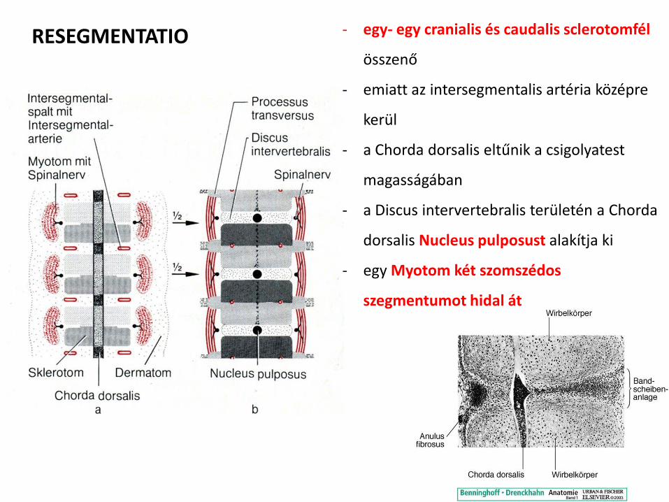
1
2
3
1
2
3
FUSION OF TWO SCLEROTOMES TO BUILD A VERTEBRA
11
STAGES OF FURTHER VERTEBRAL DEVELOPMENT A week 5 B week 6 (neural arch is the primordium of the vertebral arch) C week 7 D at birth EF puberty
Mesenchymal vertebra Chondrification of vertebra Ossification of vertebra begins
12
INDUKCIOacute 1) A chorda dorsalis indukaacutelja a sclerotomsejtek koumlzeacutepvonali vaacutendorlaacutesaacutet eacutes fuacutezioacutejaacutet Corpus vertebrae 2) A velőcső indukaacutelja a paacuteros neuraacutelis iacutevek noumlvekedeacuteseacutet eacutes dorsalisan a koumlzeacutepvonalbeli fuacutezioacutejaacutet Arcus vertebrae
A bordaacutek fejlődeacutese
CARTILAGE MODEL OF DEVELOPING BONE
DIAPHYSIAL (PRIMARY) OSSIFICATION DURING
FETAL LIFE
EPIPHYSIAL (SECONDARY) OSSIFICATION AFTER
BIRTH 15
A csigolyaacutek fejlődeacutesi rendellenesseacutegei
bull Hemivertebra (eacutek- vagy feacutelcsigolya)
csigolyatest inkomplett ossificatioja kongenitaacutelis ScoliosisKyphosisLordosis (ritkaacuten)
bull bdquoButterfly vertebrardquo
bull Blokkcsigolya
(Sacrum normaacutelisan)
bull Tranziens (aacutetmeneti) csigolya ndash lumbalizaacutecioacute
ndash szakralizaacutecioacute
bull Spina bifida
Costa cervicalis Costa lumbalis
ABNORMAL DEVELOPMENT OF VERTEBRAE AND RIBS 19
Spina bifida
a sclerotom sejtjeinek koumlzeacutepvonalhoz vaacutendorlaacutesaacutet a chorda dorsalis indukaacutelja Corpus vertebrae a csigolyaiacutevek noumlvekedeacuteseacutehez eacutes zaacuteroacutedaacutesaacutehoz normaacutelisan fejlődő velőcső szuumlkseacuteges ha ez nincs akkor spina bifida alakul ki
SPINA BIFIDA IS A DEFECT OF THE VERTEBRAE COMBINED WITH DEFECT OF THE SPINAL CORD
21
Bifid spine the neural arches of several vertebrae are missing the spinal cord is
exposed to skin
22
Case courtesy of Dr Ahmed Almuslim Radiopaediaorg
Hemivertebra
Case courtesy of Radiopaediaorg
Butterfly vertebra
Block vertebrae
Case courtesy of Dr Roberto Schubert Radiopaediaorg
Case courtesy of Dr M Osama Yonso Radiopaediaorg
Cervical ribs
Case courtesy of Dr Nafisa Shakir Batta Radiopaediaorg
Occipital vertebrae (Prebasioccipital arch)
Cleidocranial dysostosis Dr Matt A Morgan and Dr Basab Bhattacharya et al
Cleidocranial dysostosis (CCD) is a skeletal dysplasia with predominantly membranous bone involvement It carries an autosomal dominant inheritance 4
Pathology
It is characterised by incomplete ossification of skeletal structures inclusive of the clavicle as well as defective development of the pubic bones vertebral column and long bones 5
Clinical features
bull large head with large fontenelles with delayed closure
bull broad mandible
bull supernumerary teeth
bull high arched palate
bull neonatal distress due to thorax being narrowed and bell shaped
bull excessively mobile shoulders
bull may have genu valgum and short fingers
Radiographic features
Plain film
Skull
bull wormian bones
bull widened sutures andor fontanelles
bull premature fusion of the coronal suture (brachycephaly)
bull frontal andor parietal bossing
bull basilar invagination (atlantoaxial impaction)
bull persistent metopic suture
Chest
bull hypoplasiaaplasia of lateral clavicle (absent clavicles) may have two separate hypoplastic segments 5
bull supernumerary ribs
bull hemivertebrae with spondylosis
bull small and high scapulae
Pelvis
bull hypoplasia of iliac bones
bull absentdelayed ossification of the pubic bone (pseudo-widening of the symphysis pubis)
Limbs
bull short absent fibula
bull short absent radius
bull coxa vara
bull hypoplastic terminal phalanges
Radiopaediaorg Case courtesy of Dr Sharifah Intan Radiopaediaorg
Klippel-Feil Syndrome
Case courtesy of Dr Mohammed ElBeialy Radiopaediaorg
Fusion of C2 C3 and C4 vertebral bodies suggestive of type II Klippel-Feil syndrome (full spinal imaging is required to confirm the type) In addition to AP diameter at the level of the affected discovertebral joint is smaller than the diameter at the superior and inferior limits of the vertebrae adjacent to the uninvolved discs which is called the wasp-waist sign
bdquoKlippelldquo von Noble Frawley - TPNoble JMFrawley The Klippel-Feil syndrome Ann Surg 82(5) 728ndash734 (1925)[1] Lizenziert unter Gemeinfrei uumlber Wikimedia Commons - httpcommonswikimediaorgwikiFileKlippelJPGmediaviewerFileKlippelJPG
Atlas amp Axis
Lig apicis dentis
peacuteldaClavicula
Corpus desmalis csontosodaacutes ndash elsőkeacutent kezdődik Extremitasok chondralis csontosodaacutes ndash 20-24 eacuteves korban veacutegződik
peacutelda Sternum
Greyrsquos Anatomy
9 eacutev
2 hoacutenap
Sternum punctio bull voumlroumls csontvelő leveacutetel
bull Foramen sternale 4-5
bull bordakoumlz magassaacutegaacuteban
Case courtesy of Dr Chris ODonnell Radiopaediaorg
Koponya fejlődeacutese
bull Neurocranium
ndash Desmocranium
ndash Chondrocranium
bull Viscerocranium
A mesenchyma eredete
Crista neuralis
Paraxialis mesoderma (SomitaacutekSomitomerek)
Lateralis mesoderma
Desmocranium (haacutertyaacutes neurocranium)
Mesenchyma
- Crista neuralis
- Paraxialis mesoderma (SomitaacutekSomitomerek)
a mesenchyma beboriacutetja az agyat
desmalis csontosodaacutes
- csontspiculumok
terjednek a primer csontosodaacutesi magokboacutel
- noumlvekedeacutes kiacutevuumll appoziacutecioacute + beluumll reszorpcioacute
(pre- eacutes postnatalisan)
3 hoacutenap
Uacutejszuumlloumltt koponya
bull Suturae koumltőszoumlvet (Syndesmosis)
ndash ahogy a csontok uacutegy ezek is keacutefeacutele eredetűek
ndash ganglionleacutec sagittalis oumlsszekoumltteteacutesek hellipfelnőttkorig
ndash paraxialis mesoderma cronoalis struktuacuteraacutek
bull Fonticuli kiszeacutelesedett hellip3 eacutev
MOLDING a koponya deformaacuteloacutedik ideiglenesen szuumlleteacuteskor
Chondrocranium (porcos neurocranium)
bull kuumlloumlnaacutelloacute porcok chondralis csontosodaacutes + oumlsszenőnek egyseacuteges koponyaalap
(pl Synchondrosis sphenoocipitalis)
bull prechordalis chondrocranium ndash ganglionleacutec eredet
bull chordalis chondrocranium ndash occipitalis sclerotomokboacutel paraxialis
mesoderma
40
az arc fejlődeacutese keacutesőbbi előadaacutes teacutemaacuteja
Viscerocranium főkeacutent 1 eacutes 2 garatiacutevből fejlődik
Craniosynostosis (12500)
bull brachycephaly bicoronal andor bilamboid sutures bull scaphocephalydolichocephaly sagittal suture bull plagiocephaly unilateral coronal and lambdoid sutures bull frontal plagiocephaly unilateral coronal suture bull occipital plagiocephaly unilateral lambdoid suture bull trigonocephaly metopic suture bull pachycephaly lamdoid suture bull oxycephalyturricephaly sagittal coronal and lambdoid
sutures (Tower like skull) bull cloverleaf skullKleeblattschadel intrauterine sagittal
coronal lambdoid sutures (most severe) bull harlequin eye ipsilateral coronal suture
httpwwwcraniokidscozaimagesfaqlrgimgjpg
brachycephaly
scaphocephaly
plagiocephaly
trigonocephaly
Scaphocephalia Sutura sagittalis korai synostosisa hosszuacute magas koponya
BrachycephaliaTurricephalia Sutura coronalis eacutesvagy lambdoidea korai synostosisa
roumlvid magas koponya
Khagendra Thapa Magar Nepal 67 cm
Robert Pershing Wadlow
273 cm Pigmeusok IGF-I hiaacuteny (Ituri nők 135 cm feacuterfiak 143 cm)
Akromegaacutelia
csak az acrak nőnekhellip
IZOMRENDSZER
bull vaacutezizomzat paraxialis mesoderma (somitaacutek-somitomerek)
bull simaizom
ndash splanchnicus mesoderma (visceralis lateralis mesoderma) belek eacutes szaacutermazeacutekai falaacuteban
ndash ectoderma() pupilla (neuroectoderma) emlő- eacutes verejteacutekmirigyek (haacutem szrmazeacutekai) izmai
bull sziacutevizom sziacutevcső koumlruumlli splanchnicus mesoderma
Haraacutentcsiacutekolt vaacutezizomzat
bull somitaacutek SCLEROTOM kivaacutendorol + DERMOMYOTOM marad
bull az ectoderma feleacute eső DERMOTOM epitheloid bull az entoderma feleacute eső MYOTOM sejtjei a
dermomyotom keacutet eacuteleacuten (bdquoajakrdquo) talaacutelhatoacutek amik egymaacutes feleacute vaacutendorolnak eacutes bezaacuteroacutednak 1) ventrolateralis ajak (VLL) keacutesőbbi HYPOMER hypaxialis izomzat (testfal veacutegtagok) 1) dorsomedialis ajak (DML) keacutesőbbi EPIMER epaxialis izomzat (haacutetizomzat)
bull occipitalis somitomerek laza szerkezetű keacutepletek nem
kuumlloumlnuumll el bennuumlk sclerotom eacutes demomyotom de az izmok fejlődeacutese hasonloacutean zajlik
bull inak myotommal hataacuteros sclerotomboacutel szaacutermaznak
Haraacutentcsiacutekolt izomrostok
bull myoblastok fuacutezioacuteja sokmagvuacute hosszuacute izomrostokkaacute
bull myofilamentumok keacutepzeacutese
bull a 3 hoacutenapra megjelenik a haraacutentcsiacutekolat
Myoblast fusion in cultured muscle cells
Susan M Abmayr and Grace K Pavlath Development
2012139641-656
A model for the role of myostatin during muscle growth and differentiation
Brett Langley et al J Biol Chem 200227749831-49840
copy2002 by American Society for Biochemistry and Molecular Biology
Izmok fejlődeacutese bull a myoblastok vaacutendorlaacutesaacutet a koumlrnyező
(mesenchyma eredetű) koumltőszoumlvet iraacutenyiacutetja izmok mintaacutezata
bull EPIMERltHYPOMER
bull eredetileg szegmentaacutelt + szegmentaacutelt beidegzeacutes Rr posterioresanteriores
bull Epimer M erector spinae
bull Hypomer testfal + veacutegtagok izmai
FIGYELEM 1) AZ ADOTT SOMITAacuteBOacuteL SZAacuteRMAZOacute IZMOK ELVAacuteNDOROLHATNAK
DE MEGTARTJAacuteK AZ EREDETI SZEGMENTAacuteLIS (SPINALIS) BEIDEGZEacuteSUumlKET (Pl feluumlletes haacutetizmok)
2) EGY SZEGMENTAacuteLIS SOMITAacuteBOacuteL TOumlBB IZOM IS SZAacuteRMAZHAT (Pl M intercostalis externus et internus)
3) EGY IZOM TOumlBB SZEGMENTAacuteLIS SZOMITAacuteBOacuteL SZAacuteRMAZOacute REacuteSZEK OumlSSZEOLVADAacuteSAacuteVAL IS LEacuteTREJOumlHET EKKOR MINDIG AZ EREDETI SZEGMENSEKBŐL KAPJAacuteK A REacuteSZEI A BEIDEGZEacuteST (Pl veacutegtagok izmai ndash az idegeik szegmentaacutelis idegrostok kombinaacutecioacutejaacuteval joumlttek leacutetre)
Testfal izmai bull cervicalis szegmentumok Mm scaleni M geniohyoideus
et Mm prevertebrales
bull thoracalis szegmentumok 3 reacuteteg ndash Mm intercostalis externus + internus + intimusM transversus
thoracis
ndash Mm obliquus externus + internus + M transversus abdominis
mellkason szegmentaacuteltak de a hasfalon oumlsszeolvadnak
bull lumbalis szegmentumok M quadratus lumborum
bull sacraliscoccygealis szegmentumok Diaphragma pelvis M sphincter ani externus
+ ventralis longitudinalis oszlop
Mm infrahyoidei ndash (M sternalis) ndash M rectus abdominis
bdquoszilvahas-szindroacutema kivaacuteltoacute ok lehet a kivaacutelasztoacute rendszer zavara ascites hasfal atroacutefia
Veacutegtagok izomzata ld keacutesőbb
Fej izomzata
bull vaacutezizmok paraxialis mesoderma (somitomer + somita) eredetűek
bull kiveacutetel
ndash iris (szemserleg neuroectoderma)
ndash garatiacutevek izmai
Sziacutevizom bull sziacutevcső koumlruumlli splanchnicus mesodermaacuteboacutel
bull myoblastok kapcsoloacuteszerkezetekkel koumltődnek ( keacutesőbbi Discus intercalaris)
bull myofibrillumok a vaacutezizmokra jellemző elrendeződeacutesben (haraacutentcsiacutekolat) de nem fuacutezionaacutelnak a sejtek
(kiveacuteve ingeruumlletvezető rendszer rendezetlen)
httpswwwyoutubecomwatchv=Y5uKMM8Od9g
Simaizom
bull erek lateralis mesoderma + crista neuralis
bull beacutel eacutes fuumlggeleacutekek splanchnicus mesoderma
bull pupilla izmai neuroectoderma
bull emlő- eacutes verejteacutekmirigy ectoderma
bull SRF myocardinhellip indiacutetjaacutek be az utat
Felhasznaacutelt irodalom
bull T W Sadler Orvosi embrioloacutegia
Medicina Koumlnyvkiadoacute Zrt 10 kiadaacutes
bull Benninghoff-Drenckhahn Anatomie
Urban amp Fischer Verlag 17 kiadaacutes
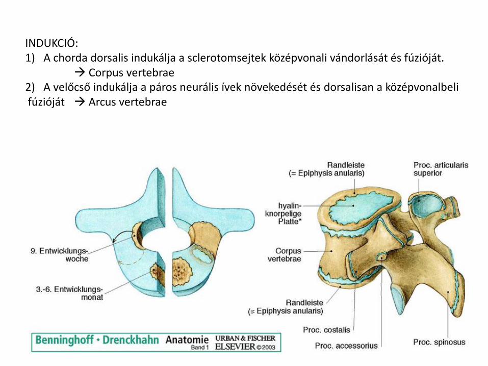
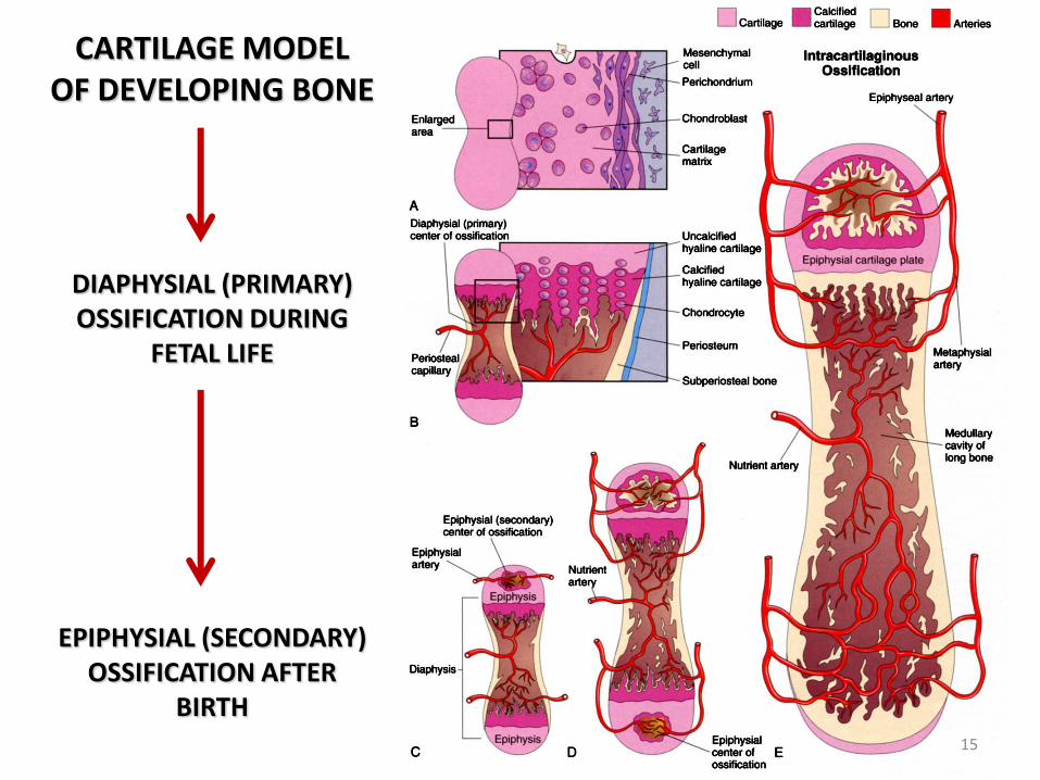
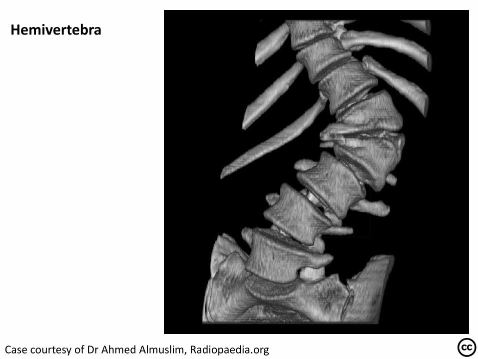
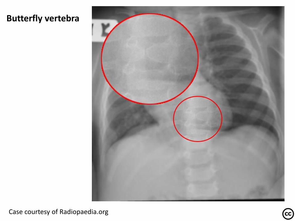
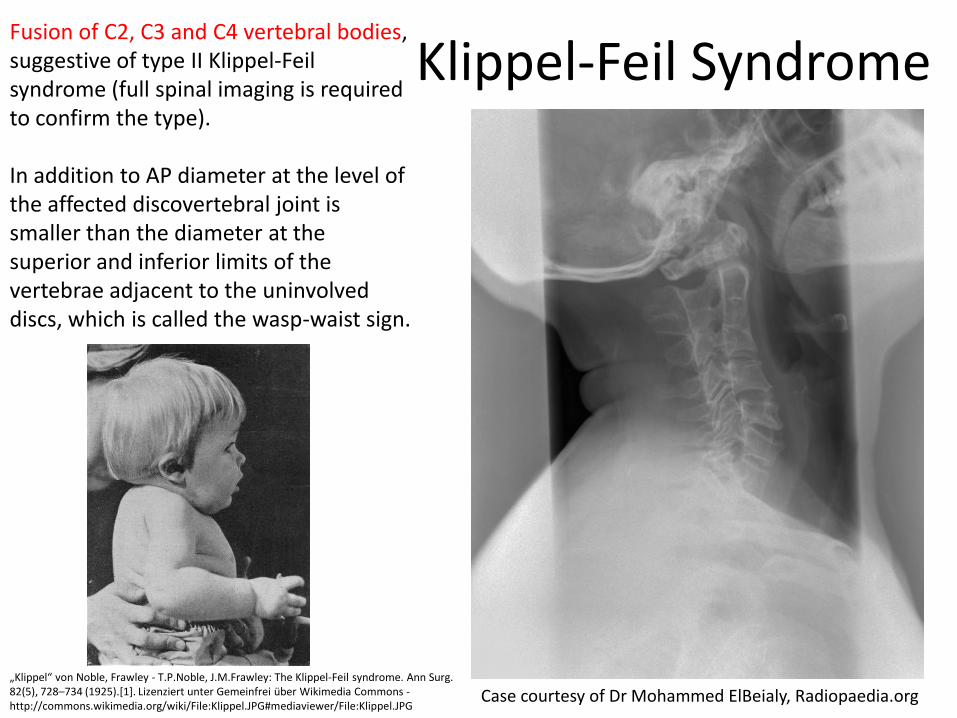
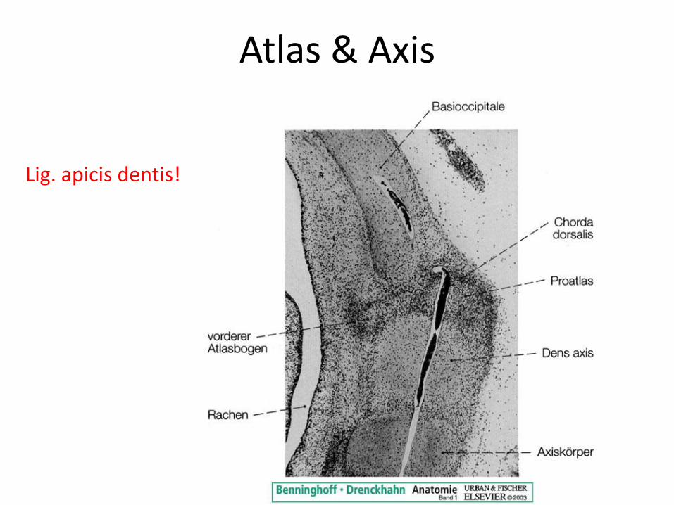
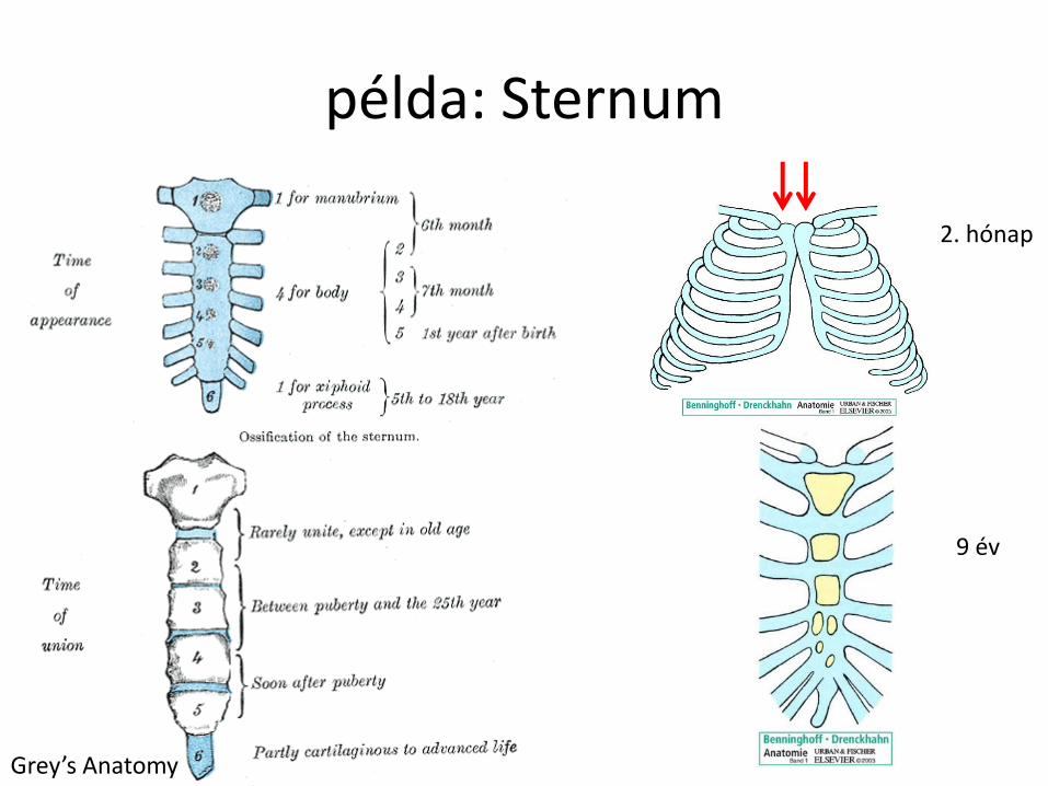
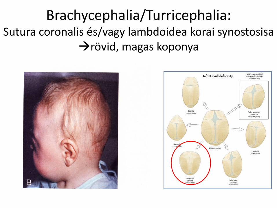
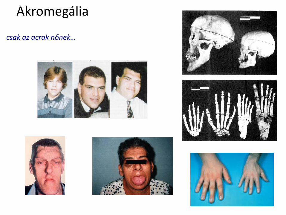

Felhasznaacutelt irodalom
bull T W Sadler Orvosi embrioloacutegia
Medicina Koumlnyvkiadoacute Zrt 10 kiadaacutes
bull Benninghoff-Drenckhahn Anatomie
Urban amp Fischer Verlag 17 kiadaacutes

![[ ] Villamos energia [ ] Földgáz és kóolaj feltárása és kitermelése [ ] Szén és más szilárd tüzelóanyag feltárása és kitermelése](https://img.pdfslide.net/doc/110x75/5e3d7dc2ad699a30c97f39ad/-villamos-energia-fldgz-s-kolaj-feltrsa-s-kitermelse.jpg)