Embed Size (px)
Citation preview
1
3
1516
5678 9
10111213
1718192021222324252627
2
4
14
2829
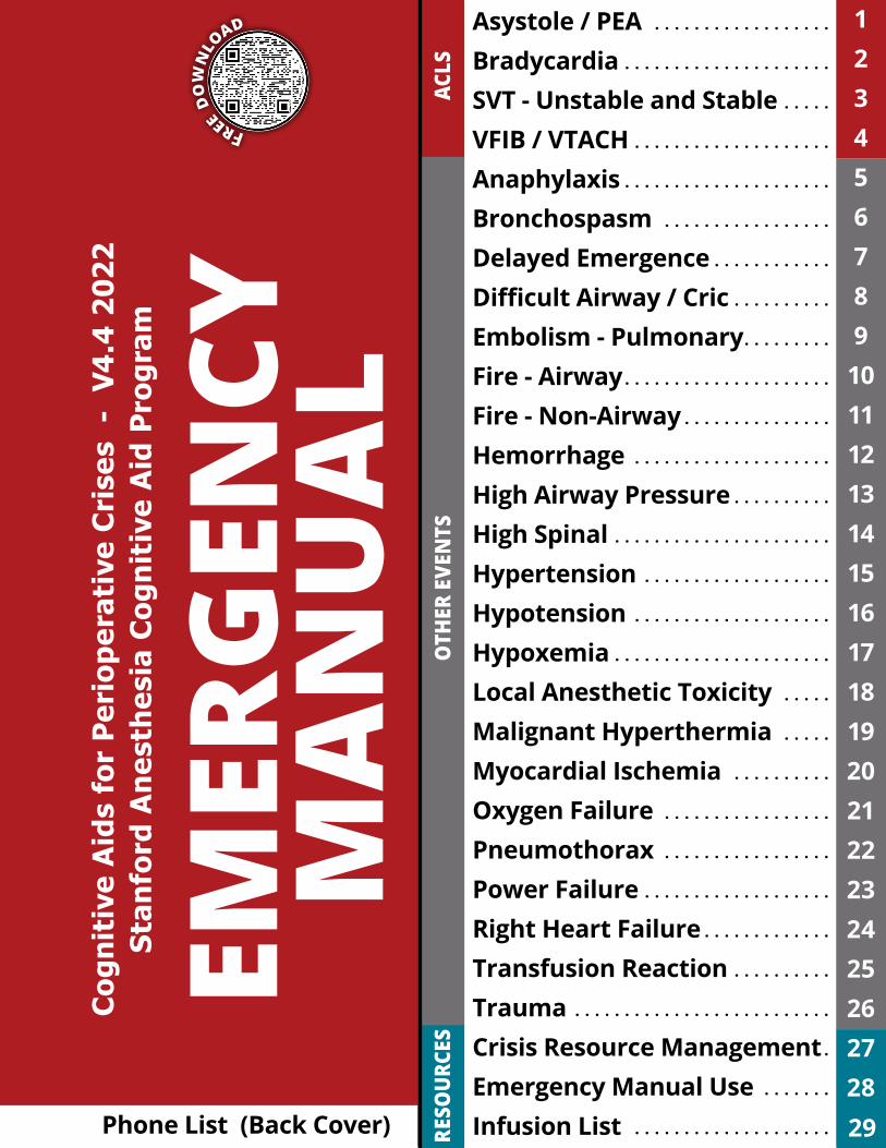
Asystole / PEA
SVT - Unstable and Stable
BronchospasmDelayed EmergenceDifficult Airway / CricEmbolism - PulmonaryFire - AirwayFire - Non-AirwayHemorrhage
High Spinal
Local Anesthetic ToxicityMalignant HyperthermiaMyocardial IschemiaOxygen FailurePneumothoraxPower FailureRight Heart FailureTransfusion ReactionTraumaCrisis Resource ManagementEmergency Manual UseInfusion List
Bradycardia
VFIB / VTACHAnaphylaxis
HypotensionHypoxemia
Hypertension
High Airway Pressure
EMER
GEN
CYM
AN
UA
L
Cog
nit
ive
Aid
s fo
r P
erio
per
ativ
e C
rise
s -
V4
.4 2
022
Sta
nfo
rd A
nes
thes
ia C
ogn
itiv
e A
id P
rogr
am
ACLS
RESO
URCE
SO
THER
EVE
NTS
Phone List (Back Cover)
. . . . . . . . . . . . . . . . . .
. . . . .
. . . . . . . . . . . . . . . . .
. . . . . . . . . . . .
. . . . . . . . . .
. . . . . . . . .
. . . . . . . . . . . . . . . . . . . . .
. . . . . . . . . . . . . . .
. . . . . . . . . . . . . . . . . . . .
. . . . . . . . . . . . . . . . . . . . . .
. . . . .
. . . . .
. . . . . . . . . .
. . . . . . . . . . . . . . . . .
. . . . . . . . . . . . . . . . .
. . . . . . . . . . . . . . . . . . .
. . . . . . . . . . . . .
. . . . . . . . . .
. . . . . . . . . . . . . . . . . . . . . . . . . .
.
. . . . . . .
. . . . . . . . . . . . . . . . . . . . .
. . . . . . . . . . . . . . . . . . . .
. . . . . . . . . . . . . . . . . . . . .
. . . . . . . . . . . . . . . . . . . .
. . . . . . . . . . . . . . . . . . . . . .
. . . . . . . . . . . . . . . . . . .
. . . . . . . . . .
. . . . . . . . . . . . . . . . . . . .
Fre
e dow
nloa
d
TREA
TMEN
T
Asystole / PEA
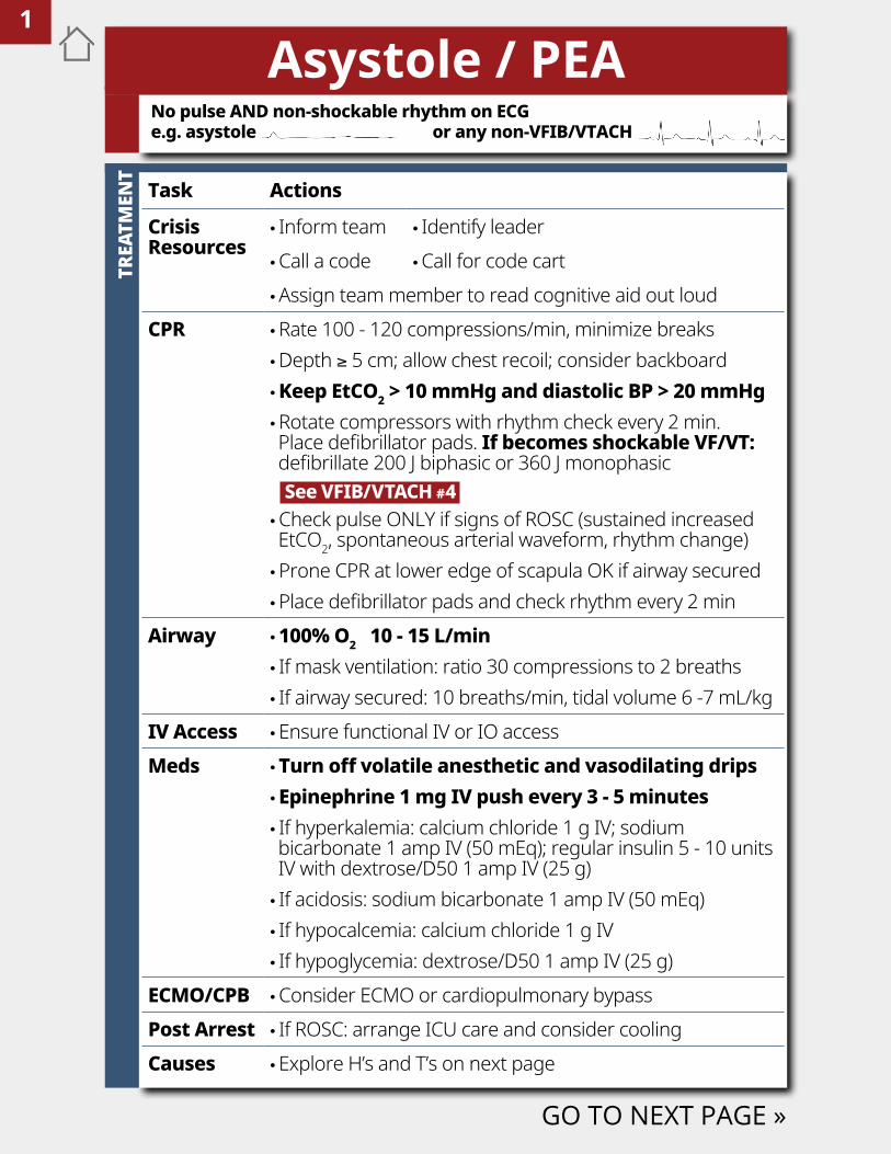
Task Actions
Crisis Resources
• Inform team • Identify leader •Call a code •Call for code cart •Assign team member to read cognitive aid out loud
CPR •Rate 100 - 120 compressions/min, minimize breaks •Depth ≥ 5 cm; allow chest recoil; consider backboard • Keep EtCO2 > 10 mmHg and diastolic BP > 20 mmHg •Rotate compressors with rhythm check every 2 min. Place defibrillator pads. If becomes shockable VF/VT: defibrillate 200 J biphasic or 360 J monophasic See VFIB/VTACH #4 •Check pulse ONLY if signs of ROSC (sustained increased EtCO2, spontaneous arterial waveform, rhythm change) •Prone CPR at lower edge of scapula OK if airway secured •Place defibrillator pads and check rhythm every 2 min
Airway • 100% O2 10 - 15 L/min • If mask ventilation: ratio 30 compressions to 2 breaths • If airway secured: 10 breaths/min, tidal volume 6 -7 mL/kg
IV Access •Ensure functional IV or IO accessMeds •Turn off volatile anesthetic and vasodilating drips
•Epinephrine 1 mg IV push every 3 - 5 minutes • If hyperkalemia: calcium chloride 1 g IV; sodium bicarbonate 1 amp IV (50 mEq); regular insulin 5 - 10 units IV with dextrose/D50 1 amp IV (25 g) • If acidosis: sodium bicarbonate 1 amp IV (50 mEq) • If hypocalcemia: calcium chloride 1 g IV • If hypoglycemia: dextrose/D50 1 amp IV (25 g)
ECMO/CPB •Consider ECMO or cardiopulmonary bypassPost Arrest • If ROSC: arrange ICU care and consider coolingCauses •Explore H’s and T’s on next page
No pulse AND non-shockable rhythm on ECG e.g. asystole or any non-VFIB/VTACH
GO TO NEXT PAGE »
1
3
1516
5678 9
10111213
1718192021222324252627
2
4
14
DI
FFER
ENTI
AL D
IAGN
OSI
S
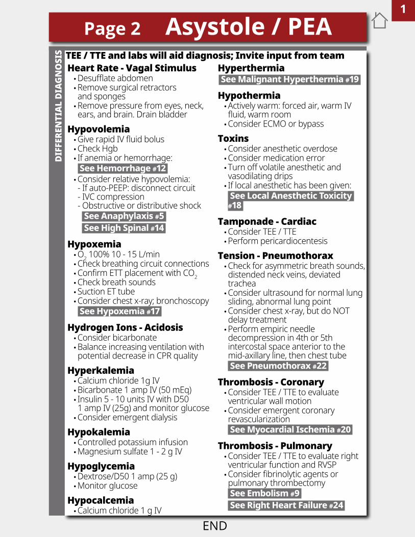
END
Page 2 Asystole / PEA/TEE aTd Labs will aid diagnosisHeart Rate - Vagal Stimulus •Desufflate abdomen •Remove surgical retractors and sponges •Remove pressure from eyes, neck, ears, and brain. Drain bladder
Hypovolemia •Give rapid IV fluid bolus •Check Hgb • If anemia or hemorrhage: See Hemorrhage #12 •Consider relative hypovolemia: - If auto-PEEP: disconnect circuit - IVC compression - Obstructive or distributive shock See Anaphylaxis #5 See High Spinal #14
Hypoxemia •O2 100% 10 - 15 L/min •Check breathing circuit connections •Confirm ETT placement with CO2 •Check breath sounds • Suction ET tube •Consider chest x-ray; bronchoscopy See Hypoxemia #17
Hydrogen Ions - Acidosis •Consider bicarbonate •Balance increasing ventilation with potential decrease in CPR quality
Hyperkalemia •Calcium chloride 1g IV •Bicarbonate 1 amp IV (50 mEq) • Insulin 5 - 10 units IV with D50 1 amp IV (25g) and monitor glucose •Consider emergent dialysis
Hypokalemia •Controlled potassium infusion •Magnesium sulfate 1 - 2 g IV
Hypoglycemia •Dextrose/D50 1 amp (25 g) •Monitor glucose
Hypocalcemia •Calcium chloride 1 g IV
Hyperthermia See Malignant Hyperthermia #19
Hypothermia •Actively warm: forced air, warm IV fluid, warm room •Consider ECMO or bypass
Toxins •Consider anesthetic overdose •Consider medication error • Turn off volatile anesthetic and vasodilating drips • If local anesthetic has been given: See Local Anesthetic Toxicity #18
Tamponade - Cardiac •Consider TEE / TTE •Perform pericardiocentesis
Tension - Pneumothorax •Check for asymmetric breath sounds, distended neck veins, deviated trachea •Consider ultrasound for normal lung sliding, abnormal lung point •Consider chest x-ray, but do NOT delay treatment •Perform empiric needle decompression in 4th or 5th intercostal space anterior to the mid-axillary line, then chest tube See Pneumothorax #22
Thrombosis - Coronary •Consider TEE / TTE to evaluate ventricular wall motion •Consider emergent coronary revascularization See Myocardial Ischemia #20
Thrombosis - Pulmonary •Consider TEE / TTE to evaluate right ventricular function and RVSP •Consider fibrinolytic agents or pulmonary thrombectomy See Embolism #9 See Right Heart Failure #24
TEE / TTE and labs will aid diagnosis; Invite input from team
1
3
1516
5678 9
10111213
1718192021222324252627
2
4
14
2829

This space is intentionally blank
3
1516
5678 9
10111213
171819202122232425
2
4
14
26272829
TREA
TMEN
T
END
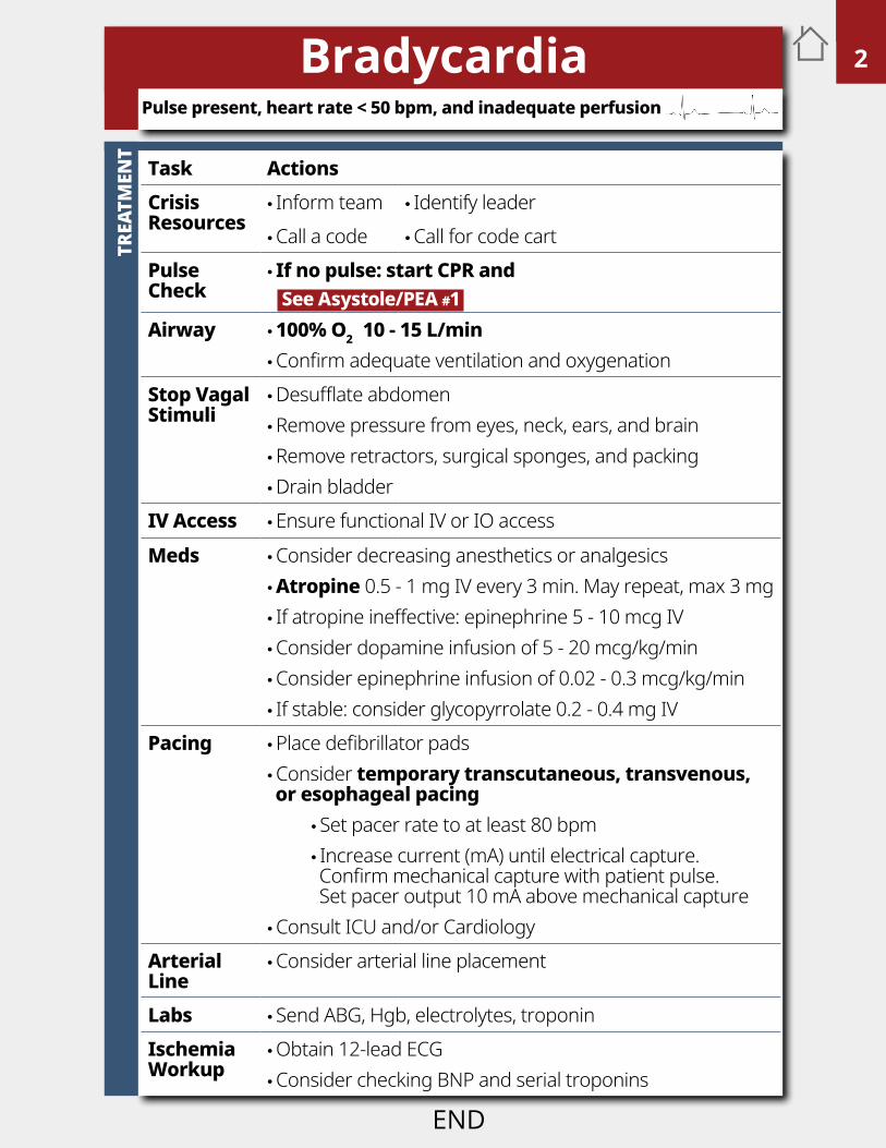
BradycardiaPulse present, heart rate < 50 bpm, and inadequate perfusion
Task ActionsCrisis Resources
• Inform team • Identify leader •Call a code •Call for code cart
Pulse Check
• If no pulse: start CPR and See Asystole/PEA #1
Airway • 100% O2 10 - 15 L/min •Confirm adequate ventilation and oxygenation
Stop Vagal Stimuli
•Desufflate abdomen •Remove pressure from eyes, neck, ears, and brain •Remove retractors, surgical sponges, and packing •Drain bladder
IV Access •Ensure functional IV or IO accessMeds •Consider decreasing anesthetics or analgesics
• Atropine 0.5 - 1 mg IV every 3 min. May repeat, max 3 mg • If atropine ineffective: epinephrine 5 - 10 mcg IV •Consider dopamine infusion of 5 - 20 mcg/kg/min •Consider epinephrine infusion of 0.02 - 0.3 mcg/kg/min • If stable: consider glycopyrrolate 0.2 - 0.4 mg IV
Pacing •Place defibrillator pads •Consider temporary transcutaneous, transvenous, or esophageal pacing
• Set pacer rate to at least 80 bpm • Increase current (mA) until electrical capture. Confirm mechanical capture with patient pulse. Set pacer output 10 mA above mechanical capture
•Consult ICU and/or CardiologyArterial Line
•Consider arterial line placement
Labs • Send ABG, Hgb, electrolytes, troponinIschemia Workup
•Obtain 12-lead ECG •Consider checking BNP and serial troponins
1
3
1516
5678 9
10111213
1718192021222324252627
2
4
14
2829
TREA
TMEN
T
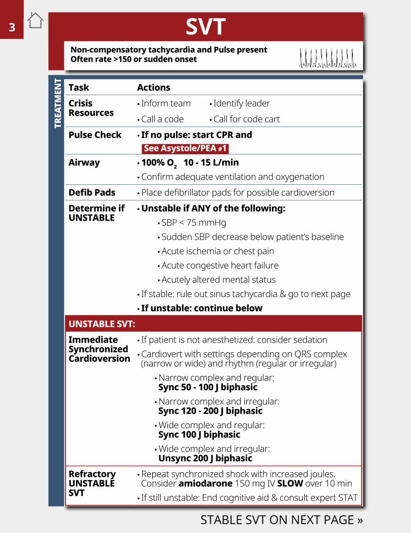
Task ActionsCrisis Resources
• Inform team • Identify leader •Call a code •Call for code cart
Pulse Check • If no pulse: start CPR and See Asystole/PEA #1
Airway • 100% O2 10 - 15 L/min •Confirm adequate ventilation and oxygenation
Defib Pads •Place defibrillator pads for possible cardioversionDetermine if UNSTABLE
• Unstable if ANY of the following: • SBP < 75 mmHg • Sudden SBP decrease below patient’s baseline •Acute ischemia or chest pain •Acute congestive heart failure •Acutely altered mental status
• If stable: rule out sinus tachycardia & go to next page • If unstable: continue below
UNSTABLE SVT:Immediate Synchronized Cardioversion
• If patient is not anesthetized: consider sedation •Cardiovert with settings depending on QRS complex (narrow or wide) and rhythm (regular or irregular)
•Narrow complex and regular: Sync 50 - 100 J biphasic •Narrow complex and irregular: Sync 120 - 200 J biphasic •Wide complex and regular: Sync 100 J biphasic •Wide complex and irregular: Unsync 200 J biphasic
Refractory UNSTABLE SVT
•Repeat synchronized shock with increased joules. Consider amiodarone 150 mg IV SLOW over 10 min • If still unstable: End cognitive aid & consult expert STAT
STABLE SVT ON NEXT PAGE »
Non-compensatory tachycardia and Pulse present Often rate >150 or sudden onset
SVT3
1516
5678 9
10111213
171819202122232425
2
4
14
26272829
TREA
TMEN
T
END
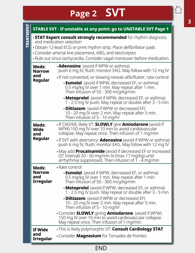
Page 2 SVTSTABLE SVT - If unstable at any point: go to UNSTABLE SVT Page 1
• STAT Expert consult strongly recommended for rhythm diagnosis and medication selection •Obtain 12-lead ECG or print rhythm strip. Place defibrillator pads •Consider arterial line placement, ABG, and electrolytes •Rule out sinus tachycardia. Consider vagal maneuver before medication
Meds: Narrow and Regular
• Adenosine (avoid if WPW or asthma) push 6 mg IV, flush; monitor EKG. May follow with 12 mg IV • If not converted, or slowing reveals afib/flutter, rate control:
• Esmolol (avoid if WPW, decreased EF, or asthma) 0.5 mg/kg IV over 1 min. May repeat after 1 min. Then infusion of 50 - 300 mcg/kg/min • Metoprolol (avoid if WPW, decreased EF, or asthma) 1 - 2.5 mg IV push. May repeat or double after 3 - 5 min • Diltiazem (avoid if WPW or decreased EF) 10 - 20 mg IV over 2 min. May repeat after 5 min. Then infusion of 5 - 10 mg/hr
Meds: Wide andRegular
• If CAD/MI, likely VT: SLOWLY give Amiodarone (avoid if WPW) 150 mg IV over 10 min to avoid cardiovascular collapse. May repeat once. Then infusion of 1 mg/min • If SVT with aberrancy: Adenosine (avoid if WPW or asthma) push 6 mg IV, flush; monitor EKG. May follow with 12 mg IV •May add Procainamide (avoid if decreased EF or increased QT interval) 20 - 50 mg/min IV (max 17 mg/kg) until arrhythmia suppressed. Then infusion of 1 - 4 mg/min
Meds: Narrow and Irregular
•Rate control: • Esmolol (avoid if WPW, decreased EF, or asthma) 0.5 mg/kg IV over 1 min. May repeat after 1 min. Then infusion of 50 - 300 mcg/kg/min • Metoprolol (avoid if WPW, decreased EF, or asthma) 1 - 2.5 mg IV push. May repeat or double after 3 - 5 min • Diltiazem (avoid if WPW or decreased EF) 10 - 20 mg IV over 2 min. May repeat after 5 min. Then infusion of 5 - 10 mg/hr
•Consider SLOWLY giving Amiodarone (avoid if WPW) 150 mg IV over 10 min to avoid cardiovascular collapse. May repeat once. Then infusion of 1 mg/min
If Wide and Irregular
• This is likely polymorphic VT: Consult Cardiology STAT •Consider Magnesium for Torsades de Pointes
1
3
1516
5678 9
10111213
1718192021222324252627
2
4
14
2829
TREA
TMEN
T
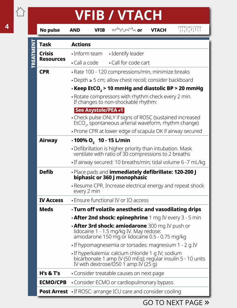
VFIB / VTACH
Task ActionsCrisis Resources
• Inform team • Identify leader •Call a code •Call for code cart
CPR •Rate 100 - 120 compressions/min, minimize breaks •Depth ≥ 5 cm; allow chest recoil; consider backboard • Keep EtCO2 > 10 mmHg and diastolic BP > 20 mmHg •Rotate compressors with rhythm check every 2 min. If changes to non-shockable rhythm: See Asystole/PEA #1 •Check pulse ONLY if signs of ROSC (sustained increased EtCO2, spontaneous arterial waveform, rhythm change) •Prone CPR at lower edge of scapula OK if airway secured
Airway • 100% O2 10 - 15 L/min •Defibrillation is higher priority than intubation. Mask ventilate with ratio of 30 compressions to 2 breaths • If airway secured: 10 breaths/min; tidal volume 6 -7 mL/kg
Defib •Place pads and immediately defibrillate: 120-200 J biphasic or 360 J monophasic •Resume CPR. Increase electrical energy and repeat shock every 2 min
IV Access •Ensure functional IV or IO accessMeds •Turn off volatile anesthetic and vasodilating drips
• After 2nd shock: epinephrine 1 mg IV every 3 - 5 min •After 3rd shock: amiodarone 300 mg IV push or lidocaine 1 - 1.5 mg/kg IV. May redose: amiodarone 150 mg or lidocaine 0.5 - 0.75 mg/kg • If hypomagnesemia or torsades: magnesium 1 - 2 g IV • If hyperkalemia: calcium chloride 1 g IV; sodium bicarbonate 1 amp IV (50 mEq); regular insulin 5 - 10 units IV with dextrose/D50 1 amp IV (25 g)
H’s & T’s •Consider treatable causes on next pageECMO/CPB •Consider ECMO or cardiopulmonary bypassPost Arrest • If ROSC: arrange ICU care and consider cooling
GO TO NEXT PAGE »
No pulse AND VFIB or VTACH
3
1516
5678 9
10111213
171819202122232425
2
4
14
26272829
DI
FFER
ENTI
AL D
IAGN
OSI
S
END
TEE / TTE and labs will aid diagnosis; Invite input from teamHeart Rate - Vagal Stimulus •Desufflate abdomen •Remove surgical retractors and sponges •Remove pressure from eyes, neck, ears, and brain. Drain bladder
Hypovolemia •Give rapid IV fluid bolus •Check Hgb • If anemia or hemorrhage: See Hemorrhage #12 •Consider relative hypovolemia: - If auto-PEEP: disconnect circuit - IVC compression - Obstructive or distributive shock See Anaphylaxis #5 See High Spinal #14
Hypoxemia •O2 100% 10 - 15 L/min •Check breathing circuit connections •Confirm ETT placement with CO2 •Check breath sounds • Suction ET tube •Consider chest x-ray; bronchoscopy See Hypoxemia #17
Hydrogen Ions - Acidosis •Consider bicarbonate •Balance increasing ventilation with potential decrease in CPR quality
Hyperkalemia •Calcium chloride 1g IV •Bicarbonate 1 amp IV (50 mEq) • Insulin 5 - 10 units IV with D50 1 amp IV (25g) and monitor glucose •Consider emergent dialysis
Hypokalemia •Controlled potassium infusion •Magnesium sulfate 1 - 2 g IV
Hypoglycemia •Dextrose/D50 1 amp (25 g) •Monitor glucose
Hypocalcemia •Calcium chloride 1 g IV
Hyperthermia See Malignant Hyperthermia #19
Hypothermia •Actively warm: forced air, warm IV fluid, warm room •Consider ECMO or bypass
Toxins •Consider anesthetic overdose •Consider medication error • Turn off volatile anesthetic and vasodilating drips • If local anesthetic has been given: See Local Anesthetic Toxicity #18
Tamponade - Cardiac •Consider TEE / TTE •Perform pericardiocentesis
Tension - Pneumothorax •Check for asymmetric breath sounds, distended neck veins, deviated trachea •Consider ultrasound for normal lung sliding, abnormal lung point •Consider chest x-ray, but do NOT delay treatment •Perform empiric needle decompression in 4th or 5th intercostal space anterior to the mid-axillary line, then chest tube See Pneumothorax #22
Thrombosis - Coronary •Consider TEE / TTE to evaluate ventricular wall motion •Consider emergent coronary revascularization See Myocardial Ischemia #20
Thrombosis - Pulmonary •Consider TEE / TTE to evaluate right ventricular function and RVSP •Consider fibrinolytic agents or pulmonary thrombectomy See Embolism #9 See Right Heart Failure #24
Page 2 VFIB / VTACH1
3
1516
5678 9
10111213
1718192021222324252627
2
4
14
2829
TREA
TMEN
T
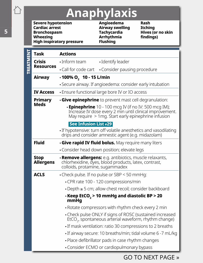
Anaphylaxis
Task ActionsCrisis Resources
• Inform team • Identify leader •Call for code cart •Consider pausing procedure
Airway • 100% O2 10 - 15 L/min • Secure airway. If angioedema: consider early intubation
IV Access •Ensure functional large bore IV or IO accessPrimaryMeds
•Give epinephrine to prevent mast cell degranulation: • Epinephrine 10 - 100 mcg IV (if no IV: 500 mcg IM); Increase IV dose every 2 min until clinical improvement. May require > 1mg. Start early epinephrine infusion • See Infusion List #29
• If hypotensive: turn off volatile anesthetics and vasodilating drips and consider amnestic agent (e.g. midazolam)
Fluid •Give rapid IV fluid bolus. May require many liters •Consider head down position; elevate legs
Stop Allergens
•Remove allergens: e.g. antibiotics, muscle relaxants, chlorhexidine, dyes, blood products, latex, contrast, colloids, protamine, sugammadex
ACLS •Check pulse. If no pulse or SBP < 50 mmHg: •CPR rate 100 - 120 compressions/min •Depth ≥ 5 cm; allow chest recoil; consider backboard • Keep EtCO2 > 10 mmHg and diastolic BP > 20 mmHg •Rotate compressors with rhythm check every 2 min •Check pulse ONLY if signs of ROSC (sustained increased EtCO2, spontaneous arterial waveform, rhythm change) • If mask ventilation: ratio 30 compressions to 2 breaths • If airway secure: 10 breaths/min; tidal volume 6 -7 mL/kg •Place defibrillator pads in case rhythm changes •Consider ECMO or cardiopulmonary bypass
GO TO NEXT PAGE »
Severe hypotensionCardiac arrestBronchospasmWheezingHigh inspiratory pressure
AngioedemaAirway swellingTachycardiaArrhythmiaFlushing
RashItchingHives (or no skin findings)
3
1516
5678 9
10111213
171819202122232425
2
4
14
26272829
END
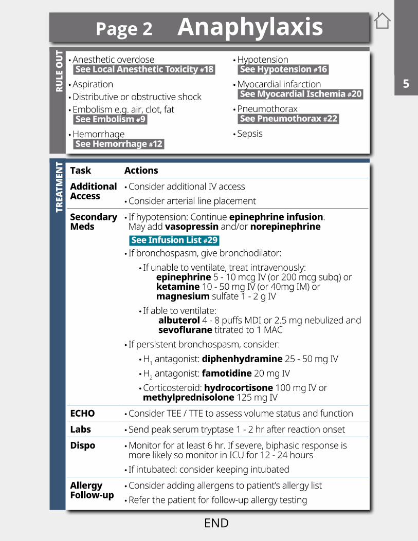
Task ActionsAdditional Access
•Consider additional IV access •Consider arterial line placement
SecondaryMeds
• If hypotension: Continue epinephrine infusion. May add vasopressin and/or norepinephrine See Infusion List #29 • If bronchospasm, give bronchodilator:
• If unable to ventilate, treat intravenously: epinephrine 5 - 10 mcg IV (or 200 mcg subq) or ketamine 10 - 50 mg IV (or 40mg IM) or magnesium sulfate 1 - 2 g IV • If able to ventilate: albuterol 4 - 8 puffs MDI or 2.5 mg nebulized and sevoflurane titrated to 1 MAC
• If persistent bronchospasm, consider: •H1 antagonist: diphenhydramine 25 - 50 mg IV •H2 antagonist: famotidine 20 mg IV •Corticosteroid: hydrocortisone 100 mg IV or methylprednisolone 125 mg IV
ECHO •Consider TEE / TTE to assess volume status and functionLabs • Send peak serum tryptase 1 - 2 hr after reaction onsetDispo •Monitor for at least 6 hr. If severe, biphasic response is
more likely so monitor in ICU for 12 - 24 hours • If intubated: consider keeping intubated
Allergy Follow-up
•Consider adding allergens to patient’s allergy list •Refer the patient for follow-up allergy testing
TREA
TMEN
T
Page 2 AnaphylaxisRU
LE O
UT
•Anesthetic overdose See Local Anesthetic Toxicity #18 •Aspiration •Distributive or obstructive shock •Embolism e.g. air, clot, fat See Embolism #9 •Hemorrhage See Hemorrhage #12
•Hypotension See Hypotension #16 •Myocardial infarction See Myocardial Ischemia #20 •Pneumothorax See Pneumothorax #22 • Sepsis
1
3
1516
5678 9
10111213
1718192021222324252627
2
4
14
2829

This space is intentionally blank
3
1516
5678 9
10111213
171819202122232425
2
4
14
26272829
TREA
TMEN
TSIGNS
END
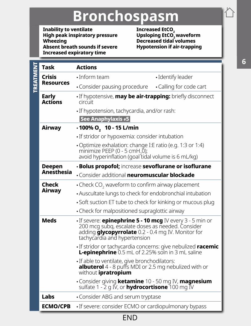
BronchospasmInability to ventilateHigh peak inspiratory pressureWheezing Absent breath sounds if severeIncreased expiratory time
Increased EtCO2Upsloping EtCO2 waveformDecreased tidal volumesHypotension if air-trapping
Task ActionsCrisis Resources
• Inform team • Identify leader •Consider pausing procedure •Calling for code cart
Early Actions
• If hypotensive, may be air-trapping: briefly disconnect circuit • If hypotension, tachycardia, and/or rash: See Anaphylaxis #5
Airway • 100% O2 10 - 15 L/min • If stridor or hypoxemia: consider intubation •Optimize exhalation: change I:E ratio (e.g. 1:3 or 1:4) minimize PEEP (0 - 5 cmH20); avoid hyperinflation (goal tidal volume is 6 mL/kg)
Deepen Anesthesia
• Bolus propofol; increase sevoflurane or isoflurane •Consider additional neuromuscular blockade
Check Airway
•Check CO2 waveform to confirm airway placement •Auscultate lungs to check for endobronchial intubation • Soft suction ET tube to check for kinking or mucous plug •Check for malpositioned supraglottic airway
Meds • If severe: epinephrine 5 - 10 mcg IV every 3 - 5 min or 200 mcg subq, escalate doses as needed. Consider adding glycopyrrolate 0.2 - 0.4 mg IV. Monitor for tachycardia and hypertension • If stridor or tachycardia concerns: give nebulized racemic L-epinephrine 0.5 mL of 2.25% soln in 3 mL saline • If able to ventilate, give bronchodilators: albuterol 4 - 8 puffs MDI or 2.5 mg nebulized with or without ipratropium •Consider giving ketamine 10 - 50 mg IV, magnesium sulfate 1 - 2 g IV, or hydrocortisone 100 mg IV
Labs •Consider ABG and serum tryptaseECMO/CPB • If severe: consider ECMO or cardiopulmonary bypass
1
3
1516
5678 9
10111213
1718192021222324252627
2
4
14
2829

This space is intentionally blank
3
1516
5678 9
10111213
171819202122232425
2
4
14
26272829
TREA
TMEN
TSIGNS
END
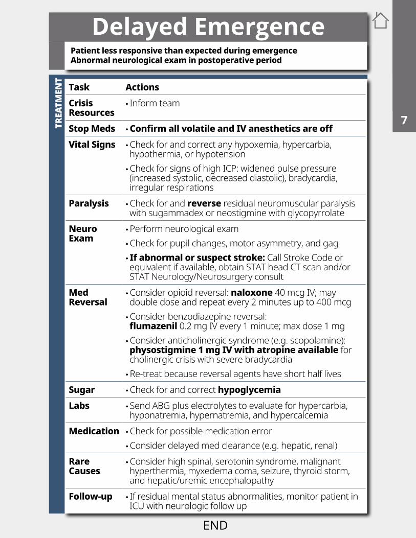
Delayed EmergencePatient less responsive than expected during emergenceAbnormal neurological exam in postoperative period
Task ActionsCrisis Resources
• Inform team
Stop Meds •Confirm all volatile and IV anesthetics are offVital Signs •Check for and correct any hypoxemia, hypercarbia,
hypothermia, or hypotension •Check for signs of high ICP: widened pulse pressure (increased systolic, decreased diastolic), bradycardia, irregular respirations
Paralysis •Check for and reverse residual neuromuscular paralysis with sugammadex or neostigmine with glycopyrrolate
Neuro Exam
•Perform neurological exam •Check for pupil changes, motor asymmetry, and gag • If abnormal or suspect stroke: Call Stroke Code or equivalent if available, obtain STAT head CT scan and/or STAT Neurology/Neurosurgery consult
Med Reversal
•Consider opioid reversal: naloxone 40 mcg IV; may double dose and repeat every 2 minutes up to 400 mcg •Consider benzodiazepine reversal: flumazenil 0.2 mg IV every 1 minute; max dose 1 mg •Consider anticholinergic syndrome (e.g. scopolamine): physostigmine 1 mg IV with atropine available for cholinergic crisis with severe bradycardia •Re-treat because reversal agents have short half lives
Sugar •Check for and correct hypoglycemiaLabs • Send ABG plus electrolytes to evaluate for hypercarbia,
hyponatremia, hypernatremia, and hypercalcemiaMedication •Check for possible medication error
•Consider delayed med clearance (e.g. hepatic, renal)Rare Causes
•Consider high spinal, serotonin syndrome, malignant hyperthermia, myxedema coma, seizure, thyroid storm, and hepatic/uremic encephalopathy
Follow-up • If residual mental status abnormalities, monitor patient in ICU with neurologic follow up
1
3
1516
5678 9
10111213
1718192021222324252627
2
4
14
2829
TREA
TMEN
T
Can Oxygenate: Cannot Intubate, Cannot Oxygenate (CICO):
•Monitor CO2 return by capnography and SpO2
• If cannot oxygenate at any time: go to red box •While oxygenating, options include:
•Awaken patient • Finish case with SGA/LMA or mask • Intubate through SGA/LMA •Combined video/fiberoptic •Other advanced airway techniques
• Priority is cutting the neck! •Call for Cric-capable help •Get Cric kit: scalpel (e.g. #10 blade), bougie, and 6.0 ET tube •Additional operator can attempt to oxygenate from above (e.g. mask, SGA/LMA, video laryngoscopy) • Start Cric/eFONA (next page)
Difficult Airway / Cric
Task ActionsCrisis Resources
• Inform team •Call for airway help •Call for anesthesia tech •Call for difficult airway cart
Optimize Conditions
•Ensure paralysis (e.g. rocuronium 1.2 mg/kg) •Ensure anesthetic depth (e.g. re-bolus or infuse propofol) •Optimize positioning (e.g. sniffing position, head of bed elevation to 30°, neck extension, bed height)
Oxygenate •Do not fixate on intubation •Monitor CO2 return by capnography and SpO2
• If SpO2 critically low at any time: go to red box below •Consider oxygenation modalities (max 2 attempts each):
• Mask: use two-handed grip; insert oral/nasal airway • Supraglottic airway SGA/LMA: optimize size and fit (change position of head or device, cuff inflation); consider 2nd generation • Laryngoscopy: video preferred. Consider alternate blade, rigid stylet, bougie, external laryngeal manipulation, release of cricoid pressure
•Choose experienced operator and familiar equipment
CRIC / eFONA ON NEXT PAGE »
Failed laryngoscopy or difficulty oxygenating or ventilating
3
1516
5678 9
10111213
171819202122232425
2
4
14
26272829
END
CRI
C /
EM
ERGE
NCY
FRO
NT
OF
NEC
K AC
CESS
(eFO
NA)
PREV
ENTI
ON
CO2
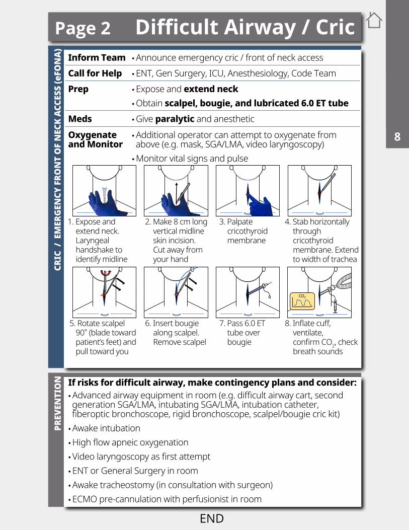
1. Expose and extend neck. Laryngeal handshake to identify midline
2. Make 8 cm long vertical midline skin incision. Cut away from your hand
3. Palpate cricothyroid membrane
4. Stab horizontally through cricothyroid membrane. Extend to width of trachea
5. Rotate scalpel 90° (blade toward patient’s feet) and pull toward you
6. Insert bougie along scalpel. Remove scalpel
7. Pass 6.0 ET tube over bougie
8. Inflate cuff, ventilate, confirm CO2, check breath sounds
If risks for difficult airway, make contingency plans and consider: •Advanced airway equipment in room (e.g. difficult airway cart, second generation SGA/LMA, intubating SGA/LMA, intubation catheter, fiberoptic bronchoscope, rigid bronchoscope, scalpel/bougie cric kit) •Awake intubation •High flow apneic oxygenation •Video laryngoscopy as first attempt •ENT or General Surgery in room •Awake tracheostomy (in consultation with surgeon) •ECMO pre-cannulation with perfusionist in room
Inform Team •Announce emergency cric / front of neck accessCall for Help •ENT, Gen Surgery, ICU, Anesthesiology, Code TeamPrep •Expose and extend neck
•Obtain scalpel, bougie, and lubricated 6.0 ET tubeMeds •Give paralytic and anestheticOxygenate and Monitor
•Additional operator can attempt to oxygenate from above (e.g. mask, SGA/LMA, video laryngoscopy) •Monitor vital signs and pulse
Page 2 Difficult Airway / Cric1
3
1516
5678 9
10111213
1718192021222324252627
2
4
14
2829
TREA
TMEN
TRU
LE O
UT
Embolism - PulmonarySudden decrease in EtCO2, BP, or SpO2Sudden increase in central venous pressureDyspnea, respiratory distress, or cough in awake patientIncreased risk in long bone orthopedic surgery, pregnancy, cancer (especially renal tumor), high BMI, laparoscopic surgery, or surgical site above level of the heart
Task ActionsCrisis Resources
• Inform team • Identify leader •Call for help •Get code cart •Consider terminating procedure
Pulse Check • If no pulse: start CPR, check rhythm, and follow appropriate algorithm See Asystole/PEA #1 VFIB/VTACH #4
Airway • 100% O2 10 - 15 L/minCirculation •Turn off volatile anesthetic and vasodilating drips
•Give IV vasopressor bolus to support circulation •Consider rapid fluid bolus
Evaluate Right Heart
• If unstable or RV function decreased on TEE / TTE, use medication and diuresis to:
•Maintain sinus rhythm •Maintain normal RV volume status •Maintain RV contractility •Decrease RV afterload See Right Heart Failure #24
ECMO/CPB • If severe decompensation: consider ECMO or cardiopulmonary bypass
GO TO NEXT PAGE »
Consider other causes: •Anaphylaxis See Anaphylaxis # 5 •Bone cement implantation syndrome •Bronchospasm See Bronchospasm #6 •Cardiac tamponade •Cardiogenic shock
•Distributive shock •Hypovolemia •Myocardial ischemia See Myocardial Ischemia #20 •Pneumothorax See Pneumothorax #22 •Pulmonary edema
3
1516
5678 9
10111213
171819202122232425
2
4
14
26272829
END
TREA
TMEN
TFurther management depends on embolism type:
Pulmonary Thromboembolism:Risk Factors •Chronic illness, neoplasm, immobility, missed
anticoagulation Treatment •Discuss feasibility and safety of urgent thrombolysis
vs. thrombectomy with surgical team • Thrombolysis: If safe, use recombinant tissue plasminogen activator (rtPA) alteplase 10 mg IV followed by infusion of 90 mg over 2 hours • Thrombectomy: Consider STAT Cardiovascular Surgery consult (open) or STAT Interventional Radiology consult (percutaneous)
• Supportive treatment: airway, breathing, circulationAir or CO2 Embolism:Signs •Air visible on TEE / TTETreatment • Limit entrainment of air: check IV lines for air; flood
surgical field with saline; consider placing surgical site below heart; consider left lateral decubitus position •Attempt removal of air: aspirate air from central line if present • Supportive treatment: airway, breathing, circulation •Consider hyperbaric oxygen treatment
Cement or Fat Embolism:Signs • Petechial rash
•Confusion or irritability if awake
Treatment • Supportive treatment: airway, breathing, circulation
Amniotic Fluid Embolism:Signs •Peripartum patient with maternal or fetal compromise:
altered mental status, hypotension, hypoxemia, seizures, coagulopathy
Treatment • Supportive treatment: airway, breathing, circulation •Monitor fetus and consider urgent Cesarean section •Monitor for and treat seizures and DIC
Page 2 Embolism - Pulmonary1
3
1516
5678 9
10111213
1718192021222324252627
2
4
14
2829
Fire - AirwaySudden pop, spark, flame, smoke, heat, or odor
TREA
TMEN
T
Task ActionsCrisis Resources
• Inform team • Identify leader •Call for help
AnesthesiaProfessionalImmediateResponse
• Disconnect breathing circuit from the anesthesia machine to prevent torch formation •Stop fresh gas flow
SurgeonImmediate Response
• If clamp immediately available: clamp ET tube. If clamp not available: fold (kink) ET tube (prevents torch formation if circuit not yet disconnected) • Immediately remove ET tube and any airway foreign bodies • Pour saline into airway and suction debris
Check Extent of Fire
• If fire spreads beyond airway (e.g. to drapes, patient): See Fire - Non-Airway #11
After Fire Extinguished
• Re-establish oxygenation when fire is extinguished •Minimize FiO2 as much as possible. Consider air ventilation • Consider prompt reintubation with ET tube ≥ 7.0 mm ID prior to swelling •Ensure adequate anesthesia: e.g. propofol infusion •Perform bronchoscopic examination of entire airway to assess injury and remove residual debris • Inspect ET tube pieces to verify none left in airway • Save all materials for later investigation •Consider steroid: e.g. dexamethasone 8 mg IV
Disposition • ICU care for prolonged mechanical ventilation and airway observation
FIRE PREVENTION ON NEXT PAGE »
3
1516
5678 9
10111213
171819202122232425
2
4
14
26272829
END
PREV
ENTI
ON
Page 2 Fire - Airway
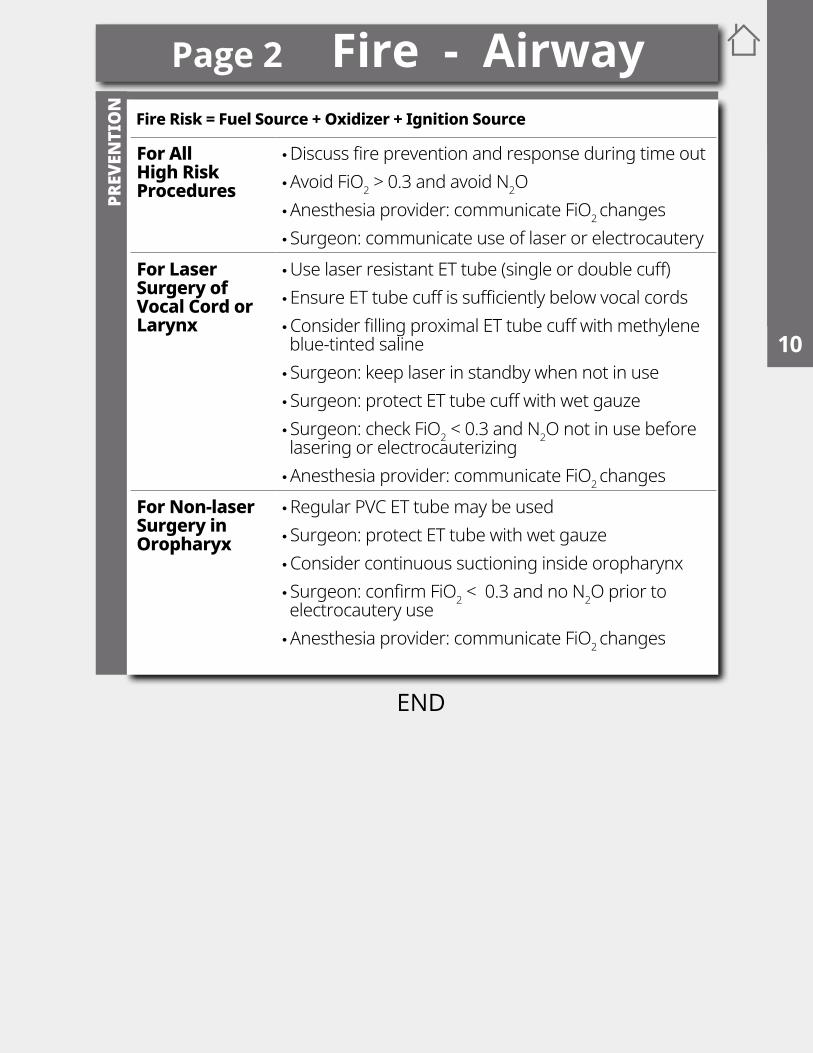
For All High Risk Procedures
•Discuss fire prevention and response during time out •Avoid FiO2 > 0.3 and avoid N2O •Anesthesia provider: communicate FiO2 changes • Surgeon: communicate use of laser or electrocautery
For Laser Surgery of Vocal Cord or Larynx
•Use laser resistant ET tube (single or double cuff) •Ensure ET tube cuff is sufficiently below vocal cords •Consider filling proximal ET tube cuff with methylene blue-tinted saline • Surgeon: keep laser in standby when not in use • Surgeon: protect ET tube cuff with wet gauze • Surgeon: check FiO2 < 0.3 and N2O not in use before lasering or electrocauterizing •Anesthesia provider: communicate FiO2 changes
For Non-laser Surgery in Oropharyx
•Regular PVC ET tube may be used • Surgeon: protect ET tube with wet gauze •Consider continuous suctioning inside oropharynx • Surgeon: confirm FiO2 < 0.3 and no N2O prior to electrocautery use •Anesthesia provider: communicate FiO2 changes
Fire Risk = Fuel Source + Oxidizer + Ignition Source
1
3
1516
5678 9
10111213
1718192021222324252627
2
4
14
2829
TREA
TMEN
T
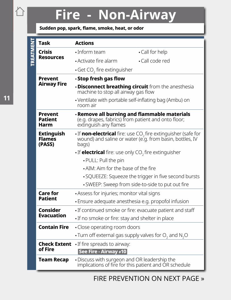
Fire - Non-AirwaySudden pop, spark, flame, smoke, heat, or odor
Task ActionsCrisis Resources
• Inform team •Call for help •Activate fire alarm •Call code red •Get CO2 fire extinguisher
Prevent Airway Fire
•Stop fresh gas flow • Disconnect breathing circuit from the anesthesia machine to stop all airway gas flow •Ventilate with portable self-inflating bag (Ambu) on room air
Prevent Patient Harm
•Remove all burning and flammable materials (e.g. drapes, fabrics) from patient and onto floor; extinguish any flames
Extinguish Flames(PASS)
• If non-electrical fire: use CO2 fire extinguisher (safe for wound) and saline or water (e.g. from basin, bottles, IV bags) • If electrical fire: use only CO2 fire extinguisher
•PULL: Pull the pin •AIM: Aim for the base of the fire • SQUEEZE: Squeeze the trigger in five second bursts • SWEEP: Sweep from side-to-side to put out fire
Care for Patient
•Assess for injuries; monitor vital signs •Ensure adequate anesthesia e.g. propofol infusion
Consider Evacuation
• If continued smoke or fire: evacuate patient and staff • If no smoke or fire: stay and shelter in place
Contain Fire •Close operating room doors • Turn off external gas supply valves for O2 and N2O
Check Extent of Fire
• If fire spreads to airway: See Fire - Airway #10
Team Recap •Discuss with surgeon and OR leadership the implications of fire for this patient and OR schedule
FIRE PREVENTION ON NEXT PAGE »
3
1516
5678 9
10111213
171819202122232425
2
4
14
26272829
END
PREV
ENTI
ON
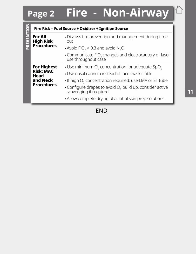
Fire Risk = Fuel Source + Oxidizer + Ignition Source
For All High Risk Procedures
•Discuss fire prevention and management during time out •Avoid FiO2 > 0.3 and avoid N2O •Communicate FiO2 changes and electrocautery or laser use throughout case
For Highest Risk: MAC Head and Neck Procedures
•Use minimum O2 concentration for adequate SpO2
•Use nasal cannula instead of face mask if able • If high O2 concentration required: use LMA or ET tube •Configure drapes to avoid O2 build up, consider active scavenging if required •Allow complete drying of alcohol skin prep solutions
Page 2 Fire - Non-Airway1
3
1516
5678 9
10111213
1718192021222324252627
2
4
14
2829
TREA
TMEN
T
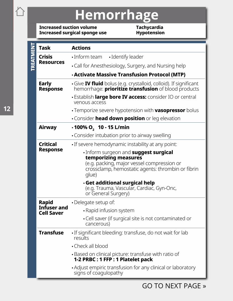
Hemorrhage
Task ActionsCrisis Resources
• Inform team • Identify leader •Call for Anesthesiology, Surgery, and Nursing help •Activate Massive Transfusion Protocol (MTP)
Early Response
•Give IV fluid bolus (e.g. crystalloid, colloid). If significant hemorrhage: prioritize transfusion of blood products •Establish large bore IV access: consider IO or central venous access • Temporize severe hypotension with vasopressor bolus •Consider head down position or leg elevation
Airway • 100% O2 10 - 15 L/min •Consider intubation prior to airway swelling
Critical Response
• If severe hemodynamic instability at any point: • Inform surgeon and suggest surgical temporizing measures (e.g. packing, major vessel compression or crossclamp, hemostatic agents: thrombin or fibrin glue) • Get additional surgical help (e.g. Trauma, Vascular, Cardiac, Gyn-Onc, or General Surgery)
Rapid Infuser and Cell Saver
•Delegate setup of: •Rapid infusion system •Cell saver (if surgical site is not contaminated or cancerous)
Transfuse • If significant bleeding: transfuse, do not wait for lab results •Check all blood •Based on clinical picture: transfuse with ratio of 1-2 PRBC : 1 FFP : 1 Platelet pack •Adjust empiric transfusion for any clinical or laboratory signs of coagulopathy
GO TO NEXT PAGE »
Increased suction volumeIncreased surgical sponge use
TachycardiaHypotension
3
1516
5678 9
10111213
171819202122232425
2
4
14
26272829
END
TREA
TMEN
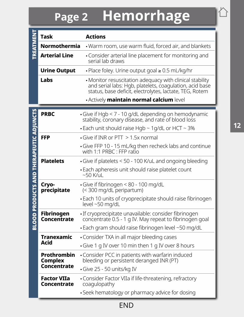
TTask ActionsNormothermia •Warm room, use warm fluid, forced air, and blanketsArterial Line •Consider arterial line placement for monitoring and
serial lab drawsUrine Output •Place foley. Urine output goal ≥ 0.5 mL/kg/hrLabs •Monitor resuscitation adequacy with clinical stability
and serial labs: Hgb, platelets, coagulation, acid base status, base deficit, electrolytes, lactate, TEG, Rotem •Actively maintain normal calcium level
BLO
OD
PRO
DUCT
S AN
D TH
ERAP
EUTI
C AD
JUN
CTS
Page 2 Hemorrhage
PRBC •Give if Hgb < 7 - 10 g/dL depending on hemodynamic stability, coronary disease, and rate of blood loss •Each unit should raise Hgb ~ 1g/dL or HCT ~ 3%
FFP •Give if INR or PTT > 1.5x normal •Give FFP 10 - 15 mL/kg then recheck labs and continue with 1:1 PRBC : FFP ratio
Platelets •Give if platelets < 50 - 100 K/uL and ongoing bleeding •Each apheresis unit should raise platelet count ~50 K/uL
Cryo-precipitate
•Give if fibrinogen < 80 - 100 mg/dL (< 300 mg/dL peripartum) •Each 10 units of cryoprecipitate should raise fibrinogen level ~50 mg/dL
Fibrinogen Concentrate
• If cryoprecipitate unavailable: consider fibrinogen concentrate 0.5 - 1 g IV. May repeat to fibrinogen goal •Each gram should raise fibrinogen level ~50 mg/dL
Tranexamic Acid
•Consider TXA in all major bleeding cases •Give 1 g IV over 10 min then 1 g IV over 8 hours
Prothrombin Complex Concentrate
•Consider PCC in patients with warfarin induced bleeding or persistent deranged INR (PT) •Give 25 - 50 units/kg IV
Factor VIIa Concentrate
•Consider Factor VIIa if life-threatening, refractory coagulopathy • Seek hematology or pharmacy advice for dosing
1
3
1516
5678 9
10111213
1718192021222324252627
2
4
14
2829
TREA
TMEN
T
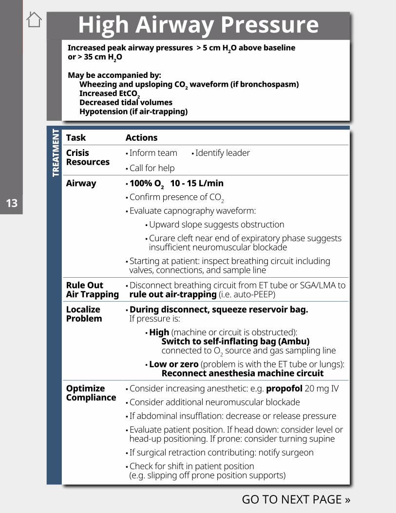
Task ActionsCrisis Resources
• Inform team • Identify leader •Call for help
Airway • 100% O2 10 - 15 L/min •Confirm presence of CO2 •Evaluate capnography waveform:
•Upward slope suggests obstruction •Curare cleft near end of expiratory phase suggests insufficient neuromuscular blockade
• Starting at patient: inspect breathing circuit including valves, connections, and sample line
Rule Out Air Trapping
•Disconnect breathing circuit from ET tube or SGA/LMA to rule out air-trapping (i.e. auto-PEEP)
Localize Problem
•During disconnect, squeeze reservoir bag. If pressure is:
• High (machine or circuit is obstructed): Switch to self-inflating bag (Ambu) connected to O2 source and gas sampling line • Low or zero (problem is with the ET tube or lungs): Reconnect anesthesia machine circuit
Optimize Compliance
•Consider increasing anesthetic: e.g. propofol 20 mg IV •Consider additional neuromuscular blockade • If abdominal insufflation: decrease or release pressure •Evaluate patient position. If head down: consider level or head-up positioning. If prone: consider turning supine • If surgical retraction contributing: notify surgeon •Check for shift in patient position (e.g. slipping off prone position supports)
GO TO NEXT PAGE »
Increased peak airway pressures > 5 cm H2O above baseline or > 35 cm H2O
May be accompanied by:Wheezing and upsloping CO2 waveform (if bronchospasm)Increased EtCO2Decreased tidal volumesHypotension (if air-trapping)
High Airway Pressure3
1516
5678 9
10111213
171819202122232425
2
4
14
26272829
END
RULE
OU
TTR
EATM
ENT
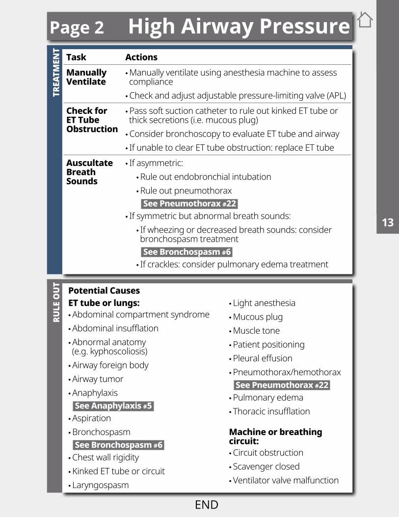
Task ActionsManually Ventilate
•Manually ventilate using anesthesia machine to assess compliance •Check and adjust adjustable pressure-limiting valve (APL)
Check for ET Tube Obstruction
•Pass soft suction catheter to rule out kinked ET tube or thick secretions (i.e. mucous plug) •Consider bronchoscopy to evaluate ET tube and airway • If unable to clear ET tube obstruction: replace ET tube
AuscultateBreath Sounds
• If asymmetric: •Rule out endobronchial intubation •Rule out pneumothorax See Pneumothorax #22
• If symmetric but abnormal breath sounds: • If wheezing or decreased breath sounds: consider bronchospasm treatment See Bronchospasm #6 • If crackles: consider pulmonary edema treatment
Potential CausesET tube or lungs: •Abdominal compartment syndrome •Abdominal insufflation •Abnormal anatomy (e.g. kyphoscoliosis) •Airway foreign body •Airway tumor •Anaphylaxis See Anaphylaxis #5 •Aspiration •Bronchospasm See Bronchospasm #6 •Chest wall rigidity •Kinked ET tube or circuit • Laryngospasm
• Light anesthesia •Mucous plug •Muscle tone •Patient positioning •Pleural effusion •Pneumothorax/hemothorax See Pneumothorax #22 •Pulmonary edema • Thoracic insufflation
Machine or breathing circuit: •Circuit obstruction • Scavenger closed •Ventilator valve malfunction
Page 2 High Airway Pressure1
3
1516
5678 9
10111213
1718192021222324252627
2
4
14
2829

This space is intentionally blank
3
1516
5678 9
10111213
171819202122232425
2
4
14
26272829
END
High SpinalTR
EATM
ENT
RULE
OU
T
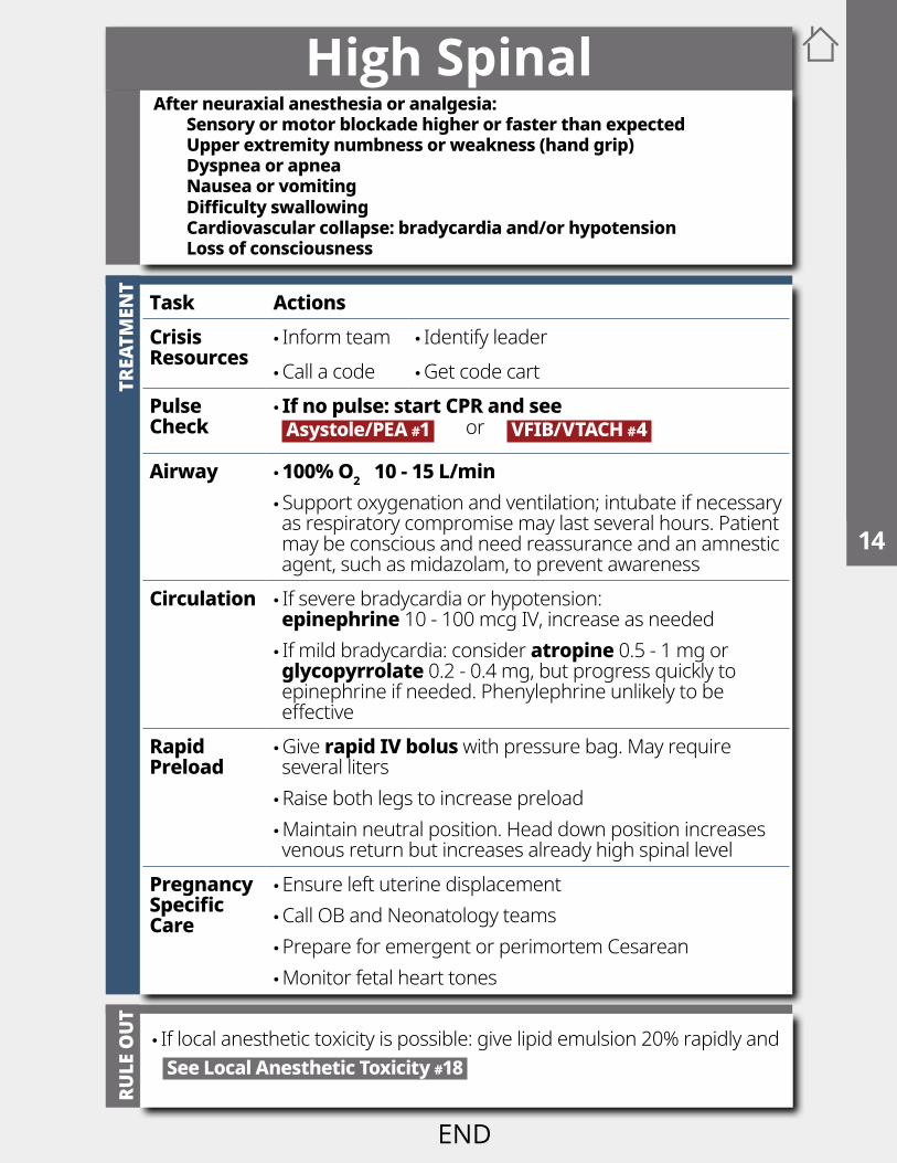
• If local anesthetic toxicity is possible: give lipid emulsion 20% rapidly and See Local Anesthetic Toxicity #18
Task ActionsCrisis Resources
• Inform team • Identify leader •Call a code •Get code cart
Pulse Check
• If no pulse: start CPR and see Asystole/PEA #1 or VFIB/VTACH #4
Airway • 100% O2 10 - 15 L/min • Support oxygenation and ventilation; intubate if necessary as respiratory compromise may last several hours. Patient may be conscious and need reassurance and an amnestic agent, such as midazolam, to prevent awareness
Circulation • If severe bradycardia or hypotension: epinephrine 10 - 100 mcg IV, increase as needed • If mild bradycardia: consider atropine 0.5 - 1 mg or glycopyrrolate 0.2 - 0.4 mg, but progress quickly to epinephrine if needed. Phenylephrine unlikely to be effective
Rapid Preload
•Give rapid IV bolus with pressure bag. May require several liters •Raise both legs to increase preload •Maintain neutral position. Head down position increases venous return but increases already high spinal level
Pregnancy Specific Care
•Ensure left uterine displacement •Call OB and Neonatology teams •Prepare for emergent or perimortem Cesarean •Monitor fetal heart tones
After neuraxial anesthesia or analgesia:Sensory or motor blockade higher or faster than expected Upper extremity numbness or weakness (hand grip)Dyspnea or apneaNausea or vomiting Difficulty swallowingCardiovascular collapse: bradycardia and/or hypotensionLoss of consciousness
1
3
1516
5678 9
10111213
1718192021222324252627
2
4
14
2829
TREA
TMEN
TRU
LE O
UT
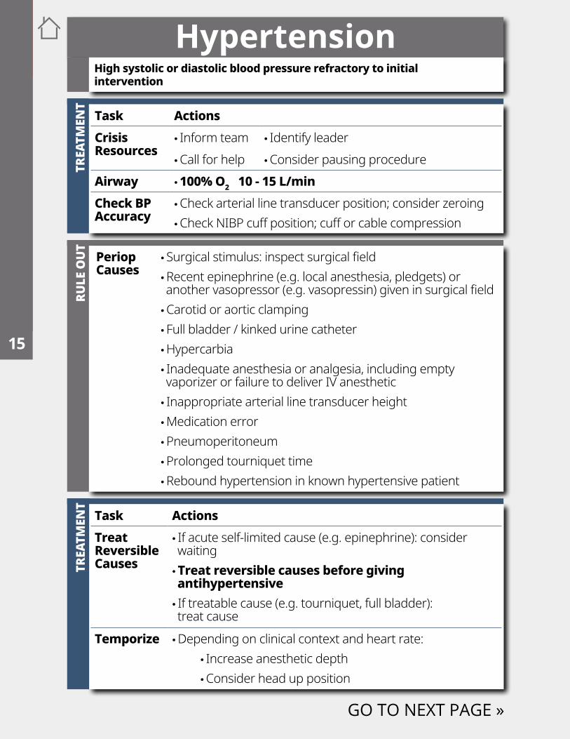
Periop Causes
• Surgical stimulus: inspect surgical field •Recent epinephrine (e.g. local anesthesia, pledgets) or another vasopressor (e.g. vasopressin) given in surgical field •Carotid or aortic clamping • Full bladder / kinked urine catheter •Hypercarbia • Inadequate anesthesia or analgesia, including empty vaporizer or failure to deliver IV anesthetic • Inappropriate arterial line transducer height •Medication error •Pneumoperitoneum •Prolonged tourniquet time •Rebound hypertension in known hypertensive patient
Hypertension
GO TO NEXT PAGE »
High systolic or diastolic blood pressure refractory to initial intervention
Task ActionsCrisis Resources
• Inform team • Identify leader •Call for help •Consider pausing procedure
Airway • 100% O2 10 - 15 L/minCheck BP Accuracy
•Check arterial line transducer position; consider zeroing •Check NIBP cuff position; cuff or cable compression
TREA
TMEN
T
Task ActionsTreat Reversible Causes
• If acute self-limited cause (e.g. epinephrine): consider waiting •Treat reversible causes before giving antihypertensive • If treatable cause (e.g. tourniquet, full bladder): treat cause
Temporize •Depending on clinical context and heart rate: • Increase anesthetic depth •Consider head up position
3
1516
5678 9
10111213
171819202122232425
2
4
14
26272829
RULE
OU
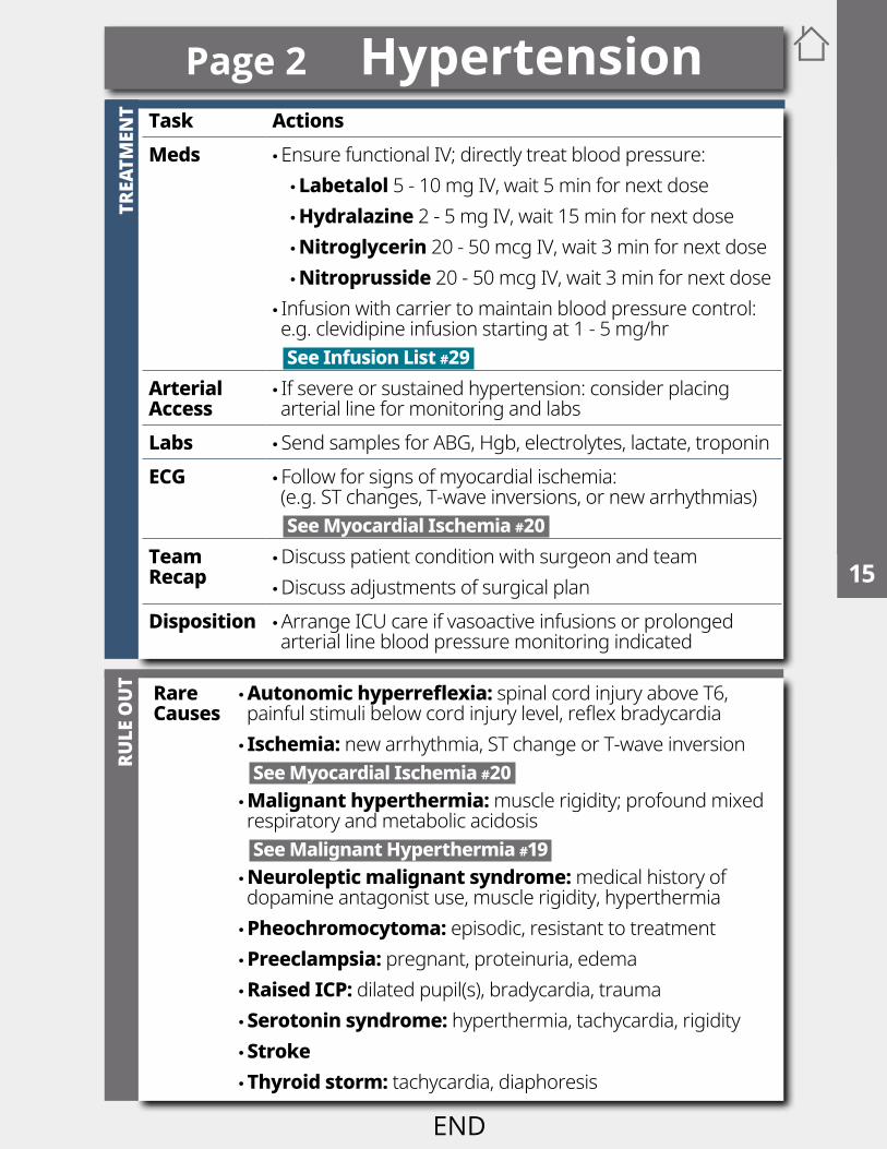
T Rare Causes
•Autonomic hyperreflexia: spinal cord injury above T6, painful stimuli below cord injury level, reflex bradycardia • Ischemia: new arrhythmia, ST change or T-wave inversion See Myocardial Ischemia #20 • Malignant hyperthermia: muscle rigidity; profound mixed respiratory and metabolic acidosis See Malignant Hyperthermia #19 • Neuroleptic malignant syndrome: medical history of dopamine antagonist use, muscle rigidity, hyperthermia • Pheochromocytoma: episodic, resistant to treatment • Preeclampsia: pregnant, proteinuria, edema • Raised ICP: dilated pupil(s), bradycardia, trauma • Serotonin syndrome: hyperthermia, tachycardia, rigidity • Stroke • Thyroid storm: tachycardia, diaphoresis
TREA
TMEN
T Task ActionsMeds •Ensure functional IV; directly treat blood pressure:
• Labetalol 5 - 10 mg IV, wait 5 min for next dose • Hydralazine 2 - 5 mg IV, wait 15 min for next dose • Nitroglycerin 20 - 50 mcg IV, wait 3 min for next dose • Nitroprusside 20 - 50 mcg IV, wait 3 min for next dose
• Infusion with carrier to maintain blood pressure control: e.g. clevidipine infusion starting at 1 - 5 mg/hr See Infusion List #29
Arterial Access
• If severe or sustained hypertension: consider placing arterial line for monitoring and labs
Labs • Send samples for ABG, Hgb, electrolytes, lactate, troponinECG • Follow for signs of myocardial ischemia:
(e.g. ST changes, T-wave inversions, or new arrhythmias) See Myocardial Ischemia #20
Team Recap
•Discuss patient condition with surgeon and team •Discuss adjustments of surgical plan
Disposition •Arrange ICU care if vasoactive infusions or prolonged arterial line blood pressure monitoring indicated
END
Page 2 Hypertension1
3
1516
5678 9
10111213
1718192021222324252627
2
4
14
2829
TREA
TMEN
T
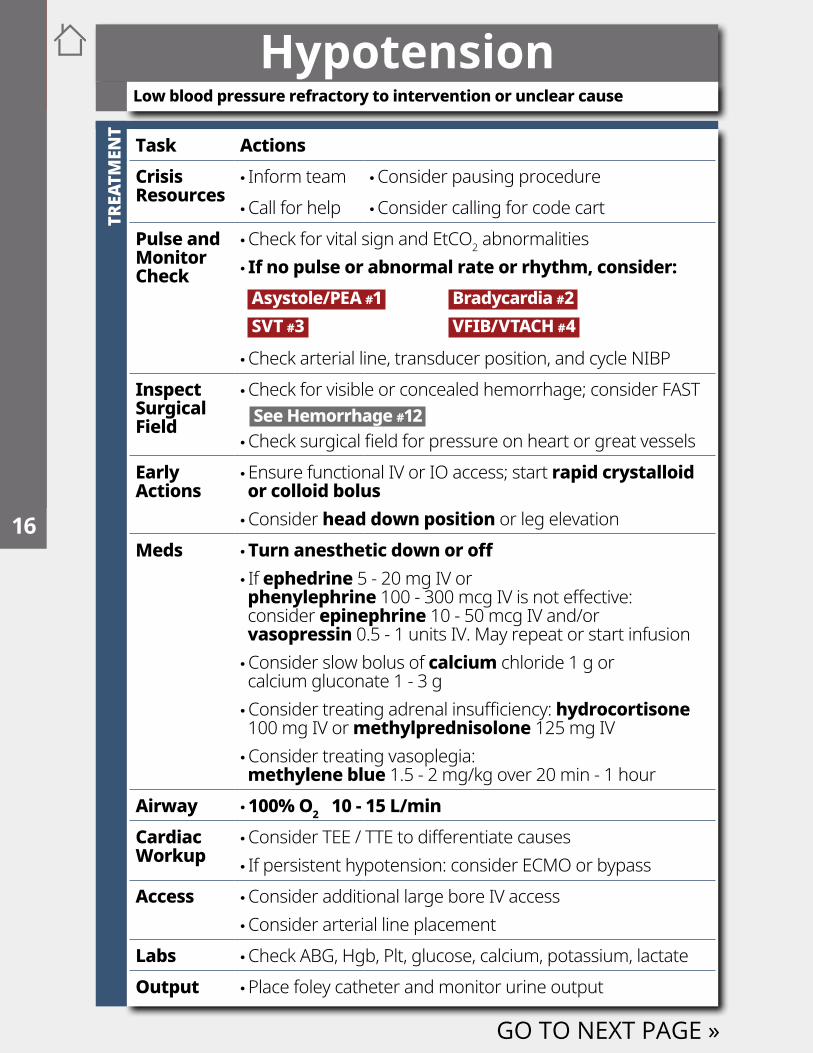
Hypotension
Task ActionsCrisis Resources
• Inform team •Consider pausing procedure •Call for help •Consider calling for code cart
Pulse and Monitor Check
•Check for vital sign and EtCO2 abnormalities • If no pulse or abnormal rate or rhythm, consider: Asystole/PEA #1 Bradycardia #2 SVT #3 VFIB/VTACH #4
•Check arterial line, transducer position, and cycle NIBPInspect Surgical Field
•Check for visible or concealed hemorrhage; consider FAST See Hemorrhage #12 •Check surgical field for pressure on heart or great vessels
Early Actions
•Ensure functional IV or IO access; start rapid crystalloid or colloid bolus •Consider head down position or leg elevation
Meds •Turn anesthetic down or off • If ephedrine 5 - 20 mg IV or phenylephrine 100 - 300 mcg IV is not effective: consider epinephrine 10 - 50 mcg IV and/or vasopressin 0.5 - 1 units IV. May repeat or start infusion •Consider slow bolus of calcium chloride 1 g or calcium gluconate 1 - 3 g •Consider treating adrenal insufficiency: hydrocortisone 100 mg IV or methylprednisolone 125 mg IV •Consider treating vasoplegia: methylene blue 1.5 - 2 mg/kg over 20 min - 1 hour
Airway • 100% O2 10 - 15 L/minCardiac Workup
•Consider TEE / TTE to differentiate causes • If persistent hypotension: consider ECMO or bypass
Access •Consider additional large bore IV access •Consider arterial line placement
Labs •Check ABG, Hgb, Plt, glucose, calcium, potassium, lactateOutput •Place foley catheter and monitor urine output
GO TO NEXT PAGE »
Low blood pressure refractory to intervention or unclear cause
3
1516
5678 9
10111213
171819202122232425
2
4
14
26272829

END
Low SVR •Anaphylaxis • Shock (septic/spinal/neurogenic) See Anaphylaxis #5 • Transfusion reaction •Neuraxial block See Transfusion Reaction #25 See High Spinal #14 •Vasodilators
Low HR •Bradycardia/heart block •High spinal •Vagal stimulus See High Spinal #14
Low Preload
•Auto-PEEP •Pericardial tamponade •Embolism e.g.air,clot,fat •Pneumothorax See Embolism #9 See Pneumothorax #22 •Hypovolemia •Right heart failure See Hemorrhage #12 See Right Heart Failure #24 • IVC compression •Vasodilators
Low Inotropy
•Acidosis • Local anesthetic toxicity •Arrhythmias See Local Anes Toxicity #18 •Cardiomyopathy •Myocardial depressants •Hypoxemia •Myocardial ischemia See Hypoxemia #17 See Myocardial Ischemia #20
High Afterload
• Stenotic valvular disease • LVOT obstruction
Low Forward Flow
•Regurgitant valvular disease
Explore Other Causes By Physiologic Differential: •Blood Pressure = Systemic Vascular Resistance (SVR) x Cardiac Output (CO) •Cardiac Output (CO) = Heart Rate (HR) x Stroke Volume (SV) • Stroke Volume (SV) components: Preload, Inotropy, Afterload
Rule out rapidly lethal causes: •Anaphylaxis See Anaphylaxis #5 •Auto-PEEP: disconnect circuit See High Airway Pressure #13 •Cardiovascular: consider TEE / TTE to evaluate volume status, LV/RV function, valvular disease, LV outflow obstruction See Embolism #9 See Myocardial Ischemia #20 See Right Heart Failure #24 •Hemorrhage or concealed hemorrhage See Hemorrhage #12
• IVC compression: prone, obese, pregnant, surgical manipulation • Local anesthetic toxicity See Local Anes Toxicity #18 •Pneumoperitoneum or pneumopericardium •Pneumothorax •Cardiac tamponade See Pneumothorax #22 •Vasodilators: check doses of volatile/IV anesthetics and drips
DIFF
EREN
TIAL
DIA
GNO
SIS
Page 2 Hypotension1
3
1516
5678 9
10111213
1718192021222324252627
2
4
14
2829
TREA
TMEN
T
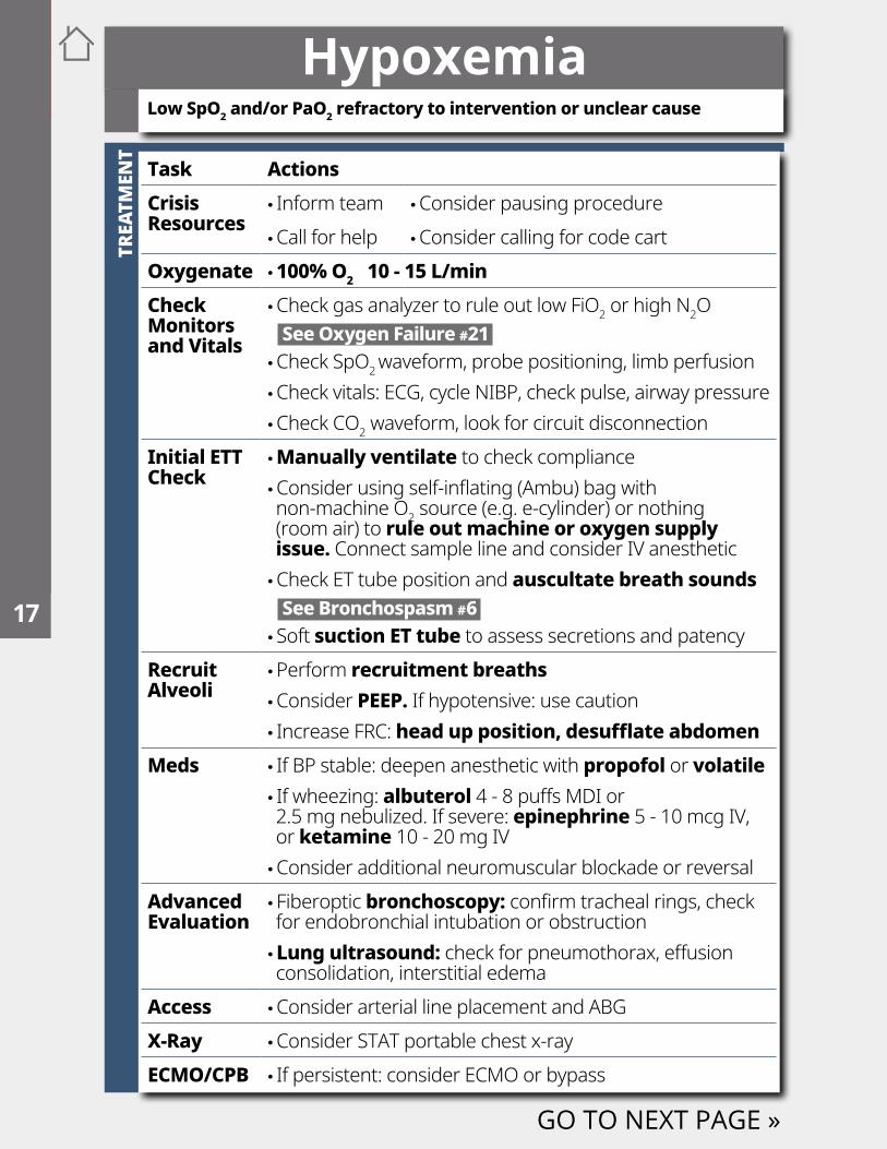
Hypoxemia
Task ActionsCrisis Resources
• Inform team •Consider pausing procedure •Call for help •Consider calling for code cart
Oxygenate • 100% O2 10 - 15 L/minCheck Monitors and Vitals
•Check gas analyzer to rule out low FiO2 or high N2O See Oxygen Failure #21 •Check SpO2 waveform, probe positioning, limb perfusion •Check vitals: ECG, cycle NIBP, check pulse, airway pressure •Check CO2 waveform, look for circuit disconnection
Initial ETT Check
•Manually ventilate to check compliance •Consider using self-inflating (Ambu) bag with non-machine O2 source (e.g. e-cylinder) or nothing (room air) to rule out machine or oxygen supply issue. Connect sample line and consider IV anesthetic •Check ET tube position and auscultate breath sounds See Bronchospasm #6 • Soft suction ET tube to assess secretions and patency
Recruit Alveoli
•Perform recruitment breaths •Consider PEEP. If hypotensive: use caution • Increase FRC: head up position, desufflate abdomen
Meds • If BP stable: deepen anesthetic with propofol or volatile • If wheezing: albuterol 4 - 8 puffs MDI or 2.5 mg nebulized. If severe: epinephrine 5 - 10 mcg IV, or ketamine 10 - 20 mg IV •Consider additional neuromuscular blockade or reversal
Advanced Evaluation
• Fiberoptic bronchoscopy: confirm tracheal rings, check for endobronchial intubation or obstruction • Lung ultrasound: check for pneumothorax, effusion consolidation, interstitial edema
Access •Consider arterial line placement and ABGX-Ray •Consider STAT portable chest x-rayECMO/CPB • If persistent: consider ECMO or bypass
GO TO NEXT PAGE »
Low SpO2 and/or PaO2 refractory to intervention or unclear cause
3
1516
5678 9
10111213
171819202122232425
2
4
14
26272829
END
DIFF
EREN
TIAL
DIA
GNO
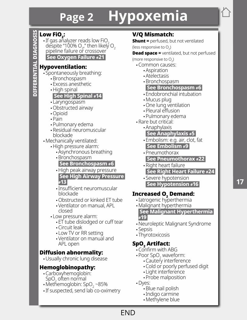
SIS Low FiO2:
• If gas analyzer reads low FiO2 despite “100% O2,” then likely O2 pipeline failure or crossover See Oxygen Failure #21
Hypoventilation: • Spontaneously breathing:
•Bronchospasm •Excess anesthetic •High spinal • See High Spinal #14 • Laryngospasm •Obstructed airway •Opioid •Pain •Pulmonary edema •Residual neuromuscular blockade
•Mechanically ventilated: •High pressure alarm: •Asynchronous breathing •Bronchospasm See Bronchospasm #6 •High peak airway pressure See High Airway Pressure #13 • Insufficient neuromuscular blockade •Obstructed or kinked ET tube •Ventilator on manual, APL closed
• Low pressure alarm: •ET tube dislodged or cuff tear •Circuit leak • Low TV or RR setting •Ventilator on manual and APL open
Diffusion abnormality: •Usually chronic lung disease
Hemoglobinopathy: •Carboxyhemoglobin: SpO2 often normal •Methemoglobin: SpO2 ~85% • If suspected, send lab co-oximetry
V/Q Mismatch: Shunt = perfused, but not ventilated (less responsive to O2) Dead space = ventilated, but not perfused (more responsive to O2) •Common causes:
•Aspiration •Atelectasis •Bronchospasm See Bronchospasm #6 •Endobronchial intubation •Mucus plug •One lung ventilation •Pleural effusion •Pulmonary edema
•Rare but critical: •Anaphylaxis See Anaphylaxis #5 •Embolism: e.g. air, clot, fat See Embolism #9 •Pneumothorax See Pneumothorax #22 •Right heart failure See Right Heart Failure #24 • Severe hypotension See Hypotension #16
Increased O2 Demand: • Iatrogenic hyperthermia •Malignant hyperthermia See Malignant Hyperthermia #19 •Neuroleptic Malignant Syndrome • Sepsis • Thyrotoxicosis
SpO2 Artifact: •Confirm with ABG •Poor SpO2 waveform:
•Cautery interference •Cold or poorly perfused digit • Light interference •Probe malposition
•Dyes: •Blue nail polish • Indigo carmine •Methylene blue
Page 2 Hypoxemia1
3
1516
5678 9
10111213
1718192021222324252627
2
4
14
2829
TREA
TMEN
T
Local Anesthetic Toxicity
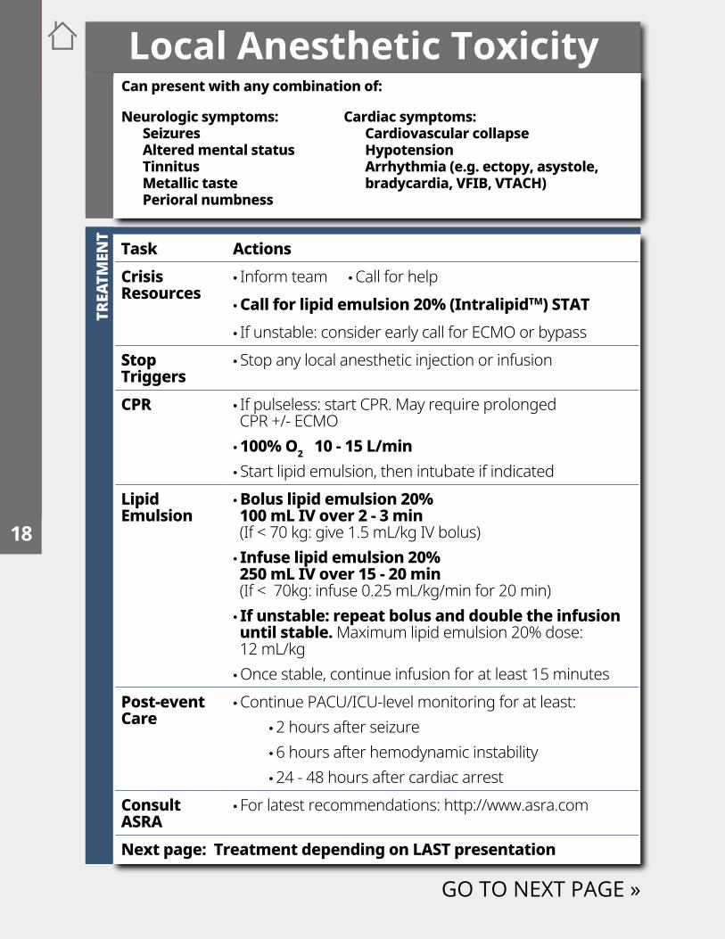
Task ActionsCrisis Resources
• Inform team •Call for help •Call for lipid emulsion 20% (IntralipidTM) STAT • If unstable: consider early call for ECMO or bypass
Stop Triggers
• Stop any local anesthetic injection or infusion
CPR • If pulseless: start CPR. May require prolonged CPR +/- ECMO • 100% O2 10 - 15 L/min • Start lipid emulsion, then intubate if indicated
Lipid Emulsion
• Bolus lipid emulsion 20% 100 mL IV over 2 - 3 min (If < 70 kg: give 1.5 mL/kg IV bolus) • Infuse lipid emulsion 20% 250 mL IV over 15 - 20 min (If < 70kg: infuse 0.25 mL/kg/min for 20 min) • If unstable: repeat bolus and double the infusion until stable. Maximum lipid emulsion 20% dose: 12 mL/kg •Once stable, continue infusion for at least 15 minutes
Post-event Care
•Continue PACU/ICU-level monitoring for at least: •2 hours after seizure •6 hours after hemodynamic instability •24 - 48 hours after cardiac arrest
Consult ASRA
• For latest recommendations: http://www.asra.com
Next page: Treatment depending on LAST presentation
GO TO NEXT PAGE »
Can present with any combination of:
Neurologic symptoms:SeizuresAltered mental statusTinnitusMetallic tastePerioral numbness
Cardiac symptoms: Cardiovascular collapse HypotensionArrhythmia (e.g. ectopy, asystole, bradycardia, VFIB, VTACH)
3
1516
5678 9
10111213
171819202122232425
2
4
14
26272829
END
TREA
TMEN
T
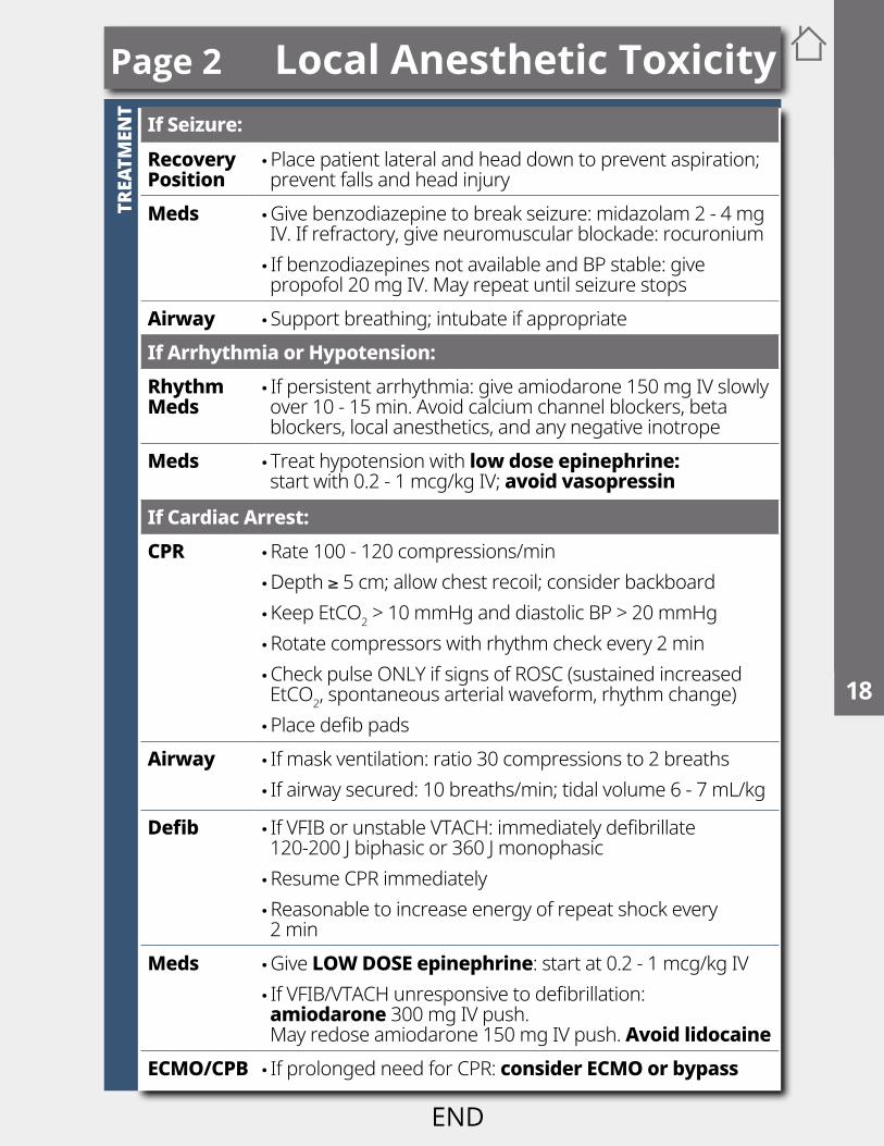
Page 2 Local Anesthetic ToxicityIf Seizure:RecoveryPosition
•Place patient lateral and head down to prevent aspiration; prevent falls and head injury
Meds •Give benzodiazepine to break seizure: midazolam 2 - 4 mg IV. If refractory, give neuromuscular blockade: rocuronium • If benzodiazepines not available and BP stable: give propofol 20 mg IV. May repeat until seizure stops
Airway • Support breathing; intubate if appropriateIf Arrhythmia or Hypotension:Rhythm Meds
• If persistent arrhythmia: give amiodarone 150 mg IV slowly over 10 - 15 min. Avoid calcium channel blockers, beta blockers, local anesthetics, and any negative inotrope
Meds • Treat hypotension with low dose epinephrine: start with 0.2 - 1 mcg/kg IV; avoid vasopressin
If Cardiac Arrest:CPR •Rate 100 - 120 compressions/min
•Depth ≥ 5 cm; allow chest recoil; consider backboard •Keep EtCO2 > 10 mmHg and diastolic BP > 20 mmHg •Rotate compressors with rhythm check every 2 min •Check pulse ONLY if signs of ROSC (sustained increased EtCO2, spontaneous arterial waveform, rhythm change) •Place defib pads
Airway • If mask ventilation: ratio 30 compressions to 2 breaths • If airway secured: 10 breaths/min; tidal volume 6 - 7 mL/kg
Defib • If VFIB or unstable VTACH: immediately defibrillate 120-200 J biphasic or 360 J monophasic •Resume CPR immediately •Reasonable to increase energy of repeat shock every 2 min
Meds •Give LOW DOSE epinephrine: start at 0.2 - 1 mcg/kg IV • If VFIB/VTACH unresponsive to defibrillation: amiodarone 300 mg IV push. May redose amiodarone 150 mg IV push. Avoid lidocaine
ECMO/CPB • If prolonged need for CPR: consider ECMO or bypass
1
3
1516
5678 9
10111213
1718192021222324252627
2
4
14
2829
TREA
TMEN
TRU
LE O
UT
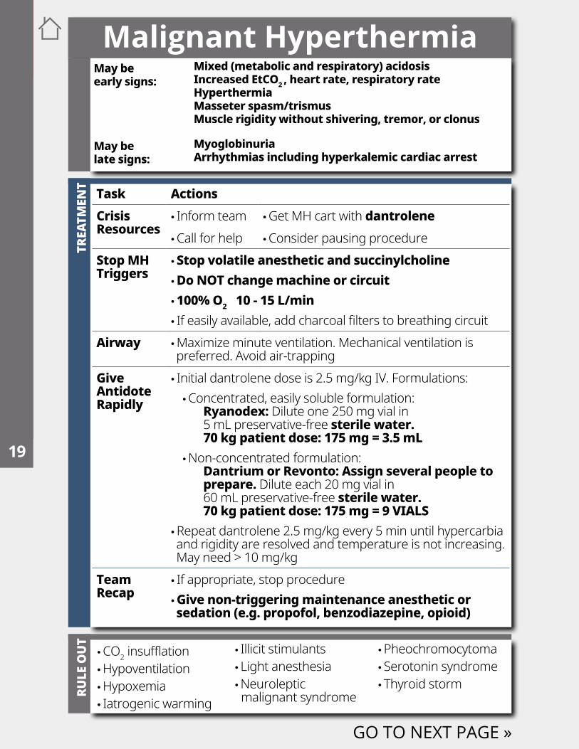
•CO2 insufflation •Hypoventilation •Hypoxemia • Iatrogenic warming
• Illicit stimulants • Light anesthesia •Neuroleptic malignant syndrome
•Pheochromocytoma • Serotonin syndrome • Thyroid storm
May be early signs:
Mixed (metabolic and respiratory) acidosis Increased EtCO2 , heart rate, respiratory rateHyperthermia Masseter spasm/trismusMuscle rigidity without shivering, tremor, or clonus
May be late signs:
Myoglobinuria Arrhythmias including hyperkalemic cardiac arrest
Malignant Hyperthermia
Task ActionsCrisis Resources
• Inform team •Get MH cart with dantrolene •Call for help •Consider pausing procedure
Stop MH Triggers
•Stop volatile anesthetic and succinylcholine • Do NOT change machine or circuit • 100% O2 10 - 15 L/min • If easily available, add charcoal filters to breathing circuit
Airway •Maximize minute ventilation. Mechanical ventilation is preferred. Avoid air-trapping
Give Antidote Rapidly
• Initial dantrolene dose is 2.5 mg/kg IV. Formulations: •Concentrated, easily soluble formulation: Ryanodex: Dilute one 250 mg vial in 5 mL preservative-free sterile water. 70 kg patient dose: 175 mg = 3.5 mL •Non-concentrated formulation: Dantrium or Revonto: Assign several people to prepare. Dilute each 20 mg vial in 60 mL preservative-free sterile water. 70 kg patient dose: 175 mg = 9 VIALS
•Repeat dantrolene 2.5 mg/kg every 5 min until hypercarbia and rigidity are resolved and temperature is not increasing. May need > 10 mg/kg
Team Recap
• If appropriate, stop procedure •Give non-triggering maintenance anesthetic or sedation (e.g. propofol, benzodiazepine, opioid)
GO TO NEXT PAGE »
3
1516
5678 9
10111213
171819202122232425
2
4
14
26272829
END
TREA
TMEN
TTask ActionsTreat Hyper- K+
• Calcium chloride 10 mg/kg IV, max 2 g •Regular insulin 5 - 10 units IV with dextrose/D50 1 amp IV (25 g); monitor glucose • Albuterol 8 - 12 puffs MDI or 2.5 mg nebulized • Sodium bicarbonate: 0.5 amp (25 mL) at a time; maintain minute ventilation to exhale additional CO2 produced • If severe: consider emergent dialysis
Treat Rhythm
• Treat arrhythmias with amiodarone 150 mg IV over 10 - 15 min, esmolol 10 - 20 mg IV bolus followed by infusion, or magnesium sulfate 1 g IV; avoid calcium channel blockers and sodium channel blockers (e.g. verapamil, diltiazem, lidocaine, procainamide) • If unstable, call for code cart and also see ACLS event: Asystole/PEA #1 Bradycardia #2 SVT #3 VFIB/VTACH #4
Active Cooling
• If core temperature > 38° C: actively cool with cold IV fluid (20 - 30 ml/kg normal saline or plasmalyte) •Additional cooling: Stop active warming; set forced air on ambient; cool room; put ice packs on head, axilla, and groin; wet skin; cool lavage if open abdomen or peritoneal catheter (avoid bladder lavage to preserve urine output measurement)
Access •Consider additional IV access and arterial line placement
Labs • Send ABG, K+, CK, urine myoglobin, coagulation panel, lactateUrine Output
•Place Foley catheter and monitor urine output: goal 1 - 2 mL/kg/hr; consider IV fluids and diuretics
MH Hotline
•Call 24/7 for expert consultation: 1-800-MH-HYPER (1-800-644-9737) http://www.mhaus.org
ICU Care
• Transport with experienced personnel after stabilization •Mechanical ventilation commonly required because 20% of MH events relapse within 16 hours. Extubate once metabolically and hemodynamically stable •Continue dantrolene: 1 mg/kg bolus every 4 - 6 hours or 0.25 mg/kg/hr infusion for up to 24 hours •Monitor for rhabdo, DIC, hyperK+, compartment syndrome
Post Event
•Complete AMRA (Adverse Metabolic Reaction to Anesthesia): https://anest.ufl.edu/namhr/ • Test genes: https://www.mhaus.org/testing/genetic-testing/
Page 2 Malignant Hyperthermia1
3
1516
5678 9
10111213
1718192021222324252627
2
4
14
2829
TREA
TMEN
T
Myocardial Ischemia
Task ActionsCrisis Resources
• Inform team •Get code cart •Call for Cardiac Anesthesiology or Cardiology help
Airway • Set supplemental O2 to maintain SpO2 ≥ 95%Monitor •Get 12-lead ECG; verify ECG leads in correct position
•Expand ECG monitor view to leads II, V5, and others •Prepare for arrhythmia: apply defibrillator pads and leads
Team Recap
•Pause or stop procedure if possible • Discuss bleeding and risk of anticoagulation
Meds • Treat any tachycardia, bradycardia, hypotension or hypertension See Infusion List #29 •Discuss with surgeon the explicit contraindications and benefits of dual antiplatelet therapy and anticoagulation:
•Aspirin 160 - 325 mg PO, nasogastric, or rectal • P2Y12 ADP receptor inhibitor: e.g. clopidogrel 300 mg PO, prasugrel 60 mg PO, or ticagrelor 180mg PO • Heparin infusion
• Treat pain with narcotics: fentanyl or morphine •Consider nitroglycerin paste or infusion. Avoid if hypotensive •Consider beta blocker to slow heart rate and allow coronary perfusion. Esmolol preferred because it can be stopped if it precipitates CHF. Avoid if bradycardia, 1st or 2nd degree heart block, or hypotensive • If acute pulmonary edema, consider diuresis: furosemide 10 - 40 mg IV. Monitor urine output
ST segment depression or elevation T-wave inversionArrhythmias: conduction abnormality (e.g. new LBBB), irregular rhythm, tachycardia, bradycardia, or hypotensionRegional wall motion abnormalityNew or worsened mitral regurgitationChest pain, dyspnea, nausea, or diaphoresis
GO TO NEXT PAGE »
3
1516
5678 9
10111213
171819202122232425
2
4
14
26272829
END
TREA
TMEN
T
Page 2 Myocardial IschemiaTask ActionsCardiology Consult
• If STEMI: consult Cardiology for possible emergent coronary revascularization or fibrinolysis •Consider emergent transfer to Cath Lab or PCI Center
Access •Consider additional IV access •Place arterial line for monitoring and labs •Consider central line placement
Labs • Send ABG, electrolytes, Hgb, troponin, coagulation panelECHO •Consider TEE / TTE to assess volume status, wall motion,
ventricular function, and valvular disease •Use contractility to guide vasoactive infusion choice
ECMO/CPB •Consider ECMO, cardiopulmonary bypass, or intra-aortic balloon pump
Disposition •May require ICU care
1
3
1516
5678 9
10111213
1718192021222324252627
2
4
14
2829

This space is intentionally blank
3
1516
5678 9
10111213
171819202122232425
2
4
14
26272829
END
TREA
TMEN
T
Oxygen Failure
Task ActionsCrisis Resources
• Inform team •Consider pausing procedure •Call for help •Get code cart with O2 cylinder
Non-MachineVentilation
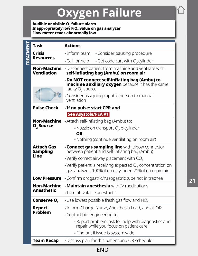
•Disconnect patient from machine and ventilate with self-inflating bag (Ambu) on room air •Do NOT connect self-inflating bag (Ambu) to machine auxiliary oxygen because it has the same faulty O2 source •Consider assigning capable person to manual ventilation
Pulse Check • If no pulse: start CPR and See Asystole/PEA #1
Non-MachineO2 Source
•Attach self-inflating bag (Ambu) to: •Nozzle on transport O2 e-cylinderOR •Nothing (continue ventilating on room air)
Attach Gas SamplingLine
• Connect gas sampling line with elbow connector between patient and self-inflating bag (Ambu) •Verify correct airway placement with CO2
•Verify patient is receiving expected O2 concentration on gas analyzer: 100% if on e-cylinder, 21% if on room air
Low Pressure •Confirm orogastric/nasogastric tube not in tracheaNon-Machine Anesthetic
• Maintain anesthesia with IV medications • Turn off volatile anesthetic
Conserve O2 •Use lowest possible fresh gas flow and FiO2
Report Problem
• Inform Charge Nurse, Anesthesia Lead, and all ORs •Contact bio-engineering to:
•Report problem; ask for help with diagnostics and repair while you focus on patient care • Find out if issue is system wide
Team Recap •Discuss plan for this patient and OR schedule
Audible or visible O2 failure alarmInappropriately low FiO2 value on gas analyzerFlow meter reads abnormally low
1
3
1516
5678 9
10111213
1718192021222324252627
2
4
14
2829

This space is intentionally blank
3
1516
5678 9
10111213
171819202122232425
2
4
14
26272829
END
TREA
TMEN
T
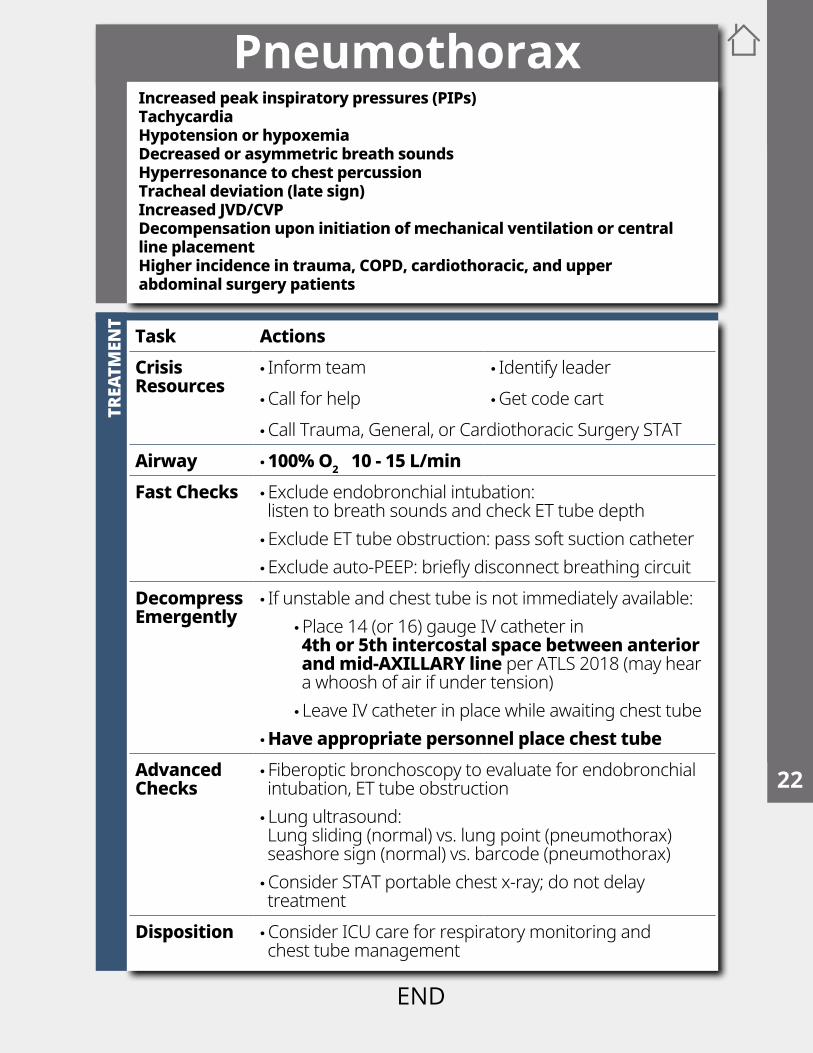
Task ActionsCrisis Resources
• Inform team • Identify leader •Call for help •Get code cart •Call Trauma, General, or Cardiothoracic Surgery STAT
Airway • 100% O2 10 - 15 L/minFast Checks •Exclude endobronchial intubation:
listen to breath sounds and check ET tube depth •Exclude ET tube obstruction: pass soft suction catheter •Exclude auto-PEEP: briefly disconnect breathing circuit
Decompress Emergently
• If unstable and chest tube is not immediately available: •Place 14 (or 16) gauge IV catheter in 4th or 5th intercostal space between anterior and mid-AXILLARY line per ATLS 2018 (may hear a whoosh of air if under tension) • Leave IV catheter in place while awaiting chest tube
•Have appropriate personnel place chest tubeAdvanced Checks
• Fiberoptic bronchoscopy to evaluate for endobronchial intubation, ET tube obstruction • Lung ultrasound: Lung sliding (normal) vs. lung point (pneumothorax) seashore sign (normal) vs. barcode (pneumothorax) •Consider STAT portable chest x-ray; do not delay treatment
Disposition •Consider ICU care for respiratory monitoring and chest tube management
PneumothoraxIncreased peak inspiratory pressures (PIPs)TachycardiaHypotension or hypoxemiaDecreased or asymmetric breath soundsHyperresonance to chest percussionTracheal deviation (late sign)Increased JVD/CVPDecompensation upon initiation of mechanical ventilation or central line placementHigher incidence in trauma, COPD, cardiothoracic, and upper abdominal surgery patients
1
3
1516
5678 9
10111213
1718192021222324252627
2
4
14
2829

This space is intentionally blank
3
1516
5678 9
10111213
171819202122232425
2
4
14
26272829
END
TREA
TMEN
T
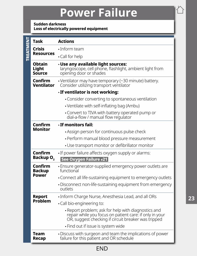
Power Failure
Task ActionsCrisis Resources
• Inform team •Call for help
Obtain Light Source
•Use any available light sources: laryngoscope, cell phone, flashlight, ambient light from opening door or shades
Confirm Ventilator
•Ventilator may have temporary (~30 minute) battery. Consider utilizing transport ventilator • If ventilator is not working:
•Consider converting to spontaneous ventilation •Ventilate with self-inflating bag (Ambu) •Convert to TIVA with battery operated pump or dial-a-flow / manual flow regulator
Confirm Monitor
• If monitors fail: •Assign person for continuous pulse check •Perform manual blood pressure measurement •Use transport monitor or defibrillator monitor
Confirm Backup O2
• If power failure affects oxygen supply or alarms: See Oxygen Failure #21
Confirm Backup Power
•Ensure generator-supplied emergency power outlets are functional •Connect all life-sustaining equipment to emergency outlets •Disconnect non-life-sustaining equipment from emergency outlets
Report Problem
• Inform Charge Nurse, Anesthesia Lead, and all ORs •Call bio-engineering to:
•Report problem; ask for help with diagnostics and repair while you focus on patient care: if only in your OR, suggest checking if circuit breaker was tripped • Find out if issue is system wide
Team Recap
•Discuss with surgeon and team the implications of power failure for this patient and OR schedule
Sudden darknessLoss of electrically powered equipment
1
3
1516
5678 9
10111213
1718192021222324252627
2
4
14
2829
TREA
TMEN
TRU
LE O
UT
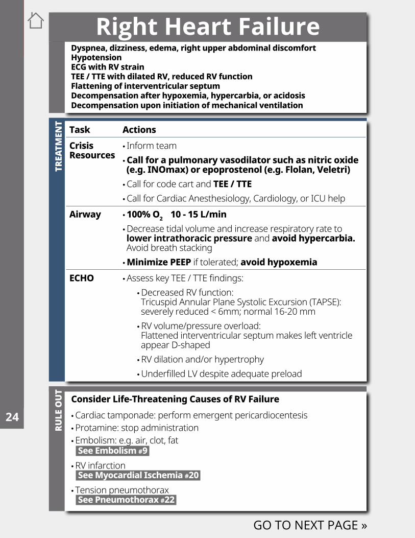
Consider Life-Threatening Causes of RV Failure •Cardiac tamponade: perform emergent pericardiocentesis •Protamine: stop administration •Embolism: e.g. air, clot, fat See Embolism #9 •RV infarction See Myocardial Ischemia #20 • Tension pneumothorax See Pneumothorax #22
Right Heart Failure
Task ActionsCrisis Resources
• Inform team •Call for a pulmonary vasodilator such as nitric oxide (e.g. INOmax) or epoprostenol (e.g. Flolan, Veletri) •Call for code cart and TEE / TTE •Call for Cardiac Anesthesiology, Cardiology, or ICU help
Airway • 100% O2 10 - 15 L/min •Decrease tidal volume and increase respiratory rate to lower intrathoracic pressure and avoid hypercarbia. Avoid breath stacking • Minimize PEEP if tolerated; avoid hypoxemia
ECHO •Assess key TEE / TTE findings: •Decreased RV function: Tricuspid Annular Plane Systolic Excursion (TAPSE): severely reduced < 6mm; normal 16-20 mm •RV volume/pressure overload: Flattened interventricular septum makes left ventricle appear D-shaped •RV dilation and/or hypertrophy •Underfilled LV despite adequate preload
GO TO NEXT PAGE »
Dyspnea, dizziness, edema, right upper abdominal discomfortHypotensionECG with RV strain TEE / TTE with dilated RV, reduced RV function Flattening of interventricular septumDecompensation after hypoxemia, hypercarbia, or acidosis Decompensation upon initiation of mechanical ventilation
3
1516
5678 9
10111213
171819202122232425
2
4
14
26272829
END
TREA
TMEN
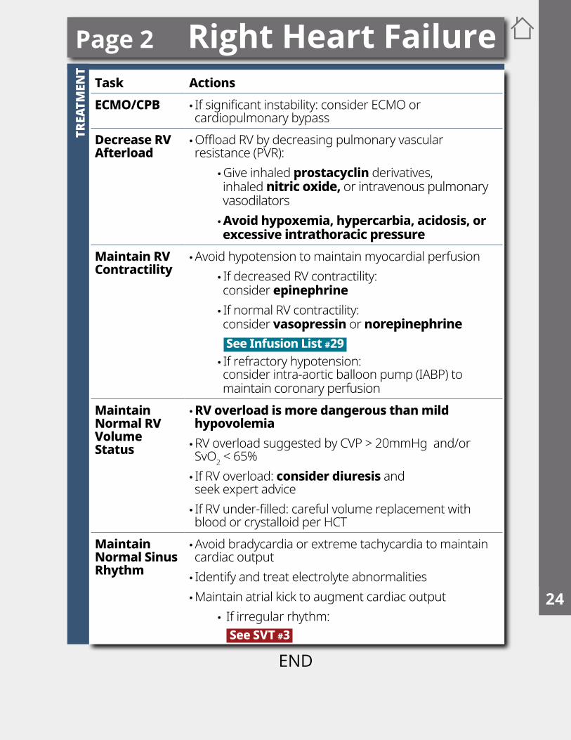
TTask ActionsECMO/CPB • If significant instability: consider ECMO or
cardiopulmonary bypassDecrease RV Afterload
•Offload RV by decreasing pulmonary vascular resistance (PVR):
•Give inhaled prostacyclin derivatives, inhaled nitric oxide, or intravenous pulmonary vasodilators •Avoid hypoxemia, hypercarbia, acidosis, or excessive intrathoracic pressure
Maintain RV Contractility
•Avoid hypotension to maintain myocardial perfusion • If decreased RV contractility: consider epinephrine • If normal RV contractility: consider vasopressin or norepinephrine See Infusion List #29 • If refractory hypotension: consider intra-aortic balloon pump (IABP) to maintain coronary perfusion
Maintain Normal RV Volume Status
•RV overload is more dangerous than mild hypovolemia •RV overload suggested by CVP > 20mmHg and/or SvO2 < 65% • If RV overload: consider diuresis and seek expert advice • If RV under-filled: careful volume replacement with blood or crystalloid per HCT
Maintain Normal Sinus Rhythm
•Avoid bradycardia or extreme tachycardia to maintain cardiac output • Identify and treat electrolyte abnormalities •Maintain atrial kick to augment cardiac output
• If irregular rhythm: See SVT #3
Page 2 Right Heart Failure1
3
1516
5678 9
10111213
1718192021222324252627
2
4
14
2829

This space is intentionally blank
3
1516
5678 9
10111213
171819202122232425
2
4
14
26272829
END
TREA
TMEN
T
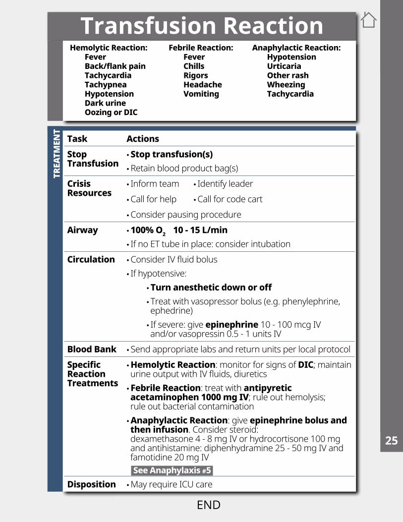
Transfusion Reaction
Task ActionsStop Transfusion
•Stop transfusion(s) •Retain blood product bag(s)
Crisis Resources
• Inform team • Identify leader •Call for help •Call for code cart •Consider pausing procedure
Airway • 100% O2 10 - 15 L/min • If no ET tube in place: consider intubation
Circulation •Consider IV fluid bolus • If hypotensive:
•Turn anesthetic down or off • Treat with vasopressor bolus (e.g. phenylephrine, ephedrine) • If severe: give epinephrine 10 - 100 mcg IV and/or vasopressin 0.5 - 1 units IV
Blood Bank • Send appropriate labs and return units per local protocolSpecific Reaction Treatments
• Hemolytic Reaction: monitor for signs of DIC; maintain urine output with IV fluids, diuretics • Febrile Reaction: treat with antipyretic acetaminophen 1000 mg IV; rule out hemolysis; rule out bacterial contamination • Anaphylactic Reaction: give epinephrine bolus and then infusion. Consider steroid: dexamethasone 4 - 8 mg IV or hydrocortisone 100 mg and antihistamine: diphenhydramine 25 - 50 mg IV and famotidine 20 mg IV See Anaphylaxis #5
Disposition •May require ICU care
Hemolytic Reaction:FeverBack/flank painTachycardiaTachypneaHypotensionDark urineOozing or DIC
Febrile Reaction:FeverChillsRigorsHeadacheVomiting
Anaphylactic Reaction:HypotensionUrticariaOther rashWheezingTachycardia
1
3
1516
5678 9
10111213
1718192021222324252627
2
4
14
2829
TREA
TMEN
T - T
RAU
MA
BAY
Trauma
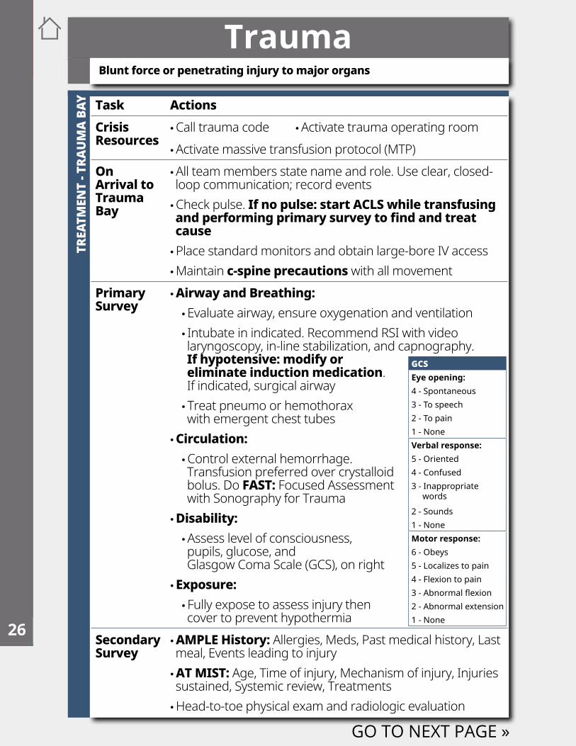
Task ActionsCrisis Resources
•Call trauma code •Activate trauma operating room •Activate massive transfusion protocol (MTP)
On Arrival to Trauma Bay
•All team members state name and role. Use clear, closed-loop communication; record events •Check pulse. If no pulse: start ACLS while transfusing and performing primary survey to find and treat cause •Place standard monitors and obtain large-bore IV access •Maintain c-spine precautions with all movement
Primary Survey
• Airway and Breathing: •Evaluate airway, ensure oxygenation and ventilation • Intubate in indicated. Recommend RSI with video laryngoscopy, in-line stabilization, and capnography. If hypotensive: modify or eliminate induction medication. If indicated, surgical airway • Treat pneumo or hemothorax with emergent chest tubes
• Circulation: •Control external hemorrhage. Transfusion preferred over crystalloid bolus. Do FAST: Focused Assessment with Sonography for Trauma
• Disability: •Assess level of consciousness, pupils, glucose, and Glasgow Coma Scale (GCS), on right
• Exposure: • Fully expose to assess injury then cover to prevent hypothermia
Secondary Survey
• AMPLE History: Allergies, Meds, Past medical history, Last meal, Events leading to injury • AT MIST: Age, Time of injury, Mechanism of injury, Injuries sustained, Systemic review, Treatments •Head-to-toe physical exam and radiologic evaluation
GO TO NEXT PAGE »
Blunt force or penetrating injury to major organs
GCSEye opening:4 - Spontaneous3 - To speech2 - To pain1 - NoneVerbal response:5 - Oriented4 - Confused3 - Inappropriate words
2 - Sounds1 - NoneMotor response:6 - Obeys5 - Localizes to pain4 - Flexion to pain3 - Abnormal flexion2 - Abnormal extension1 - None
3
1516
5678 9
10111213
171819202122232425
2
4
14
26272829
END
TREA
TMEN
T - T
RAU
MA
OR
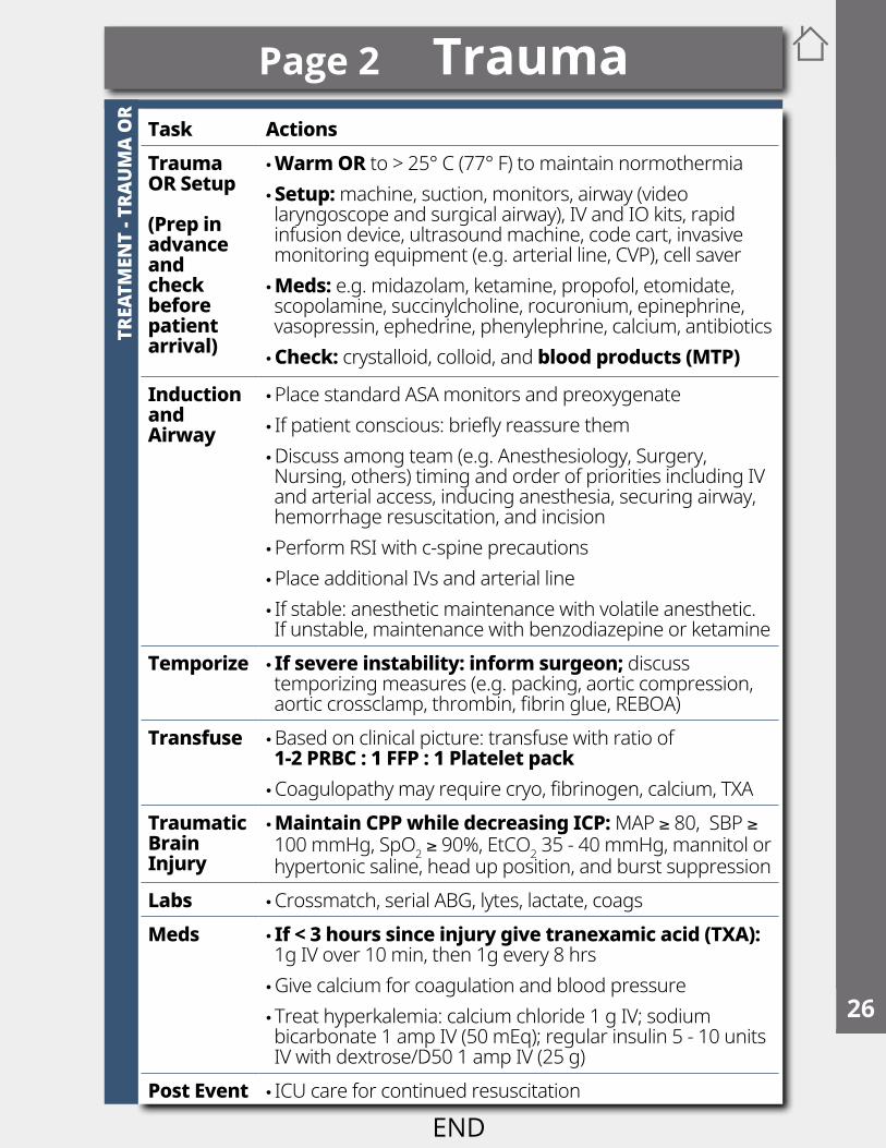
Task ActionsTrauma OR Setup
(Prep in advance and check before patient arrival)
• Warm OR to > 25° C (77° F) to maintain normothermia • Setup: machine, suction, monitors, airway (video laryngoscope and surgical airway), IV and IO kits, rapid infusion device, ultrasound machine, code cart, invasive monitoring equipment (e.g. arterial line, CVP), cell saver • Meds: e.g. midazolam, ketamine, propofol, etomidate, scopolamine, succinylcholine, rocuronium, epinephrine, vasopressin, ephedrine, phenylephrine, calcium, antibiotics • Check: crystalloid, colloid, and blood products (MTP)
Induction and Airway
•Place standard ASA monitors and preoxygenate • If patient conscious: briefly reassure them •Discuss among team (e.g. Anesthesiology, Surgery, Nursing, others) timing and order of priorities including IV and arterial access, inducing anesthesia, securing airway, hemorrhage resuscitation, and incision •Perform RSI with c-spine precautions •Place additional IVs and arterial line • If stable: anesthetic maintenance with volatile anesthetic. If unstable, maintenance with benzodiazepine or ketamine
Temporize • If severe instability: inform surgeon; discuss temporizing measures (e.g. packing, aortic compression, aortic crossclamp, thrombin, fibrin glue, REBOA)
Transfuse •Based on clinical picture: transfuse with ratio of 1-2 PRBC : 1 FFP : 1 Platelet pack •Coagulopathy may require cryo, fibrinogen, calcium, TXA
Traumatic Brain Injury
• Maintain CPP while decreasing ICP: MAP ≥ 80, SBP ≥ 100 mmHg, SpO2 ≥ 90%, EtCO2 35 - 40 mmHg, mannitol or hypertonic saline, head up position, and burst suppression
Labs •Crossmatch, serial ABG, lytes, lactate, coagsMeds • If < 3 hours since injury give tranexamic acid (TXA):
1g IV over 10 min, then 1g every 8 hrs •Give calcium for coagulation and blood pressure • Treat hyperkalemia: calcium chloride 1 g IV; sodium bicarbonate 1 amp IV (50 mEq); regular insulin 5 - 10 units IV with dextrose/D50 1 amp IV (25 g)
Post Event • ICU care for continued resuscitation
Page 2 Trauma1
3
1516
5678 9
10111213
1718192021222324252627
2
4
14
2829
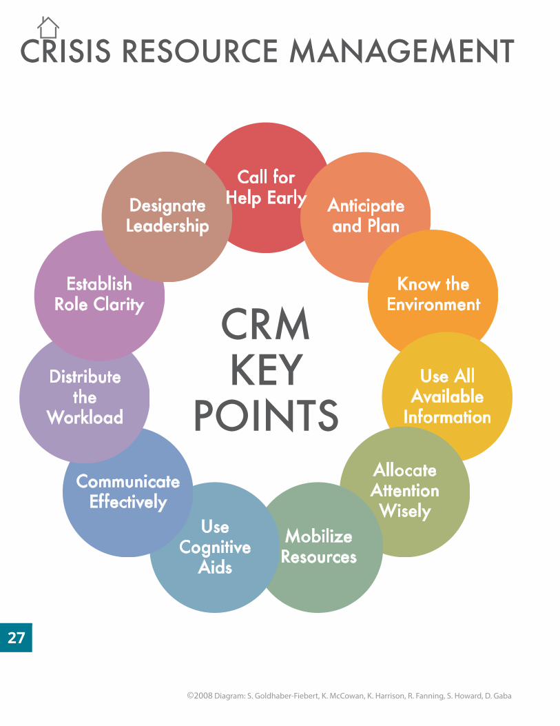
3
1516
5678 9
10111213
171819202122232425
2
4
14
26272829
Call for Help Early Anticipate
and Plan
Know the Environment
Use All Available
Information
Allocate Attention Wisely
Mobilize Resources
Use Cognitive
Aids
Communicate Effectively
Distribute the
Workload
CRMKEY
POINTS
Establish Role Clarity
Designate Leadership
CRISIS RESOURCE MANAGEMENT
©2008 Diagram: S. Goldhaber-Fiebert, K. McCowan, K. Harrison, R. Fanning, S. Howard, D. Gaba
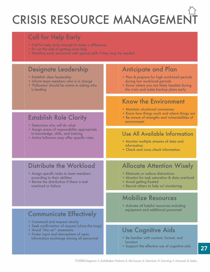
CRISIS RESOURCE MANAGEMENTCall for Help Early • Call for help early enough to make a difference • Err on the side of getting more help • Mobilize early personnel with special skills if they may be needed
Anticipate and Plan • Plan & prepare for high work-load periods during low work-load periods • Know where you are likely headed during the crisis and make backup plans early
Know the Environment • Maintain situational awareness • Know how things work and where things are • Be aware of strengths and vulnerabilities of environment
Use All Available Information• Monitor multiple streams of data and information • Check and cross check information
Allocate Attention Wisely• Eliminate or reduce distractions • Monitor for task saturation & data overload • Avoid getting fixated• Recruit others to help w/ monitoring
Mobilize Resources • Activate all helpful resources including equipment and additional personnel
Use Cognitive Aids • Be familiar with content, format, and location • Support the effective use of cognitive aids
Establish Role Clarity • Determine who will do what • Assign areas of responsibility appropriate to knowledge, skills, and training • Active followers may offer specific roles
Distribute the Workload • Assign specific tasks to team members according to their abilities• Revise the distribution if there is task overload or failure
Communicate Effectively • Command and request clearly • Seek confirmation of request (close the loop) • Avoid “thin air” statements• Foster input and atmosphere of open information exchange among all personnel
Designate Leadership • Establish clear leadership • Inform team members who is in charge • 'Followers' should be active in asking who is leading
©2008 Diagram: S. Goldhaber-Fiebert, K. McCowan, K. Harrison, R. Fanning, S. Howard, D. Gaba
1
3
1516
5678 9
10111213
1718192021222324252627
2
4
14
2829
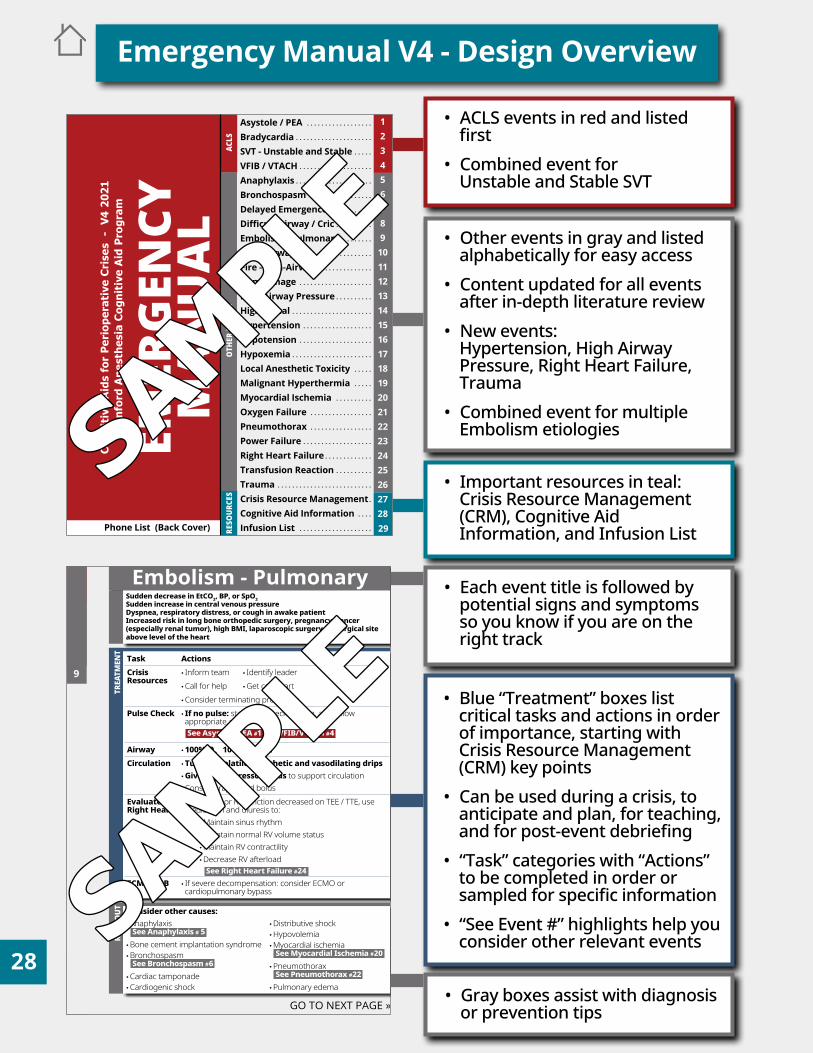
TREA
TMEN
TRU
LE O
UT
Embolism - PulmonarySudden decrease in EtCO2, BP, or SpO2Sudden increase in central venous pressureDyspnea, respiratory distress, or cough in awake patientIncreased risk in long bone orthopedic surgery, pregnancy, cancer (especially renal tumor), high BMI, laparoscopic surgery, or surgical site above level of the heart
Task ActionsCrisis Resources
• Inform team • Identify leader •Call for help •Get code cart •Consider terminating procedure
Pulse Check • If no pulse: start CPR, check rhythm, and follow appropriate algorithm See Asystole/PEA #1 VFIB/VTACH #4
Airway • 100% O2 10 - 15 L/minCirculation •Turn off volatile anesthetic and vasodilating drips
• Give IV vasopressor bolus to support circulation •Consider rapid fluid bolus
Evaluate Right Heart
• If unstable or RV function decreased on TEE / TTE, use medication and diuresis to:
•Maintain sinus rhythm •Maintain normal RV volume status •Maintain RV contractility •Decrease RV afterload See Right Heart Failure #24
ECMO/CPB • If severe decompensation: consider ECMO or cardiopulmonary bypass
GO TO NEXT PAGE »
Consider other causes: •Anaphylaxis See Anaphylaxis # 5 •Bone cement implantation syndrome •Bronchospasm See Bronchospasm #6 •Cardiac tamponade •Cardiogenic shock
•Distributive shock •Hypovolemia •Myocardial ischemia See Myocardial Ischemia #20 •Pneumothorax See Pneumothorax #22 •Pulmonary edema
3
1516
5678 9
10111213
171819202122232425
2
4
14
26272829
• ACLS events in red and listed first
• Combined event for Unstable and Stable SVT
• Other events in gray and listed alphabetically for easy access
• Content updated for all events after in-depth literature review
• New events: Hypertension, High Airway Pressure, Right Heart Failure, Trauma
• Combined event for multiple Embolism etiologies
• Important resources in teal: Crisis Resource Management (CRM), Cognitive Aid Information, and Infusion List
• Each event title is followed by potential signs and symptoms so you know if you are on the right track
• Blue “Treatment” boxes list critical tasks and actions in order of importance, starting with Crisis Resource Management (CRM) key points
• Can be used during a crisis, to anticipate and plan, for teaching, and for post-event debriefing
• “Task” categories with “Actions” to be completed in order or sampled for specific information
• “See Event #” highlights help you consider other relevant events
• Gray boxes assist with diagnosis or prevention tips
Emergency Manual V4 - Design Overview
1
3
1516
5678 9
10111213
1718192021222324252627
2
4
14
2829
Asystole / PEA
SVT - Unstable and Stable
BronchospasmDelayed EmergenceDifficult Airway / CricEmbolism - PulmonaryFire - AirwayFire - Non-AirwayHemorrhage
High Spinal
Local Anesthetic ToxicityMalignant HyperthermiaMyocardial IschemiaOxygen FailurePneumothoraxPower FailureRight Heart FailureTransfusion ReactionTraumaCrisis Resource ManagementCognitive Aid Information Infusion List
Bradycardia
VFIB / VTACHAnaphylaxis
HypotensionHypoxemia
Hypertension
High Airway Pressure
EMER
GEN
CYM
AN
UA
L
Cog
nit
ive
Aid
s fo
r P
erio
per
ativ
e C
rise
s -
V4
202
1S
tan
ford
An
esth
esia
Cog
nit
ive
Aid
Pro
gram
ACLS
RESO
URCE
SO
THER
EVE
NTS
Phone List (Back Cover)
. . . . . . . . . . . . . . . . . .
. . . . .
. . . . . . . . . . . . . . . . .
. . . . . . . . . . . .
. . . . . . . . . .
. . . . . . . . .
. . . . . . . . . . . . . . . . . . . . .
. . . . . . . . . . . . . . .
. . . . . . . . . . . . . . . . . . . .
. . . . . . . . . . . . . . . . . . . . . .
. . . . .
. . . . .
. . . . . . . . . .
. . . . . . . . . . . . . . . . .
. . . . . . . . . . . . . . . . .
. . . . . . . . . . . . . . . . . . .
. . . . . . . . . . . . .
. . . . . . . . . .
. . . . . . . . . . . . . . . . . . . . . . . . . .
.
. . . .
. . . . . . . . . . . . . . . . . . . . .
. . . . . . . . . . . . . . . . . . . .
. . . . . . . . . . . . . . . . . . . . .
. . . . . . . . . . . . . . . . . . . .
. . . . . . . . . . . . . . . . . . . . . .
. . . . . . . . . . . . . . . . . . .
. . . . . . . . . .
. . . . . . . . . . . . . . . . . . . .
3
1516
5678 9
10111213
171819202122232425
2
4
14
26272829
SAMPLE
SAMPLE
SAMPLE
SAMPLE
Emergency Manual V4 - Use and Implementation
Use: Recent research suggests that Emergency Manual (EM) use improves teamwork, facilitates coordination, decreases stress, and enables delivery of better patient care.1 The Stanford EM, and other similar tools, are used effectively in both clinical and educational settings:
Clinical: • Pre-event for ‘just in time’ review for at-risk patients • During event for crisis management • Post-event for team debriefing
Educational: • Self-review • 1:1 or small group teaching • Studying for oral exams • During simulation cases and debrief sessions
We welcome your feedback and continuously learn from our community of users.
Implementation: These websites include a number of tips and free resources to support EM implementation by your interprofessional team:
• Stanford EM - https://emergencymanual.stanford.edu/ Download the EM in English or another language, find implementation tips, and learn more• Emergency Manual Implementation Collaborative (EMIC) - https://www.emergencymanuals.org/• EM Implementation Toolkit - https://www.implementingemergencychecklists.org This resource-rich guide, developed with Ariadne Labs, includes videos you can use or adapt, and other training materials.
Stanford EM Formats:• Large (8 ½ x 11”) hanging printed version (most popular), with or without event number tabs*• Small (4 ¼ x 5 ½”) printed pocket version*• PDF with hyperlinks (accessible on a computer, mobile device, or an electronic health record) • E-book*Our printed versions are operating room safe (wipeable, and MRI-safe). You can use any printer you choose. See our website for information about our printer.
EM Customization - See our website for templates to customize for your setting:• Infusion List (inside back cover)• Phone List (outside back cover)
EM Training:• EM Reader: reads aloud to team/leader to interactively ensure vital actions are performed, medication doses correct, diagnoses considered, find specific desired information, while allowing leader to maintain situational awareness and team communication.2 • Train team members to ask empowering questions: Would you like me to get/read the emergency manual? Which event in the emergency manual are we dealing with? These can help the leader remember the EM is available and can trigger its use. In our experience, leaders often answer, “Yes,” and had forgotten about the EM due to the stress of the crisis.
1. Goldhaber-Fiebert SN, Howard SK, Gaba DM, et al. Clinical Uses and Impacts of Emergency Manuals During Perioperative Crises. Anesth Analg. 2020 Dec;131(6):1815-1826. 2. Burden AR, et al. Does every code need a “reader?” Improvement of rare event management with a cognitive aid “reader” during a simulated emergency: a pilot study. Simul Healthc. 2012 Feb;7(1):1-9.
1
3
1516
5678 9
10111213
1718192021222324252627
2
4
14
2829
Emergency Manual V4 - Publication Details
Stanford Anesthesia Cognitive Aid Program (SACAP) EM V4 Team:
Ellile Sultan, MD Amanda Burden, MD Barbara K. Burian, PhD Naola S. Austin, MD Kyle Harrison, MD Sara Goldhaber-Fiebert, MD Steven K. Howard, MD David M. Gaba, MD Summer Reid, BA (Administrative Associate) Acknowledgements: Iterative simulation testing and integration of feedback from clinical uses of V3 were integral to the development of V4. Meredith Hutton MD and Vladimir Nekhendzy MD made author-level contributions to content and usability of the Difficult Airway / Cric event. We appreciate the leadership, faculty, trainees, and staff at Stanford for their support of the development, implementation, and improvement of this emergency manual over the past decade. Many individuals contributed over the years. For this version, we especially appreciate the detailed reviews of content, design, and usability, by 38 global expert physicians from 16 institutions and 8 countries. For more information on current and past contributions, please visit our website: https://emergencymanual.stanford.edu/
References: Citations are not written on each event given usability priorities, but you can visit our website if interested in relevant content literature. We strive to integrate the most pertinent clinical information from published literature and clinical practice guidelines.
Citation: Stanford Anesthesia Cognitive Aid Program,* Emergency Manual: Cognitive aids for perioperative crises, Version 4, 2021. See http://emergencymanual.stanford.edu for latest version. Creative Commons BY-NC-ND (https://creativecommons.org/licenses/by-nc-nd/4.0/legalcode). *Goldhaber-Fiebert SN, Austin N, Sultan E, Burian BK, Burden A, Howard SK, Gaba DM, Harrison TK.
Local Modifications and Creative Commons Licensing: Research supports that local customization of cognitive aids is helpful for many reasons. We allow all needed modifications for use at your local institution, without further permissions. You should keep original authorship attribution and add ‘Adapted By ___.’ For more than minor PDF modifications or local phone list for back cover, we suggest requesting our original InDesign file: Email [email protected]. We are not responsible for any errors introduced and caution that there are usability cons to adding too much information. See this review for an overview of effective cognitive aid design.1 No derivatives may be shared beyond local use without explicit permission (e.g. translations or hospital systems that contact us first), and all use must be non-commercial. We use Creative Commons 4.0 International Licensing, With Attribution, Non-Commercial, and No Derivatives; See details at https://creativecommons.org/licenses/by-nc-nd/4.0/
Disclaimers: The material in this Manual is not intended to be a substitute for sound medical knowledge and training. Clinicians should always use their clinical judgment and decision making for patient management. Departure from the information presented here is encouraged as appropriate, since situations can vary widely.
We use generic medication names whenever possible and include some brand names, which might be better known to clinicians, to support effective use during crises. To reduce potentially distracting visual clutter, TM superscripts have not been included with brand name medications in the cognitive aids.
Enabling clinical uses during crises requires systematic implementation efforts beyond simply hanging emergency manuals (EM) in operating rooms, as EMs can be forgotten when under stress. Use the resources on the previous page to efficiently and effectively integrate EMs into your practice.
1. Burian BK, Clebone A, Dismukes K, Ruskin KJ. More Than a Tick Box: Medical Checklist Development, Design, and Use. Anesth Analg. 2018 Jan;126(1):223-232.
3
1516
5678 9
10111213
171819202122232425
2
4
14
26272829
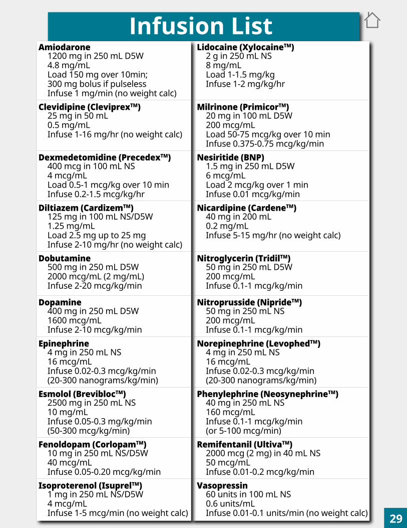
1
3
1516
5678 9
10111213
1718192021222324252627
2
4
14
2829
Infusion ListAmiodarone
1200 mg in 250 mL D5W4.8 mg/mLLoad 150 mg over 10min; 300 mg bolus if pulselessInfuse 1 mg/min (no weight calc)
Lidocaine (XylocaineTM)2 g in 250 mL NS 8 mg/mLLoad 1-1.5 mg/kgInfuse 1-2 mg/kg/hr
Clevidipine (CleviprexTM)25 mg in 50 mL0.5 mg/mLInfuse 1-16 mg/hr (no weight calc)
Milrinone (PrimicorTM)20 mg in 100 mL D5W200 mcg/mLLoad 50-75 mcg/kg over 10 minInfuse 0.375-0.75 mcg/kg/min
Dexmedetomidine (PrecedexTM)400 mcg in 100 mL NS4 mcg/mLLoad 0.5-1 mcg/kg over 10 minInfuse 0.2-1.5 mcg/kg/hr
Nesiritide (BNP)1.5 mg in 250 mL D5W6 mcg/mLLoad 2 mcg/kg over 1 minInfuse 0.01 mcg/kg/min
Diltiazem (CardizemTM)125 mg in 100 mL NS/D5W1.25 mg/mLLoad 2.5 mg up to 25 mgInfuse 2-10 mg/hr (no weight calc)
Nicardipine (CardeneTM)40 mg in 200 mL0.2 mg/mLInfuse 5-15 mg/hr (no weight calc)
Dobutamine500 mg in 250 mL D5W2000 mcg/mL (2 mg/mL)Infuse 2-20 mcg/kg/min
Nitroglycerin (TridilTM)50 mg in 250 mL D5W200 mcg/mLInfuse 0.1-1 mcg/kg/min
Dopamine400 mg in 250 mL D5W1600 mcg/mLInfuse 2-10 mcg/kg/min
Nitroprusside (NiprideTM)50 mg in 250 mL NS200 mcg/mLInfuse 0.1-1 mcg/kg/min
Epinephrine4 mg in 250 mL NS16 mcg/mLInfuse 0.02-0.3 mcg/kg/min(20-300 nanograms/kg/min)
Norepinephrine (LevophedTM)4 mg in 250 mL NS16 mcg/mLInfuse 0.02-0.3 mcg/kg/min(20-300 nanograms/kg/min)
Esmolol (BreviblocTM)2500 mg in 250 mL NS10 mg/mLInfuse 0.05-0.3 mg/kg/min(50-300 mcg/kg/min)
Phenylephrine (NeosynephrineTM)40 mg in 250 mL NS160 mcg/mLInfuse 0.1-1 mcg/kg/min(or 5-100 mcg/min)
Fenoldopam (CorlopamTM)10 mg in 250 mL NS/D5W40 mcg/mLInfuse 0.05-0.20 mcg/kg/min
Remifentanil (UltivaTM)2000 mcg (2 mg) in 40 mL NS50 mcg/mLInfuse 0.01-0.2 mcg/kg/min
Isoproterenol (IsuprelTM)1 mg in 250 mL NS/D5W4 mcg/mLInfuse 1-5 mcg/min (no weight calc)
Vasopressin60 units in 100 mL NS0.6 units/mLInfuse 0.01-0.1 units/min (no weight calc)

Phone listplaceholder
Interv. Platform Number Operating Rms NumberAnes Scheduler 500P (XX = OR#)
500P/After 5pm 6-0249 Circulator 6-37XX300P 3-3430 Circ. (OR 29) 7-6251
Lane Surgery Ctr (LSC) 4-8812 Emergency MDs Number Anes. OR 1 7-3083Anes Tech 300P Airway Pgr #12640 OR 2 7-3085
500P 4-0219 500P Airway Pgr #12605 OR 3 7-3007300P 6-1850 ENT Pgr #27087 OR 4 7-3091
LSC 6-2120 Interv. Rad. Pgr #27237 OR 6 7-3475ENDO/MRI 4-6359 Neurology Pgr #27191 OR 7 7-3508
CATH/CT 4-4587 Regional Block Pgr #25625 OR 8 7-3512Biomed. Engineering Acute Pain Pgr #27246 OR 9 7-3483
500P 628-273-3352 Periop Resident Pgr #22637 OR 10 7-3480300P 5-7454 OR 11 7-3477
LSC 5-8186 LPCH Number OR 12 7-3119Cath Lab 500P/300P 7-2595 Peds Control Desk 1-2820 OR 14 7-3120
Charge Nurse Peds Anes Scheduler 1-9705 OR 15 7-3124500P 650-788-5784 Peds Anes Emergency 1-9706 OR 16 7-3126300P 4-4590 OB/L&D Front Desk 3-5403 OR 17 7-3210
LSC 4-9420 OB Anes Attending 1-0865 OR 18 7-3212Clinical Lab 3-6671/3-6111 OB Anes Fellow 1-0866 OR 19 7-3097Control Desk OB Anes Resident 1-0867 OR 20 7-3101
500P 3-7251 OR 21 7-3104300P 7-5468 ICU Number OR 22 7-3093
LSC 5-6102 E2 (300P) Front Desk 5-7122 OR 23 7-3094Cath Lab 500P 7-1896 M4 Front Desk 7-3294 OR 24 7-3096Cath Lab 300P 3-7676 J4 Front Desk 7-3301 OR 25 7-3113
LSC IR Scheduler 5-3615 K4 Front Desk 7-3313 OR 26 7-3118LSC Brachytherapy 5-4371 J2 (CVICU) Front Desk 7-3325 OR 27 7-3468
Endoscopy 650-823-6841 MICU Triage Fellow 4-8820 OR 28 7-3472 PET-CT/NucMed 5-8263 SICU Resident 5-3234 OR 29 7-6241
MRI 500P/300P 3-6335 OR 30 7-3105Emergency Dept 3-7337 Location Number 300P (XX = OR#)
Pharmacy Blood Bank (MTG) 3-6444/3-6445 Circulator 4-70XX500P 7-1723 Transfusion MD Pgr #12027 Anes. ORs 5-72XX
300P/LSC 5-5923 Charge Nurse 7-1351 Anes. (OR 21) 8-2121Preop/Holding Blood Gas 5-4382 LSC (XX = OR#)
500P 7-3187 EPIC/IT Help 3-3333 Circulator 6-88XX300P 5-8742 Interpreter Service 7-7780 Anes. A1 6-8813
LSC 3-5991 Interpreter Phone 1-800-523-1786 A2 6-8814Cath Lab 500P 7-3181 Code 201273 A3 5-9869Cath Lab 300P 4-5428 Occup. Health 3-5922 A4 8-7973
PACU Needle Stick Pgr #17849 A5 5-5349500P 7-3133 Preop Clinic 4-1429 A6 5-5350300P 3-6631 Radiology Rdg Room 6-1173 A7 5-5354
LSC 3-5917 RT Resource 3-7709 A8 5-3067Cath Lab 500P 7-3113 RT Supervisor 650-867-9312 A9 5-3066Cath Lab 300P 4-5428 Risk Management 3-6824 A10 3-7815
288 for on-call manager or pgr #16210 A11 3-4901Security 3-7222 A12 5-9391
Stanford Phone Numbers
Operator Inside: 288Operator
Operator Outside (650) 723-4000
Full Prefix: (650) 721-, 723-, 724-, 725-, 736-, 497-, 498- EMERGENCY or CODE, including Stroke Code, STEMI, Code Blue, Airway Code → Dial 211
Phone ListPlaceholder
EMERGENCY MANUAL