Embed Size (px)
Citation preview
A discrete population of squamocolumnar junction cellsimplicated in the pathogenesis of cervical cancerMichael Herfsa,b, Yusuke Yamamotoc, Anna Lauryd, Xia Wange, Marisa R. Nuccia, Margaret E. McLaughlin-Drubinf,Karl Müngerf, Sarah Feldmang, Frank D. McKeonc,e,1, Wa Xiana,h,1,2, and Christopher P. Cruma,1,2
aDivision of Women’s and Perinatal Pathology, Department of Pathology, Brigham and Women’s Hospital, Boston, MA 02115; bDepartment of Pathology,Groupe Interdisciplinaire de Génoprotéomique Appliquée (GIGA)-Cancer, University of Liege, Liege, Belgium; cGenome Institute of Singapore, Agency forScience, Technology and Research (A-STAR), Singapore 138672; dDepartment of Pathology, School of Medicine, University of California, Los Angeles, CA90095; eDepartment of Cell Biology, Harvard Medical School, Boston, MA 02115; fDepartment of Medicine, Channing Laboratory, Brigham and Women’sHospital, Boston, MA 02115; gDepartment of Obstetrics and Gynecology and Reproductive Biology, Brigham and Women’s Hospital, Boston, MA 02115; andhInstitute of Medical Biology, A-STAR, Singapore 138648
Edited by Douglas R. Lowy, National Cancer Institute, Bethesda, MD, and approved May 8, 2012 (received for review February 19, 2012)
Infection by carcinogenic human papillomaviruses (HPV) results inprecancers [cervical intraepithelial neoplasia (CIN)] and cancers nearthe ectoendocervical squamocolumnar (SC) junction of the cervix.However, the specific cells targetedbyHPV have not been identifiedand the cellular origin of cervical cancer remains elusive. In thisstudy,weuncovered a discrete population of SC junctional cellswithunique morphology and gene-expression profile. We also demon-strated that the selected junctional biomarkers were expressed bya high percentage of high-grade CIN and cervical cancers associatedwith carcinogenic HPVs but rarely in ectocervical/transformationzone CINs or those associated with noncarcinogenic HPVs. That theoriginal SC junction immunophenotypewas not regenerated at newSC junctions following excision, not induced by expression of viraloncoproteins in foreskin keratinocytes, and not seen in HPV-relatedprecursors of the vagina, vulva, and penis further support the notionthat junctional cells are the source of cervical cancer. Taken together,our findings suggest that carcinogenic HPV-related CINs and cervicalcancers are linked to a small, discrete cell population that localizes tothe SC junction of the cervix, expresses a unique gene expressionsignature, and is not regenerated after excision. The findings in thisstudy uncover a potential target for cervical cancer prevention,provide insight into the risk assessment of cervical lesions, andestablish a model for elucidating the pathway to cervical cancerfollowing carcinogenic HPV infection.
gynecology | oncology | embryogenesis
Carcinogenic human papillomaviruses (HPVs) cause cervicalcancer and its precursor, cervical intraepithelial neoplasia
(CIN), at the squamocolumnar (SC) junction (1–6). Because ofboth cervical anatomy and hormonal status, the position of thisjunction varies. In most women, vaginal pH acidification occur-ring during adolescence induces the replacement of a portion ofthe endocervical columnar epithelium by a metaplastic squa-mous epithelium. This area of replacement [the transformationzone (TZ)] leads to the proximal migration of the SC junction.In the last decade, the relationship between HPV infection and
neoplasia has spawned new paradigms of cervical cancer preventionviaHPV screening andPap smear triage (7, 8).Moreover, successfulconstruction of papilloma virions in vitro has resulted in vaccinesthat prevent many CINs and presumably, cervical cancer (9).Although infection withHPV is necessary for the development of
CIN, the components of early cervical carcinogenesis have yet to befully assembled. HPV infection presumably initiates when the virusenters basal keratinocytes via defects in the epithelial covering,attachment to the basement membrane, and viral capsid confor-mational change (10). Models of HPV-mediated oncogenesis useHPV-immortalized keratinocytes or transgenicmice (11, 12). Thesesystems have uncovered important molecular pathways; however,they have not shed light on why cervical neoplasms are topo-graphically restricted to the SC junction (5, 6). Rather, it was gen-erally assumed that the TZ squamous epithelium was the site of
neoplastic change.Recently, it was reported that Barrett esophagus,a precursor of esophageal adenocarcinoma, is derived from a dis-crete population of embryonic cells residing at the gastroesophagealSC junction (13). In this report, wedescribe a similar, and previouslyunreported, population of SC junction cells in the cervix with aunique expression profile, embryonic characteristics, and relation-ship to carcinogenic HPV-associated cervical neoplasia.
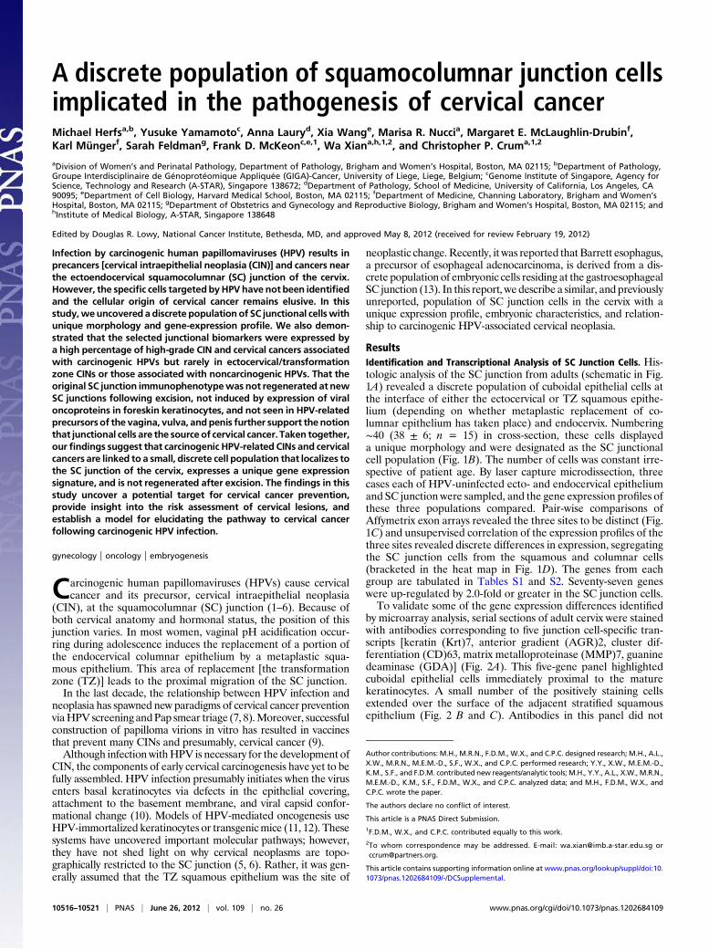
ResultsIdentification and Transcriptional Analysis of SC Junction Cells. His-tologic analysis of the SC junction from adults (schematic in Fig.1A) revealed a discrete population of cuboidal epithelial cells atthe interface of either the ectocervical or TZ squamous epithe-lium (depending on whether metaplastic replacement of co-lumnar epithelium has taken place) and endocervix. Numbering∼40 (38 ± 6; n = 15) in cross-section, these cells displayeda unique morphology and were designated as the SC junctionalcell population (Fig. 1B). The number of cells was constant irre-spective of patient age. By laser capture microdissection, threecases each of HPV-uninfected ecto- and endocervical epitheliumand SC junction were sampled, and the gene expression profiles ofthese three populations compared. Pair-wise comparisons ofAffymetrix exon arrays revealed the three sites to be distinct (Fig.1C) and unsupervised correlation of the expression profiles of thethree sites revealed discrete differences in expression, segregatingthe SC junction cells from the squamous and columnar cells(bracketed in the heat map in Fig. 1D). The genes from eachgroup are tabulated in Tables S1 and S2. Seventy-seven geneswere up-regulated by 2.0-fold or greater in the SC junction cells.To validate some of the gene expression differences identified
by microarray analysis, serial sections of adult cervix were stainedwith antibodies corresponding to five junction cell-specific tran-scripts [keratin (Krt)7, anterior gradient (AGR)2, cluster dif-ferentiation (CD)63, matrix metalloproteinase (MMP)7, guaninedeaminase (GDA)] (Fig. 2A). This five-gene panel highlightedcuboidal epithelial cells immediately proximal to the maturekeratinocytes. A small number of the positively staining cellsextended over the surface of the adjacent stratified squamousepithelium (Fig. 2 B and C). Antibodies in this panel did not
Author contributions: M.H., M.R.N., F.D.M., W.X., and C.P.C. designed research; M.H., A.L.,X.W., M.R.N., M.E.M.-D., S.F., W.X., and C.P.C. performed research; Y.Y., X.W., M.E.M.-D.,K.M., S.F., and F.D.M. contributed new reagents/analytic tools; M.H., Y.Y., A.L., X.W., M.R.N.,M.E.M.-D., K.M., S.F., F.D.M., W.X., and C.P.C. analyzed data; and M.H., F.D.M., W.X., andC.P.C. wrote the paper.
The authors declare no conflict of interest.
This article is a PNAS Direct Submission.1F.D.M., W.X., and C.P.C. contributed equally to this work.2To whom correspondence may be addressed. E-mail: [email protected] [email protected].
This article contains supporting information online at www.pnas.org/lookup/suppl/doi:10.1073/pnas.1202684109/-/DCSupplemental.
10516–10521 | PNAS | June 26, 2012 | vol. 109 | no. 26 www.pnas.org/cgi/doi/10.1073/pnas.1202684109
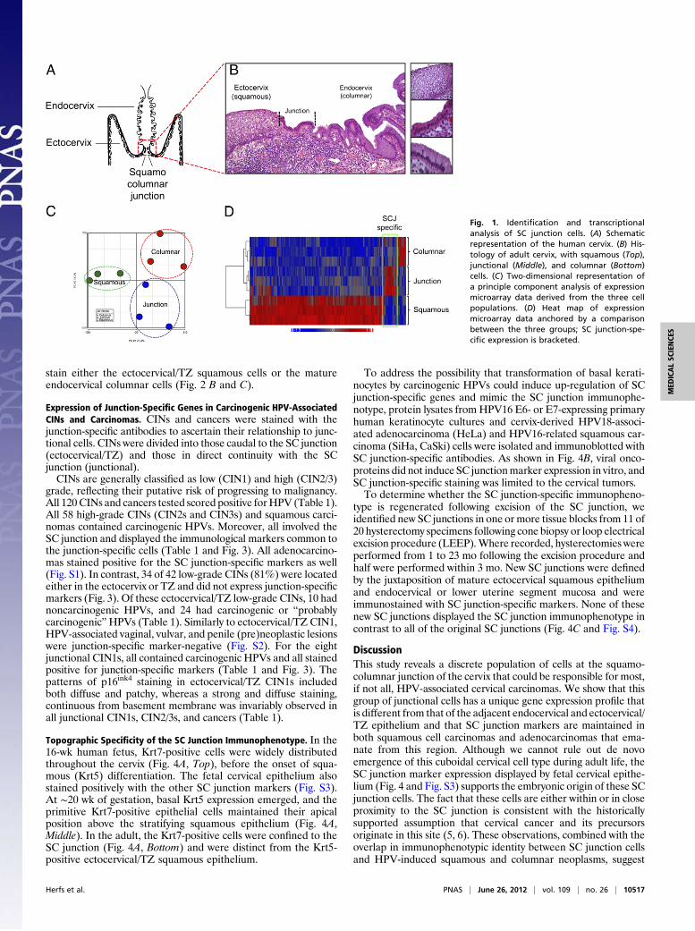
stain either the ectocervical/TZ squamous cells or the matureendocervical columnar cells (Fig. 2 B and C).
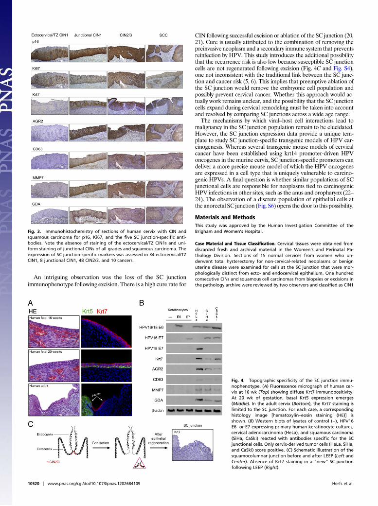
Expression of Junction-Specific Genes in Carcinogenic HPV-AssociatedCINs and Carcinomas. CINs and cancers were stained with thejunction-specific antibodies to ascertain their relationship to junc-tional cells. CINs were divided into those caudal to the SC junction(ectocervical/TZ) and those in direct continuity with the SCjunction (junctional).CINs are generally classified as low (CIN1) and high (CIN2/3)
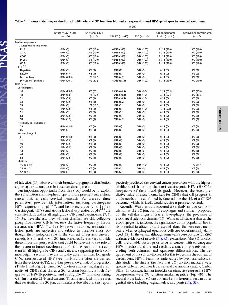
grade, reflecting their putative risk of progressing to malignancy.All 120 CINs and cancers tested scored positive forHPV (Table 1).All 58 high-grade CINs (CIN2s and CIN3s) and squamous carci-nomas contained carcinogenic HPVs. Moreover, all involved theSC junction and displayed the immunological markers common tothe junction-specific cells (Table 1 and Fig. 3). All adenocarcino-mas stained positive for the SC junction-specific markers as well(Fig. S1). In contrast, 34 of 42 low-grade CINs (81%) were locatedeither in the ectocervix or TZ and did not express junction-specificmarkers (Fig. 3). Of these ectocervical/TZ low-grade CINs, 10 hadnoncarcinogenic HPVs, and 24 had carcinogenic or “probablycarcinogenic”HPVs (Table 1). Similarly to ectocervical/TZ CIN1,HPV-associated vaginal, vulvar, and penile (pre)neoplastic lesionswere junction-specific marker-negative (Fig. S2). For the eightjunctional CIN1s, all contained carcinogenic HPVs and all stainedpositive for junction-specific markers (Table 1 and Fig. 3). Thepatterns of p16ink4 staining in ectocervical/TZ CIN1s includedboth diffuse and patchy, whereas a strong and diffuse staining,continuous from basement membrane was invariably observed inall junctional CIN1s, CIN2/3s, and cancers (Table 1).
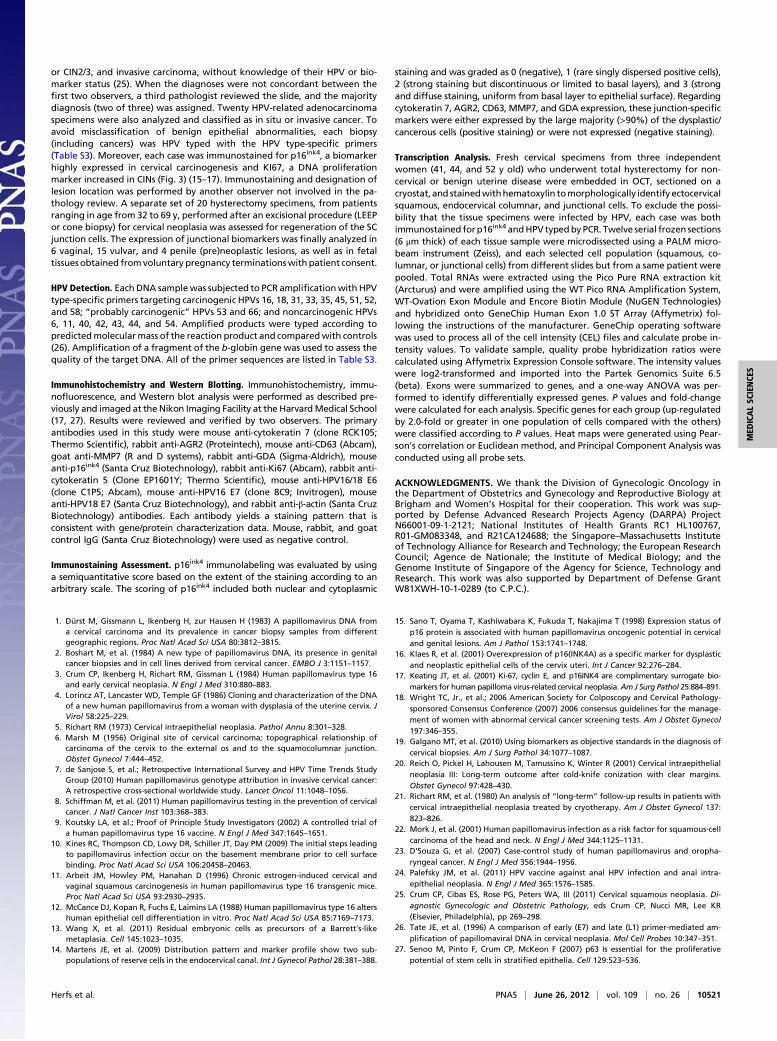
Topographic Specificity of the SC Junction Immunophenotype. In the16-wk human fetus, Krt7-positive cells were widely distributedthroughout the cervix (Fig. 4A, Top), before the onset of squa-mous (Krt5) differentiation. The fetal cervical epithelium alsostained positively with the other SC junction markers (Fig. S3).At ∼20 wk of gestation, basal Krt5 expression emerged, and theprimitive Krt7-positive epithelial cells maintained their apicalposition above the stratifying squamous epithelium (Fig. 4A,Middle). In the adult, the Krt7-positive cells were confined to theSC junction (Fig. 4A, Bottom) and were distinct from the Krt5-positive ectocervical/TZ squamous epithelium.
To address the possibility that transformation of basal kerati-nocytes by carcinogenic HPVs could induce up-regulation of SCjunction-specific genes and mimic the SC junction immunophe-notype, protein lysates from HPV16 E6- or E7-expressing primaryhuman keratinocyte cultures and cervix-derived HPV18-associ-ated adenocarcinoma (HeLa) and HPV16-related squamous car-cinoma (SiHa, CaSki) cells were isolated and immunoblotted withSC junction-specific antibodies. As shown in Fig. 4B, viral onco-proteins did not induce SC junctionmarker expression in vitro, andSC junction-specific staining was limited to the cervical tumors.To determine whether the SC junction-specific immunopheno-
type is regenerated following excision of the SC junction, weidentified new SC junctions in one or more tissue blocks from 11 of20 hysterectomy specimens following cone biopsy or loop electricalexcision procedure (LEEP).Where recorded, hysterectomies wereperformed from 1 to 23 mo following the excision procedure andhalf were performed within 3 mo. New SC junctions were definedby the juxtaposition of mature ectocervical squamous epitheliumand endocervical or lower uterine segment mucosa and wereimmunostained with SC junction-specific markers. None of thesenew SC junctions displayed the SC junction immunophenotype incontrast to all of the original SC junctions (Fig. 4C and Fig. S4).
DiscussionThis study reveals a discrete population of cells at the squamo-columnar junction of the cervix that could be responsible for most,if not all, HPV-associated cervical carcinomas. We show that thisgroup of junctional cells has a unique gene expression profile thatis different from that of the adjacent endocervical and ectocervical/TZ epithelium and that SC junction markers are maintained inboth squamous cell carcinomas and adenocarcinomas that ema-nate from this region. Although we cannot rule out de novoemergence of this cuboidal cervical cell type during adult life, theSC junction marker expression displayed by fetal cervical epithe-lium (Fig. 4 and Fig. S3) supports the embryonic origin of these SCjunction cells. The fact that these cells are either within or in closeproximity to the SC junction is consistent with the historicallysupported assumption that cervical cancer and its precursorsoriginate in this site (5, 6). These observations, combined with theoverlap in immunophenotypic identity between SC junction cellsand HPV-induced squamous and columnar neoplasms, suggest
Fig. 1. Identification and transcriptionalanalysis of SC junction cells. (A) Schematicrepresentation of the human cervix. (B) His-tology of adult cervix, with squamous (Top),junctional (Middle), and columnar (Bottom)cells. (C) Two-dimensional representation ofa principle component analysis of expressionmicroarray data derived from the three cellpopulations. (D) Heat map of expressionmicroarray data anchored by a comparisonbetween the three groups; SC junction-spe-cific expression is bracketed.
Herfs et al. PNAS | June 26, 2012 | vol. 109 | no. 26 | 10517
MED
ICALSC
IENCE
S
that this unique population of cells at the SC junction forms a coregroup that can spawn multiple tumor phenotypes (Table 1).The cell of origin for cervical cancer has been subject to specu-
lation for some time but has traditionally been linked to the squa-mous epithelium in the TZ. Interestingly, we found that the SCjunction population is not a stratified squamous epithelium (theprototype formost models of HPV infection in the TZ) but a single
layer of cuboidal epithelial cells (Figs. 1, 2, and 4). The observationin this report that SC junction cells are the prime target for cervicalcarcinogenesis does not deny the prevailing theory that epithelialdisruption and basal keratinocyte infection lead to HPV infection(10), because it is likely the mechanism for HPV-related lesionsdeveloping in the TZ, ectocervix, and other mucosal sites. More-over, columnar or reserve cells have also been proposed as targets
Fig. 2. Characterization of several SC junction-specific biomarkers. (A) Fold differences in ex-pression of five genes within the squamous, co-lumnar, and junctional cells. These results arederived from microarray analysis. (B) Low-magni-fication image showing the entire cervix. Krt7staining highlights a discrete population of cu-boidal cells at the interface of the transformationzone squamous epithelium and endocervix. (C)Antibodies stain an identical cell population atthe SC junction. The positive staining extendsfrom the surface of the stratified squamous epi-thelium onto the adjacent basement membrane.Note the absence of staining of either the maturekeratinocytes or the endocervical columnar cells.
10518 | www.pnas.org/cgi/doi/10.1073/pnas.1202684109 Herfs et al.
of infection (14). However, their broader topographic distributionargues against a unique role in cancer development.An important opportunity from this study would be to exploit
the SC junction immunophenotype to more precisely understandcancer risk in early cervical neoplasia. At present, threeparameters provide risk information, including carcinogenicHPV, expression of p16ink4, and histologic grade (7, 8, 15–19).Carcinogenic HPVs and strong lesional expression of p16ink4 areconsistently found in all high grade CINs and carcinomas (7, 8,15–19); nevertheless, they will not discriminate this collectivegroup from most CIN1s because the latter frequently harborcarcinogenic HPVs (17, 19). Moreover histologic estimates oflesion grade are subjective and subject to observer error. Al-though their biological role in the context of cervical carcino-genesis is still unknown, SC junction-specific markers providethree important perspectives that could be relevant to the role ofthis region in tumor development. First, they seem to be a con-stant in all high-grade CINs and cancers, supporting their com-mon origin. Second, they are virtually absent in most low-gradeCINs, irrespective of HPV type, implying the latter are derivedfrom the ectocervix/TZ, and thus pose a lower risk of progression(Table 1 and Fig. 3). Third, SC junction markers highlight a mi-nority of CIN1s that shares a SC junction location, a high fre-quency of HPV16 positivity, and strong p16ink4 immunostainingwith high grade CINs and cancers. Thus, in nearly all of the casesthat we studied, the SC junction markers described in this report
precisely predicted the cervical cancer precursors with the highestlikelihood of harboring the most carcinogenic HPV (HPV16),irrespective of their histologic grade. However, the exact pre-dictive value of these biomarkers for CIN1s that will progress ingrade needs to be confirmed by determining the risk of a CIN2/3outcome, which, in itself, would require a prospective study.Recently, Wang et al. uncovered a similarly unique cell pop-
ulation at the SC junction of esophagus and stomach proposedas the cellular origin of Barrett’s esophagus, the precursor ofesophageal adenocarcinoma (13). Wang et al. suggest that at theesophagogastric junction, the significance of this population lies inits potential to attach to and expand along the basement mem-brane when esophageal squamous cells are experimentally dam-aged (13). In the cervix, although some cells score positive for Ki67without evidence of mitosis (Fig. S5), expansion of the SC junctioncells presumably occurs prior to or in concert with carcinogenicHPV infection, and the end result is a range of phenotypes, in-cluding both columnar and squamous differentiation. The re-quirement of the SC junction cells for this to occur in the context ofcarcinogenic HPV infection is underscored by two observations inthis study. The first is the fact that SC junction markers werespecific only for cell lines from cervical carcinomas (HeLa, CasKi,SiHa). In contrast, human foreskin keratinocytes expressing HPVoncoproteins were SC junction marker-negative (Fig. 4B). Thesecond is the lack of SC junctionmarkers in lesions arising in othergenital sites, including vagina, vulva, and penis (Fig. S2).
Table 1. Immunostaining evaluation of p16ink4a and SC Junction biomarker expression and HPV genotypes in cervical specimens
N (%)
Ectocervical/TZ CIN 1(n = 34)
Junctional CIN 1(n = 8) CIN 2/3 (n = 48) SCC (n = 10)
Adenocarcinomain situ (n = 11)
Invasive adenocarcinoma(n = 9)
Protein expressionSC junction-specific genesKrt7 0/34 (0) 8/8 (100) 48/48 (100) 10/10 (100) 11/11 (100) 9/9 (100)AGR2 0/34 (0) 8/8 (100) 48/48 (100) 10/10 (100) 11/11 (100) 9/9 (100)CD63 0/34 (0) 8/8 (100) 48/48 (100) 10/10 (100) 11/11 (100) 9/9 (100)MMP7 0/34 (0) 8/8 (100) 48/48 (100) 10/10 (100) 11/11 (100) 9/9 (100)GDA 0/34 (0) 8/8 (100) 48/48 (100) 10/10 (100) 11/11 (100) 9/9 (100)
p16ink4a
Negative 0/34 (0) 0/8 (0) 0/48 (0) 0/10 (0) 0/11 (0) 0/9 (0)Patchy 16/34 (47) 0/8 (0) 0/48 (0) 0/10 (0) 0/11 (0) 0/9 (0)Diffuse basal 8/34 (23.5) 1/8 (12.5) 2/48 (4.2) 0/10 (0) 0/11 (0) 0/9 (0)Diffuse full thickness 10/34 (29.5) 7/8 (87.5) 46/48 (95.8) 10/10 (100) 11/11 (100) 9/9 (100)
HPV typeCarcinogenic16 8/34 (23.6) 6/8 (75) 29/48 (60.4) 6/10 (60) 7/11 (63.6) 5/9 (55.6)18 3/34 (8.8) 1/8 (12.5) 5/48 (10.4) 1/10 (10) 3/11 (27.3) 3/9 (33.3)31 3/34 (8.8) 0/8 (0) 5/48 (10.4) 1/10 (10) 0/11 (0) 0/9 (0)33 1/34 (2.9) 0/8 (0) 3/48 (6.2) 0/10 (0) 0/11 (0) 0/9 (0)35 0/34 (0) 1/8 (12.5) 1/48 (2.1) 0/10 (0) 0/11 (0) 0/9 (0)45 1/34 (2.9) 0/8 (0) 0/48 (0) 1/10 (10) 1/11 (9.1) 0/9 (0)51 0/34 (0) 0/8 (0) 1/48 (2.1) 0/10 (0) 0/11 (0) 0/9 (0)52 2/34 (5.9) 0/8 (0) 0/48 (0) 0/10 (0) 0/11 (0) 0/9 (0)58 2/34 (5.9) 0/8 (0) 2/48 (4.2) 0/10 (0) 0/11 (0) 0/9 (0)
“Probably carcinogenic”53 4/34 (11.8) 0/8 (0) 0/48 (0) 0/10 (0) 0/11 (0) 0/9 (0)66 0/34 (0) 0/8 (0) 0/48 (0) 0/10 (0) 0/11 (0) 0/9 (0)
Noncarcinogenic6 4/34 (11.8) 0/8 (0) 0/48 (0) 0/10 (0) 0/11 (0) 0/9 (0)11 2/34 (5.9) 0/8 (0) 0/48 (0) 0/10 (0) 0/11 (0) 0/9 (0)40 1/34 (2.9) 0/8 (0) 0/48 (0) 0/10 (0) 0/11 (0) 0/9 (0)42 1/34 (2.9) 0/8 (0) 0/48 (0) 0/10 (0) 0/11 (0) 0/9 (0)43 0/34 (0) 0/8 (0) 0/48 (0) 0/10 (0) 0/11 (0) 0/9 (0)44 0/34 (0) 0/8 (0) 0/48 (0) 0/10 (0) 0/11 (0) 0/9 (0)54 2/34 (5.9) 0/8 (0) 0/48 (0) 0/10 (0) 0/11 (0) 0/9 (0)
Multiple16 and 18 0/30 (0) 0/8 (0) 0/48 (0) 1/10 (10) 0/11 (0) 1/9 (11.1)35 and 6 0/30 (0) 0/8 (0) 1/48 (2.1) 0/10 (0) 0/11 (0) 0/9 (0)52 and 6 0/30 (0) 0/8 (0) 1/48 (2.1) 0/10 (0) 0/11 (0) 0/9 (0)
Herfs et al. PNAS | June 26, 2012 | vol. 109 | no. 26 | 10519
MED
ICALSC
IENCE
S
An intriguing observation was the loss of the SC junctionimmunophenotype following excision. There is a high cure rate for
CIN following successful excision or ablation of the SC junction (20,21). Cure is usually attributed to the combination of removing thepreinvasive neoplasm and a secondary immune system that preventsreinfection by HPV. This study introduces the additional possibilitythat the recurrence risk is also low because susceptible SC junctioncells are not regenerated following excision (Fig. 4C and Fig. S4),one not inconsistent with the traditional link between the SC junc-tion and cancer risk (5, 6). This implies that preemptive ablation ofthe SC junction would remove the embryonic cell population andpossibly prevent cervical cancer. Whether this approach would ac-tually work remains unclear, and the possibility that the SC junctioncells expand during cervical remodeling must be taken into accountand resolved by comparing SC junctions across a wide age range.The mechanisms by which viral–host cell interactions lead to
malignancy in the SC junction population remain to be elucidated.However, the SC junction expression data provide a unique tem-plate to study SC junction-specific transgenic models of HPV car-cinogenesis. Whereas several transgenic mouse models of cervicalcancer have been established using krt14 promoter-driven HPVoncogenes in the murine cervix, SC junction-specific promoters candeliver a more precise mouse model of which the HPV oncogenesare expressed in a cell type that is uniquely vulnerable to carcino-genic HPVs. A final question is whether similar populations of SCjunctional cells are responsible for neoplasms tied to carcinogenicHPV infections in other sites, such as the anus and oropharynx (22–24). The observation of a discrete population of epithelial cells atthe anorectal SC junction (Fig. S6) opens the door to this possibility.
Materials and MethodsThis study was approved by the Human Investigation Committee of theBrigham and Women’s Hospital.
Case Material and Tissue Classification. Cervical tissues were obtained fromdiscarded fresh and archival material in the Women’s and Perinatal Pa-thology Division. Sections of 15 normal cervices from women who un-derwent total hysterectomy for non-cervical-related neoplasms or benignuterine disease were examined for cells at the SC junction that were mor-phologically distinct from ecto- and endocervical epithelium. One hundredconsecutive CINs and squamous cell carcinomas from biopsies or excisions inthe pathology archive were reviewed by two observers and classified as CIN1
Fig. 3. Immunohistochemistry of sections of human cervix with CIN andsquamous carcinoma for p16, Ki67, and the five SC junction-specific anti-bodies. Note the absence of staining of the ectocervical/TZ CIN1s and uni-form staining of junctional CINs of all grades and squamous carcinoma. Theexpression of SC junction-specific markers was assessed in 34 ectocervical/TZCIN1, 8 junctional CIN1, 48 CIN2/3, and 10 cancers.
Fig. 4. Topographic specificity of the SC junction immu-nophenotype. (A) Fluorescence micrograph of human cer-vix at 16 wk (Top) showing diffuse Krt7 immunopositivity.At 20 wk of gestation, basal Krt5 expression emerges(Middle). In the adult cervix (Bottom), the Krt7 staining islimited to the SC junction. For each case, a correspondinghistology image [hematoxylin–eosin staining (HE)] isshown. (B) Western blots of lysates of control (−), HPV16E6- or E7-expressing primary human keratinocyte cultures,cervical adenocarcinoma (HeLa), and squamous carcinoma(SiHa, CaSki) reacted with antibodies specific for the SCjunctional cells. Only cervix-derived tumor cells (HeLa, SiHa,and CaSki) score positive. (C) Schematic illustration of thesquamocolumnar junction before and after LEEP (Left andCenter). Absence of Krt7 staining in a “new” SC junctionfollowing LEEP (Right).
10520 | www.pnas.org/cgi/doi/10.1073/pnas.1202684109 Herfs et al.
or CIN2/3, and invasive carcinoma, without knowledge of their HPV or bio-marker status (25). When the diagnoses were not concordant between thefirst two observers, a third pathologist reviewed the slide, and the majoritydiagnosis (two of three) was assigned. Twenty HPV-related adenocarcinomaspecimens were also analyzed and classified as in situ or invasive cancer. Toavoid misclassification of benign epithelial abnormalities, each biopsy(including cancers) was HPV typed with the HPV type-specific primers(Table S3). Moreover, each case was immunostained for p16ink4, a biomarkerhighly expressed in cervical carcinogenesis and KI67, a DNA proliferationmarker increased in CINs (Fig. 3) (15–17). Immunostaining and designation oflesion location was performed by another observer not involved in the pa-thology review. A separate set of 20 hysterectomy specimens, from patientsranging in age from 32 to 69 y, performed after an excisional procedure (LEEPor cone biopsy) for cervical neoplasia was assessed for regeneration of the SCjunction cells. The expression of junctional biomarkers was finally analyzed in6 vaginal, 15 vulvar, and 4 penile (pre)neoplastic lesions, as well as in fetaltissues obtained fromvoluntary pregnancy terminationswith patient consent.
HPV Detection. EachDNA samplewas subjected to PCR amplificationwithHPVtype-specific primers targeting carcinogenic HPVs 16, 18, 31, 33, 35, 45, 51, 52,and 58; “probably carcinogenic” HPVs 53 and 66; and noncarcinogenic HPVs6, 11, 40, 42, 43, 44, and 54. Amplified products were typed according topredictedmolecularmass of the reaction product and comparedwith controls(26). Amplification of a fragment of the b-globin gene was used to assess thequality of the target DNA. All of the primer sequences are listed in Table S3.
Immunohistochemistry and Western Blotting. Immunohistochemistry, immu-nofluorescence, and Western blot analysis were performed as described pre-viously and imaged at the Nikon Imaging Facility at the HarvardMedical School(17, 27). Results were reviewed and verified by two observers. The primaryantibodies used in this study were mouse anti-cytokeratin 7 (clone RCK105;Thermo Scientific), rabbit anti-AGR2 (Proteintech), mouse anti-CD63 (Abcam),goat anti-MMP7 (R and D systems), rabbit anti-GDA (Sigma-Aldrich), mouseanti-p16ink4 (Santa Cruz Biotechnology), rabbit anti-Ki67 (Abcam), rabbit anti-cytokeratin 5 (Clone EP1601Y; Thermo Scientific), mouse anti-HPV16/18 E6(clone C1P5; Abcam), mouse anti-HPV16 E7 (clone 8C9; Invitrogen), mouseanti-HPV18 E7 (Santa Cruz Biotechnology), and rabbit anti-β-actin (Santa CruzBiotechnology) antibodies. Each antibody yields a staining pattern that isconsistent with gene/protein characterization data. Mouse, rabbit, and goatcontrol IgG (Santa Cruz Biotechnology) were used as negative control.
Immunostaining Assessment. p16ink4 immunolabeling was evaluated by usinga semiquantitative score based on the extent of the staining according to anarbitrary scale. The scoring of p16ink4 included both nuclear and cytoplasmic
staining and was graded as 0 (negative), 1 (rare singly dispersed positive cells),2 (strong staining but discontinuous or limited to basal layers), and 3 (strongand diffuse staining, uniform from basal layer to epithelial surface). Regardingcytokeratin 7, AGR2, CD63, MMP7, and GDA expression, these junction-specificmarkers were either expressed by the large majority (>90%) of the dysplastic/cancerous cells (positive staining) or were not expressed (negative staining).
Transcription Analysis. Fresh cervical specimens from three independentwomen (41, 44, and 52 y old) who underwent total hysterectomy for non-cervical or benign uterine disease were embedded in OCT, sectioned on acryostat, and stainedwithhematoxylin tomorphologically identify ectocervicalsquamous, endocervical columnar, and junctional cells. To exclude the possi-bility that the tissue specimens were infected by HPV, each case was bothimmunostained forp16ink4 andHPV typedbyPCR. Twelve serial frozen sections(6 μm thick) of each tissue sample were microdissected using a PALM micro-beam instrument (Zeiss), and each selected cell population (squamous, co-lumnar, or junctional cells) from different slides but from a same patient werepooled. Total RNAs were extracted using the Pico Pure RNA extraction kit(Arcturus) and were amplified using the WT Pico RNA Amplification System,WT-Ovation Exon Module and Encore Biotin Module (NuGEN Technologies)and hybridized onto GeneChip Human Exon 1.0 ST Array (Affymetrix) fol-lowing the instructions of the manufacturer. GeneChip operating softwarewas used to process all of the cell intensity (CEL) files and calculate probe in-tensity values. To validate sample, quality probe hybridization ratios werecalculated using Affymetrix Expression Console software. The intensity valueswere log2-transformed and imported into the Partek Genomics Suite 6.5(beta). Exons were summarized to genes, and a one-way ANOVA was per-formed to identify differentially expressed genes. P values and fold-changewere calculated for each analysis. Specific genes for each group (up-regulatedby 2.0-fold or greater in one population of cells compared with the others)were classified according to P values. Heat maps were generated using Pear-son’s correlation or Euclidean method, and Principal Component Analysis wasconducted using all probe sets.
ACKNOWLEDGMENTS. We thank the Division of Gynecologic Oncology inthe Department of Obstetrics and Gynecology and Reproductive Biology atBrigham and Women’s Hospital for their cooperation. This work was sup-ported by Defense Advanced Research Projects Agency (DARPA) ProjectN66001-09-1-2121; National Institutes of Health Grants RC1 HL100767,R01-GM083348, and R21CA124688; the Singapore–Massachusetts Instituteof Technology Alliance for Research and Technology; the European ResearchCouncil; Agence de Nationale; the Institute of Medical Biology; and theGenome Institute of Singapore of the Agency for Science, Technology andResearch. This work was also supported by Department of Defense GrantW81XWH-10-1-0289 (to C.P.C.).
1. Dürst M, Gissmann L, Ikenberg H, zur Hausen H (1983) A papillomavirus DNA froma cervical carcinoma and its prevalence in cancer biopsy samples from differentgeographic regions. Proc Natl Acad Sci USA 80:3812–3815.
2. Boshart M, et al. (1984) A new type of papillomavirus DNA, its presence in genitalcancer biopsies and in cell lines derived from cervical cancer. EMBO J 3:1151–1157.
3. Crum CP, Ikenberg H, Richart RM, Gissman L (1984) Human papillomavirus type 16and early cervical neoplasia. N Engl J Med 310:880–883.
4. Lorincz AT, Lancaster WD, Temple GF (1986) Cloning and characterization of the DNAof a new human papillomavirus from a woman with dysplasia of the uterine cervix. JVirol 58:225–229.
5. Richart RM (1973) Cervical intraepithelial neoplasia. Pathol Annu 8:301–328.6. Marsh M (1956) Original site of cervical carcinoma; topographical relationship of
carcinoma of the cervix to the external os and to the squamocolumnar junction.Obstet Gynecol 7:444–452.
7. de Sanjose S, et al.; Retrospective International Survey and HPV Time Trends StudyGroup (2010) Human papillomavirus genotype attribution in invasive cervical cancer:A retrospective cross-sectional worldwide study. Lancet Oncol 11:1048–1056.
8. Schiffman M, et al. (2011) Human papillomavirus testing in the prevention of cervicalcancer. J Natl Cancer Inst 103:368–383.
9. Koutsky LA, et al.; Proof of Principle Study Investigators (2002) A controlled trial ofa human papillomavirus type 16 vaccine. N Engl J Med 347:1645–1651.
10. Kines RC, Thompson CD, Lowy DR, Schiller JT, Day PM (2009) The initial steps leadingto papillomavirus infection occur on the basement membrane prior to cell surfacebinding. Proc Natl Acad Sci USA 106:20458–20463.
11. Arbeit JM, Howley PM, Hanahan D (1996) Chronic estrogen-induced cervical andvaginal squamous carcinogenesis in human papillomavirus type 16 transgenic mice.Proc Natl Acad Sci USA 93:2930–2935.
12. McCance DJ, Kopan R, Fuchs E, Laimins LA (1988) Human papillomavirus type 16 altershuman epithelial cell differentiation in vitro. Proc Natl Acad Sci USA 85:7169–7173.
13. Wang X, et al. (2011) Residual embryonic cells as precursors of a Barrett’s-likemetaplasia. Cell 145:1023–1035.
14. Martens JE, et al. (2009) Distribution pattern and marker profile show two sub-populations of reserve cells in the endocervical canal. Int J Gynecol Pathol 28:381–388.
15. Sano T, Oyama T, Kashiwabara K, Fukuda T, Nakajima T (1998) Expression status ofp16 protein is associated with human papillomavirus oncogenic potential in cervicaland genital lesions. Am J Pathol 153:1741–1748.
16. Klaes R, et al. (2001) Overexpression of p16(INK4A) as a specific marker for dysplasticand neoplastic epithelial cells of the cervix uteri. Int J Cancer 92:276–284.
17. Keating JT, et al. (2001) Ki-67, cyclin E, and p16INK4 are complimentary surrogate bio-markers for human papilloma virus-related cervical neoplasia.Am J Surg Pathol 25:884–891.
18. Wright TC, Jr., et al.; 2006 American Society for Colposcopy and Cervical Pathology-sponsored Consensus Conference (2007) 2006 consensus guidelines for the manage-ment of women with abnormal cervical cancer screening tests. Am J Obstet Gynecol197:346–355.
19. Galgano MT, et al. (2010) Using biomarkers as objective standards in the diagnosis ofcervical biopsies. Am J Surg Pathol 34:1077–1087.
20. Reich O, Pickel H, Lahousen M, Tamussino K, Winter R (2001) Cervical intraepithelialneoplasia III: Long-term outcome after cold-knife conization with clear margins.Obstet Gynecol 97:428–430.
21. Richart RM, et al. (1980) An analysis of “long-term” follow-up results in patients withcervical intraepithelial neoplasia treated by cryotherapy. Am J Obstet Gynecol 137:823–826.
22. Mork J, et al. (2001) Human papillomavirus infection as a risk factor for squamous-cellcarcinoma of the head and neck. N Engl J Med 344:1125–1131.
23. D’Souza G, et al. (2007) Case-control study of human papillomavirus and oropha-ryngeal cancer. N Engl J Med 356:1944–1956.
24. Palefsky JM, et al. (2011) HPV vaccine against anal HPV infection and anal intra-epithelial neoplasia. N Engl J Med 365:1576–1585.
25. Crum CP, Cibas ES, Rose PG, Peters WA, III (2011) Cervical squamous neoplasia. Di-agnostic Gynecologic and Obstetric Pathology, eds Crum CP, Nucci MR, Lee KR(Elsevier, Philadelphia), pp 269–298.
26. Tate JE, et al. (1996) A comparison of early (E7) and late (L1) primer-mediated am-plification of papillomaviral DNA in cervical neoplasia. Mol Cell Probes 10:347–351.
27. Senoo M, Pinto F, Crum CP, McKeon F (2007) p63 Is essential for the proliferativepotential of stem cells in stratified epithelia. Cell 129:523–536.
Herfs et al. PNAS | June 26, 2012 | vol. 109 | no. 26 | 10521
MED
ICALSC
IENCE
S