Embed Size (px)
Citation preview

1
A DISINTEGRIN AND METALLOPROTEINASE WITH THROMBOSPONDIN
MOTIFS-LIKE-5 (ADAMTSL-5) IS A NOVEL EXTRACELLULAR MATRIX PROTEIN THAT
BINDS TO FIBRILLIN-1 AND PROMOTES FIBRILLIN-1 FIBRIL FORMATION*
Ko Tsutsui1,2
, Ri-ichiroh Manabe1,2
, Tomiko Yamada1, Itsuko Nakano
1,2, Yasuko Oguri
1, Douglas R.
Keene3, Gerhard Sengle
4, Lynn Y. Sakai
3,4, and Kiyotoshi Sekiguchi
1,2
From 1Sekiguchi Biomatrix Signaling Project, Exploratory Research for Advanced Technology, Japan
Science and Technology Agency, c/o Aichi Medical University, Nagakute, Aichi 480-1195, Japan
and 2Institute for Protein Research, Osaka University, Suita, Osaka 565-0871, Japan
and 3Shriners Hospital for Children and
4Department of Biochemistry and Molecular Biology,
Oregon Health & Science University, Portland, OR 97239, USA
Running title: ADAMTSL-5 promotes fibrillin-1 microfibril assembly
Address correspondence to: Kiyotoshi Sekiguchi, Institute for Protein Research, Osaka University, 3-2
Yamadaoka, Suita, Osaka 565-0871, Japan. Fax: +81-6-6879-8619; E-mail:
A disintegrin and metalloproteinase with
thrombospondin motifs (ADAMTS)-like
(ADAMTSL) proteins, a subgroup of the
ADAMTS superfamily, share several domains
with ADAMTS proteinases, including
thrombospondin type I repeats, a cysteine-rich
domain, and an ADAMTS spacer, but lack a
catalytic domain. We identified two new
members of ADAMTSL proteins,
ADAMTSL-5 and -5 , which differ in their
N-terminal amino acid sequences but have
common C-terminal regions. When transfected
into MG63 osteosarcoma cells, both isoforms
were secreted and deposited into pericellular
matrices, although ADAMTSL-5 , in contrast
to -5 , was barely detectable in the conditioned
medium. Immunolabeling at the light and
electron microscopic levels showed their close
association with fibrillin-1-rich microfibrils in
elastic connective tissues. Surface plasmon
resonance analyses demonstrated that
ADAMTSL-5 binds to the N-terminal half of
fibrillin-1 with a dissociation constant of ~80
nM. When MG63 cells were transfected or
exogenously supplemented with ADAMTSL-5,
fibrillin-1 matrix assembly was promoted in
the early but not the late stage of the assembly
process. Furthermore, ADAMTSL-5
transgenic mice exhibited excessive fibrillin-1
fibril formation in tissues where ADAMTSL-5
was over-expressed. All together, these results
indicated that ADAMTSL-5 is a novel
microfibril-associated protein that binds
directly to fibrillin-1 and promotes fibrillin-1
matrix assembly.
Microfibrils are multifunctional fibrillar
extracellular matrices (ECMs1) that contribute to
the elastic properties of connective tissues and
also control the bioavailabilities of transforming
growth factor- (TGF- ) superfamily molecules
(1,2). The major component of microfibrils is
fibrillin-1, a 350 kD cysteine-rich glycoprotein,
which has a modular structure composed of arrays
of calcium-binding epidermal growth factor-like
(cbEGF) domains separated by 8-cysteine
domains (also designated TB domain) and hybrid
domains (3,4). This modular domain structure
gives rise to an extended 150 nm rod with flexible
regions (5). Linear and lateral self-association of
fibrillin-1 monomers creates the backbone of the
microfibril, onto which various combinations of
microfibril-associated proteins assemble,
followed by binding and crosslinking of elastin in
elastic tissues (6). Electron microscopic
observations revealed that tissue microfibrils
display periodic beaded string structures with a
mean diameter of 10-12 nm and 50-56 nm
intervals (7,8). Several models have been
proposed for fibrillin-1 microfibril assembly,
including parallel unstaggered and staggered
models (8-10), but the mechanism by which
http://www.jbc.org/cgi/doi/10.1074/jbc.M109.076919The latest version is at JBC Papers in Press. Published on November 23, 2009 as Manuscript M109.076919
Copyright 2009 by The American Society for Biochemistry and Molecular Biology, Inc.
by guest on April 10, 2019
http://ww
w.jbc.org/
Dow
nloaded from

2
fibrillin-1 monomers assemble into microfibrils
remains controversial.
Accumulated evidence indicates that fibrillin-1
fibrils elongate in a parallel, head-to-tail manner.
Epitope mapping of monoclonal antibodies
directed to the N-terminal as well as C-terminal
regions of fibrillin-1 supports an N-to-C linear
alignment in tissue microfibrils (7,9). Consistent
with this alignment, N-terminal recombinant
fragments exhibit high affinity binding to
C-terminal fragments, typically containing the last
three cbEGF domains (11,12). Not only N-to-C
interactions but also N-to-N and C-to-C
interactions have been suggested to occur in
microfibril assembly (11-15). Linear and lateral
associations of fibrillin-1 monomers do not seem
to proceed independently, as lateral association
into disulfide-bonded multimers strongly
potentiates the binding affinity of the C-terminal
half of fibrillin-1 to the N-terminal half adsorbed
onto a solid surface (12). Furthermore, multimers,
but not monomers, of the C-terminal fragment are
capable of inhibiting fibrillin-1 fibril formation by
cultured dermal fibroblasts (12), corroborating the
importance of lateral association of the C-terminal
region in fibrillin-1 fibril formation. N-terminal
fragments may also undergo disulfide-bonded
dimer formation as a very early event in fibrillin-1
assembly (13,15).
Fibrillin-1 binds to a variety of ECM
molecules, and some of these may play a role in
microfibril assembly. Fibrillin-1 and -2 have been
shown to bind to fibronectin at their C-terminal
regions (16). Interaction of fibrillin-1 with cell
surface fibronectin deposits is critical for
fibrillin-1 microfibril assembly, as inhibition of
fibronectin matrix assembly results in diminished
fibrillin-1 microfibril assembly (16,17).
Fibrillin-1 also binds to heparin and heparan
sulfate chains at multiple sites, of which the
N-terminal region encoded by exons 1-11 exhibits
the highest affinity for heparin (18,19).
Interactions of fibrillin-1 with cell surface
heparan sulfate proteoglycans may also be
involved in microfibril assembly, since
exogenously added heparin strongly inhibits
fibrillin-1 microfibril assembly, as is also the case
when heparan sulfate attachment to core proteins
or sulfation of glycosaminoglycan chains are
inhibited (20,21). Fibrillin-1 also binds to other
microfibril-associated proteins including latent
TGF- binding proteins (LTBPs (22,23)),
microfibril-associated glycoproteins (MAGPs
(24,25)) and fibulins (23,26), thereby contributing
to tissue-specific heterogeneity of microfibril
composition (22,27). However, none of these
associated proteins have been demonstrated to
regulate fibrillin-1 matrix assembly so far.
Mutations in the fibrillin-1 gene (FBN1) cause
fibrillinopathies, heritable disorders of connective
tissues that include Marfan syndrome and related
diseases (1). The Marfan syndrome demonstrates
pleiotropic features in multiple organ systems.
Major features include cardiovascular, ocular and
skeletal anomalies (OMIM #154700). Fibroblasts
from various types of Marfan patients show
reduced and/or aberrant depositions of fibrillin-1
(28,29). Most missense mutations in Marfan
syndrome affect cbEGF domains, frequently
disrupting one of the six conserved cysteine
residues or the residues involved in calcium
binding, and hence leading to aberrant
conformation of fibrillin-1 monomers and
enhanced proteolytic degradation of microfibrils
(4,30,31). FBN1 is the gene responsible not only
for Marfan syndrome, but also for autosomal
dominant Weill-Marchesani syndrome (OMIM
#608328), a disorder characterized by short
stature, acromelic dysplasia, brachydactyly, joint
stiffness, and lens abnormalities (32,33).
ADAMTSL proteins are a subgroup of
ADAMTS superfamily proteins, composed of an
N-terminal thrombospondin type I repeat (TSR), a
cysteine-rich (Cys-rich) repeat, an ADAMTS
spacer, and C-terminal tandem TSRs,
occasionally with a PLAC domain at their
C-terminal ends, but lacking the catalytic domain
that is shared by ADAMTS and ADAM
proteinases (34). Currently four ADAMTSL
proteins have been identified (see Fig. 1A), i.e.,
ADAMTSL-1/punctin (35), ADAMTSL-2 (36),
ADAMTSL-3/punctin-2 (37), and
ADAMTSL-4/TSRC1 (38). Various ADAMTSL
and ADAMTS proteins have been shown to
associate with ECM molecules through their TSR
by guest on April 10, 2019
http://ww
w.jbc.org/
Dow
nloaded from

3
domains after secretion (35,37,39-44).
ADAMTS-1 and ADAMTS-4 are capable of
binding to sulfated glycosaminoglycan chains via
the first TSR (39,44) and ADAMTSL-2 was
recently shown to bind to LTBP-1 (42). Although
the physiological functions of ADAMTSL
proteins remain only poorly defined, mutations in
ADAMTSL2 have been found in patients of
geleophysic dysplasia, an autosomal recessive
disorder characterized by short stature,
brachydactyly, thick skin, and cardiac valvular
anomalies (42). Mutations in ADAMTSL4 have
been associated with ectopia lentis, a rare
autosomal-recessive disorder characterized by
subluxation of the lens resulting from the
disruption of the zonular fibers (45). Hence,
genetic evidence indicates tissue-specific roles for
ADAMTSL proteins in organs in which fibrillin-1
also performs regulatory or mechanical functions.
Recently, we developed a methodology for
comprehensive identification of unknown ECM
proteins by combining in silico screening of
secreted proteins with in vitro functional
screening and immunohistochemical analysis (46).
We identified 16 hitherto unknown ECM proteins,
among which we found one with a domain
structure homologous to ADAMTSL proteins.
Here, we show that this protein, now designated
ADAMTSL-5, is associated with microfibrils
through direct interaction with fibrillin-1 and
promotes fibrillin-1 matrix assembly in vitro and
in vivo.
EXPERIMENTAL PROCEDURES
Computational Screening for Novel ECM
Proteins ADAMTSL-5 cDNAs were isolated by
our systematic computational screening for ECM
proteins from a RIKEN mouse full-length cDNA
collection (46). cDNAs encoding putative
secreted proteins were selected based on the
presence of an N-terminal signal sequence and the
absence of any transmembrane domains using
PSORT II (47) and SOSUI (48), respectively. A
homology search using FASTY (49) was
performed to eliminate any functionally known
proteins. Two ADAMTSL-5 cDNAs were
identified as those encoding putative ECM
proteins with an N-terminal signal sequence and
C-terminal tandem TSR repeats.
Cell Culture MG63 cells were obtained from
Health Science Research Resources Bank (Tokyo,
Japan) and maintained in Dulbecco’s modified
Eagle’s medium containing 10% (v/v) fetal
bovine serum supplemented with penicillin and
streptomycin at 37°C in a humidified atmosphere
containing 5% CO2. FreeStyle 293F cells
(Invitrogen, Carlsbad, CA) were maintained in
FreeStyle 293 Expression Medium (Invitrogen)
according to the manufacturer’s instructions.
Animals ICR and C57BL/6 mice were
purchased from Japan SLC Inc. (Hamamatsu,
Japan). Adamtsl5 transgenic mice were generated
as described below. The animals and procedures
used in this study were in accordance with the
guidelines of Aichi Medical University and Osaka
University Animal Care and Use Committees.
RT-PCR Mouse tissue total RNA was
purchased from Clontech (Mountain View, CA).
Total RNA of transfected MG63 cells was
isolated by using RNeasy kit (Qiagen, Valencia,
CA). cDNAs were reverse-transcribed from total
RNA and amplified by PCR with target-specific
primers using RedTaq polymerase (Sigma, St.
Louis, MO). Transcripts encoding -actin,
fibrillin-1, and transfected ADAMTSL-5 and
-5 were amplified using the following pairs of
primers;
5’-GCGGTTCCGATGCCCTGAGGC-3’ and
5’-CGCCTAGAAGCACTTGCGGTGC-3’ (for
-actin); 5’-GGAGAAGCACAAACGAAACT-3’
and 5’-CCCCAATGGAAATACACGTC-3’ (for
fibrillin-1);
5’-CTGGACTGCCACCTTCTGAGGC-3’ and
5’-CCTGGCAGCCCGTGTTATCACC-3’ (for
ADAMTSL-5 ); and
5’-GCACTTGGCAGACGGAGTCTCC-3’ and
5’-CCTGGCAGCCCGTGTTATCACC-3’ (for
ADAMTSL-5 ).
Antibodies An affinity-purified polyclonal
antibody against ADAMTSL-5 was prepared as
follows. A cDNA fragment encoding the
C-terminal half of the ADAM spacer domain
(amino acid residues 534-618 of ADAMTSL-5 ),
by guest on April 10, 2019
http://ww
w.jbc.org/
Dow
nloaded from

4
which exhibits the least homology to any known
ADAMTS or ADAMTSL proteins, was amplified
by PCR with the following primers;
5’-CCGGAATTCAGCCCACAGGTGCCACCA
CAC-3’ and
5’-AACTCGAGTCAGTCCCGGCTTCGCCTTG
GG-3’. The PCR product was cleaved with EcoRI
and XhoI and cloned into pGEX4T1 (GE
Healthcare, Giles, U.K.), followed by expression
as a glutathione S-transferase (GST) fusion
protein in Eschericia coli BL21. The resulting
fusion protein was purified using glutathione
Sepharose (GE Healthcare) according to the
manufacturer’s instructions and used as an
immunogen. The antibody against ADAMTSL-5
was raised in rabbits and affinity-purified on an
immunogen-immobilized column, followed by
depletion of antibodies against GST on a
GST-immobilized affinity column, as described
previously (50). The specific immunoreactivity of
the resulting antibody was verified by ELISA and
Western blotting using the immunogen and
recombinant mouse and human full-length
ADAMTSL-5 expressed in mammalian cells.
Monoclonal antibodies against FLAG tag (M2)
and fibrillin-1 (clone 69) were purchased from
Sigma and Chemicon (Temecula, CA),
respectively. Rabbit polyclonal anti-fibrillin-1
(pAb9543) antibody was previously described
(51). Alexa Fluor 488 or 546-conjugated
secondary antibodies were purchased from
Molecular Probes (Eugene, OR).
Expression and Purification of Recombinant
ADAMTSL-5 A cDNA fragment encoding the
entire coding region of ADAMTSL-5 was
amplified using KOD-Plus DNA polymerase
(TOYOBO, Osaka, Japan) with a primer pair of
5’-GCTAGCGAATTCGCCACCATGTTTGTCA
GCTACC-3’ and
5’-GCGGCCGCAGGTCGACCTCGGCTCCCC-
3’ so that an artificial Kozak sequence was
included before the first codon. The amplified
fragment was subcloned into the NheI/SalI sites
of a eukaryotic expression vector, pSecTag2A
(Invitrogen). The vector contains myc and His6
epitopes in tandem downstream of the SalI site,
and therefore, the resulting construct expresses
full-length ADAMTSL-5 fused with a
myc-His6-tag at its C-terminus. For expression of
ADAMTSL-5 , an ADAMTSL-5 -specific
sequence was amplified by PCR with a primer
pair of 5’-
GCTTCGAATTCGCCACCATGGTTTCCTATC
TCACGAGC-3’ and 5’-
GCGAGAACAGGCAGACCAGGGG-3’ and
replaced into the EcoRI/PvuI sites of the
ADAMTSL-5 expression construct. The
nucleotide sequences of the amplified cDNAs
were verified by sequencing from both directions.
For purification of ADAMTSL-5 , 293F cells
were transfected with the cDNA encoding
ADAMTSL-5 and cultured for three days.
Conditioned media were clarified by
centrifugation and then applied to a Ni-agarose
column (Qiagen). After washing with 50 mM
sodium phosphate buffer (pH 8.0) containing 0.3
M NaCl and 10 mM imidazole, bound proteins
were eluted with 50 mM sodium phosphate buffer
(pH 8.0) containing 0.3 M NaCl and 250 mM
imidazole. The eluates were dialyzed against PBS
and stored at 80°C until use.
Generation of Adamtsl5 Transgenic Mice A
cDNA fragment encoding the multicloning site
and the franking FLAG tag sequence of
pFLAG-CMV-5a (Sigma) was amplified by PCR
using a following pair of primers,
5’-GAACTCGAGGCGGCCGCGAATTCAAGC
TTGCGG-3’ and
5’-GAAGAGCTCGCGGCCGCTACTTGTCAT
CATCG-3’, which contained XhoI-NotI and
SacI-NotI sites, respectively, and subcloned into
the XhoI/SacI sites of pBluescriptII KS+
(Stratagene, La Jolla, CA). A cDNA fragment
encoding an entire open reading frame of
ADAMTSL-5 was amplified using a primer pair
of
5’-CCGGAATTCGCCGCCATGTTTGTCAGCT
ACCTGTTATTAAC-3’ and
5’-TTTTCCTTTTGCGGCCGCCTCGGCTCCC
CAGAAAGCCCAC-3’, cleaved with EcoRI and
NotI and then subcloned into EcoRI/PspOMI sites
of pEGFPN3 vector (Clontech). The resulting
plasmid was cleaved with EcoRI and BamHI, and
the fragment containing ADAMTSL-5 cDNA
by guest on April 10, 2019
http://ww
w.jbc.org/
Dow
nloaded from

5
was subcloned into a pBluescriptII KS+ plasmid
containing multicloning sites of pFLAG-CMV-5a
described above. The NotI fragment containing an
entire coding region of ADAMTSL-5 was
inserted into the NotI site of pKN185 (kindly
provided by Dr. Noriyuki Tsumaki, Osaka
University, Japan) containing the mouse Col2a1
promoter/enhancer sequence (52). A
HindIII-NdeI fragment of the resulting construct,
which contained the Col2aI promoter/enhancer
followed by ADAMTSL-5 cDNA fused with a
FLAG tag, was microinjected into fertilized
C57BL/6 oocytes to generate transgenic mice.
Genotyping by PCR identified a total of 24 lines
of founder mice, of which two lines were
arbitrarily chosen and used for
immunohistochemical analyses.
Southern Blot Hybridization Genomic DNA
was isolated from adult mouse liver using
DNeasy Tissue Kit (Qiagen). Serially diluted
genomic DNA digested with EcoRI and BamHI
were hybridized with a DIG-labeled probe
amplified with a primer pair of
5’-AGCATTGGCTGTGACGACTTCC-3’ and
5’-CTAAGTAGTTGTTGCTCTTGTGC-3’ using
PCR DIG Probe Synthesis kit (Roche Applied
Science, Indianapolis, IN). Hybridized probe was
detected using DIG-High Prime DNA Labeling
and Detection Starter Kit II (Roche Applied
Science). Intensities of signals from endogenous
Adamtsl5 and the transgene were quantified using
ImageJ software.
Immunoblotting MG63 cells seeded on 35
mm dishes were transfected with pSecTag-based
expression plasmids using Lipofectamine 2000
(Invitrogen), incubated for two hours, and then
the media were replaced with Opti-MEM
(Invitrogen). In some experiments, tunicamycin
(Calbiochem, Darmstadt, Germany) was added to
the medium to inhibit N-glycosylation. After
cultivation for indicated periods of time,
conditioned medium was pooled, and cells
adhering to culture dishes were harvested with
PBS containing 10 mM EDTA for 30 minutes at
37°C. A complete detachment of cells was
visually confirmed under a microscope. The ECM
remaining on the dishes was extracted with 2
SDS-PAGE sample treatment buffer. Tissue
samples were obtained from E15.5 littermates
after crossing wild-type female and transgenic
male mice. Tissues were homogenized in 10
volume of 1 SDS-PAGE sample treatment
buffer. Aliquots of cell lysates, ECM fractions,
conditioned media, and tissue extracts were
boiled in the sample treatment buffer with or
without 100 mM dithiothreitol and subjected to
SDS-PAGE using 5-20% gradient gels. Separated
proteins were electrotransferred to HybondECL
membranes (GE Healthcare) and probed with
anti-ADAMTSL5 antibody (0.2-0.5 μg/ml).
Bound antibodies were detected using horseradish
peroxidase-conjugated secondary antibodies and
an ECL-plus detection system (GE Healthcare).
Immunohistochemical Analysis Whole
mouse embryos at gestational day 16.5 (E16.5)
were embedded in OCT compound (Sakura,
Japan) and frozen in 2-methylbutane chilled by
liquid nitrogen. For immunolocalization of
ADAMTSL-5, 10 μm-thick cryosections were
prepared, fixed in 4% formaldehyde in PBS, and
blocked with 1.5% goat serum (DAKO) in PBS,
followed by incubation with anti-ADAMTSL5
antibody (0.2 μg/ml) for one hour. After washing
with PBS containing 0.05% Tween 20 three times,
the specimens were treated with EnVision+
system HRP (DAKO), followed by color
development using 3,3’-diaminobenzidine. After
counterstaining with haematoxylin, the sections
were examined under a Nikon Eclipse E800
microscope. The specificities of the antibody
against ADAMTSL-5 were verified by absorption
of the immunoreactivities with recombinant
full-length ADAMTSL-5 protein. For
double-immunofluoresence staining of
ADAMTSL-5 and fibrillin-1, sections were fixed
in cold acetone and incubated with anti-fibrillin-1
antibody (pAb9543, 1:200 dilution) for one hour.
After washing, the specimens were treated with
Alexa Fluor 546-conjugated goat anti-rabbit IgG
(1:500 dilution) for one hour, washed with PBS
and fixed with 4% formaldehyde in PBS. The
specimens were then incubated with
anti-ADAMTSL-5 antibody labeled by a Zenon
Alexa Fluor 488 rabbit IgG labeling kit
by guest on April 10, 2019
http://ww
w.jbc.org/
Dow
nloaded from

6
(Molecular Probes). Nuclei were visualized with
Hoechst 33342 (Molecular Probes). All staining
procedures were performed at room temperature.
The stained images were recorded using a Zeiss
LSM5 confocal microscope.
Immunocytochemistry MG63 cells were
seeded at a density of 2.5 104 cells per well on
8-well Lab-Tek chamber slides (Nunc, Roskilde,
Denmark) and cultured overnight at 37°C. The
cells were transfected with ADAMTSL-5 or -5 ,
incubated for two hours, and then the media were
replaced with Opti-MEM with or without soluble
heparin. Alternatively, MG63 cells were cultured
in Opti-MEM supplemented with indicated
concentrations of purified ADAMTSL-5 . After
incubation for indicated periods of time, cells
were fixed with 4% formaldehyde in PBS and
then incubated with primary antibodies, followed
by incubation with Alexa Fluor 488-conjugated
anti-rabbit IgG and Alexa Fluor 546-conjugated
anti-mouse IgG antibodies. Nuclei were
visualized with Hoechst 33342. Fluorescent
images were recorded using a Zeiss LSM 5
PASCAL confocal microscope.
Electron Microscopy Immunoelectron
microscopy was performed as described
previously (53). In brief, fresh tissue blocks from
neonatal mice were incubated with dilutions of
anti-ADAMTSL-5 antibody, followed by a
gold-conjugated second antibody, and then
embedded and prepared for electron microscopy.
Surface Plasmon Resonance (SPR) binding
Assay Recombinant fibrillin-1 fragments rF6
and rF90 (rF90 is identical to rF11, but it has a
C-terminal His6 tag) were produced and purified
as previously described (7). Binding analysis was
performed using a BIAcoreX (BIAcore AB,
Uppsala, Sweden). These fragments were
covalently coupled to CM5 sensor chips (research
grade; BIAcore AB) using the amine coupling kit
following the manufacturer’s instructions.
Background binding to plain control flow cells
was subtracted from total responses to yield
binding responses due to analyte interaction with
the surface-coupled ligand. Binding assays were
performed at 25°C in 10 mM Hepes buffer, pH
7.4, containing 0.15 M NaCl, 3 mM EDTA, and
0.005% (v/v) Surfactant P20 (HBS-EP buffer;
BIAcore AB). ADAMTSL-5 or monoclonal
antibody mAb69 was diluted in HBS-EP buffer
and then injected at several concentrations and
different flow rates over immobilized fibrillin-1
fragments. The surface was regenerated with a
pulse of 10 mM glycine (pH 1.7). Kinetic
constants were calculated by nonlinear fitting (1:1
interaction model with mass transfer) to the
association and dissociation curves according to
the manufacture’s instructions (BIAevaluation
3.0). Apparent equilibrium dissociation constants
were calculated as the ratio of kd/ka.
RESULTS
Identification of a New Member of the
ADAMTSL Family of Proteins We performed in
silico screening to identify novel ECM proteins
from over 60,000 varieties of mouse full-length
cDNAs (54). Multiple criteria were applied to
screen for secreted proteins, i.e. size >300 amino
acid residues, the presence of characteristic
domain(s) frequently found in ECM proteins, and
the absence of domains exclusively found in
non-ECM proteins. Among the clones that were
selected, two cDNAs (AK138976 and
AK045414), encoding 1018 and 658 amino acids,
shared almost the identical sequence over 630
residues in their C-terminal regions, comprising
an ADAMTS spacer domain, six TSRs and a
PLAC domain in tandem. Since this domain
structure is similar to that of the C-terminal region
of ADAMTSL proteins, we designated them
ADAMTSL-5 and -5 (Fig. 1A). The N-terminal
structures of ADAMTSL-5 and -5 are distinct
from each other and also from those of other
ADAMTSL proteins. ADAMTSL-5 contains a
signal sequence and a Cys-rich domain but does
not contain a typical first TSR domain that is
conserved in all ADAMTSL and ADAMTS
proteins, with the exception of
ADAMTSL-4/TSRC1 (Fig. 1A). Nevertheless,
sequence alignment of ADAMTSL-5 with other
ADAMTS/ADAMTSL proteins revealed that a
consensus tryptophan motif (WXXWXXW)
found in the first TSR domain of
by guest on April 10, 2019
http://ww
w.jbc.org/
Dow
nloaded from

7
ADAMTS/ADAMTSL proteins is present in the
N-terminal region of ADAMTSL-5 along with
the conserved three cysteine residues that follow
the tryptophan motif (Fig. 1B). Furthermore, the
three cysteine residues preceding the Cys-rich
domain are also present in ADAMTSL-5 ,
indicating that ADAMTSL-5 contains a
TSR-like domain, in which the six cysteine
residues conserved in the first TSR domain of
ADAMTS/ADAMTSL proteins were split into
the N-terminal and C-terminal halves with an
insertion of ~200 amino acid residues. A similar
TSR-like domain with a long insertion has also
been found in ADAMTSL-4/TSRC1. Although
the overall sequence homology between
ADAMTSL-4 and -5 at the inserted sequences is
rather weak, a stretch of 26 amino acid residues
that are enriched in arginines, prolines, and
glycines are highly conserved between these two
ADAMTSL proteins (Fig. 1B).
Unlike ADAMTSL-5 , the N-terminal region
of ADAMTSL-5 contains neither the first
TSR/TSR-like domain nor the Cys-rich domain,
and hence, the signal sequence is directly
followed by an ADAMTS spacer domain (Fig.
1A). Since all ADAMTS/ADAMTSL proteins
known to date contain both TSR/TSR-like and
Cys-rich domains in their N-terminal region,
ADAMTSL-5 is the first example of an
ADAMTS/ADAMTSL protein that lacks both of
these conserved domains.
Mapping of ADAMTSL-5 and -5 cDNAs to
mouse genomic DNA sequences revealed that
both proteins are encoded by an identical gene
located on chromosome 9. The mouse
ADAMTSL-5 gene spans more than 500
kilo-base pairs of DNA with the exon/intron
structure shown in Fig. 1C. ADAMTSL-5 is
encoded by 18 exons, of which the eleven 3’
exons are shared with ADAMTSL-5 and the
seven 5’ exons are unique to the form. The
ADAMTSL-5 -specific region is encoded by a
single exon mapped between the 7th and 8th
exons of ADAMTSL-5 , suggesting that
transcription of ADAMTSL-5 is driven by a
promoter distinct from that for ADAMTSL-5 .
To explore the expression patterns of the two
variant forms of ADAMTSL-5 in fetal and adult
tissues, RT-PCR products of ADAMTSL-5 and
-5 transcripts were analyzed. Although both
transcripts were detected in mouse embryos from
E7 through E17, they differed significantly in
their expression levels in different adult tissues
(Fig. 2). ADAMTSL-5 transcripts were
ubiquitously expressed in all tissues examined,
while ADAMTSL-5 transcripts were detected in
brain, spinal cord, eye, kidney, stomach, and
uterus, but not in other tissues. Distinct
tissue-specific expression levels for these
ADAMTSL-5 variants further support the
possibility that transcription of these variants is
driven by distinct promoters.
ADAMTSL-5 is an ECM-associated
Glycoprotein Because both ADAMTSL-5
proteins were predicted to possess typical signal
sequences at their N-termini, we examined
whether ADAMTSL-5 proteins were secreted and
deposited in the ECM by transfecting MG63
human osteosarcoma cells with individual cDNAs.
Transfected cells were detached with 10 mM
EDTA and the ECM remaining on the dishes
were dissolved in SDS-PAGE sample treatment
buffer. The resulting ECM extracts were
subjected to Western blotting analyses along with
cell lysates and conditioned medium. Under
reducing conditions, ADAMTSL-5 and -5 gave
major bands of 145 kDa and 95 kDa, respectively
(Fig. 3A). Under non-reducing conditions, these
major bands were detected at positions slightly
lower than those obtained under reducing
conditions (Fig. 3B), reflecting the presence of
multiple domains with intramolecular disulfide
bonds. Multiple higher molecular weight bands
were also detected for both variant forms under
reducing and non-reducing conditions, suggesting
that both proteins tend to form multimers. Under
non-reducing conditions, significant amounts of
both proteins stayed at the top of gels, and some
of these materials moved farther into the gels after
reduction, indicating that a part of the multimers
are crosslinked by intermolecular disufide bonds.
Nevertheless, some of the high molecular weight
materials remained in the reduced gels,
by guest on April 10, 2019
http://ww
w.jbc.org/
Dow
nloaded from

8
suggesting that some multimers are either
partially resistant to dissociation under the
reducing conditions employed or are covalently
crosslinked via non-disulfide bond(s). It should be
noted that a fraction of ADAMTSL-5 was
recovered in the ECM extracts, but no
ADAMTSL-5 was apparent in the conditioned
medium, in striking contrast to ADAMTSL-5 , a
major fraction of which was recovered in the
conditioned medium. The secreted
ADAMTSL-5 gave predominantly the 95 kDa
monomer band under reducing conditions with
smaller amounts of high molecular weight
multimers. These results indicate that, although
both ADAMTSL-5 proteins tend to form high
molecular multimers, ADAMTSL-5 is less
prone to do so than ADAMTSL-5 and stays
soluble primarily as monomers in the conditioned
medium. Secretion of ADAMTSL-5 proteins was
dependent on N-glycosylation, since secretion
into the conditioned medium was completely
blocked when transfected cells were grown in the
presence of tunicamycin, an inhibitor of
N-glycosylation (Supplementary Figure S1).
ADAMTSL-5 Proteins Are Extracellular
Matrix Proteins Associated with Microfibrils To
confirm the secretion and extracellular deposition
of ADAMTSL-5 proteins in vivo, we examined
their tissue distributions by
immunohistochemistry using cryosections of
mouse whole embryos and adult tissues. In E16.5
mouse embryos, ADAMTSL-5 proteins were
detected in fibrillar structures in various elastic
tissues including developing dermis, perichondria
surrounding cartilages, and the vessel walls of
aortae and ligaments (Fig. 4A-D). ADAMTSL-5
proteins were also localized, though to a lesser
extent, in the vessel walls of arteries in adult
kidney and the basement membrane zones of
mitral valve in adult heart (Fig. 4E and F,
respectively). Because these
immunohistochemical data indicate that
ADAMTSL-5 proteins are mainly localized in
fibrillar ECMs in elastic tissues, we next
examined whether the ADAMTSL-5-positive
fibrils contained fibrillin-1, a major component of
microfibrils and elastic fibers. Double
immunofluoresence staining of E16.5 mouse
embryos showed that ADAMTSL-5 stained a
subset of tissues containing fibrillin-1. In the
fibrillar matrices of the perichondrium,
ADAMTSL-5 was colocalized with fibrillin-1
(Fig. 4G-I), as well as in other tissues where
ADAMTSL-5 was expressed (data not shown).
To further analyze the localization of
ADAMTSL-5, we employed immunoelectron
microscopy. Neonatal elastic cartilage and
perichondrium were labeled with
anti-ADAMTSL-5 antibody, followed by
incubation with gold-conjugated secondary
antibody (Fig. 5). Gold labeling was detected
along microfibrils in both tissues irrespective of
the presence or absence of amorphous elastin. No
labeling was detected on collagen fibers or
amorphous elastin. Similar labeling of
microfibrils was observed in the skin and the
tendon of newborn mice (data not shown). These
results provide further evidence that the
ADAMTSL-5 proteins are microfibril-associated
proteins.
ADAMTSL-5 Binds to the N-terminal Half of
Fibrillin-1 Because immunolocalization
analyses indicated that ADAMTSL-5 proteins are
associated with fibrillin-1-containing microfibrils,
we examined whether ADAMTSL-5 could
directly bind to fibrillin-1. Recombinant
ADAMTSL-5 was expressed with a His 6 tag in
293F cells and purified from the conditioned
medium using Ni-agarose columns. Binding of
the recombinant ADAMTSL-5 to fibrillin-1 was
assessed by SPR analyses using fibrillin-1
fragments, rF90 and rF6, which represent the
N-terminal and C-terminal halves of fibrillin-1,
respectively (Fig. 6A). Recombinant
ADAMTSL-5 was found to bind to rF90, but not
to rF6, demonstrating that ADAMTSL-5 is
capable of binding to the N-terminal half of
fibrillin-1 (Fig. 6B). Proper immobilization of rF6
on sensor chips was verified by the significant
binding of a monoclonal antibody against the
C-terminal region of fibrillin-1. These data
indicate the inability of ADAMTSL-5 to bind to
the C-terminal half of fibrillin-1. The binding of
ADAMTSL-5 to the N-terminal half of
by guest on April 10, 2019
http://ww
w.jbc.org/
Dow
nloaded from

9
fibrillin-1 was dose-dependent with a dissociation
constant of 78.8 nM (Fig. 6C). The direct binding
of ADAMTSL-5 to the N-terminal half of
fibrillin-1 was also confirmed in solid phase
ELISA binding assays (data not shown).
ADAMTSL-5 Promotes Fibrillin-1 Matrix
Assembly in Vitro Direct binding of
ADAMTSL-5 to fibrillin-1, together with
colocalization of ADAMTSL-5 proteins with
fibrillin-1 in vivo, raised the possibility that
ADAMTSL-5 proteins are involved in the
assembly of fibrillin-1 into microfibrils. To
address this possibility, we examined the effects
of exogenous expression of ADAMTSL-5
proteins on fibrillin-1 matrix assembly in MG63
cells, which have been shown to produce
fibrillin-1-containing fibrillar matrices (55).
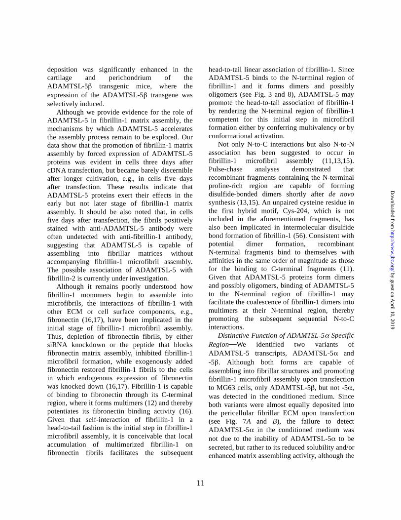
When we transfected MG63 cells with expression
vectors encoding ADAMTSL-5 proteins, both
ADAMTSL-5 and -5 proteins were detected in
fibrillar matrices where fibrillin-1 fibrils were
colocalized (Fig. 7A). No ADAMTSL-5 fibrils
were detectable in mock transfected cells,
indicating that the endogenous expression of
ADAMTSL-5 proteins was negligible in MG63
cells. It was noted that fibrillin-1 fibrils were
barely detectable in mock transfected cells but
clearly detected in ADAMTSL-5 transfected cells
three days after transfection, suggesting that the
forced expression of ADAMTSL-5 proteins
promotes fibrillin-1 fibril formation. Such effects
of ADAMTSL-5 proteins to promote fibrillin-1
fibril formation were not clearly discernible five
days after transfection, when endogenous
fibrillin-1 fibril formation became evident in
mock transfected cells (Fig. 7B). No differences
were detected in the expression levels of
fibrillin-1 transcripts between mock transfected
and ADAMTSL-5 transfected cells, irrespective
of variant type (Fig. 7C), indicating that the
promotion of fibrillin-1 fibril formation by forced
expression of ADAMTSL-5 proteins was not due
to the up-regulation of fibrillin-1 expression, but
likely due to the promotion of fibrillin-1
fibrillogenesis per se.
To further address the role of ADAMTSL-5
proteins in fibrillin-1 fibril formation, MG63 cells
were incubated with increasing concentrations of
purified ADAMTSL-5 for three days. The
exogenously added ADAMTSL-5 was found
incorporated into fibrillar matrices in a
dose-dependent manner (Fig. 7D). Concomitant
with the assembly of ADAMTSL-5 into fibrils,
fibrillin-1 fibril formation became more evident
as more exogenous ADAMTSL-5 was added to
the culture medium. These results support the
possibility that ADAMTSL-5 proteins promote
fibrillin-1 fibril formation.
ADAMTSL-5 Promotes Fibrillin-1 Matrix
Assembly in Vivo To further explore the role of
ADAMTSL-5 in fibrillin-1 microfibril assembly
in vivo, we generated transgenic mice
overexpressing FLAG-tagged ADAMTSL-5
under the control of the type II collagen
promoter/enhancer, which restricts the transgene
expression in cartilagenous tissues. Southern blot
analyses demonstrated that more than twenty
copies of the transgene were integrated into the
genome of the mice (Fig. 8A). The type II
collagen promoter/enhancer-driven
overexpression of the recombinant
ADAMTSL-5 was confirmed by Western
blotting of tissue extracts prepared from
cartilaginous and non-cartilagenous tissues of
control and transgenic mice. A significant amount
of the transgene product was detected in the
costae from transgenic mice, when compared with
ADAMTSL-5 proteins endogenously expressed in
the costae from wild-type mice and the whisker
pad and the gut tube from transgenic mice (Fig.
8B). The recombinant ADAMTSL-5 expressed
in the costae of transgenic mice gave multiple
bands migrating at the positions of monomers,
dimers, trimers, and their processed forms under
reducing conditions, consistent with the results
obtained with recombinant ADAMTSL-5
proteins expressed in MG63 cells (see Fig. 3). No
clear difference was detected in the expression
level of ADAMTSL-5 proteins between control
and transgenic mice in non-cartilageneous tissues
(i.e., the whisker pad and the gut tube),
confirming the specificity of the type II collagen
promoter/enhancer. Interestingly, immunoblot
band patterns significantly differed between the
by guest on April 10, 2019
http://ww
w.jbc.org/
Dow
nloaded from

10
whisker pad and the gut tube, the former
characterized by ~85 kDa and ~150 kDa bands
and the latter characterized with triplet bands at
100-120 kDa region and a ~240 kDa band. Given
that the former banding pattern is reminiscent to
that of the recombinant ADAMTSL-5
overexpressed in the costae of transgenic mice, it
seems likely that the whisker pads express mainly
ADAMTSL-5 while ADAMTSL-5 is
predominantly expressed in the gut tube.
Overexpression of the transgene was further
corroborated by immunohistochemistry using
anti-ADAMTSL-5 antibody. ADAMTSL-5
proteins were highly expressed in the cartilage
and the surrounding perichondrium of the
transgenic embryos, where the type II collagen
promoter/enhancer was expected to be highly
active, although endogenous ADAMTSL-5 was
only faintly detected in these cartilaginous tissues
of wild-type embryos at this stage of development
(E12.5) (Fig. 8, C and D). In line with the
overexpression of ADAMTSL-5 in these
cartilaginous tissues, fibrillin-1 deposition in the
ECM was significantly enhanced in both the
cartilage and perichondrium of transgenic
embryos (Fig. 8, E-J). Fibrillin-1 was barely
detected in the cartilage matrix of wild-type
embryos but was clearly detectable as pericellular
deposits in the cartilage of transgenic embryos,
overlapping with the ADAMTSL-5 deposits.
Similarly, fibrillin-1 was detected as short fibrillar
deposits in the perichondrium of wild-type
embryos but it had coalesced into thick fibrils in
the transgenic embryos. No clear difference in the
staining intensity of fibrillin-1 was observed
between wild-type and transgenic embryos in
tissues where transgene expression was not
induced. These results further support a role for
ADAMTSL-5 in promoting fibrillin-1 matrix
assembly in vivo.
DISCUSSION
Our data show that ADAMTSL-5 is a new
member of the ADAMTSL protein family, which
is secreted and deposited on fibrillin-1 containing
microfibrils. There are two forms of Adamtsl5
gene products, ADAMTSL-5 and -5 ; these
isoforms share the same domain structure
comprising an ADAMTS spacer domain, six TSR
repeats, and the C-terminal PLAC domain but
differ in their N-terminal regions where
ADAMTSL-5 , but not ADAMTSL-5 , contains
a TSR repeat split by an insertion of ~200 amino
acid residues and the following Cys-rich domain.
Immunohistochemistry demonstrated that
ADAMTSL-5 proteins are localized to fibrillar
structures in various elastic tissues, where they
codistribute with fibrillin-1. Association of
ADAMTSL-5 proteins with microfibrils was
further confirmed by immunoelectron microscopy.
When expressed in MG63 cells, ADAMTSL-5
proteins assemble into fibrillar matrices where
they colocalize with fibrillin-1. We also provide
evidence that ADAMTSL-5 proteins bind directly
to the N-terminal half of fibrillin-1 with moderate
affinity (a dissociation constant of ~80 nM).
These results point to the conclusion that
ADAMTSL-5 is an ECM protein that associates
with microfibrils through direct binding to
fibrillin-1.
ADAMTSL-5 Promotes Fibrillin-1 Matrix
Assembly Several lines of evidence indicate that
ADAMTSL-5 proteins promote the assembly of
fibrillin-1 microfibrils. Transfection of MG63
cells with cDNAs encoding ADAMTSL-5
proteins resulted in an accelerated assembly of
fibrillin-1 into fibrillar matrices, to which the
expressed ADAMTSL-5 proteins were also
aligned. The expression levels of fibrillin-1
transcripts remained unaffected in the transfected
cells, indicating that the accelerated matrix
assembly of fibrillin-1 was not due to the
up-regulation of fibrillin-1 gene expression but to
the enhancement of the fibrillin-1 matrix
assembly per se. In support of this conclusion,
matrix assembly of fibrillin-1 was promoted by
the exogenous addition of ADAMTSL-5 in a
dose-dependent manner. Further support for the
role of ADAMTSL-5 in fibrillin-1 matrix
assembly was obtained from transgenic mice in
which recombinant ADAMTSL-5 was
overexpressed under the control of the type II
collagen promoter/enhancer. Fibrillin-1
by guest on April 10, 2019
http://ww
w.jbc.org/
Dow
nloaded from
11
deposition was significantly enhanced in the
cartilage and perichondrium of the
ADAMTSL-5 transgenic mice, where the
expression of the ADAMTSL-5 transgene was
selectively induced.
Although we provide evidence for the role of
ADAMTSL-5 in fibrillin-1 matrix assembly, the
mechanisms by which ADAMTSL-5 accelerates
the assembly process remain to be explored. Our
data show that the promotion of fibrillin-1 matrix
assembly by forced expression of ADAMTSL-5
proteins was evident in cells three days after
cDNA transfection, but became barely discernible
after longer cultivation, e.g., in cells five days
after transfection. These results indicate that
ADAMTSL-5 proteins exert their effects in the
early but not later stage of fibrillin-1 matrix
assembly. It should be also noted that, in cells
five days after transfection, the fibrils positively
stained with anti-ADAMTSL-5 antibody were
often undetected with anti-fibrillin-1 antibody,
suggesting that ADAMTSL-5 is capable of
assembling into fibrillar matrices without
accompanying fibrillin-1 microfibril assembly.
The possible association of ADAMTSL-5 with
fibrillin-2 is currently under investigation.
Although it remains poorly understood how
fibrillin-1 monomers begin to assemble into
microfibrils, the interactions of fibrillin-1 with
other ECM or cell surface components, e.g.,
fibronectin (16,17), have been implicated in the
initial stage of fibrillin-1 microfibril assembly.
Thus, depletion of fibronectin fibrils, by either
siRNA knockdown or the peptide that blocks
fibronectin matrix assembly, inhibited fibrillin-1
microfibril formation, while exogenously added
fibronectin restored fibrillin-1 fibrils to the cells
in which endogenous expression of fibronectin
was knocked down (16,17). Fibrillin-1 is capable
of binding to fibronectin through its C-terminal
region, where it forms multimers (12) and thereby
potentiates its fibronectin binding activity (16).
Given that self-interaction of fibrillin-1 in a
head-to-tail fashion is the initial step in fibrillin-1
microfibril assembly, it is conceivable that local
accumulation of multimerized fibrillin-1 on
fibronectin fibrils facilitates the subsequent
head-to-tail linear association of fibrillin-1. Since
ADAMTSL-5 binds to the N-terminal region of
fibrillin-1 and it forms dimers and possibly
oligomers (see Fig. 3 and 8), ADAMTSL-5 may
promote the head-to-tail association of fibrillin-1
by rendering the N-terminal region of fibrillin-1
competent for this initial step in microfibril
formation either by conferring multivalency or by
conformational activation.
Not only N-to-C interactions but also N-to-N
association has been suggested to occur in
fibrillin-1 microfibril assembly (11,13,15).
Pulse-chase analyses demonstrated that
recombinant fragments containing the N-terminal
proline-rich region are capable of forming
disulfide-bonded dimers shortly after de novo
synthesis (13,15). An unpaired cysteine residue in
the first hybrid motif, Cys-204, which is not
included in the aforementioned fragments, has
also been implicated in intermolecular disulfide
bond formation of fibrillin-1 (56). Consistent with
potential dimer formation, recombinant
N-terminal fragments bind to themselves with
affinities in the same order of magnitude as those
for the binding to C-terminal fragments (11).
Given that ADAMTSL-5 proteins form dimers
and possibly oligomers, binding of ADAMTSL-5
to the N-terminal region of fibrillin-1 may
facilitate the coalescence of fibrillin-1 dimers into
multimers at their N-terminal region, thereby
promoting the subsequent sequential N-to-C
interactions.
Distinctive Function of ADAMTSL-5 Specific
Region We identified two variants of
ADAMTSL-5 transcripts, ADAMTSL-5 and
-5 . Although both forms are capable of
assembling into fibrillar structures and promoting
fibrillin-1 microfibril assembly upon transfection
to MG63 cells, only ADAMTSL-5 , but not -5 ,
was detected in the conditioned medium. Since
both variants were almost equally deposited into
the pericellular fibrillar ECM upon transfection
(see Fig. 7A and B), the failure to detect
ADAMTSL-5 in the conditioned medium was
not due to the inability of ADAMTSL-5 to be
secreted, but rather to its reduced solubility and/or
enhanced matrix assembling activity, although the
by guest on April 10, 2019
http://ww
w.jbc.org/
Dow
nloaded from

12
expression and/or secretion of ADAMTSL-5
could be less efficient than those of
ADAMTSL-5 . These differences between
ADAMTSL-5 and -5 are likely due to their
distinctive N-terminal regions. It should be noted
that the insertion in the first TSR-like domain in
ADAMTSL-5 is enriched in arginine residues
(~13% of a total of 209 residues) and contains
two RRXR sequences (see Fig. 1B), which are
putative heparin binding motifs (57). Thus, the
insertion in the first TSR-like domain may bind to
heparan sulfate and other glycosaminoglycan
chains and may thereby contribute to the removal
of ADAMTSL-5 from the conditioned medium.
The Cys-rich domain may also be involved in the
removal of ADAMTSL-5 , as recombinant
fragments of ADAMTS-1 and ADAMTS-5 that
harbor only the Cys-rich and ADAMTS spacer
domains were recovered only in the ECM
deposits but not in the conditioned medium
(39,58).
Although the precise structure of the first TSR
of ADAMTSL-5 with its insertion remains to be
defined, it is interesting to note that the inserted
sequence is mapped between strands B and C,
which in TSR domains of thrombospondin-1 are
connected by a relatively long loop that is secured
by a conserved disulfide bond (59). Given that the
tryptophan motif (WXXWXXW) and six cysteine
residues including those that secure the loop
between strands B and C, which together
contribute to the core structure of TSRs (59), are
conserved in the first TSR of ADAMTSL-5 , it
seems likely that the inserted sequence loops out
of the core structure and folds separately. A
similar long insertion with >200 amino acid
residues has been found in the first TSR of
ADAMTSL-4/TSRC1 (38). Although overall
homology in the inserted sequence between
ADAMTSL-4 and -5 is only moderate, a stretch
of 26 amino acid residues, which are enriched in
basic residues, prolines, and glycines, are highly
conserved between two ADAMTSL proteins,
indicative of their roles in biological functions of
these proteins, e.g., binding to glycosaminoglycan
chains.
ADAMTSL5 As a Potential Candidate Gene
Associated with Fibrillinopathies Mutations in
the fibrillin-1 gene cause fibrillinopathies,
inherited disorders of connective tissues typically
represented by the Marfan syndrome. Since
ADAMTSL-5 proteins associate with fibrillin-1
microfibrils and promote microfibril assembly in
vitro and in vivo, it is tempting to speculate that
mutations in the ADAMTSL-5 gene could lead to
aberrant microfibril assembly in elastic and
nonelastic tissues and thereby cause conditions
similar to fibrillinopathies. Consistent with this
possibility, mutations in the ADAMTSL-2 gene
have been associated with an acromelic dysplasia
called geleophysic dysplasia, a rare autosomal
recessive disorder characterized by short stature,
brachydactyly, thick skin, and cardiac valvular
anomalies (42). These clinical features are similar
to those of Weill-Marchesani syndrome, which is
also an acromeric dysplasia associated with
mutations in fibrillin-1. Recently, mutations in the
ADAMTSL-4 gene were found in patients with
ectopia lentis (45), displacement of the lens due to
abnormalities in the ciliary zonule, a cardinal
feature of the Marfan syndrome. In addition,
familial forms of isolated ectopia lentis are also
caused by mutations in fibrillin-1, further
corroborating the potential involvement of
ADAMTSL proteins in fibrillinopathies.
Although it remains to be defined how
mutations in ADAMTSL-2 and ADAMTSL-4
genes lead to the symptoms associated with
fibrillinopathies, aberrant microfibril assembly
and the resulting effects on TGF- bioavailability
may be an integral part of the pathogenetic
processes. Le Goff et al (42) observed a
significant increase in active TGF- in the culture
medium of fibroblasts derived from individuals
with geleophysic dysplasia, along with an
increased phosphorylation of SMAD2 and its
nuclear localization, indicative of increased
TGF- signaling events. They also showed that
ADAMTSL-2 associates with LTBP-1, which
performs a major role in the storage of latent
TGF- in the ECM and is similar to fibrillins in
its domain structure (60). Although it is unknown
whether ADAMTSL-2 directly binds to fibrillin-1,
our results that ADAMTSL-5 binds to fibrillin-1
by guest on April 10, 2019
http://ww
w.jbc.org/
Dow
nloaded from

13
and promotes its assembly into microfibrils raise
the possibility that ADAMTSL-5 is also involved
in the ECM-based network regulating TGF-
bioavailability. It remains an open question
whether any defects in the ADAMTSL-5 gene
dysregulate the bioavailability of TGF- and
hence lead to conditions associated with
fibrillinopathies. Mouse models in which
Adamtsl5 is mutated should provide important
insight into the physiological functions of
ADAMTSL-5 and other ADAMTSL family
proteins and their relevance to pathogenesis of
fibrillinopathies.
REFERENCES
1. Charbonneau, N. L., Ono, R. N., Corson, G. M., Keene, D. R., and Sakai, L. Y. (2004) Birth
Defects Res. C Embryo Today 72, 37-50
2. Chaudhry, S. S., Cain, S. A., Morgan, A., Dallas, S. L., Shuttleworth, C. A., and Kielty, C. M.
(2007) J. Cell Biol. 176, 355-367
3. Sakai, L. Y., Keene, D. R., and Engvall, E. (1986) J. Cell Biol. 103, 2499-2509
4. Handford, P. A., Downing, A. K., Reinhardt, D. P., and Sakai, L. Y. (2000) Matrix Biol. 19,
457-470
5. Sakai, L. Y., Keene, D. R., Glanville, R. W., and Bachinger, H. P. (1991) J. Biol. Chem. 266,
14763-14770
6. Kielty, C. M., Sherratt, M. J., and Shuttleworth, C. A. (2002) J. Cell Sci. 115, 2817-2828
7. Reinhardt, D. P., Keene, D. R., Corson, G. M., Poschl, E., Bachinger, H. P., Gambee, J. E., and
Sakai, L. Y. (1996) J. Mol. Biol. 258, 104-116
8. Kuo, C. L., Isogai, Z., Keene, D. R., Hazeki, N., Ono, R. N., Sengle, G., Peter Bachinger, H., and
Sakai, L. Y. (2007) J. Biol. Chem. 282, 4007-4020
9. Baldock, C., Koster, A. J., Ziese, U., Rock, M. J., Sherratt, M. J., Kadler, K. E., Shuttleworth, C.
A., and Kielty, C. M. (2001) J. Cell Biol. 152, 1045-1056
10. Lee, S. S., Knott, V., Jovanovic, J., Harlos, K., Grimes, J. M., Choulier, L., Mardon, H. J., Stuart,
D. I., and Handford, P. A. (2004) Structure 12, 717-729
11. Marson, A., Rock, M. J., Cain, S. A., Freeman, L. J., Morgan, A., Mellody, K., Shuttleworth, C.
A., Baldock, C., and Kielty, C. M. (2005) J. Biol. Chem. 280, 5013-5021
12. Hubmacher, D., El-Hallous, E. I., Nelea, V., Kaartinen, M. T., Lee, E. R., and Reinhardt, D. P.
(2008) Proc. Nat. Acad. Sci. U. S. A. 105, 6548-6553
13. Trask, T. M., Ritty, T. M., Broekelmann, T., Tisdale, C., and Mecham, R. P. (1999) Biochem. J.
340, 693-701
by guest on April 10, 2019
http://ww
w.jbc.org/
Dow
nloaded from

14
14. Lin, G., Tiedemann, K., Vollbrandt, T., Peters, H., Batge, B., Brinckmann, J., and Reinhardt, D. P.
(2002) J. Biol. Chem. 277, 50795-50804
15. Ashworth, J. L., Kelly, V., Wilson, R., Shuttleworth, C. A., and Kielty, C. M. (1999) J. Cell Sci.
112, 3549-3558
16. Sabatier, L., Chen, D., Fagotto-Kaufmann, C., Hubmacher, D., McKee, M. D., Annis, D. S.,
Mosher, D. F., and Reinhardt, D. P. (2009) Mol. Biol. Cell 20, 846-858
17. Kinsey, R., Williamson, M. R., Chaudhry, S., Mellody, K. T., McGovern, A., Takahashi, S.,
Shuttleworth, C. A., and Kielty, C. M. (2008) J. Cell Sci. 121, 2696-2704
18. Cain, S. A., Baldock, C., Gallagher, J., Morgan, A., Bax, D. V., Weiss, A. S., Shuttleworth, C. A.,
and Kielty, C. M. (2005) J. Biol. Chem. 280, 30526-30537
19. Cain, S. A., Baldwin, A. K., Mahalingam, Y., Raynal, B., Jowitt, T. A., Shuttleworth, C. A.,
Couchman, J. R., and Kielty, C. M. (2008) J. Biol. Chem. 283, 27017-27027
20. Tiedemann, K., Batge, B., Muller, P. K., and Reinhardt, D. P. (2001) J. Biol. Chem. 276,
36035-36042
21. Ritty, T. M., Broekelmann, T. J., Werneck, C. C., and Mecham, R. P. (2003) Biochem. J. 375,
425-432
22. Isogai, Z., Ono, R. N., Ushiro, S., Keene, D. R., Chen, Y., Mazzieri, R., Charbonneau, N. L.,
Reinhardt, D. P., Rifkin, D. B., and Sakai, L. Y. (2003) J. Biol. Chem. 278, 2750-2757
23. Ono, R. N., Sengle, G., Charbonneau, N. L., Carlberg, V., Bachinger, H. P., Sasaki, T.,
Lee-Arteaga, S., Zilberberg, L., Rifkin, D. B., Ramirez, F., Chu, M. L., and Sakai, L. Y. (2009) J.
Biol. Chem. in press
24. Jensen, S. A., Reinhardt, D. P., Gibson, M. A., and Weiss, A. S. (2001) J. Biol. Chem. 276,
39661-39666
25. Penner, A. S., Rock, M. J., Kielty, C. M., and Shipley, J. M. (2002) J. Biol. Chem. 277,
35044-35049
26. El-Hallous, E., Sasaki, T., Hubmacher, D., Getie, M., Tiedemann, K., Brinckmann, J., Batge, B.,
Davis, E. C., and Reinhardt, D. P. (2007) J. Biol. Chem. 282, 8935-8946
27. Charbonneau, N. L., Dzamba, B. J., Ono, R. N., Keene, D. R., Corson, G. M., Reinhardt, D. P.,
and Sakai, L. Y. (2003) J. Biol. Chem. 278, 2740-2749
28. Aoyama, T., Francke, U., Dietz, H. C., and Furthmayr, H. (1994) J. Clin. Invest. 94, 130-137
by guest on April 10, 2019
http://ww
w.jbc.org/
Dow
nloaded from

15
29. Godfrey, M., Raghunath, M., Cisler, J., Bevins, C. L., DePaepe, A., Di Rocco, M., Gregoritch, J.,
Imaizumi, K., Kaplan, P., Kuroki, Y., Silberbach, M., Superti-Furga, A., Van Thienen, M., Vetter,
U., and Steinmann, B. (1995) Am. J. Pathol. 146, 1414-1421
30. Robinson, P. N., Arteaga-Solis, E., Baldock, C., Collod-Beroud, G., Booms, P., De Paepe, A.,
Dietz, H. C., Guo, G., Handford, P. A., Judge, D. P., Kielty, C. M., Loeys, B., Milewicz, D. M.,
Ney, A., Ramirez, F., Reinhardt, D. P., Tiedemann, K., Whiteman, P., and Godfrey, M. (2006) J.
Med. Genet. 43, 769-787
31. Handford, P. A. (2000) Biochim. Biophys. Acta 1498, 84-90
32. Faivre, L., Megarbane, A., Alswaid, A., Zylberberg, L., Aldohayan, N., Campos-Xavier, B., Bacq,
D., Legeai-Mallet, L., Bonaventure, J., Munnich, A., and Cormier-Daire, V. (2002) Hum. Genet.
110, 366-370
33. Faivre, L., Gorlin, R. J., Wirtz, M. K., Godfrey, M., Dagoneau, N., Samples, J. R., Le Merrer, M.,
Collod-Beroud, G., Boileau, C., Munnich, A., and Cormier-Daire, V. (2003) J. Med. Genet. 40,
34-36
34. Porter, S., Clark, I. M., Kevorkian, L., and Edwards, D. R. (2005) Biochem. J. 386, 15-27
35. Hirohata, S., Wang, L. W., Miyagi, M., Yan, L., Seldin, M. F., Keene, D. R., Crabb, J. W., and
Apte, S. S. (2002) J. Biol. Chem. 277, 12182-12189
36. Koo, B. H., Le Goff, C., Jungers, K. A., Vasanji, A., O'Flaherty, J., Weyman, C. M., and Apte, S.
S. (2007) Matrix Biol. 26, 431-441
37. Hall, N. G., Klenotic, P., Anand-Apte, B., and Apte, S. S. (2003) Matrix Biol. 22, 501-510
38. Buchner, D. A., and Meisler, M. H. (2003) Gene 307, 23-30
39. Kuno, K., and Matsushima, K. (1998) J. Biol. Chem. 273, 13912-13917
40. Kashiwagi, M., Enghild, J. J., Gendron, C., Hughes, C., Caterson, B., Itoh, Y., and Nagase, H.
(2004) J. Biol. Chem. 279, 10109-10119
41. Somerville, R. P., Longpre, J. M., Jungers, K. A., Engle, J. M., Ross, M., Evanko, S., Wight, T.
N., Leduc, R., and Apte, S. S. (2003) J. Biol. Chem. 278, 9503-9513
42. Le Goff, C., Morice-Picard, F., Dagoneau, N., Wang, L. W., Perrot, C., Crow, Y. J., Bauer, F.,
Flori, E., Prost-Squarcioni, C., Krakow, D., Ge, G., Greenspan, D. S., Bonnet, D., Le Merrer, M.,
Munnich, A., Apte, S. S., and Cormier-Daire, V. (2008) Nat. Genet. 40, 1119-1123
43. Hashimoto, G., Shimoda, M., and Okada, Y. (2004) J. Biol. Chem. 279, 32483-32491
by guest on April 10, 2019
http://ww
w.jbc.org/
Dow
nloaded from

16
44. Tortorella, M., Pratta, M., Liu, R. Q., Abbaszade, I., Ross, H., Burn, T., and Arner, E. (2000) J.
Biol. Chem. 275, 25791-25797
45. Ahram, D., Sato, T. S., Kohilan, A., Tayeh, M., Chen, S., Leal, S., Al-Salem, M., and El-Shanti,
H. (2009) Am. J. Hum. Genet. 84, 274-278
46. Manabe, R., Tsutsui, K., Yamada, T., Kimura, M., Nakano, I., Shimono, C., Sanzen, N., Furutani,
Y., Fukuda, T., Oguri, Y., Shimamoto, K., Kiyozumi, D., Sato, Y., Sado, Y., Senoo, H.,
Yamashina, S., Fukuda, S., Kawai, J., Sugiura, N., Kimata, K., Hayashizaki, Y., and Sekiguchi, K.
(2008) Proc. Nat. Acad. Sci. U. S. A. 105, 12849-12854
47. Nakai, K., and Horton, P. (1999) Trends Biochem. Sci. 24, 34-36
48. Hirokawa, T., Boon-Chieng, S., and Mitaku, S. (1998) Bioinformatics 14, 378-379
49. Pearson, W. R., Wood, T., Zhang, Z., and Miller, W. (1997) Genomics 46, 24-36
50. Koff, A., Giordano, A., Desai, D., Yamashita, K., Harper, J. W., Elledge, S., Nishimoto, T.,
Morgan, D. O., Franza, B. R., and Roberts, J. M. (1992) Science 257, 1689-1694
51. Pereira, L., Andrikopoulos, K., Tian, J., Lee, S. Y., Keene, D. R., Ono, R., Reinhardt, D. P., Sakai,
L. Y., Biery, N. J., Bunton, T., Dietz, H. C., and Ramirez, F. (1997) Nat. Genet. 17, 218-222
52. Tsumaki, N., Tanaka, K., Arikawa-Hirasawa, E., Nakase, T., Kimura, T., Thomas, J. T., Ochi, T.,
Luyten, F. P., and Yamada, Y. (1999) J. Cell Biol. 144, 161-173
53. Sakai, L. Y., and Keene, D. R. (1994) Methods Enzymol. 245, 29-52
54. Team, T. F. C. a. t. R. G. E. R. G. P. I. I. (2002) Nature 420, 563-573
55. Dzamba, B. J., Keene, D. R., Isogai, Z., Charbonneau, N. L., Karaman-Jurukovska, N., Simon,
M., and Sakai, L. Y. (2001) J. Invest. Dermatol. 117, 1612-1620
56. Reinhardt, D. P., Gambee, J. E., Ono, R. N., Bachinger, H. P., and Sakai, L. Y. (2000) J. Biol.
Chem. 275, 2205-2210
57. Hileman, R. E., Fromm, J. R., Weiler, J. M., and Linhardt, R. J. (1998) BioEssays 20, 156-167
58. Gendron, C., Kashiwagi, M., Lim, N. H., Enghild, J. J., Thogersen, I. B., Hughes, C., Caterson,
B., and Nagase, H. (2007) J. Biol. Chem. 282, 18294-18306
59. Tan, K., Duquette, M., Liu, J. H., Dong, Y., Zhang, R., Joachimiak, A., Lawler, J., and Wang, J.
H. (2002) J. Cell Biol. 159, 373-382
60. Rifkin, D. B. (2005) J. Biol. Chem. 280, 7409-7412
by guest on April 10, 2019
http://ww
w.jbc.org/
Dow
nloaded from

17
FOOTNOTES
*We are grateful to Dr. Yoshihide Hayashizaki for mouse ADAMTSL-5 cDNAs and Dr. Noriyuki
Tsumaki for the Col2aI promoter/enhancer DNA. This study was supported in part by Grants-in-Aid for
Scientific Research from the Ministry of Education, Culture, Sports, Science, and Technology of Japan
(17082005 and 20370046, to K. S.), from the Shriners Hospitals for Children (to D.R.K. and to L.Y.S.),
and from the National Institutes of Health (P01AR049698 to L.Y.S.).
1The abbreviations used are: ADAMTSL, ADAMTS-like; cbEGF, calcium-binding epidermal growth
factor; DIG, digoxigenin; ECM, extracellular matrix; LTBP, latent TGF- binding protein; MAGP,
microfibril-associated protein; SPR, surface plasmon resonance; TGF, transforming growth factor; TSR,
thrombospondin type I repeat.
FIGURE LEGENDS
FIGURE 1. Structures of ADAMTSL-5 proteins. A, schematic representations of the domain structures of
ADAMTSL-5 and -5 and other ADAMTS-like proteins. The domain structure of ADAMTS-1
proteinase is included as a reference. Note that the first TSR domains of ADAMTSL-5 and
ADAMTSL-4 are split by ~200 amino acids insertions (detailed in B). The region used as an immunogen
for antibody production is indicated by a bracket. The putative N-linked glycosylation sites are marked
with Y. B, alignment of amino acid sequences of the first TSR domain and a part of the following
Cys-rich domain of ADAMTSL-5 and other ADAMTS-like proteins (upper panel). Species origins are
indicated by prefixes m (mouse) and h (human). The conserved residues are boxed. Three tryptophans
and six cysteines, which have been shown to be critical in the layered fold of TSR domains (59), are
boxed with gray background. Residue numbers are shown in the left and right ends. The insertion
sequences that split the first TSR domain of ADAMTSL-5 and ADAMTSL-4 into N-terminal and
C-terminal halves are aligned using the CLUSTAL-W algorithm
(http://www.ebi.ac.uk/Tools/clustalw2/index.html; lower panel). Conserved and semiconserved residues
are indicated by “*” and “:”, respectively. A stretch of 26 amino acid residues that are highly
homologous between ADAMTSL-5 and ADAMTSL-4 are underlined. C, a schematic diagram of
exon-intron structure of Adamtsl5 gene on mouse chromosome 9E. Exon numbers encoding
ADAMTSL-5 are shown under the diagram. The first exon encoding ADAMTSL-5 -specific sequence
including the signal peptide is located between exon 7 and exon 8 of ADAMTSL-5 .
FIGURE 2. Expression levels of ADAMTSL-5 and -5 transcripts in mouse embryos and adult tissues.
Transcripts for ADAMTSL-5 and -5 were reverse-transcribed from total RNA extracted from mouse
embryos at different gestation periods and from adult tissues, and then subjected to PCR amplification (33
cycles) using specific primer sets described under Experimental Procedures. Transcripts for -actin were
by guest on April 10, 2019
http://ww
w.jbc.org/
Dow
nloaded from

18
also amplified by RT-PCR (25 cycles) as a control. Arrows indicate the predicted positions of the PCR
products. An apparent discrepancy in the size of PCR products for ADAMTSL-5 was due to distortion
of the gel.
FIGURE 3. Expression and secretion of recombinant ADAMTSL-5 proteins. ADAMTSL-5 (TSL5 )
and -5 (TSL5 ) proteins were transiently expressed in MG63 cells for three days and then detected by
immunoblotting with anti-ADAMTSL-5 antibody under reducing (A) and non-reducing (B) conditions
after fractionation into cell lysates (C), extracellular deposits (E), and spent medium (M). Cells
transfected with empty vector (Mock) were also analyzed as controls.
FIGURE 4. Immunolocalization of ADAMTSL-5 in embryos and adult tissues. Cryosections of E16.5
mouse embryos (A-D) and those of kidney and heart from 10 week-old female mice (E, F) were incubated
with anti-ADAMTSL-5 antibody, followed by color development as described under Experimental
Procedures. The following areas in Panel A were magnified in B-D: skin with vibrissa follicles (B);
vertebral primordium (c, cartilage; arrowheads, perichondrium) in (C); aorta (a) and vein (v) in (D). In E
and F, arterial wall (a) in the kidney and the basement membrane zones of mitral valve in the heart were
positively stained, respectively. (G-I) Double immunofluorescence staining of ADAMTSL-5 and
fibrillin-1 in the fetal cartilage. Cryosections of E16.5 mouse embryos were double immunostained with
anti-ADAMTSL-5 (G; green) and anti-fibrillin-1 (H; red) antibodies. Merged image is shown in I. The
staining patterns of ADAMTSL-5 overlapped with that of fibrillin-1 in the perichondrium (arrowheads).
Other tissues (e.g., skeletal muscle shown in H and I) demonstrated staining for fibrillin-1, without
staining for ADAMTSL-5. Bars represent 100 μm.
FIGURE 5. Electron microscopic immunolocalization of ADAMTSL-5 in neonatal mouse cartilage and
perichondrium. Immunogold particles were localized on microfibrils associated with amorphous elastin
cores (asterisk) in the ear cartilage (A) and on microfibril bundles in perichondrium (B). There was no
immunogold labeling of collagen fibers (bracket). Insets in A and B are magnified views of boxed areas.
Bars represent 500 nm.
FIGURE 6. Binding of ADAMTSL-5 to the N-terminal fragment of fibrillin-1. A, schematic domain
structures of fibrillin-1 and its recombinant halves. B, sensorgrams of binding of ADAMTSL-5 to
fibrillin-1 fragments. Purified ADAMTSL-5 (200 nM) was infused over sensor chips on which
recombinant fibrillin-1 fragments representing either the N-terminal half (rF90) or C-terminal half (rF6)
were immobilized. Binding was recorded as Resonance Units (RU). C, increasing concentrations of
purified ADAMTSL-5 were infused over a sensor chip on which rF90 was immobilized. The
dissociation constant was calculated to be 78.8 10-9
M.
FIGURE 7. Promotion of fibrillin-1 microfibril assembly by ADAMTSL-5. MG63 cells were transfected
with cDNAs encoding ADAMTSL-5 or -5 or with empty vector (Mock) and then labeled with
antibodies against fibrillin-1 and ADAMTSL-5 three (A) or five (B) days after transfection. Merged
images are shown in the right column. Nuclei were visualized with Hoechst 33342. Closed and open
arrowheads in B point to the fibrils in which ADAMTSL-5 and fibrillin-1 were predominantly stained,
respectively. Note that clear fibrillin-1 depositions, which were not detected until day 5 in mock
transfected cells, were detected on day 3 after transfection in ADAMTSL-5 transfected cells. C, the
expression levels of fibrillin-1 transcripts in cells transfected with cDNAs encoding ADAMTSL-5 or
-5 or with empty vector (Mock) were determined by RT-PCR. Total RNA was collected from cells on
day 3 after transfection and subjected to RT-PCR amplification of the fibrillin-1 transcripts for indicated
by guest on April 10, 2019
http://ww
w.jbc.org/
Dow
nloaded from

19
reaction cycles. Transcripts for -actin were also subjected to RT-PCR amplification as controls. D,
MG63 cells were incubated with increasing concentrations of ADAMTSL-5 for three days to examine
the effects of exogenously added ADAMTSL-5 on fibrillin-1 matrix assembly. Cells were
immunolabeled with antibodies against ADAMTSL-5 (left; green) and fibrillin-1 (right; red). Scale bar =
50 μm.
FIGURE 8. Promotion of fibrillin-1 fibril formation by ADAMTSL-5 in vivo. A, Southern hybridization
of DNA from ADAMTSL-5 transgenic mice. Digestion of genomic DNA with EcoRI and BamHI
produced 1.6 kilobase (Kb) fragments containing the Adamtsl5 exon 8 from chromosome 9 (indicated
with an arrow) and 2.3 Kb fragments containing full length ADAMTSL-5 transgenes (indicated with an
arrowhead). Lane 1, DIG-labeled molecular weight markers; lane 2, DNA from wild-type mouse (12 μg);
lane 3, DNA from wild-type mouse (3 μg); lane 4, DNA from transgenic mouse (12 μg); lane 5, DNA
from transgenic mouse (3 μg); lane 6, DNA from transgenic mouse (0.75 μg). The 2.3 kb band was
detected only with the DNA from transgenic mice, while the 1.6 kb band, although very faint, was
detected with the DNA from wild-type and transgenic mice. Quantification of band intensities using
Image J software indicated that more than twenty copies of the transgene were integrated into the
genomic DNA of transgenic mice. The sizes of DNA molecular weight markers are shown in the left
margin. B, tissue extracts from E15.5 wild-type (W) and ADAMTSL-5 transgenic mice (Tg) were
subjected to SDS-PAGE under reducing conditions, followed by blotting with anti-ADAMTSL-5
antibody. Ten-fold diluted extracts were also subjected to Western blotting for the costae from transgenic
mice, where the transgene was highly expressed. Positions of molecular mass markers (kDa) are shown in
the left margin. Open and closed triangles point to the bands characteristic of the whisker pads and the gut
tube, respectively. C-J, immunofluorescence detection of ADAMTSL-5 proteins and fibrillin-1. Sagittal
cryosections were prepared from E12.5 wild-type (C, E, G, I) and transgenic (D, F, H, J) littermates and
subjected to double immunostaining with antibodies against ADAMTSL-5 (green) and fibrillin-1 (red).
The regions of rib cartilage (C) and surrounding perichondrium (P) are labeled in E and F. G and I,
magnified views in the boxed areas in (E). H and J, magnified views of the boxed areas in (F). The
ADAMTSL-5 transgene was highly expressed in the rib cartilage and surrounding perichondrium of
transgenic embryos (D and F), where fibrillin-1 depositions were also enhanced (H and J). Scale bar =
100 μm.
by guest on April 10, 2019
http://ww
w.jbc.org/
Dow
nloaded from

A
ADAMTS-1
ADAMTSL1
ADAMTSL-2
ADAMTSL-3
ADAMTSL-4
ADAMTSL-5
ADAMTSL-5
catalytic domain disintegrin-like domain
signal peptide Cys-rich domain ADAMTS spacerthrombospondin type I repeat (TSR)
PLAC domain
C
100 kb
exon 1 1 8 187
B
1st TSR Cys-rich
mADAMTSL-5 57-WGSWGPWSACSRSCSGGVMEQTRPCLPS-(203aa)-AISCIGAYRQYKLCNTNACPES-GRSIREVQCAS-320hADAMTSL-1 36-WDAWGPWSECSRTCGGGASYSLRRCL----------SSKSCEGRNIRYRTCSNVDCPPEAG-DFRAQQCSA- 95mADAMTSL-2 50-WGEWTKWTACSRSCGGGVTSQERHCLQQRRKSVPGTGNRTCVGTSKRYQLCRVQECPPD-GRSFREEQCVS-119hADAMTSL-3 78-WDAWGDWSDCSRTCGGGASYSLRRCL----------TGRNCEGQNIRYKTCSNHDCPPDAE-DFRAQQCSA-137mADAMTSL-4 46-WGPWGRWASCSQPCGVGVQRRSRTCELH-(251aa)-APNCSGESEQMRACSQEPCPPE-QPDPRALQCAA-357mADAMTS-1 563-WGPWGPWGDCSRTCGGGVQYTMREC----DNPVPKNGGKYCEGKRVRYRSCNIEDCPDNNGKTFREEQCEA-629
Tsutsui et al. Figure 1
mADAMTSL-5 82 LPSSYRARGGSRPNGRALSITGHVVSAVRTSVPLHRSQEDQRALAGSNASRQGPAVVRGSRHPQARGREP : * * * * : * : ** * *
mADAMTSL-4 71 --ELHPALPLPPRPPRHPEAHRPRGQGSRPQTPRDPQSLYRPQPRGRGGPLRAPASQVGREETQE---PQ
mADAMTSL-5 152 SERRSRTRGPIGPGKYGYGKAPYILPLQTDTTHTPQR------------LRRQRPSSRHSRSQEASASKQ :* * * ** ** :***: *: ***: * : : * **: : : :
mADAMTSL-4 136 GAQRFRVRDPIKPGMFGYGRVPFALPLHRSRRHPHRPGQPKNSSTGEGMVPSQPPSTELASEKHGPHMQP
mADAMTSL-5 210 GYRPPTHQFSHSQPLYQSDSGPRSGLPPSEASIYQLPLTHDQSYPAASSLFHRPELSSHHGARPHGAAQA : *:: **: ** : : :* ** * * : *:** :
mADAMTSL-4 206 PEPRSHSAETPRSGTAQTEVLPRTSSAPSYTGTPAPTSSFGDSRSFQGSLGPRMPPSPGSWSSPQGAERR
mADAMTSL-5 280 FPQHLRSTAIS----------------------------------------- 290 * : *
mADAMTSL-4 276 HPPPFSPVPRSQQSRRHWRPPGPHRSPDGWLPLTRDSSPLWSIFAPSIPAPN 327
by guest on April 10, 2019
http://ww
w.jbc.org/
Dow
nloaded from

Tsutsui et al. Figure 2
ADAMTSL-5
ADAMTSL-5
-actin
E7
E11
E17
Bra
in
Eye
Heart
Kid
ney
Liv
er
Lym
ph n
ode
Subm
axill
ary
gla
nd
Spin
al c
ord
Sple
en
Sto
mach
Ute
rus
Embryos Adult tissues
by guest on April 10, 2019
http://ww
w.jbc.org/
Dow
nloaded from

250
150
100
75
50
37
25
Mock TSL5 TSL5
Non-reduced
250
150
100
75
50
37
25
Reduced
A B
Tsutsui et al. Figure 3
C E M
Mock TSL5 TSL5
C E MC E M C E M C E MC E M
by guest on April 10, 2019
http://ww
w.jbc.org/
Dow
nloaded from

B
C
D
v
a
c
A
B
C
D
E
IG H
a
c
Tsutsui et al. Figure 4
F
by guest on April 10, 2019
http://ww
w.jbc.org/
Dow
nloaded from

A B
Tsutsui et al. Figure 5
by guest on April 10, 2019
http://ww
w.jbc.org/
Dow
nloaded from

0
20
40
60
80
100
0 50 100 150 200 250 300
370
185
9246230
ADAMTSL-5
(nM)
Time (sec)
Response D
iffe
rence (
RU
)
A
C
B
0 50 100 150 200 250 300
Time (sec)
0
20
40
60
50
10
30
rF90
rF6
Tsutsui et al. Figure 6
Fibrillin-1 full length
rF90
rF6
EGF-like domain cb EGF-like domain
unique region BM40 signal peptideproline rich region
hybrid motif 8-cysteine domain
6 histidine residues
Response D
iffe
rence (
RU
)
by guest on April 10, 2019
http://ww
w.jbc.org/
Dow
nloaded from
Mock
ADAMTSL-5
ADAMTSL-5
Fibrillin-1 ADAMTSL-5 Merged
Tsutsui et al. Figure 7
A
by guest on April 10, 2019
http://ww
w.jbc.org/
Dow
nloaded from

Mock
ADAMTSL-5
ADAMTSL-5
Tsutsui et al. Figure 7 (continued)
C
Transfection Cycle No.
Mock
ADAMTSL-5
ADAMTSL-5
18 21 24 27 30 18 21 24 27 30
Fibrillin-1 -actin
B Fibrillin-1 ADAMTSL-5 Merged
by guest on April 10, 2019
http://ww
w.jbc.org/
Dow
nloaded from

0
2.5
10
ADAMTSL-5 Fibrillin-1ADAMTSL-5 added
(μg/ml)
0.6
D
Tsutsui et al. Figure 7 (continued)
by guest on April 10, 2019
http://ww
w.jbc.org/
Dow
nloaded from

Tsutsui et al. Figure 8
W Tg W Tg W Tg
1 11/10 1 1 1 1
costaewhisker
padgut
tube
75
150
250
100
50
37
A B
2027
4361
23130
1 2 3 4 5 6
2322
564
amount of tissue
extract applied
by guest on April 10, 2019
http://ww
w.jbc.org/
Dow
nloaded from

Tsutsui et al. Figure 8 (continued)
ADAMTSL-5 ADAMTSL-5
C D
G
I J
H
H
JG
I
Fibrillin-1 Fibrillin-1
Fibrillin-1 Fibrillin-1
CP
CP
C
PC
P
ADAMTSL-5+Fibrillin-1 ADAMTSL-5+Fibrillin-1
E F
Transgenic mouseWild-type mouse
by guest on April 10, 2019
http://ww
w.jbc.org/
Dow
nloaded from

R. Keene, Gerhard Sengle, Lynn Y. Sakai and Kiyotoshi SekiguchiKo Tsutsui, Ri-ichiroh Manabe, Tomiko Yamada, Itsuko Nakano, Yasuko Oguri, Douglas
promotes fibrillin-1 fibril formation(ADAMTSL-5) is a novel extracellular matrix protein that binds to fibrillin-1 and
A disintegrin and metalloproteinase with thrombospondin motifs-like-5
published online November 23, 2009J. Biol. Chem.
10.1074/jbc.M109.076919Access the most updated version of this article at doi:
Alerts:
When a correction for this article is posted•
When this article is cited•
to choose from all of JBC's e-mail alertsClick here
Supplemental material:
http://www.jbc.org/content/suppl/2009/11/23/M109.076919.DC1
by guest on April 10, 2019
http://ww
w.jbc.org/
Dow
nloaded from