Embed Size (px)
Citation preview
A DISSERTATION ON
EVALUATION OF MODIFIED ALVARADO SCORING
SYSTEM
IN ACUTE APPENDICITIS
Dissertation Submitted in partial fulfillment of
M.S. DEGREE EXAMINATION
M.S. GENERAL SURGERY—BRANCH I
CHENGALPATTU MEDICAL COLLEGE, CHENGALPATTU.
THE TAMILNADU DR.M.G.R. MEDICAL UNIVERSITY
CHENNAI, TAMILNADU
MARCH 2O1O
CERTIFICATE
This is to certify that this dissertation titled “EVALUATION OF MODIFIED ALVARADO
SCORING SYSTEM IN ACUTE APPENDICITIS” has been prepared by
Dr.A.P.DEEBAPRIYA under my supervision in the Department of General
surgery,Chengalpattu Medical College, Chengalpattu during the academic period 2007-2010
and is being submitted to the Tamil Nadu DR.M.G.R. Medical University, Chennai in partial
fulfillment of the University regulation for the award of the Degree of Master of
surgery(M.S.General surgery) and her dissertation is a bonafide work.
Prof.Dr. P. Ramanujam.M.S Prof.Dr.G. Raja Billy Graham.M.S
Unit Chief, Head of the Department,
Department of General Surgery, Department of General Surgery,
Chengalpattu Medical College & Chengalpattu Medical College &
Hospital. Hospital.
DEAN
Chengalpattu Medical College & Hospital,
Chengalpattu.
DECLARATION
I, A.P.DEEBAPRIYA, solemnly declare that the dissertation “EVALUATION OF
MODIFIED ALVARADO SCORING SYSTEM IN ACUTE APPENDICITIS” a bonafide work
done by me in the Department of General surgery, Chengalpattu Medical College,
Chengalpattu, under the able guidance of Prof.Dr.P.Ramanujam, Associate professor,
Chengalpattu Medical College, Chengalpattu.
Place: Chengalpattu.
Date:
(A.P.Deebapriya)
ACKNOWLEDGEMENT
I wish to express my sincere thanks to Dr.shanmugam.M.S. Mch., Dean,
Chengalpattu Medical College & Hospital, Chengalpattu for having kindly permitted me to
utilize the hospital facilities.
I wish to express my grateful thanks to :
Prof. Dr. G. Raja Billy Graham, M.S., Head Of the Department., Department
of General surgery, Chengalpattu Medical College, Chengalpattu for his immense help,
encouragement and constant supervision.
Prof. Dr. P. Ramanujam, M.S., Associate Professor of General surgery, for
his valuable guidance, supervision and immense help during every phase of this study.
I wish to thank my unit Assistant professors Dr. S. Selvaganapathy. M.S., Dr.
V. T. Arasu, M.S., and Dr. S. Vetrichandar, M.S., for their valuable suggestions, guidance,
great care and attention to prepare this dissertation.
I owe great debt of gratitude to all the Assistant Professors and Tutors for their able
help and support. They have been a source of great encouragement throughout my
Postgraduate course.
And I can never forget theatre personnel for their willing cooperation and assistance.
I thank all the patients who took part in my study and their relatives.

CONTENTS
No. Topics
1. Introduction
2. Aim of study
3. Materials and methods
4. Review of literature
5. Review of Appendicitis &
Management
6. Observation and results
7. Charts
8. Discussion
9. Conclusion
10. Bibliography
Master chart
INTRODUCTION
Acute appendicitis remains one of the most common surgical diseases
encountered. When appendicitis manifests in its classical form it is easily
diagnosed & treated. Unfortunately these classic symptoms occur in one half of
patients with acute appendicitis. Accurate & timely diagnosis of atypical
appendicitis remains clinically challenging and one of the most commonly
missed problems in the emergency department. Furthermore the consequence
of missing appendicitis thus leading to perforation significantly increases
morbidity & prolongs hospitalization.
AIM OF STUDY
To evaluate the value of MODIFIED ALVARADO SCORIING SYSTEM
(MASS) as a diagnostic tool to aid early and accurate diagnosis of acute
appendicitis.
MATERIALS AND METHODS
A prospective study was conducted from august 2007 to October 2009 in
patients admitted with suspected acute appendicitis in the surgical wards of
chengalpattu medical college hospital.
Patients with suspected acute appendicitis were assessed by Modified
alvarado scoring system.
Age group comprised of 10 years to 70 Years. Both sexes were included.
Patients included in the study were haemodynamically stable without any
concurrent illness.
Thorough clinical examination was done along with total leucocyte count.
REVIEW OF LITERATURE
HISTORICAL REVIEW
The appendix was probably first noted as early as the Egyptian civilization
(3000 B.C.) During the mummification process, abdominal parts were removed
and placed in Coptic jars with inscriptions describing the contents. When these
jars were uncovered inscriptions referring to the “worm of the intestine” were
discovered.
Aristotle and Galen did not identify the appendix because they both
dissected lower animals. Which do not have appendices. Celsus, however,
probably discovered the appendix because he was allowed to dissect criminals
executed by caeser.
Leonardo Da vinci first depicted the appendix in anatomic drawings in
1472.
In 1521 Jacopo beregari da Capri a professor of anatomy in bologra
identified the appendix as an anatomical structure.
In 1500, Vesalius (1343) and Pare (1582) Referred to the appendix as the
Caecum.
Laurentine Compared the appendix to a twisted worm in 1600,
phillipe coined the term appendix vermiformis in 1710.
In 1886 Reginald H. Fitz a Harvard pathologist first described the clinical
condition of acute appendicitis. He correctly pointed out the importance of its
early diagnosis and timely treatment based on his analysis of 257 cases of
perforating inflammation of the appendix and 209 cases of typhilitis (or)
Perityphilitis (Fitz, 1886), A few yrs later, Charles Mcburney described the
clinical findings prior to rupture and advocated early surgical intervention.
Despite aggressive intervention, mortality & morbidity rates remained high
throughout the rest of the 19th century and the first half of 20th century.
The mortality rates associated with appendicitis declined with the
introduction of antibiotic, with the development of anesthesia and better
preoperative care.
1492
Leonardo da vinci clearly depicted the organ in his anatomical drawings.
1521
Berengario Da Capri first described the organ.
1530
Vidovidius first named the worm like organ as the vermiform appendix.
1543
Andrews Vesalius well illustrated in ‘De humani corporis fabrica’
1711
Lorenz Heister gave the first good description of a case of acute
appendicitis in a post mortem of an executed criminal
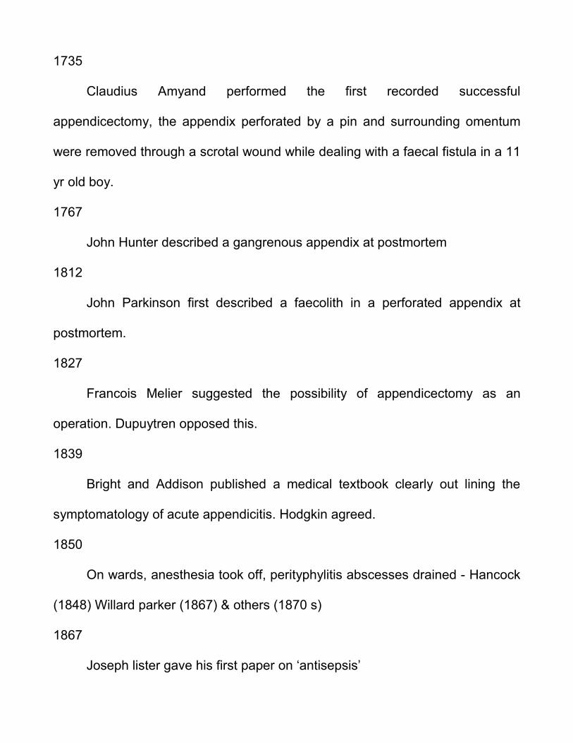
1735
Claudius Amyand performed the first recorded successful
appendicectomy, the appendix perforated by a pin and surrounding omentum
were removed through a scrotal wound while dealing with a faecal fistula in a 11
yr old boy.
1767
John Hunter described a gangrenous appendix at postmortem
1812
John Parkinson first described a faecolith in a perforated appendix at
postmortem.
1827
Francois Melier suggested the possibility of appendicectomy as an
operation. Dupuytren opposed this.
1839
Bright and Addison published a medical textbook clearly out lining the
symptomatology of acute appendicitis. Hodgkin agreed.
1850
On wards, anesthesia took off, perityphylitis abscesses drained - Hancock
(1848) Willard parker (1867) & others (1870 s)
1867
Joseph lister gave his first paper on ‘antisepsis’
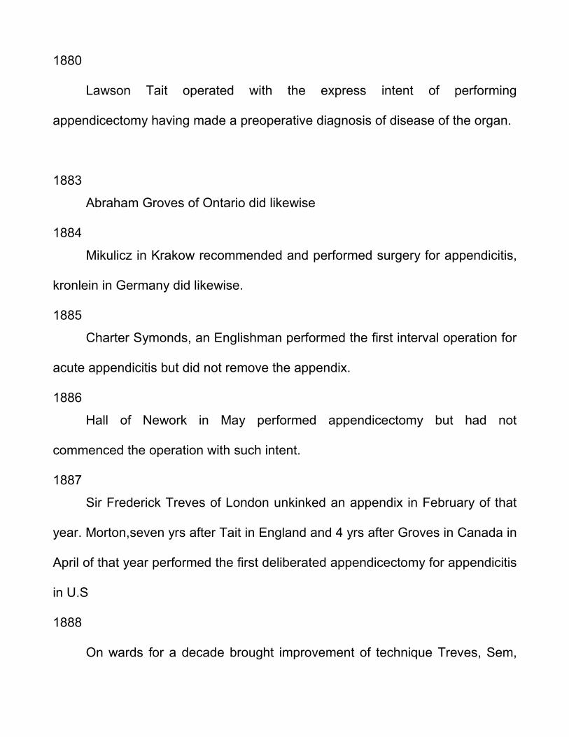
1880
Lawson Tait operated with the express intent of performing
appendicectomy having made a preoperative diagnosis of disease of the organ.
1883
Abraham Groves of Ontario did likewise
1884
Mikulicz in Krakow recommended and performed surgery for appendicitis,
kronlein in Germany did likewise.
1885
Charter Symonds, an Englishman performed the first interval operation for
acute appendicitis but did not remove the appendix.
1886
Hall of Nework in May performed appendicectomy but had not
commenced the operation with such intent.
1887
Sir Frederick Treves of London unkinked an appendix in February of that
year. Morton,seven yrs after Tait in England and 4 yrs after Groves in Canada in
April of that year performed the first deliberated appendicectomy for appendicitis
in U.S
1888
On wards for a decade brought improvement of technique Treves, Sem,
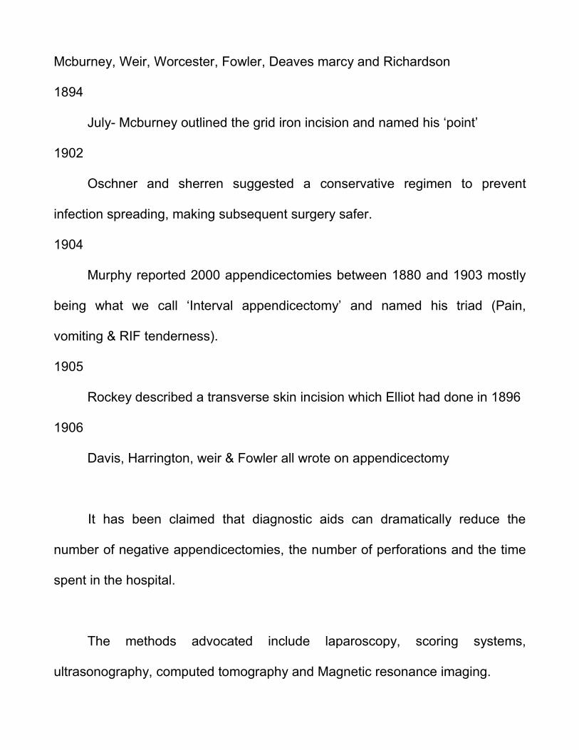
Mcburney, Weir, Worcester, Fowler, Deaves marcy and Richardson
1894
July- Mcburney outlined the grid iron incision and named his ‘point’
1902
Oschner and sherren suggested a conservative regimen to prevent
infection spreading, making subsequent surgery safer.
1904
Murphy reported 2000 appendicectomies between 1880 and 1903 mostly
being what we call ‘Interval appendicectomy’ and named his triad (Pain,
vomiting & RIF tenderness).
1905
Rockey described a transverse skin incision which Elliot had done in 1896
1906
Davis, Harrington, weir & Fowler all wrote on appendicectomy
It has been claimed that diagnostic aids can dramatically reduce the
number of negative appendicectomies, the number of perforations and the time
spent in the hospital.
The methods advocated include laparoscopy, scoring systems,
ultrasonography, computed tomography and Magnetic resonance imaging.
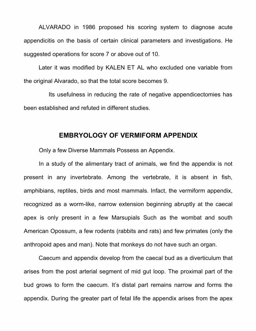
ALVARADO in 1986 proposed his scoring system to diagnose acute
appendicitis on the basis of certain clinical parameters and investigations. He
suggested operations for score 7 or above out of 10.
Later it was modified by KALEN ET AL who excluded one variable from
the original Alvarado, so that the total score becomes 9.
Its usefulness in reducing the rate of negative appendicectomies has
been established and refuted in different studies.
EMBRYOLOGY OF VERMIFORM APPENDIX
Only a few Diverse Mammals Possess an Appendix.
In a study of the alimentary tract of animals, we find the appendix is not
present in any invertebrate. Among the vertebrate, it is absent in fish,
amphibians, reptiles, birds and most mammals. Infact, the vermiform appendix,
recognized as a worm-like, narrow extension beginning abruptly at the caecal
apex is only present in a few Marsupials Such as the wombat and south
American Opossum, a few rodents (rabbits and rats) and few primates (only the
anthropoid apes and man). Note that monkeys do not have such an organ.
Caecum and appendix develop from the caecal bud as a diverticulum that
arises from the post arterial segment of mid gut loop. The proximal part of the
bud grows to form the caecum. It’s distal part remains narrow and forms the
appendix. During the greater part of fetal life the appendix arises from the apex
of Caecum. Subsequently the lateral wall of the Caecum grows much more
rapidly than the medial wall with the result the point of attachment of appendix
comes to lie on medial side into a retrocaecal and intraperitoneal position.
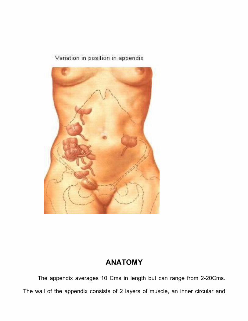
Rarely the caecum does not migrate during development to its normal
position in the right lower quadrant of abdomen. In such cases, we come across
a sub-hepatic appendix or situs inversus totalis,in which the appendix is in the
left iliac fossa, causing diagnostic difficulty if appendicitis develops.
VARIATION IN POSITION IN APPENDIX
ANATOMY
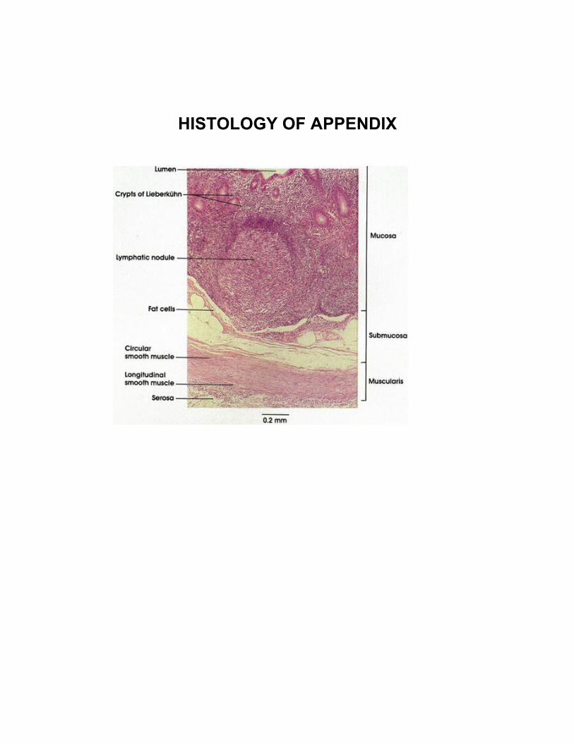
The appendix averages 10 Cms in length but can range from 2-20Cms.
The wall of the appendix consists of 2 layers of muscle, an inner circular and
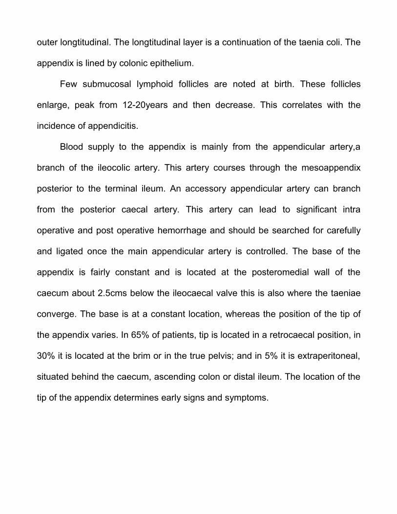
outer longtitudinal. The longtitudinal layer is a continuation of the taenia coli. The
appendix is lined by colonic epithelium.
Few submucosal lymphoid follicles are noted at birth. These follicles
enlarge, peak from 12-20years and then decrease. This correlates with the
incidence of appendicitis.
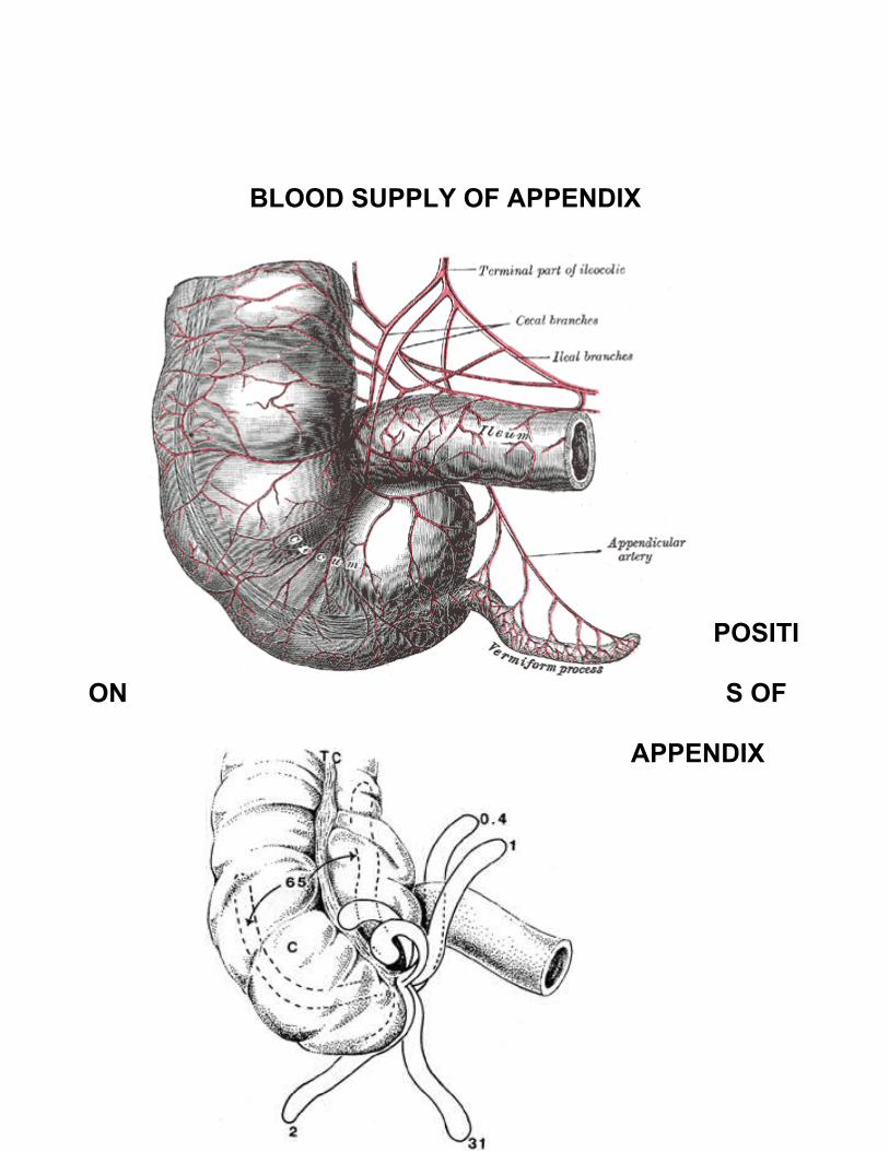
Blood supply to the appendix is mainly from the appendicular artery,a
branch of the ileocolic artery. This artery courses through the mesoappendix
posterior to the terminal ileum. An accessory appendicular artery can branch
from the posterior caecal artery. This artery can lead to significant intra
operative and post operative hemorrhage and should be searched for carefully
and ligated once the main appendicular artery is controlled. The base of the
appendix is fairly constant and is located at the posteromedial wall of the
caecum about 2.5cms below the ileocaecal valve this is also where the taeniae
converge. The base is at a constant location, whereas the position of the tip of
the appendix varies. In 65% of patients, tip is located in a retrocaecal position, in
30% it is located at the brim or in the true pelvis; and in 5% it is extraperitoneal,
situated behind the caecum, ascending colon or distal ileum. The location of the
tip of the appendix determines early signs and symptoms.
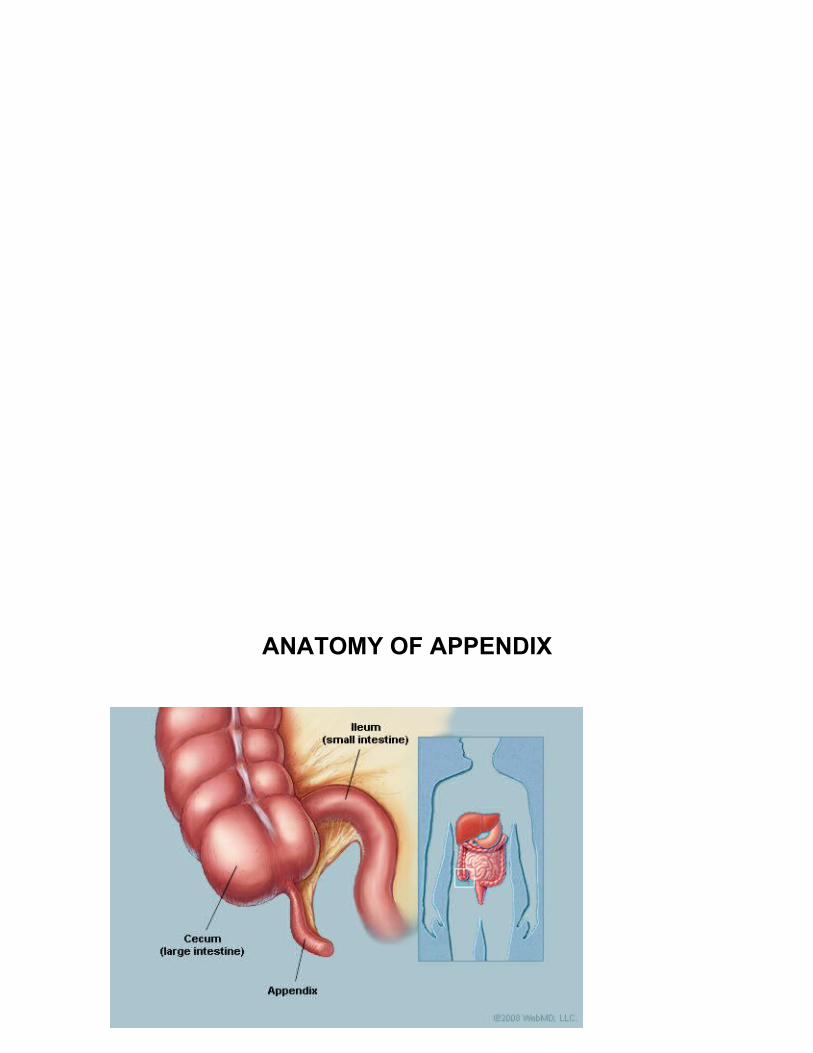
ANATOMY OF APPENDIX
BLOOD SUPPLY OF APPENDIX
POSITI
ON S OF
APPENDIX
HISTOLOGY OF APPENDIX
AETIO PATHOGENESIS
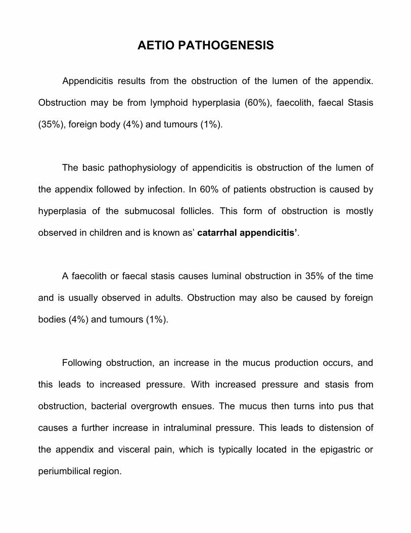
Appendicitis results from the obstruction of the lumen of the appendix.
Obstruction may be from lymphoid hyperplasia (60%), faecolith, faecal Stasis
(35%), foreign body (4%) and tumours (1%).
The basic pathophysiology of appendicitis is obstruction of the lumen of
the appendix followed by infection. In 60% of patients obstruction is caused by
hyperplasia of the submucosal follicles. This form of obstruction is mostly
observed in children and is known as’ catarrhal appendicitis’.
A faecolith or faecal stasis causes luminal obstruction in 35% of the time
and is usually observed in adults. Obstruction may also be caused by foreign
bodies (4%) and tumours (1%).
Following obstruction, an increase in the mucus production occurs, and
this leads to increased pressure. With increased pressure and stasis from
obstruction, bacterial overgrowth ensues. The mucus then turns into pus that
causes a further increase in intraluminal pressure. This leads to distension of
the appendix and visceral pain, which is typically located in the epigastric or
periumbilical region.
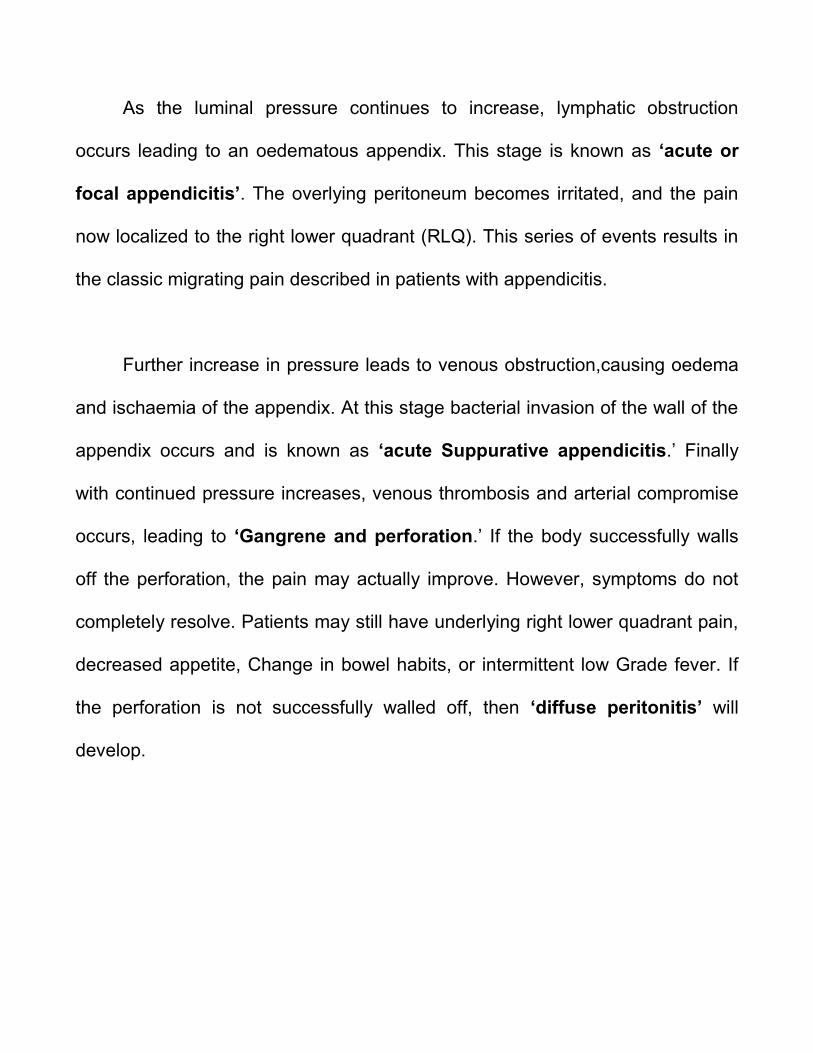
As the luminal pressure continues to increase, lymphatic obstruction
occurs leading to an oedematous appendix. This stage is known as ‘acute or
focal appendicitis’. The overlying peritoneum becomes irritated, and the pain
now localized to the right lower quadrant (RLQ). This series of events results in
the classic migrating pain described in patients with appendicitis.
Further increase in pressure leads to venous obstruction,causing oedema
and ischaemia of the appendix. At this stage bacterial invasion of the wall of the
appendix occurs and is known as ‘acute Suppurative appendicitis.’ Finally
with continued pressure increases, venous thrombosis and arterial compromise
occurs, leading to ‘Gangrene and perforation.’ If the body successfully walls
off the perforation, the pain may actually improve. However, symptoms do not
completely resolve. Patients may still have underlying right lower quadrant pain,
decreased appetite, Change in bowel habits, or intermittent low Grade fever. If
the perforation is not successfully walled off, then ‘diffuse peritonitis’ will
develop.
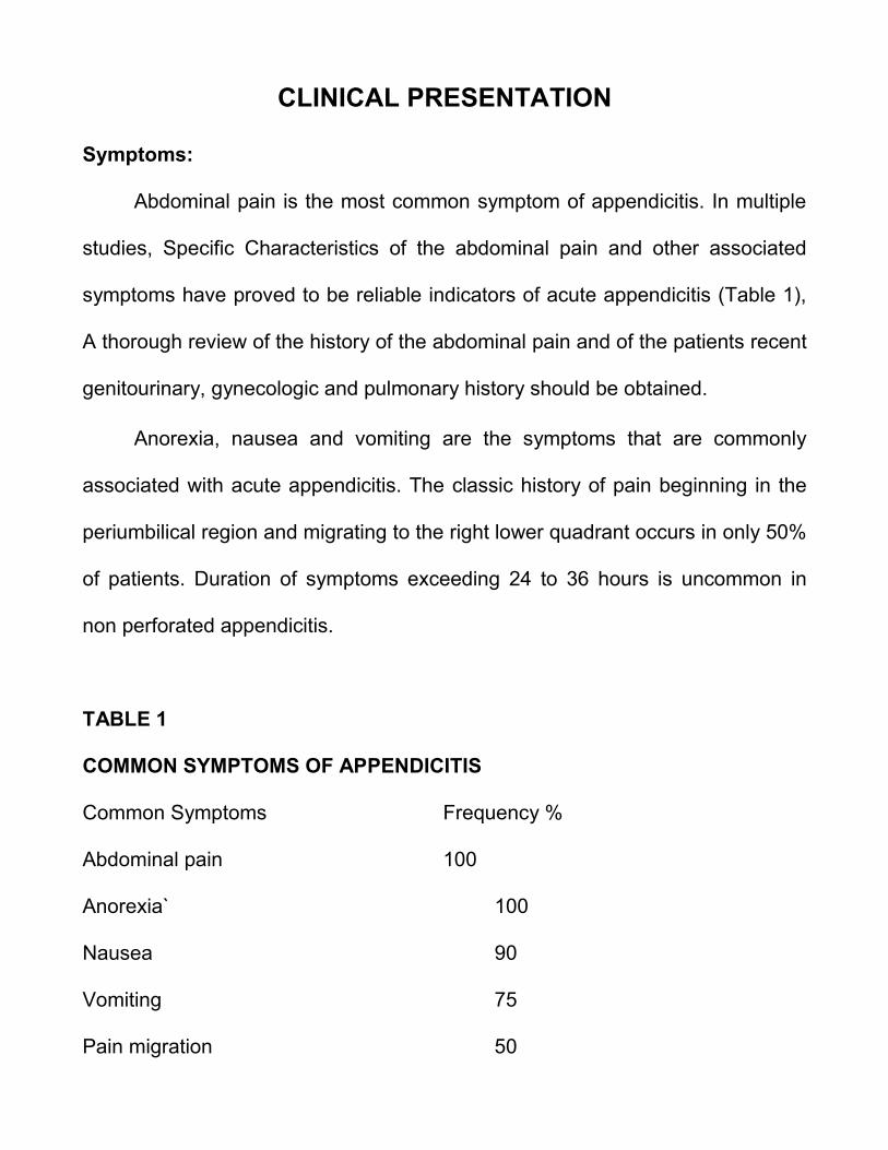
CLINICAL PRESENTATION
Symptoms:
Abdominal pain is the most common symptom of appendicitis. In multiple
studies, Specific Characteristics of the abdominal pain and other associated
symptoms have proved to be reliable indicators of acute appendicitis (Table 1),
A thorough review of the history of the abdominal pain and of the patients recent
genitourinary, gynecologic and pulmonary history should be obtained.
Anorexia, nausea and vomiting are the symptoms that are commonly
associated with acute appendicitis. The classic history of pain beginning in the
periumbilical region and migrating to the right lower quadrant occurs in only 50%
of patients. Duration of symptoms exceeding 24 to 36 hours is uncommon in
non perforated appendicitis.
TABLE 1
COMMON SYMPTOMS OF APPENDICITIS
Common Symptoms Frequency %
Abdominal pain 100
Anorexia` 100
Nausea 90
Vomiting 75
Pain migration 50
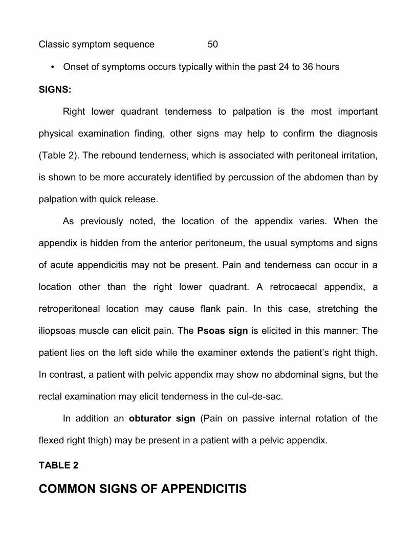
Classic symptom sequence 50
• Onset of symptoms occurs typically within the past 24 to 36 hours
SIGNS:
Right lower quadrant tenderness to palpation is the most important
physical examination finding, other signs may help to confirm the diagnosis
(Table 2). The rebound tenderness, which is associated with peritoneal irritation,
is shown to be more accurately identified by percussion of the abdomen than by
palpation with quick release.
As previously noted, the location of the appendix varies. When the
appendix is hidden from the anterior peritoneum, the usual symptoms and signs
of acute appendicitis may not be present. Pain and tenderness can occur in a
location other than the right lower quadrant. A retrocaecal appendix, a
retroperitoneal location may cause flank pain. In this case, stretching the
iliopsoas muscle can elicit pain. The Psoas sign is elicited in this manner: The
patient lies on the left side while the examiner extends the patient’s right thigh.
In contrast, a patient with pelvic appendix may show no abdominal signs, but the
rectal examination may elicit tenderness in the cul-de-sac.
In addition an obturator sign (Pain on passive internal rotation of the
flexed right thigh) may be present in a patient with a pelvic appendix.
TABLE 2
COMMON SIGNS OF APPENDICITIS
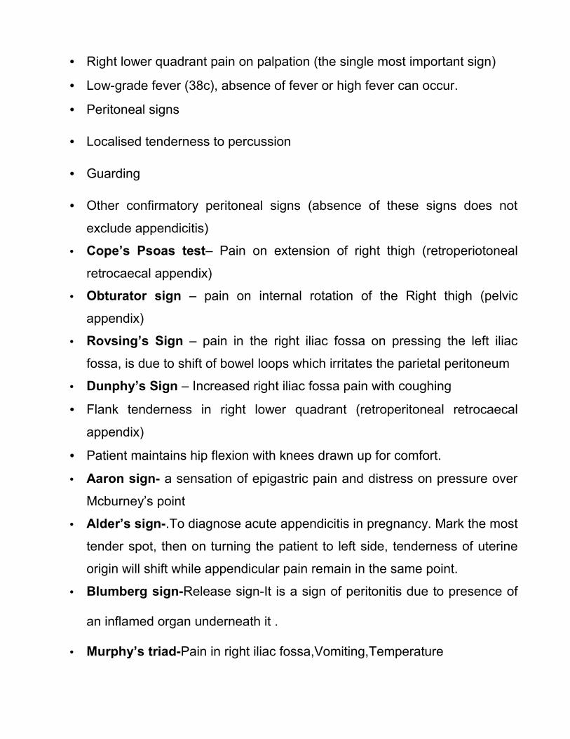
• Right lower quadrant pain on palpation (the single most important sign)
• Low-grade fever (38c), absence of fever or high fever can occur.
• Peritoneal signs
• Localised tenderness to percussion
• Guarding
• Other confirmatory peritoneal signs (absence of these signs does not
exclude appendicitis)
• Cope’s Psoas test– Pain on extension of right thigh (retroperiotoneal
retrocaecal appendix)
• Obturator sign – pain on internal rotation of the Right thigh (pelvic
appendix)
• Rovsing’s Sign – pain in the right iliac fossa on pressing the left iliac
fossa, is due to shift of bowel loops which irritates the parietal peritoneum
• Dunphy’s Sign – Increased right iliac fossa pain with coughing
• Flank tenderness in right lower quadrant (retroperitoneal retrocaecal
appendix)
• Patient maintains hip flexion with knees drawn up for comfort.
• Aaron sign- a sensation of epigastric pain and distress on pressure over
Mcburney’s point
• Alder’s sign-.To diagnose acute appendicitis in pregnancy. Mark the most
tender spot, then on turning the patient to left side, tenderness of uterine
origin will shift while appendicular pain remain in the same point.
• Blumberg sign-Release sign-It is a sign of peritonitis due to presence of
an inflamed organ underneath it .
• Murphy’s triad-Pain in right iliac fossa,Vomiting,Temperature
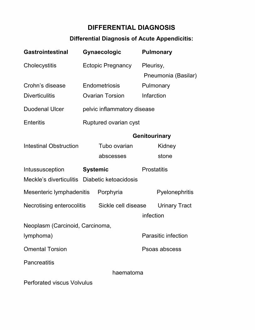
DIFFERENTIAL DIAGNOSIS
Differential Diagnosis of Acute Appendicitis:
Gastrointestinal Gynaecologic Pulmonary
Cholecystitis Ectopic Pregnancy Pleurisy,
Pneumonia (Basilar)
Crohn’s disease Endometriosis Pulmonary
Diverticulitis Ovarian Torsion Infarction
Duodenal Ulcer pelvic inflammatory disease
Enteritis Ruptured ovarian cyst
Genitourinary
Intestinal Obstruction Tubo ovarian Kidney
abscesses stone
Intussusception Systemic Prostatitis
Meckle’s diverticulitis Diabetic ketoacidosis
Mesenteric lymphadenitis Porphyria Pyelonephritis
Necrotising enterocolitis Sickle cell disease Urinary Tract
infection
Neoplasm (Carcinoid, Carcinoma,
lymphoma) Parasitic infection
Omental Torsion Psoas abscess
Pancreatitis
haematoma
Perforated viscus Volvulus
LAB AND RADIOLOGIC EVALUATION
LAB TESTS
Leukocytosis
Increase of C – Reactive protein
PLAIN X-RAY FILM
i. To elucidate the cause of abdominal pain
SIGNS OF ACUTE APPENDICITIS
a) Appendix calculus (0.5-6cm)
b) Sentinel loop – dilated atonic ileum containing a fluid level
c) Dilated caecum
d) Widening of the preperitoneal fat line
e) Right lower quadrant haze due to fluid and oedema.
f) Scoliosis concave to the Right
g) Right lower quadrant mass indenting the caecum.
h) Blurring of the right psoas outline – unreliable
i) Gas in the appendix – Rare, unreliable
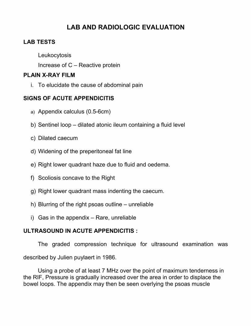
ULTRASOUND IN ACUTE APPENDICITIS :
The graded compression technique for ultrasound examination was
described by Julien puylaert in 1986.
Using a probe of at least 7 MHz over the point of maximum tenderness in the RIF, Pressure is gradually increased over the area in order to displace the bowel loops. The appendix may then be seen overlying the psoas muscle
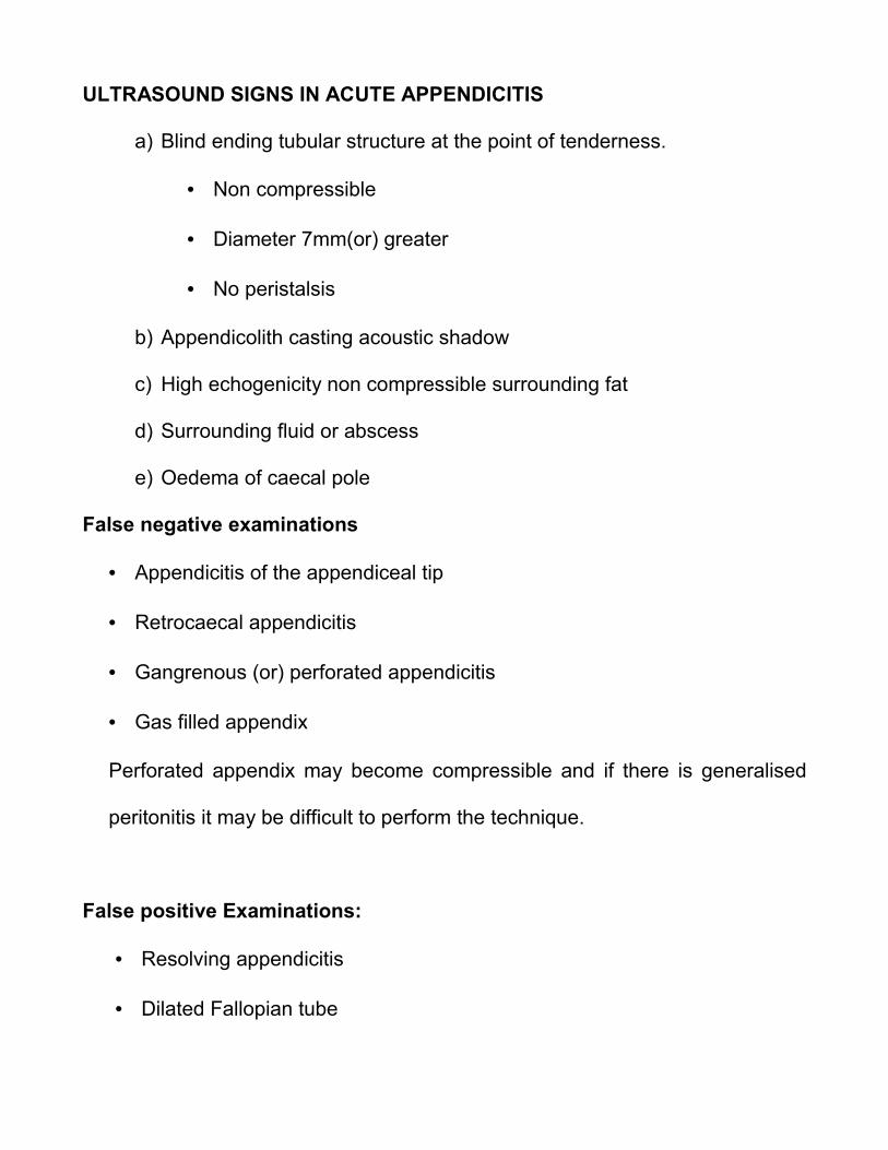
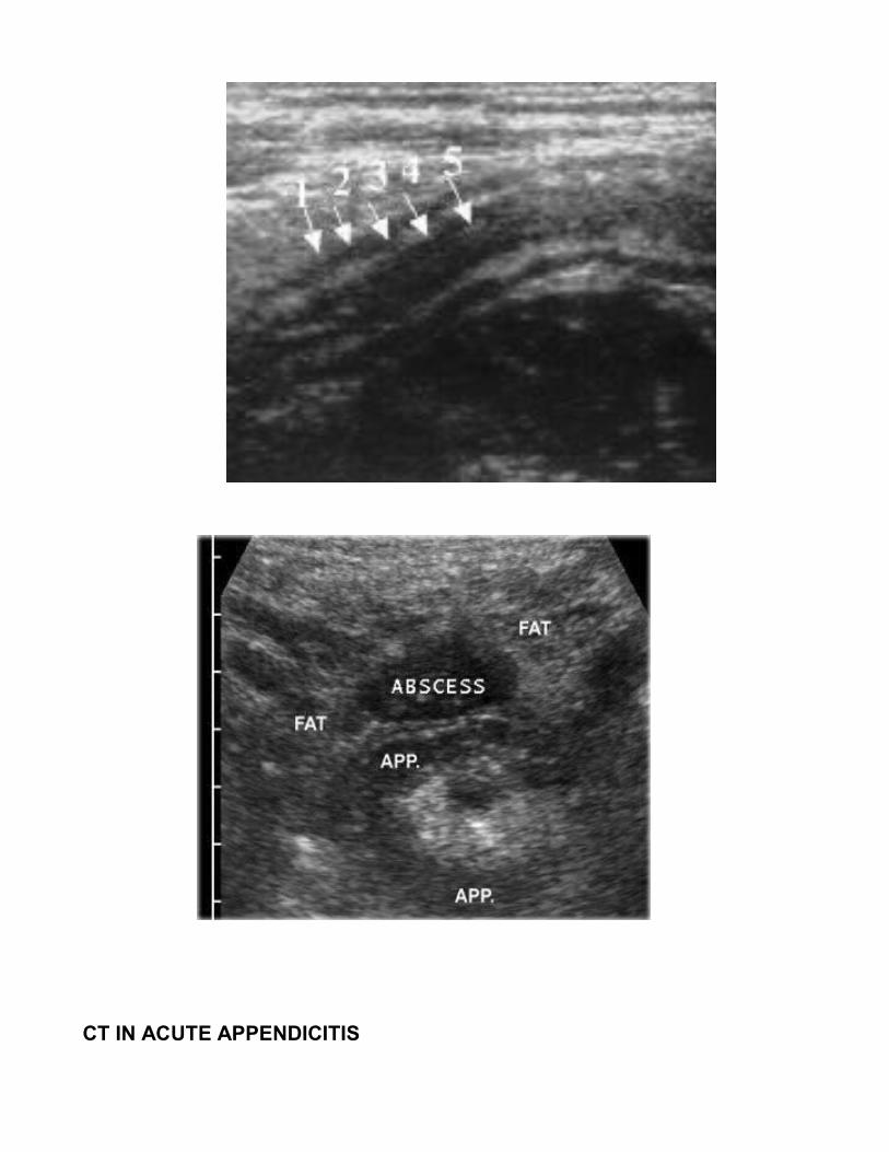
ULTRASOUND SIGNS IN ACUTE APPENDICITIS
a) Blind ending tubular structure at the point of tenderness.
• Non compressible
• Diameter 7mm(or) greater
• No peristalsis
b) Appendicolith casting acoustic shadow
c) High echogenicity non compressible surrounding fat
d) Surrounding fluid or abscess
e) Oedema of caecal pole
False negative examinations
• Appendicitis of the appendiceal tip
• Retrocaecal appendicitis
• Gangrenous (or) perforated appendicitis
• Gas filled appendix
Perforated appendix may become compressible and if there is generalised
peritonitis it may be difficult to perform the technique.
False positive Examinations:
• Resolving appendicitis
• Dilated Fallopian tube
• Inflammatory Bowel disease
• Inspissated stool mimicking an appendicolith
Major drawback in the investigation is, Normal appendix is not visualized
by these techniques. Although a positive diagnosis can be made when an
abnormal appendix is seen appendicitis cannot be excluded when an appendix
has not been found. ultrasound or CT examination should not be a substitute for
a good clinical history and examination. However there are many conditions,
which mimic appendicitis clinically and may be diagnosed at ultrasound of the
abdomen & Pelvis. It is reasonable to perform an ultrasound in young women
with suspected appendicitis in order to exclude gynecological conditions.
Ultrasound has not been shown to be of proven clinical benefit in some studies.
Any delays in treatment while scans are being organized may have an adverse
effect on the clinical outcome.
USG IN ACUTE APPENDICITIS
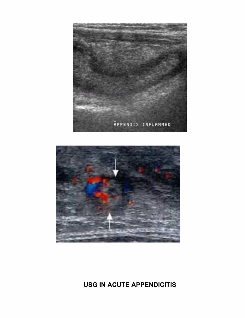
USG IN ACUTE APPENDICITIS
CT IN ACUTE APPENDICITIS
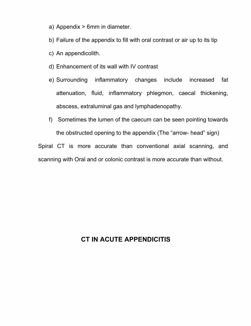
a) Appendix > 6mm in diameter.
b) Failure of the appendix to fill with oral contrast or air up to its tip
c) An appendicolith.
d) Enhancement of its wall with IV contrast
e) Surrounding inflammatory changes include increased fat
attenuation, fluid, inflammatory phlegmon, caecal thickening,
abscess, extraluminal gas and lymphadenopathy.
f) Sometimes the lumen of the caecum can be seen pointing towards
the obstructed opening to the appendix (The “arrow- head” sign)
Spiral CT is more accurate than conventional axial scanning, and
scanning with Oral and or colonic contrast is more accurate than without.
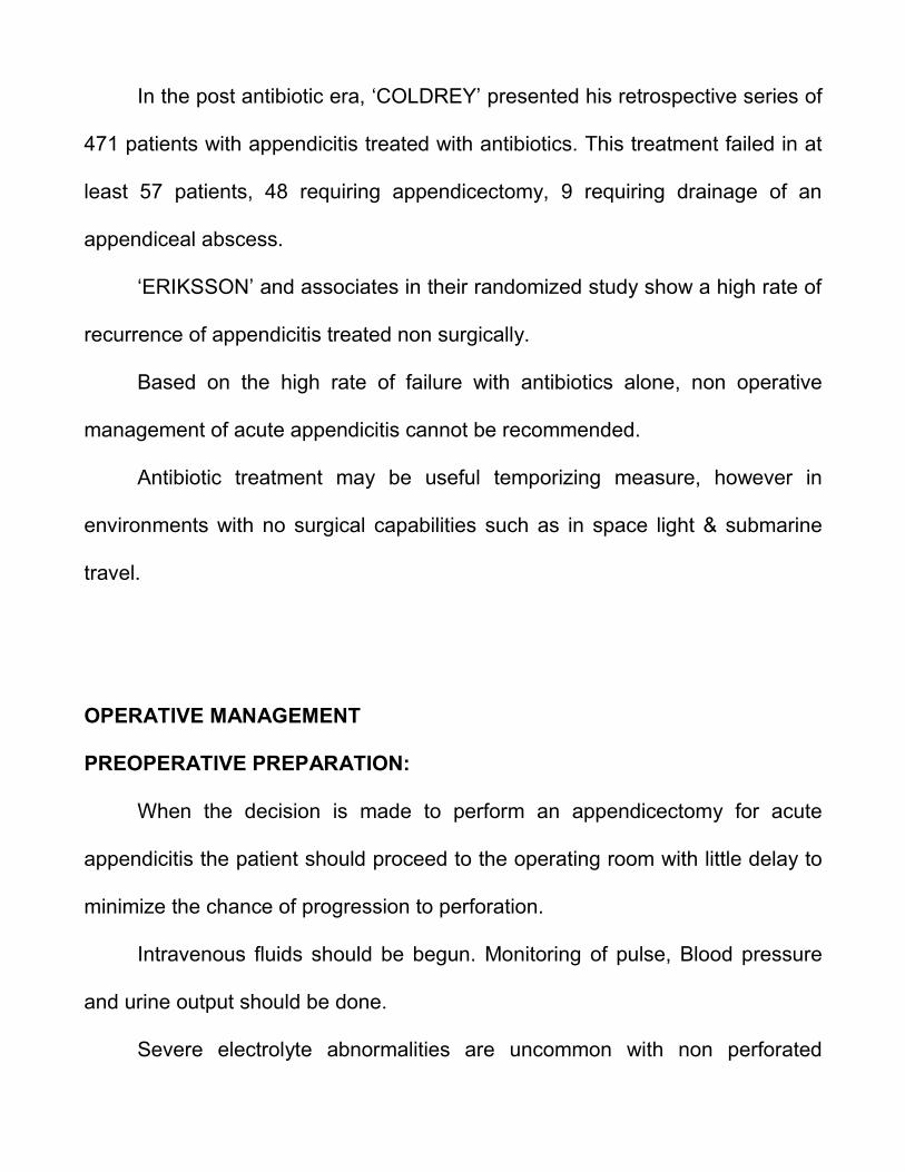
CT IN ACUTE APPENDICITIS
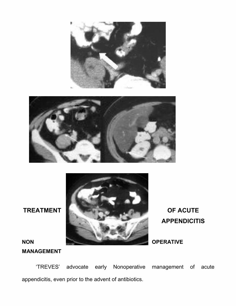
TREATMENT OF ACUTE
APPENDICITIS
NON OPERATIVE
MANAGEMENT
‘TREVES’ advocate early Nonoperative management of acute
appendicitis, even prior to the advent of antibiotics.
In the post antibiotic era, ‘COLDREY’ presented his retrospective series of
471 patients with appendicitis treated with antibiotics. This treatment failed in at
least 57 patients, 48 requiring appendicectomy, 9 requiring drainage of an
appendiceal abscess.
‘ERIKSSON’ and associates in their randomized study show a high rate of
recurrence of appendicitis treated non surgically.
Based on the high rate of failure with antibiotics alone, non operative
management of acute appendicitis cannot be recommended.
Antibiotic treatment may be useful temporizing measure, however in
environments with no surgical capabilities such as in space light & submarine
travel.
OPERATIVE MANAGEMENT
PREOPERATIVE PREPARATION:
When the decision is made to perform an appendicectomy for acute
appendicitis the patient should proceed to the operating room with little delay to
minimize the chance of progression to perforation.
Intravenous fluids should be begun. Monitoring of pulse, Blood pressure
and urine output should be done.
Severe electrolyte abnormalities are uncommon with non perforated
appendicitis, as vomiting and fever have typically been present for 24hrs or less,
but may be significant in cases of perforation. Any electrolyte deficiencies
should be corrected prior to the induction of general anesthesia.
Antibiotics should be administered 30 minutes prior to incision to achieve
adequate tissue levels.
Acceptable antibiotics include a second generation cephalosporin or
combination of antibiotics directed at gram negative and anaerobes.
OPEN APPENDICECTOMY
Appendicectomy for the free lying appendix
INCISION
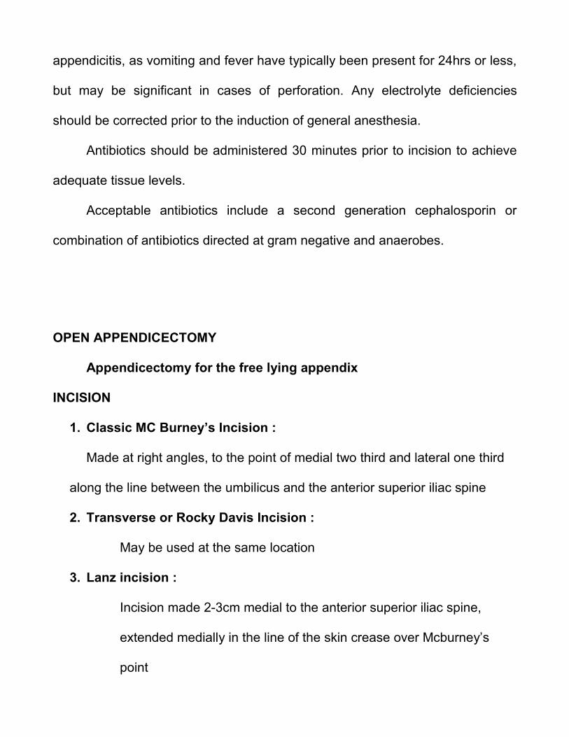
1. Classic MC Burney’s Incision :
Made at right angles, to the point of medial two third and lateral one third
along the line between the umbilicus and the anterior superior iliac spine
2. Transverse or Rocky Davis Incision :
May be used at the same location
3. Lanz incision :
Incision made 2-3cm medial to the anterior superior iliac spine,
extended medially in the line of the skin crease over Mcburney’s
point
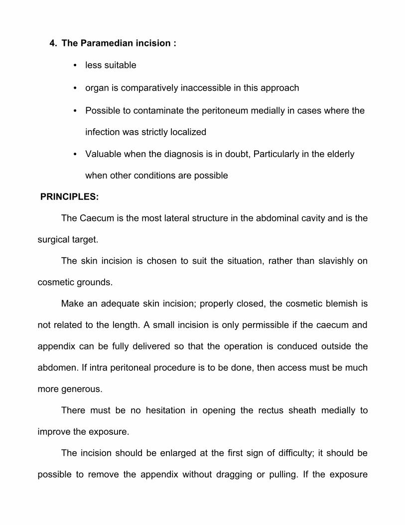
4. The Paramedian incision :
• less suitable
• organ is comparatively inaccessible in this approach
• Possible to contaminate the peritoneum medially in cases where the
infection was strictly localized
• Valuable when the diagnosis is in doubt, Particularly in the elderly
when other conditions are possible
PRINCIPLES:
The Caecum is the most lateral structure in the abdominal cavity and is the
surgical target.
The skin incision is chosen to suit the situation, rather than slavishly on
cosmetic grounds.
Make an adequate skin incision; properly closed, the cosmetic blemish is
not related to the length. A small incision is only permissible if the caecum and
appendix can be fully delivered so that the operation is conduced outside the
abdomen. If intra peritoneal procedure is to be done, then access must be much
more generous.
There must be no hesitation in opening the rectus sheath medially to
improve the exposure.
The incision should be enlarged at the first sign of difficulty; it should be
possible to remove the appendix without dragging or pulling. If the exposure
proves inadequate it is often only the muscular and facial layers that need to be
further incised as the skin wound is relatively mobile.
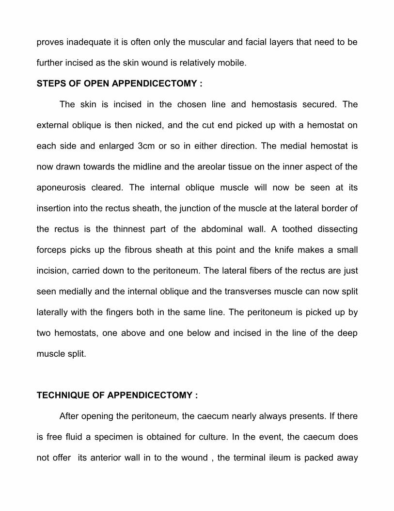
STEPS OF OPEN APPENDICECTOMY :
The skin is incised in the chosen line and hemostasis secured. The
external oblique is then nicked, and the cut end picked up with a hemostat on
each side and enlarged 3cm or so in either direction. The medial hemostat is
now drawn towards the midline and the areolar tissue on the inner aspect of the
aponeurosis cleared. The internal oblique muscle will now be seen at its
insertion into the rectus sheath, the junction of the muscle at the lateral border of
the rectus is the thinnest part of the abdominal wall. A toothed dissecting
forceps picks up the fibrous sheath at this point and the knife makes a small
incision, carried down to the peritoneum. The lateral fibers of the rectus are just
seen medially and the internal oblique and the transverses muscle can now split
laterally with the fingers both in the same line. The peritoneum is picked up by
two hemostats, one above and one below and incised in the line of the deep
muscle split.
TECHNIQUE OF APPENDICECTOMY :
After opening the peritoneum, the caecum nearly always presents. If there
is free fluid a specimen is obtained for culture. In the event, the caecum does
not offer its anterior wall in to the wound , the terminal ileum is packed away
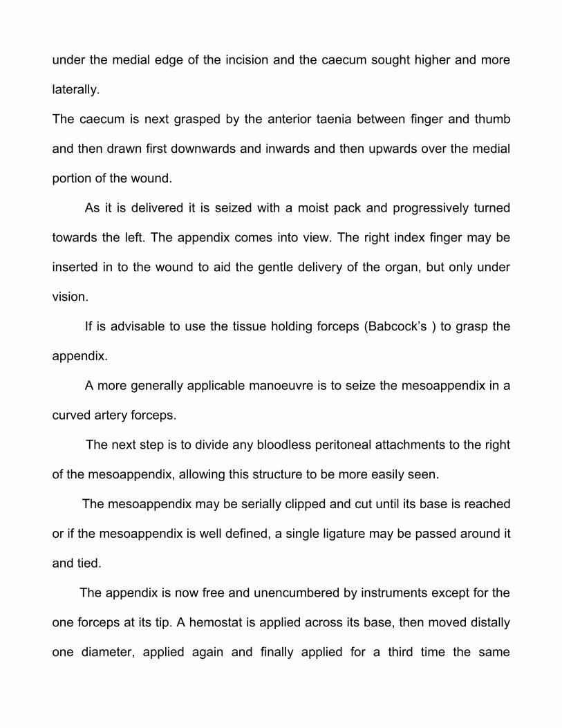
under the medial edge of the incision and the caecum sought higher and more
laterally.
The caecum is next grasped by the anterior taenia between finger and thumb
and then drawn first downwards and inwards and then upwards over the medial
portion of the wound.
As it is delivered it is seized with a moist pack and progressively turned
towards the left. The appendix comes into view. The right index finger may be
inserted in to the wound to aid the gentle delivery of the organ, but only under
vision.
If is advisable to use the tissue holding forceps (Babcock’s ) to grasp the
appendix.
A more generally applicable manoeuvre is to seize the mesoappendix in a
curved artery forceps.
The next step is to divide any bloodless peritoneal attachments to the right
of the mesoappendix, allowing this structure to be more easily seen.
The mesoappendix may be serially clipped and cut until its base is reached
or if the mesoappendix is well defined, a single ligature may be passed around it
and tied.
The appendix is now free and unencumbered by instruments except for the
one forceps at its tip. A hemostat is applied across its base, then moved distally
one diameter, applied again and finally applied for a third time the same
distance along the appendix. The organ is ligated across the first crush and will
be cut through the second.
Residual appendiceal stump should be no longer than 3cm to minimize the
possibility of stump appendicitis in the future.
Much debate has gone for years about whether or not to invaginate the
appendix stump.
Appendicular stump abscess in the caecal wall is so rare that it should not
be regarded as a contraindication to invagination. In that the gut heals best by
the formation of granulation tissue and collagen from serosal layers, it seems
rational to invaginate.
Invagination is done using either purse string or z- stitch suture placed at
least 1.5 cm away from the stump. If the caecal wall is oedematous, one must
not attempt invagination. The appendix base is cut with knife.
The tension on the caecum is now relaxed and the line of the
mesoappendix checked for bleeding. If all is well the caecum is allowed to fall
back into the wound.
Following is carried out if the appendix is with doubt.
I. In a female, Palpate right ovary and tube. The glove is examined for blood.
II. The last meter of the ileum is withdrawn to
• See for mesenteric nodes
• Meckel’s diverticulam
• Reasonabley certain that there are no other lesions
III. A finger is passed to the left and downwards to seek the inflammed loop of
sigmoid colon which is a seat of diverticular disease.
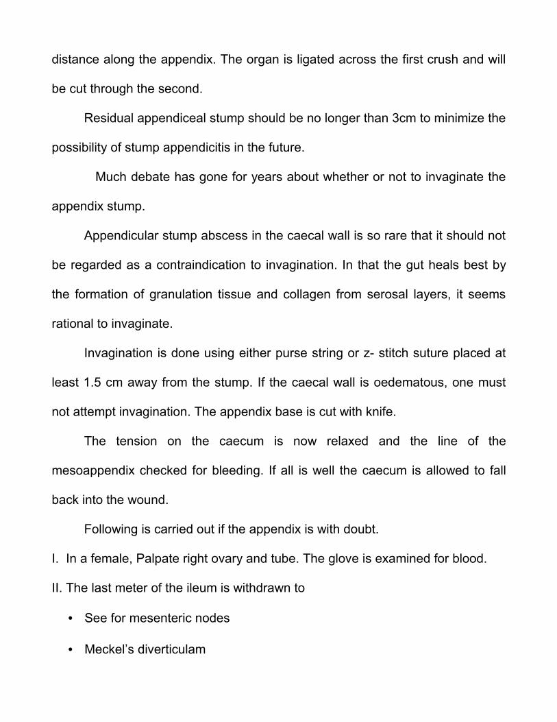
STEPS IN OPEN APPENDICECTOMY
External Oblique Aponeurosis Opened
Peritoneum opened
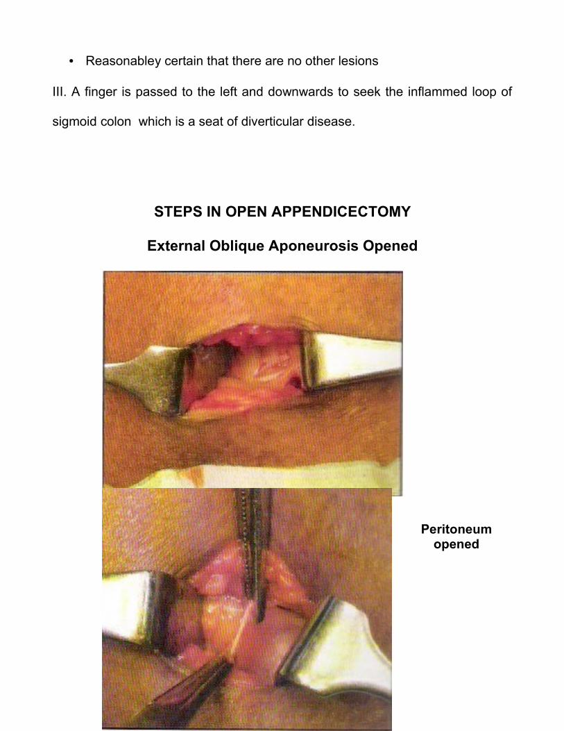
Appendix delivered
Appendix
delivered
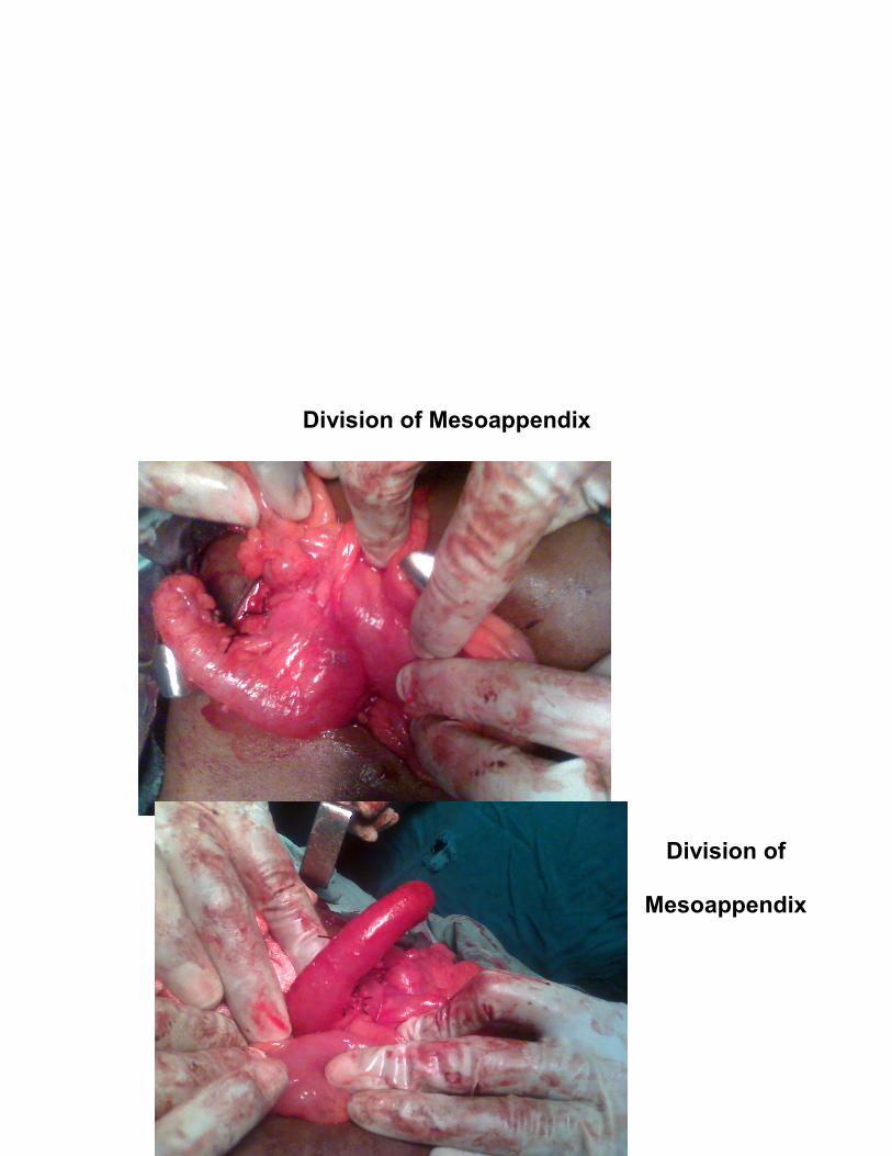
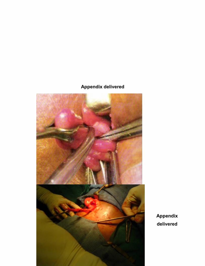
Division of Mesoappendix
Division of
Mesoappendix
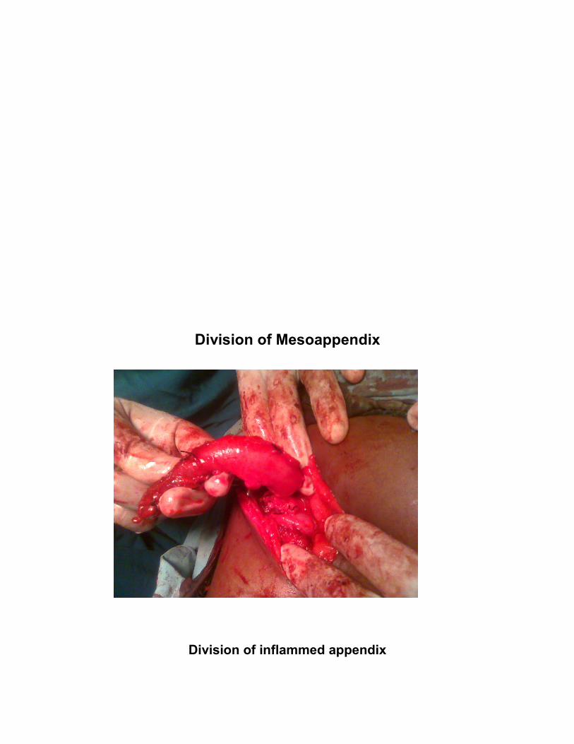
Division of Mesoappendix
Division of inflammed appendix
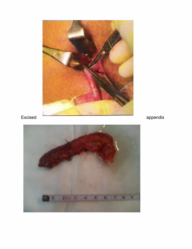
Excised appendix
PROBLEMS :
1. The caecum cannot be found.
• Either not descended fully or malrotation of the intestine
• Extension of the incision upward
2. Caecum cannot be delivered :
• Adequate access and vision. The peritoneal reflection around the
lower pole may be divided bearing in mind, gonadal vessels &
ureter lie medially just deep to the peritoneum
3. Appendix cannot be found :
• Make certain that it is the caecum that has been delivered.
Transverse colon recognised by attachment of greater omentum,
sigmoid colon by appendices epiploicae.
• Trace the taeniacoli of the caeeum, leads to the base of the
appendix. Back or undersurface of the caecum palpated, the
appendix may be buried in the caecal wall.
• If previous appendicectomy excluded, only possibility is, organ has
become inverted (or) intussuscepted.
4. Appendix has sloughed off :
• The mesoappendix anchors the organ in the field of operation
• It may be in 2 portions if a faecolith has perforated through the wall.
• Both portions must be removed and the faecolith retrieved usually
from the pelvis.
5 .The appendix lies Buried Retrocaecally :
• Enlarge the wound
• Caecum is retracted to the left.
• Reflection of the peritoneum on the lateral aspect of the caecum is
in view, a hockey- stick shaped incision is made in the parietal
peritoneum, after a little blunt dissection, in the retroperitoneal
space the caecum can be retracted still further to the left rendered
far more mobile and rotated, the combined effects of which result in
bringing the greater portion of a hidden appendix.
6. Appendix clothed with adherent Greater omentum:
• Not to disturb adherent omentum, when within it lies a gangrenous
or perforated appendix.
• Greater omentum divided between hemostats at a convenient
distance from the appendix and then appendicectomy conducted.
7. Appendix is gangrenous near its junction with caecum
• Possibility of sudden gush of liquid faeces from the caecum, to
avoid this, if the caecum is ballooned deflate the caecum before
appendicectomy.
• The method of closing the stump is, by two sutures transfixing the
caecal wall. These must be inserted before the appendix is
amputated and are later oversewn by interrupted seromuscular
sutures.
8. The mesoappendix is gangrenous and cuts out
• If a ligature will not hold, a stitch applied directly beneath a spurting
vessel may stop the bleeding.
RETROGRADE APPENDICECTOMY :
Indication

• Base of the appendix is accessible and difficulty is experienced in
identifying or delivering the distal part of the organ completely.
• In Retrocaecal appendicitis
Technique :
• Base of the appendix is held between finger and thumb so that its junction
with caecal wall apparent.
• Fine hemostat passed between caecum and appendix to create a space
and 2 simlar instruments are applied across the appendix, which is divided
between them
• The mesoappendix is then clamped & divided working distally.
• Purse string suture is inserted with the hemostat grasps the stump.
• Appendicular stump ligated.
• Base of the appendix buried
Closure :
• There is no absolute need to close the peritoneum separately
• Transverse slit in the peritoneum and deep muscle may be closed as one
layer with either continuous or interrupted absorbable ‘o’ gauge or nil
gauge
• A muscle cutting incision should be closed with continuous or interrupted
absorbable monofilament sutures.
• Skin closed with fine, interrupted monofilament sutures or clips
POST OPERATIVE COMPLICATIONS
Occur in 5% of patients with the unperforated appendix, but in more than
30% of the patients with a gangrenous (or) perforated appendix.
Most frequent complications
a) Wound infection
b) Intra abdominal abscess
c) Faecal fistula
d) Pylephlebitis
e) Intestinal obstruction
A) Wound Infection
• Anaerobic bacteroides species, Aerobes klebsiella, Enterobacter, E.
Coli
• Early signs of wound infection (Undue pain & Edema) are present
• wound should be opened and packed with saline soaked gauze and
reclosed with steri strips in 4-5 days.
B) Intra abdominal abscesses
• Pelvic, subphrenic (or) intraabdominal abscesses Occur in upto 20% of
pts with gangrenous (or) perforated appendicitis.
• Recurrent fever, malaise and anorexia of insidious onset
• CT Scan is of help in diagnosing intraabdminal abscess
• When an abscess is diagnosed it should be drained ether operatively
(or) Percutaneously.
C) Faecal Fistula
Some close spontaneously, provided that there is no anatomic reason for
the fistula remaining open. Those that do not close spontaneously require
operation.
D) Pylephlebitis or Portal pyaemia
• Characterized by jaundice, chills & high fever it is a serious illness
that leads to multiple liver abscesses.
• The infesting organism is usually E.coli
• This complication becomes rare with routine use of antibiotics in
complicated appendicitis.
E) Intestinal Obstruction
• Although not infrequent true mechanical bowel obstruction may
occur as a complication of acute appendicitis
• Operative therapy is indicated as any other mechanical small bowel
obstruction.
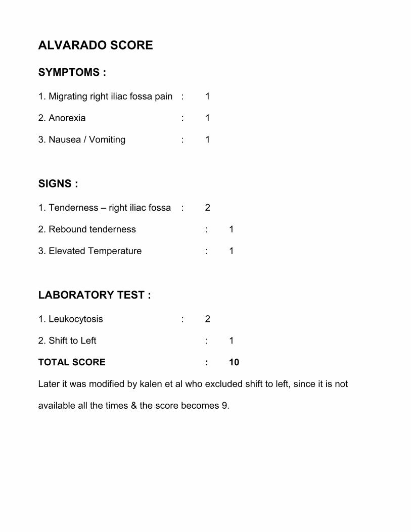
ALVARADO SCORE
SYMPTOMS :
1. Migrating right iliac fossa pain : 1
2. Anorexia : 1
3. Nausea / Vomiting : 1
SIGNS :
1. Tenderness – right iliac fossa : 2
2. Rebound tenderness : 1
3. Elevated Temperature : 1
LABORATORY TEST :
1. Leukocytosis : 2
2. Shift to Left : 1
TOTAL SCORE : 10
Later it was modified by kalen et al who excluded shift to left, since it is not
available all the times & the score becomes 9.
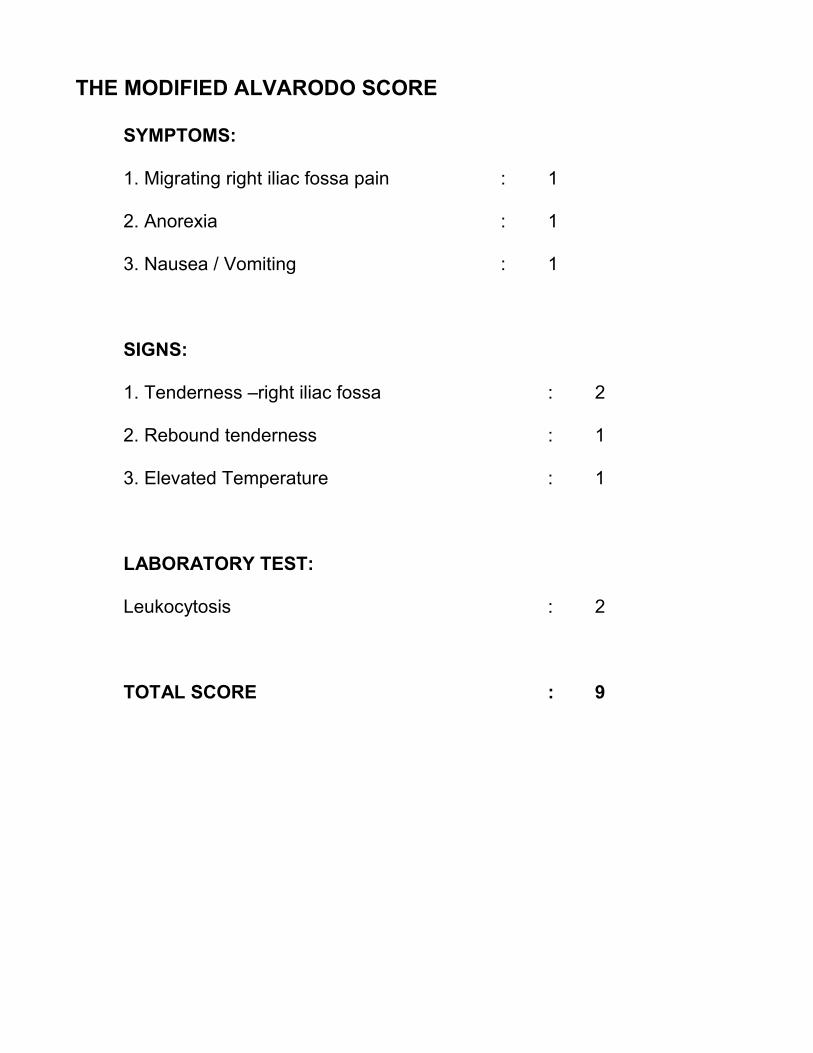
THE MODIFIED ALVARODO SCORE
SYMPTOMS:
1. Migrating right iliac fossa pain : 1
2. Anorexia : 1
3. Nausea / Vomiting : 1
SIGNS:
1. Tenderness –right iliac fossa : 2
2. Rebound tenderness : 1
3. Elevated Temperature : 1
LABORATORY TEST:
Leukocytosis : 2
TOTAL SCORE : 9
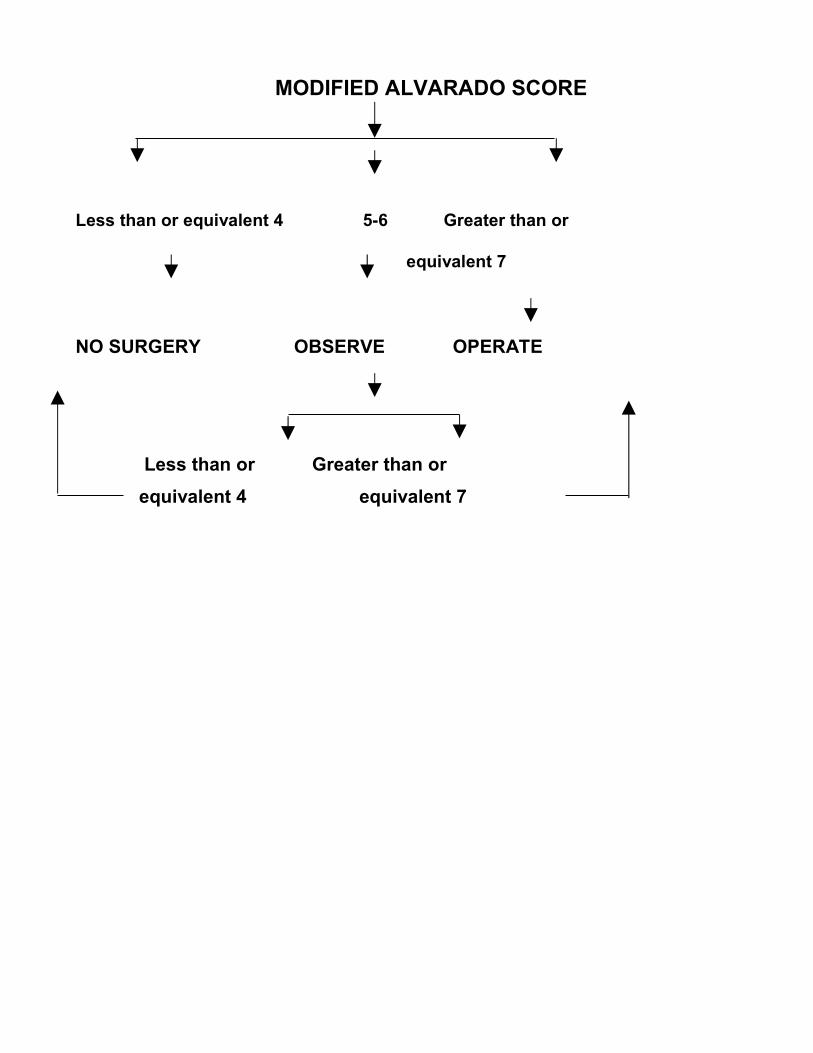
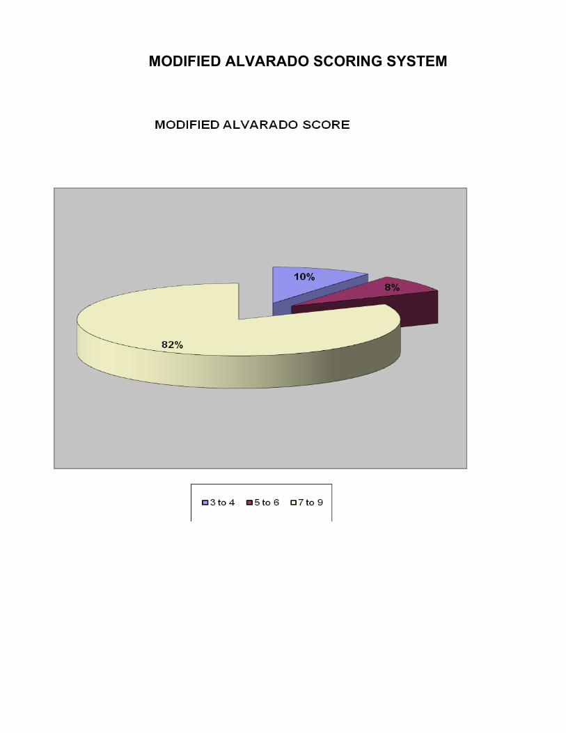
MODIFIED ALVARADO SCORE
Less than or equivalent 4 5-6 Greater than or
equivalent 7
NO SURGERY OBSERVE OPERATE
Less than or Greater than or
equivalent 4 equivalent 7
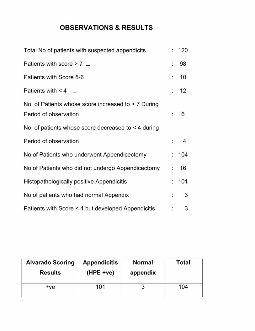
OBSERVATIONS & RESULTS
Total No of patients with suspected appendicits : 120
Patients with score > 7 : 98
Patients with Score 5-6 : 10
Patients with < 4 : 12
No. of Patients whose score increased to > 7 During
Period of observation : 6
No. of patients whose score decreased to < 4 during
Period of observation : 4
No.of Patients who underwent Appendicectomy : 104
No.of Patients who did not undergo Appendicectomy : 16
Histopathologically positive Appendicitis : 101
No.of patients who had normal Appendix : 3
Patients with Score < 4 but developed Appendicitis : 3
Alvarado Scoring
Results
Appendicitis
(HPE +ve)
Normal
appendix
Total
+ve 101 3 104
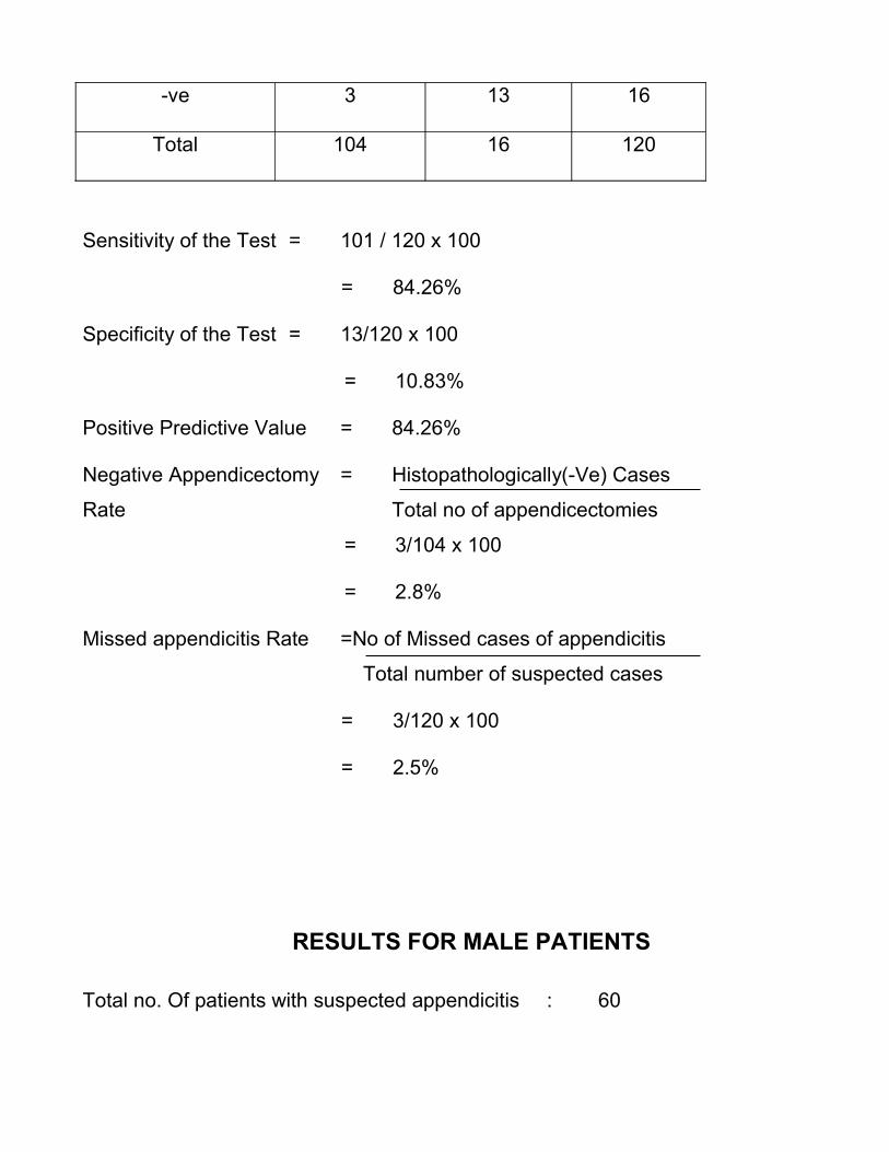
-ve 3 13 16
Total 104 16 120
Sensitivity of the Test = 101 / 120 х 100
= 84.26%
Specificity of the Test = 13/120 х 100
= 10.83%
Positive Predictive Value = 84.26%
Negative Appendicectomy = Histopathologically(-Ve) Cases
Rate Total no of appendicectomies
= 3/104 х 100
= 2.8%
Missed appendicitis Rate =No of Missed cases of appendicitis
Total number of suspected cases
= 3/120 х 100
= 2.5%
RESULTS FOR MALE PATIENTS
Total no. Of patients with suspected appendicitis : 60
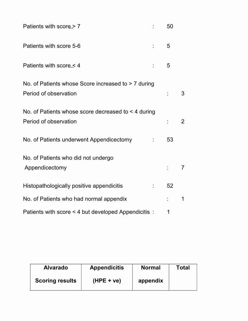
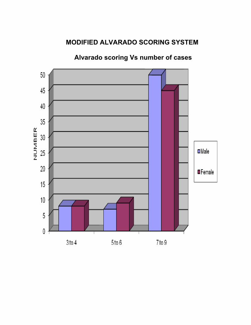
Patients with score > 7 : 50
Patients with score 5-6 : 5
Patients with score < 4 : 5
No. of Patients whose Score increased to > 7 during
Period of observation : 3
No. of Patients whose score decreased to < 4 during
Period of observation : 2
No. of Patients underwent Appendicectomy : 53
No. of Patients who did not undergo
Appendicectomy : 7
Histopathologically positive appendicitis : 52
No. of Patients who had normal appendix : 1
Patients with score < 4 but developed Appendicitis : 1
Alvarado
Scoring results
Appendicitis
(HPE + ve)
Normal
appendix
Total
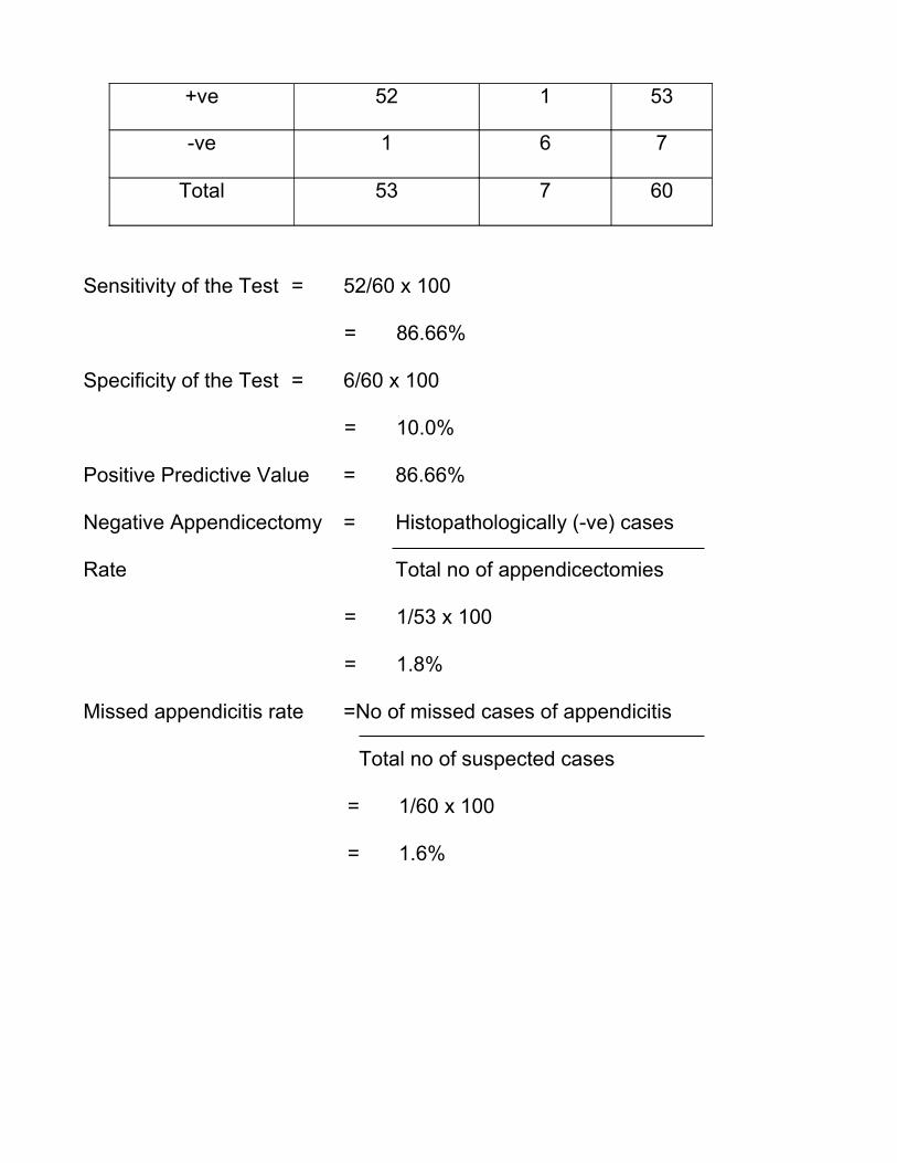
+ve 52 1 53
-ve 1 6 7
Total 53 7 60
Sensitivity of the Test = 52/60 х 100
= 86.66%
Specificity of the Test = 6/60 х 100
= 10.0%
Positive Predictive Value = 86.66%
Negative Appendicectomy = Histopathologically (-ve) cases
Rate Total no of appendicectomies
= 1/53 х 100
= 1.8%
Missed appendicitis rate =No of missed cases of appendicitis
Total no of suspected cases
= 1/60 x 100
= 1.6%
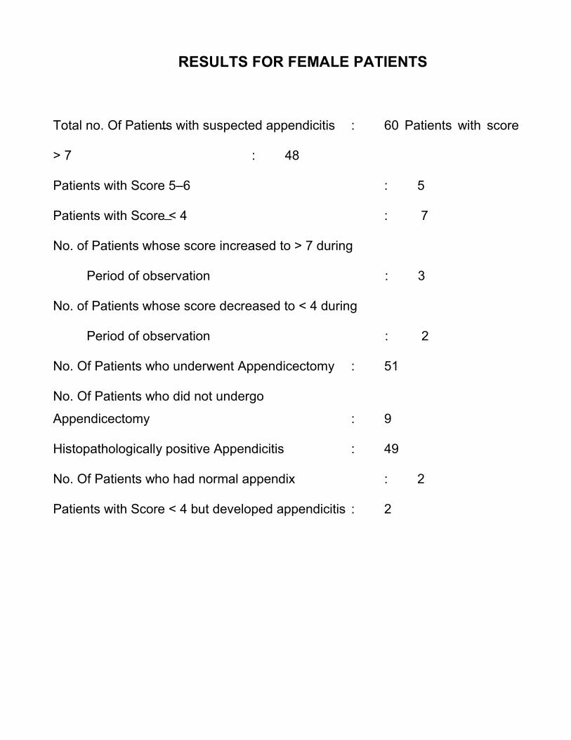
RESULTS FOR FEMALE PATIENTS
Total no. Of Patients with suspected appendicitis : 60 Patients with score
> 7 : 48
Patients with Score 5–6 : 5
Patients with Score < 4 : 7
No. of Patients whose score increased to > 7 during
Period of observation : 3
No. of Patients whose score decreased to < 4 during
Period of observation : 2
No. Of Patients who underwent Appendicectomy : 51
No. Of Patients who did not undergo
Appendicectomy : 9
Histopathologically positive Appendicitis : 49
No. Of Patients who had normal appendix : 2
Patients with Score < 4 but developed appendicitis : 2
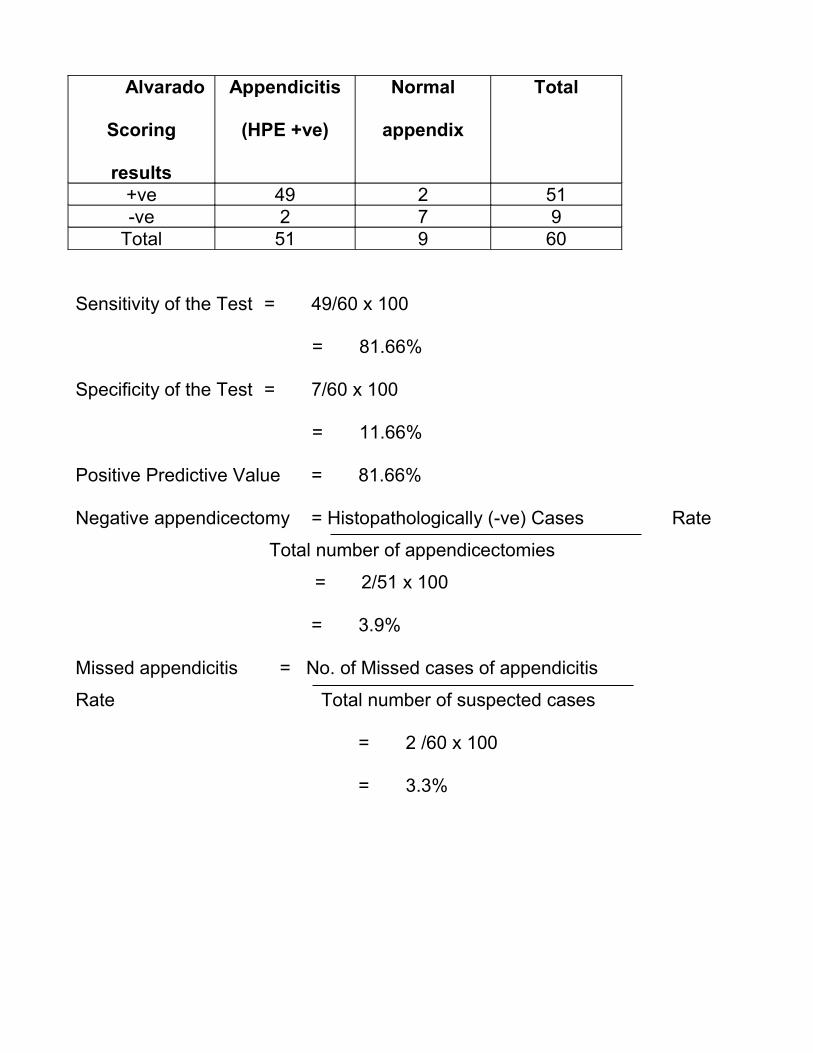
Alvarado
Scoring
results
Appendicitis
(HPE +ve)
Normal
appendix
Total
+ve 49 2 51-ve 2 7 9
Total 51 9 60
Sensitivity of the Test = 49/60 х 100
= 81.66%
Specificity of the Test = 7/60 х 100
= 11.66%
Positive Predictive Value = 81.66%
Negative appendicectomy = Histopathologically (-ve) Cases Rate
Total number of appendicectomies
= 2/51 х 100
= 3.9%
Missed appendicitis = No. of Missed cases of appendicitis
Rate Total number of suspected cases
= 2 /60 х 100
= 3.3%
MODIFIED ALVARADO SCORING SYSTEM
CHARTS
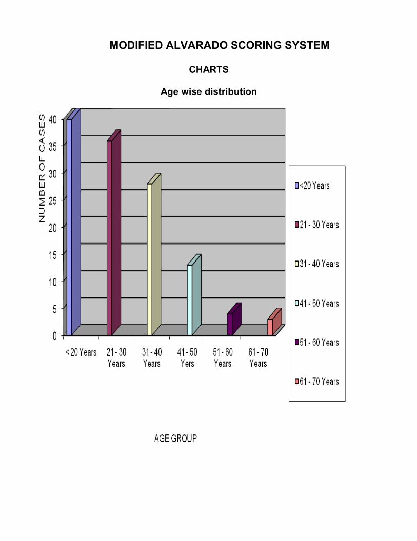
Age wise distribution
MODIFIED ALVARADO SCORING SYSTEM
Alvarado scoring Vs number of cases
MODIFIED ALVARADO SCORING SYSTEM
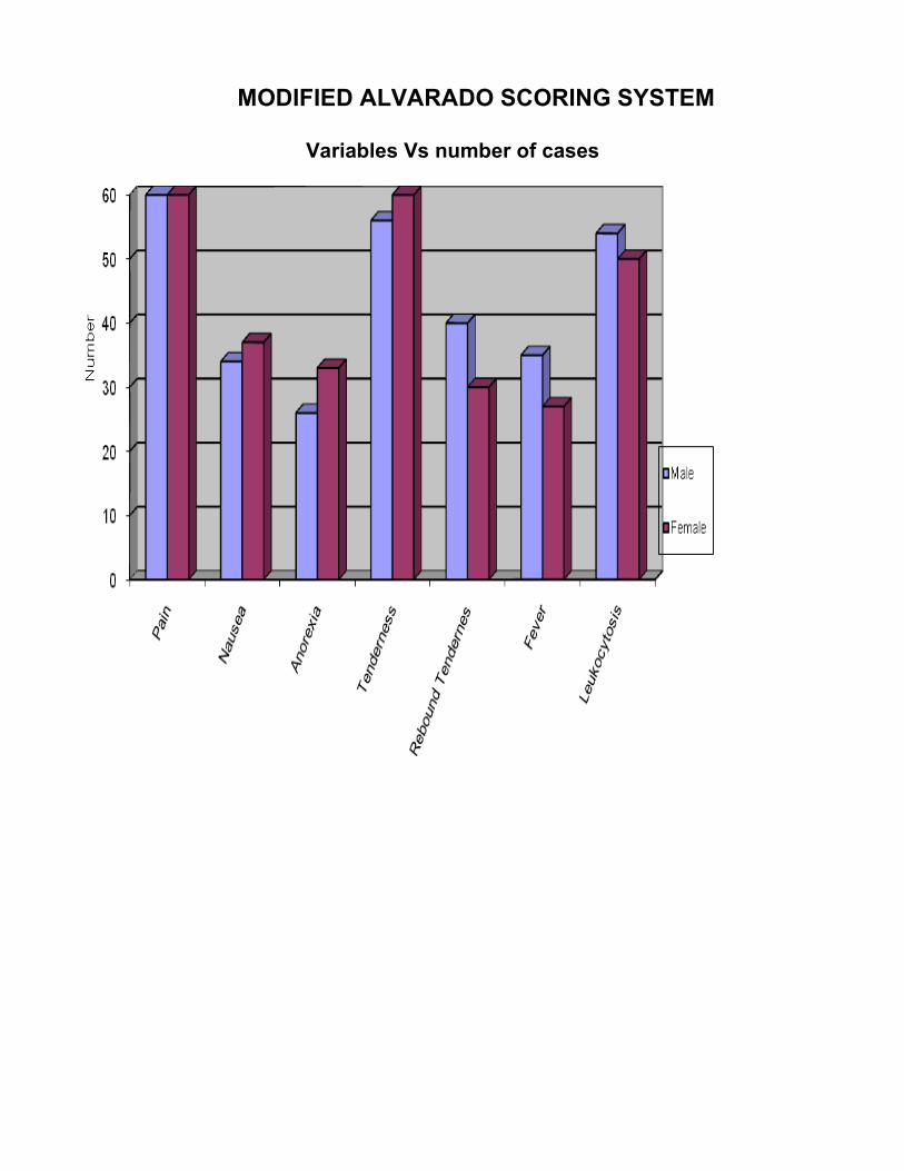
Variables Vs number of cases
MODIFIED ALVARADO SCORING SYSTEM
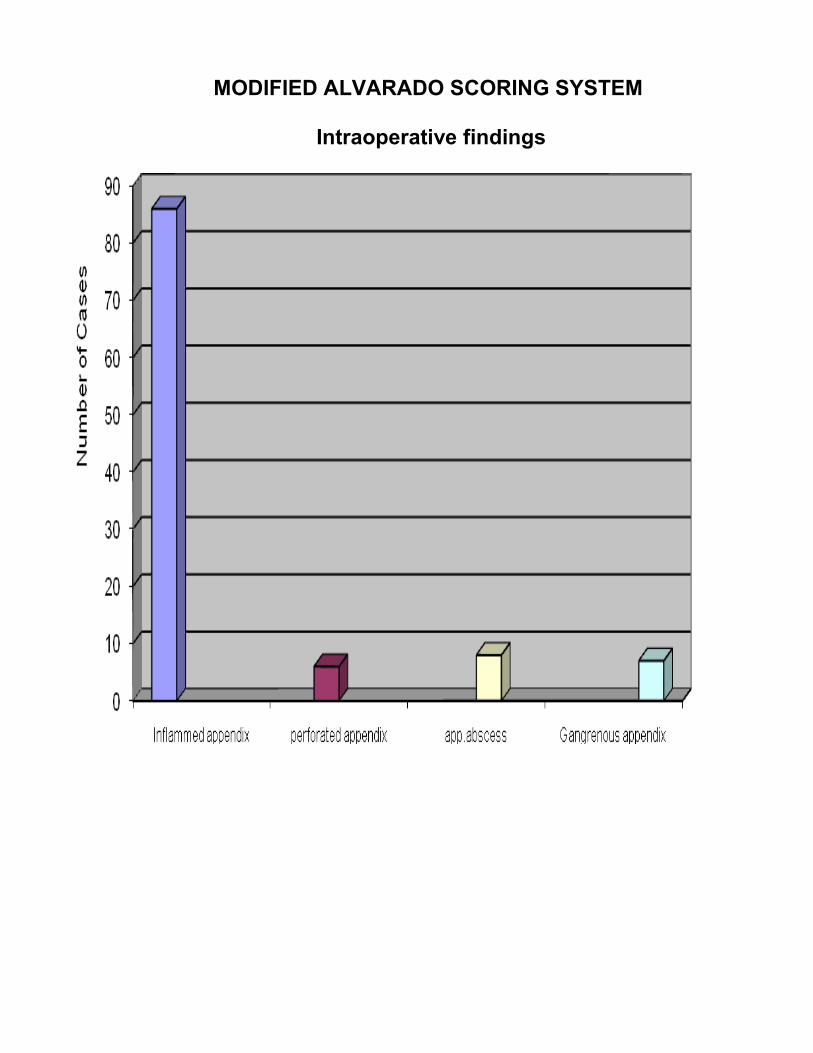
Intraoperative findings
MODIFIED ALVARADO SCORING SYSTEM
DISCUSSION
The diagnosis of acute appendicitis continues to be difficult due to the
variable presentation of the disease and the lack of reliable diagnostic test.
Although there has been some improvement in the diagnosis of acute
appendicitis over the past several decades, the percentage of normal
appendices reported in various series varies from 8 to 33%.
Clinical Scoring systems have proved useful in the management of
number of surgical conditions. In the past few years various scores have been
developed to aid the diagnosis of acute appendicitis. Although many diagnostic
scoring have been described, those are difficunt to implement in the clinical
situations. The modified Alvarado score, is a simple scoring system that can be
instituted easily.
In a prospective study of 215 adults and children in Cardiff, use of the
Alvarado score decreased an unusually high false-positive Appendicectomy rate
of 44% to 14%. 18 Fenyo 11, reported in one study a sensitivity of 90.l2% and
specificity of 91.4% and others reported a sensitivity of 73%, specificity of 87%
with negative appendicectomy rate of 17.5%.
To be useful, a scoring system must be both sensitive and specific. The
Modified Alvarado score proved to be effective in adult male patients with acute
appendicitis but not useful to the same extent in females of reproductive age
group.
Our study demonstrates that Modified Alvarado score is substantially
superior in diagnosis of Acute Appendicitis in adult with a sensitivity of 84.26%
and a specificity of 10.83%. For male patients sensitivity of 86.66%, specificity of
10.0%. For female patients sensitivity of 81.66%, specificity of 11.66%. The
Alvarado score is both simple to remember and to use. This Scoring system
seems ideal for the diagnosis of Acute Appendicitis because it is non-invasive,
requires no special equipment and can be easily used by a JUNIOR RESIDENT
as clinical routine in a peripheral hospital.
Negative Appendicectomy rate in this study is 2.8% where as in general
the negative Appendicectomy rate reported in literature is 15-30% thus it grossly
reduces the negative Appendicectomy rates. In comparison the abdominal
ultrasound has shown results with an average sensitivity of 86% and a
specificity of 94% under the condition of well-controlled clinical trials, namely in
the hands of experienced person.
CT Scans have excellent Sensitivity and specificity, ranging 87-100% and
91-97% respectively.
Leukocyte count has a sensitivity of 85% and abdominal radiography 40%.
BUT,
Abdominal ultrasound requires special equipment and it is operator
dependant.
Computed Tomography is expensive and not readily available
everywhere.
It is the same with radioisotope studies. Abdominal X-Ray is of limited
used and has the risk of radiation exposure.
In our study (98/120) 81.66% presented with a score of > 7 of the
remaining 10 observed 4 had a score of > 7 within 6 hours and 2 within 12
hours. The remaining 5 persons who were observed did not have an increase in
the score further. So 85% of Appendicectomies can be clinically decided within
first 6 hours.
Of the 12 who had a score of < 4, 3 developed acute appendicitis at a later
date.
Missed Appendicectomy rate is 2.5%. Better clinical experience and
recent radiological investigation may reduce this value.
SUMMARY AND CONCLUSION
CONCLUSION
Modified Alvarado Scoring system with a diagnostic accuracy of 97%
seems to be ideal for supporting the diagnosis of acute appendicitis because it
is noninvasive, does not require special equipments, and is simple to remember,
and use in a peripheral set up by a junior resident where radiological
investigation are difficult to perform.
The sensitivity and specificity of the test is good for the male population
compared with the females. This can be easily attributed to the pelvic
pathological conditions which require a diagnostic ultrasound in addition.
In conclusion Modified Alvarado scoring along with an abdominal and
pelvic ultrasound may be the ideal tool to diagnose acute appendicitis in males.
Acute appendicitis is a common cause of abdominal pain in patients
attending emergency departments. Nevertheless, a correct diagnosis based on
Clinical and laboratory findings is not easy.
Promising results have been published for the use of ultrasonography and
to inquire the diagnostic accuracy. However, these investigations are highly
investigator dependant or they involve exposure to radiation, respectively.
History taking and physical examination on the other hand require no
special equipment and are readily available.
It is also conceivable that imaging techniques will gain wider acceptance,
but careful history taking and clinical diagnosis are important measures.
(i) Determining which patients would benefit from these investigation,
and
(ii) Providing the clinical contest that is necessary for correct
interpretation of imaging findings.
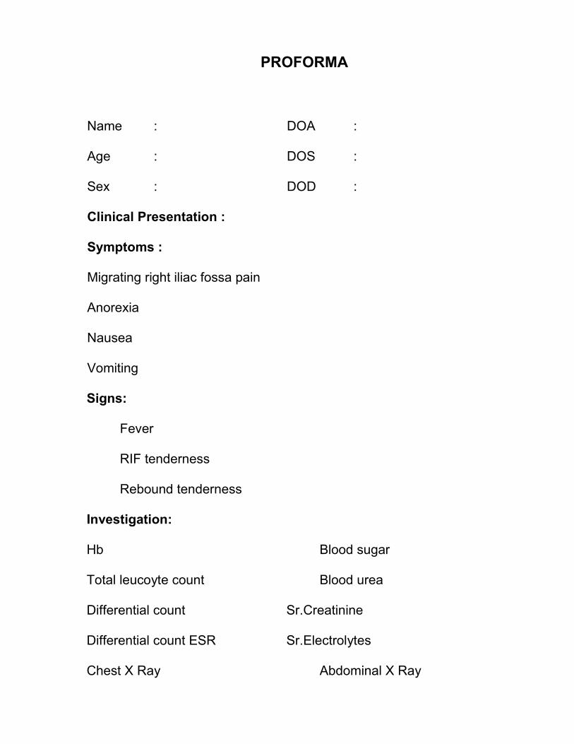
PROFORMA
Name : DOA :
Age : DOS :
Sex : DOD :
Clinical Presentation :
Symptoms :
Migrating right iliac fossa pain
Anorexia
Nausea
Vomiting
Signs:
Fever
RIF tenderness
Rebound tenderness
Investigation:
Hb Blood sugar
Total leucoyte count Blood urea
Differential count Sr.Creatinine
Differential count ESR Sr.Electrolytes
Chest X Ray Abdominal X Ray
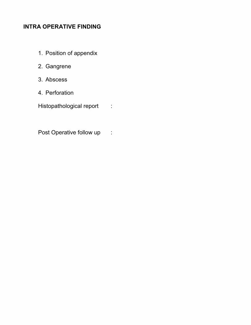
INTRA OPERATIVE FINDING
1. Position of appendix
2. Gangrene
3. Abscess
4. Perforation
Histopathological report :
Post Operative follow up :
BIBLIOGRAPHY
1. Bailey &love’short practice of surgery - 25th edition
2. Maingot’s Abdominal Operations -11th edition
3. Shackelford’s surgery of alimentary tract - 6th Edition
4. Mastery of Surgery - 5th edition
5. Sabiston’s text book of surgery - 18th edition
6. Schwartz’s principles of surgery - 8th edition
7. Diagnostic Radiology –
by Grainger and Allison - 5th edition
8. Gastrointestinal Radiology –
by Ronald–l.elsenbery - 4th edition
9. Hamilton Bailey’s Emergency Surgery - 13th edition
10. Essential surgical practice –
by Sir Alfred Cuschieri - 4th edition
11. Americaln College of Surgery Principles and practice
12. ASI Textbook of Surgery - 1st edition
13. Current Surgical diagnosis and treatment - 11th edition
14. Imaging of Acute Abdomen Radiologic Clinics of North America.
15. Last’s Anatomy - 10th edition
16. Lee Mc Gregors Synopsis of
Surgical anatomy - 12th edition
17. Harrison’s Principles of Internal medicine - 16th edition
18. Farquharson text book of
operative surgery - 9th edition
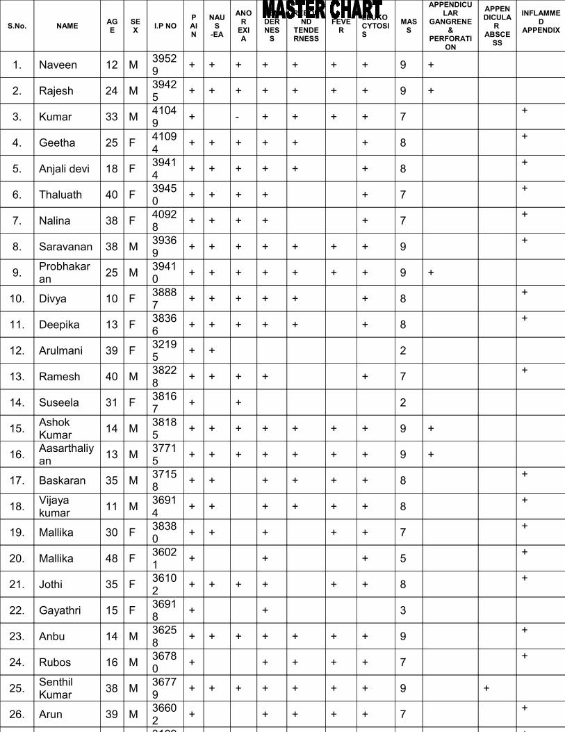
S.No. NAMEAGE
SEX
I.P NOPAIN
NAUS
-EA
ANOR
EXIA
TENDERNES
S
REBOUND
TENDERNESS
FEVER
LEUKO CYTOSIS
MASS
APPENDICULAR
GANGRENE &
PERFORATION
APPENDICULA
R ABSCE
SS
INFLAMMED
APPENDIX
1. Naveen 12 M39529
+ + + + + + + 9 +
2. Rajesh 24 M39425
+ + + + + + + 9 +
3. Kumar 33 M41049
+ - + + + + 7+
4. Geetha 25 F41094
+ + + + + + 8+
5. Anjali devi 18 F39414
+ + + + + + 8+
6. Thaluath 40 F39450
+ + + + + 7+
7. Nalina 38 F40928
+ + + + + 7+
8. Saravanan 38 M39369
+ + + + + + + 9+
9.Probhakaran
25 M39410
+ + + + + + + 9 +
10. Divya 10 F38887
+ + + + + + 8+
11. Deepika 13 F38366
+ + + + + + 8+
12. Arulmani 39 F32195
+ + 2
13. Ramesh 40 M38228
+ + + + + 7+
14. Suseela 31 F38167
+ + 2
15.Ashok Kumar
14 M38185
+ + + + + + + 9 +
16.Aasarthaliyan
13 M37715
+ + + + + + + 9 +
17. Baskaran 35 M37158
+ + + + + + 8+
18.Vijaya kumar
11 M36914
+ + + + + + 8+
19. Mallika 30 F38380
+ + + + + 7+
20. Mallika 48 F36021
+ + + 5+
21. Jothi 35 F36102
+ + + + + + 8+
22. Gayathri 15 F36918
+ + 3
23. Anbu 14 M36258
+ + + + + + + 9+
24. Rubos 16 M36780
+ + + + + 7+
25.Senthil Kumar
38 M36779
+ + + + + + + 9 +
26. Arun 39 M36602
+ + + + + 7+
3109 +