Embed Size (px)
Citation preview

pubs.acs.org/JAFCPublished on Web 07/27/2010© 2010 American Chemical Society
J. Agric. Food Chem. 2010, 58, 8895–8903 8895
DOI:10.1021/jf101778t
A Fluoroimmunoassay Based on Quantum Dot-StreptavidinConjugate for the Detection of Chlorpyrifos
YIPING CHEN,† HE LING REN,† NAN LIU,‡ NA SAI,§ XIAOYU LIU,† ZHEN LIU,†
ZHIXIAN GAO,*,†,‡ AND BAO AN NING*,‡
†College of Food Science and Technology, Huazhong Agricultural University, Wuhan, 430070, China,‡Tianjin Key Laboratory of Risk Assessment and Control Technology for Environment & Food Safety,Institute of Hygiene and Environmental Medicine, Tianjin, 300050, China, and §School of Chemical
Engineering and Technology, Tian Jin University, Tianjin, 300072, China
A rapid and sensitive competitive fluorescence-linked immunosorbent assay (cFLISA) based on quantum
dot-streptavidin conjugate (QDs-SA) was developed for the detection of chlorpyrifos in drinking water.
The QDs-SA conjugate, which consists of 3-mercaptopropyl acid-stabilized CdTe nanoparticle QDs and
streptavidin (SA) made through the active ester method, was employed to improve the sensitivity of
QDs-SA-cFLISA. The 50% inhibition concentration (IC50) and the limit of detection (LOD) were 28.5 and
3.8 ng mL-1, respectively. QDs-SA-cFLISA increased sensitivity 5.5-fold and reduced detection time by
1 h compared with conventional enzyme-linked immunosorbent assay (ELISA). With chlorpyrifos
concentrations of 100, 50, and 20 ng mL-1, recoveries ranged from 85.9% to 105.3% with coefficients
of variation ranging from 6.3% to 13.5%. This study demonstrated that QDs-SA-cFLISA was more rapid
and sensitive than conventional ELISA. Therefore, it can be used as a novel screening method for the
detection of pesticide residues.
KEYWORDS: Quantum dot-streptavidin; fluoroimmunoassay; chlorpyrifos
INTRODUCTION
The issue regarding the presence of pesticide residues in food andthe environment has attracted extensive public attention in recentyears (1). Serious environmental and health problems are arisingdue to the widespread use and abuse of pesticides (2). Thus, it hasbecome increasingly important to detect and monitor the level ofpesticide residues in food and the environment. At present, theidentification and quantification of pesticides are generally basedon chromatographic methods, such as gas chromatography (GC)and high-performance liquid chromatography coupled with massspectroscopy (HPLC-MS)(3). Although these methods are sensi-tive and reliable, they are time-consuming and highly costly.Moreover, they can only be performed by well-trained techniciansand are not convenient for on-site or in-field detection. Therefore,rapid and sensitive assays should be developed for detectingpesticide residues in food and the environment. With the develop-ment of nanotechnology, obtaining a new class of highly fluores-cent homogeneous semiconductor nanocrystals called “quantumdot” has become possible (4).
QDs, such as CdSe-ZnS core shell nanocrystals, are somewhatspherical nanocrystals with diameters ranging from 1 to 10 nm (5).These comprise a series of luminescent inorganic fluorophores withseveral important advantages over traditional fluorescent dyes (6).For example, QDs have long-term photostability, high quantumyield, narrow emission and broad excitation spectra, making it
possible to excite a number of different QDs using a singleexcitation laser wavelength. Moreover, the emission color of QDsis tunable by changing the nanocrystal size and the type of corematerial used, hence, simultaneous multianalyte detection can beachieved easily using multicolor QDs. Recently, QDs have beensuccessfully used for a variety of bioanalytical purposes, such asbiosensing (7), DNA hybridization detection (8), cellular label-ing (9), biological imaging (10) and immunoassays. Direct, sand-wich and competitive assays have likewise been developed to detectprotein toxins and veterinary drug residues such as staphylococcalenterotoxin B (SEB) (11), trinitrotoluene (TNT) (12), sulfametha-zine and enrofloxacin residue (13-15)).
Over the past decade, enzyme-linked immunosorbent assay(ELISA) has become an important alternative detection methodfor pesticides, particularly in analyzing a large number of sam-ples. It is also being used as a screening tool, because of itsnumerous practical benefits, including cost efficiency, consis-tency, portability, and ease of use (16).
QDs, as relatively new fluorescent probes, have particularlyplayed an important role in fluoroimmunoassay (17). At the sametime, the biotin-streptavidin system (B-S system), a signal ampli-fication system, has beenwidely used in immunohistochemistry (18)and immunoassays (19) for its high specificity and strong affinity.Streptavidin (SA) contains four binding sites with an extraordinaryaffinity (dissociation constant: about 10-15M) for the capture of thesmall molecule biotin which can be easily covalently coupled toproteins to enable a solid binding between those proteins and SA.
Chlorpyrifos, a kind of phosphate pesticide and environmentalendocrine disruptor, has been widely used in agriculture to kill
*Corresponding authors. Z.G.: phone, þ86-022-84655191; fax,þ86-022-84655191; e-mail, [email protected]. B.a.N.: phone, þ86-022-84655403; fax, þ86-022-84655191; e-mail, [email protected].

8896 J. Agric. Food Chem., Vol. 58, No. 16, 2010 Chen et al.
pests. Its residue is found mainly in farm soil and environmentalwater (20). Nowadays, numerous facts suggest that residual chlor-pyrifos poses a possible risk to the environment and humanhealth (21). Its presence in drinking water, specifically, is harmfulto humans. Thus, the Environmental Protection Agency (EPA)Method 525 has set the maximum allowable risk level for organo-phosphorus pesticides (OPs) in drinking water within the range of0.001 to 0.25 mg mL-1. Conventional analytical methods for thedetection of chlorpyrifos in environmental water samples includegas chromatographic-mass spectrometric (GC-MS) and HPLC,both of which are costly and time-intensive (22). Therefore, it isnecessary to develop a rapid, simple and sensitive method tomonitor residual chlorpyrifos in drinking water.
In this study, a rapid and sensitive fluoroimmunoassay (QDs-SA-cFLISA) based onQDs-SA conjugate was developed for thedetection of chlorpyrifos in drinking water. To our knowledge,this is the first study focused on the analysis of chlorpyrifosresidue using QDs and the B-S system. In this assay, QDs-SAconjugate was used as a fluorescence signal system and a signalamplification system to improve test sensitivity, conventionalELISA and HPLC methods were employed for methodologycomparison.
MATERIALS AND METHODS
Reagents. Chlorpyrifos and its analogues and intermediates werepurchased from Sigma Chemical Co (St. Louis,MO). The monoclonal anti-body against chlorpyrifos (Ab1) was purchased from Wanghua Biotechno-logy (Beijing, China). SA was purchased in Biosynthesis Biotechnologyco.(Beijing, China). N-Hydroxysulfosuccinimide sodium salt (sulfo-NHS),1-ethyl-3-(3-dimethylaminopropyl)carbodiimide (EDC) and 3-mercapto-propyl acid (MPA) were purchased from Sigma. Tellurium powder (200mesh, 99.8%), CdCl2 (99%) and NaBH4 (99%) were purchased fromAldrich. In order to determine quantum yields of QDs, rhodamine 6G witha quantum yield of 95% in ethanol was obtained from Sinopharm chemicalReagent Beijing Co. Ltd. (Beijing, China). Common solvents and salts weresupplied by Tianjin Regent Corp. (Tianjin, China). The coating buffer usedwas 0.05 mol L-1 carbonate buffer (pH 9.6), washing buffer was 0.01 molL-1 phosphate-buffered saline (PBS) with 0.05%Tween 20 (PBST) (pH 7.4)and blocking buffer was PBS buffer with 3% skimmilk power. Chlorpyrifosand its analogues were prepared by dissolving known amounts of purifiedsubstances in methanol. The stock solution (10 mg mL-1) was stored at-20 �C before being used for the preparation of standard solutions, whichwere stored at 4 �C. Pure water was prepared using the Milli-Q system fromMillipore (Bedford, MA).
Apparatus. Costar opaque black polystyrene microtiter plates werepurchased fromCorning, Inc. (New York). The 96-well ELISAmicrotiterplates were obtained from Wanger Biotech Co (Beijing, China). Ultra-filtration units (15 mL, 4 and 0.5 mL) and 0.2 μm PES membrane unitswere also purchased from Millipore. Biotinylated monoclonal antibody(Biotinylated-Ab1) was separated and purified using YM-10 columns(MWCO, 10,000; pack size, 8;Millipore, USA). Cary Eclipse fluorescencespectrophotometer (Palo Alto, CA) and a SpectraFluor Plus microtiterplate reader (Massachusetts) were used to obtain the fluorescence signal.A protein electrophoresis apparatus from Bio-Rad. Inc. (California) wasused to characterize the complete antigen (chlorpyrifos-BSA conjugate).A centrifuge, Mikro 22R from Hettich Co. (Kirchlengern, Germany) andHPLC system (Waters, USA) were also used in the study.
Synthesis ofWater-Soluble CdTeQDs.QDswere synthesized in anaqueous solution according to a previous report (23). In brief, freshlyprepared oxygen-free NaHTe solution was added to a nitrogen saturated1.25� 10-3 mol L-1 CdCl2 aqueous solution (pH=11.4) in the presenceof MPA as stabilizing agent. NaHTe was prepared by the reaction ofNaBH4 with tellurium powder at a 2:1 molar ratio. The molar ratio ofCd2þ toMPA to Te-was 1:2.4:0.5. The solutionwas refluxed for differenttime periods to control the size of the QDs.
The obtained QDs solution was further purified via ultrafiltrationaccording to a previous report (24). The first stepwas carried out on a filterwith a size of 10,000MW, after which the solution was centrifuged at 4000rpm (10 min, 4 �C) to remove nonreacted MPA. The QDs were washed
three times with 50 mmol L-1 PBS (pH 7.4). The upper phase wascollected, dissolved in PBS, and subjected to a second ultrafiltration stepthrough a 50,000MWfilter. After the second ultrafiltration step, the lowerand upper phases were collected for the next stage of the experiment. Thepurified water-soluble QDs were characterized by bright fluorescence andgood stability in PBS buffer.
OpticalCharacterization of theCdTeQDs.Fluorescence spectra ofthe QDs was taken using a Cary Eclipse fluorescence spectrophotometerequipped with a 20 kW xenon discharge lamp as a light source. Spectrawere typically taken at a scanning rate of 1000 nm/min with 10 nmexcitation/emission slits and a 700 V photomultiplier tube voltage. Thedispersion and sizes of CdTe QDs were evaluated with a transmissionelectron microscopy (TEM).
Quantum Yield (QY) of the CdTe QDs. QY was calculatedaccording to a previous report (25). Rhodamine 6G was used as thefluorescence standard to measure the fluorescence QY of the CdTe QDs.Specifically, rhodamine 6G was dissolved in anhydrous alcohol with anabsorbance rate of around 0.02 and the normal excitation wavelength of470 nm. By comparing the integrated areas of emissions from rhodamin6G and QDs, respectively, the fluorescence QY values of QDs werecalculated by taking rhodamine 6G in diluted solution.
Formation and Purification of the QDs-SA Conjugate. Theconjugate of CdTe QDs with SA was prepared through the active estermethod described in a previous work with some modifications (26). The200 μL 10 μM QDs solution produced through purification was mixed
with 200 μL of SA solution in PBS (0.01 mol L-1, pH 7.4), and the molarratio of QDs/SA was optimized. Thereafter, 20 μL of freshly prepared 10mgmL-1 EDC solution and 30 μL of freshly prepared 10 mgmL-1 sulfo-NHS solution were added to the mixture quickly. The samples wereincubated for 2 h at room temperature (RT) under continuous shaking inthe dark. The conjugate solution was filtered through a 0.2 μm PESmembrane unit to remove the protein conjugates; this was transferred to aclean centrifugal ultrafiltration unit (a 50,000 MW filter), where it was
centrifuged at 8000 rpm for 15min at 4 �C. The free nonconjugated SA, aswell as the isourea byproduct of the conjugation reaction, was removed byultrafiltration. The upper phase, which contained the QDs-SA conjugate,was decanted and diluted into 2.0 mL of PBS, after which 0.5 mL of50 mmol L-1 PBS with 0.5% bovine serum albumin (BSA) was thenadded. The QDs-SA conjugate solution (8 mM) was stored at 4 �C.
Preparation of the Chlorpyrifos-BSA Conjugate (Ag). Thechlorpyrifos-BSA conjugate was produced according to a previous reportwith some modifications (27). The synthesis pathway of the chlorpyrifos-BSA conjugate is shown in Figure 1. First, a chlorpyrifos-propionic acidderivative was synthesized through the reaction between chlorpyrifos and3-mercaptopropyl acid (Figure 1a). Second, the derivative was coupled withBSA by the carbodiimide method using EDC and sulfo-NHS (Figure 1b).After conjugation, the mixture was transferred to a clean centrifugalultrafiltration unit (a 10,000 MW filter) and centrifuged at 8000 rpm(15 min at 4 �C). Unreacted hapten (chlorpyrifos) and the byproduct wereremoved by ultrafiltration. The conjugate was then characterized by sodiumdodecyl sulfate-polyacrylamide gel electrophoresis (SDS-PAGE), andwas freeze-dried and stored at -20 �C.
Formation and Purification of the Biotinylated-Ab1 Conjugate.
NHS-LC-Biotin was diluted to 1 mg mL-1 by DMF and Ab1 was dilutedto1mgmL-1 by 0.1 gL-1NaHCO3, (pH9.6). The two solutionsweremixedat a 10:1 ratio by volume (NHS-LC-Biotin/Ab1) and stirred for 3 to 4 h forthe biotinylation ofAb1.After labeling, themixed solutionwas transferred toa YM-10 column and centrifuged at 10,000 rpm (15 min, 4 �C). Excessiveunreacted biotin and ions in the aqueous solution were removed by YM-10column, and the biotin-labeled Ab1 was eluted with 10 mmol L-1 PBS (pH7.2). It was then aliquoted and stored at -20 �C until use.
The QDs-SA-cFLISA Method. The schematic diagram of theQDs-SA-cFLISA procedure is shown in Figure 2. A 96-well opaqueblack microtiter plate was coated with 100 μL/well of the coating antigen(chlorpyrifos-BSA), diluted with coating buffer (0.05 mol L-1 NaHCO3,pH 9.6) and incubated at 37 �C for 2 h. The unbound antigen was washedfour times with 0.05% Tween-20 in a phosphate buffer solution (PBST),and the excess binding sites were blocked by 150 μL/well of 3% skimmilkpower in PBS at 37 �C for 1 h. Subsequently, 50 μL of the samples(drinkingwater sample or standard serial dilutions of chlorpyrifos inPBS),togetherwith 50 μLof the optimal biotinylated-Ab1 dilution, was added to

Article J. Agric. Food Chem., Vol. 58, No. 16, 2010 8897
the wells (in triplicate). The plate was then incubated for 1.5 h at 37 �C.After washing five times, the 100 μL/well QDs-SA conjugate was added
to the wells and incubated for 0.5 h at 37 �C. After incubation, the platewas washed seven times and patted dry. A fluorescence microplate readerwith an excitation/emission at 300/600 nm was utilized to measure thefluorescence signal. A four-parameter logistic equation was used to fit theQDs-SA-cFLISA data. Calculations were performed with OriginPro7.5software. The IC50 and LOD served as criteria to evaluate the QDs-SA-cFLISA; these different characteristics represented the analyte con-centrations obtained at 50% and 10%, respectively.
TheConventional ELISAMethod.The procedure before adding theQDs-SA conjugate was the same as that for QDs-SA-cFLISA. Afterwashing five times with PBST, 100 μL/well goat polyclonal antibody tomouse IgG-HRP (1:4000) was added and incubated for 1 h at 37 �C, andthe plate was then washed seven times. Subsequently, 100 μL of tetra-methylbenzidine substrate system was added into the well and incubatedfor 15min at 37 �C in the dark. The enzymatic reaction was then quenchedby adding 50 μL/well of 2 mol L-1 H2SO4. The absorbance was measuredwith an ELISA microplate reader at 450 nm, and the chlorpyrifos contentin unknown samples was calculated based on the calibration curve, whichwas generated during the same run.
Cross-Reactivity (CR). In our research, the CR of the compoundsstructurally related to chlorpyrifos was investigated with the Ab1 byQDs-SA-cFLISA and conventional ELISA. The CR values were calcu-lated according to eq 1.
CR ð%Þ ¼ IC50 of chlorpyrifos
IC50 of chlorpyrifos analogues� 100 ð1Þ
Optimal Concentration of Biotinylated-Ab1 and Ag in ELISA.Atfirst, optimum concentration of biotinylated-Ab1 and Ag was selected bycheckerboard titration in ELISA. At the base of checkerboard titration,different concentrations of biotinylated-Ab1 (30, 20, 15 ng mL-1) and Ag(0.30, 0.20, 0.15 μg mL-1) were observed during the competitive stepin ELISA.
Optimization of the Molar Ratio of QDs/SA. 100 μL/wellQDs-SA conjugate solutions with different QDs/SA ratios (2:1, 1:1, 1:2and 1:3) were tested using QDs-SA-cFLISA under the optimized con-centrations of biotinylated-Ab1 and Ag, respectively.
Effect of pH on the Coupled Reaction of QDs and SA. A series ofpH gradients (5.5, 6.0, 6.5, 7.0, 7.5 and 8.0) were set in order to observe theeffect on the QDs and SA coupling reaction. The resulting conjugates weretested by QDs-SA-cFLISA.
Optimized Dilution Level of the QDs-SAConjugate. 100 μL/wellserial dilutions of the QDs-SA conjugate solution (1:10, 1:20, 1:50) wereobserved to select the optimal level of the QDs-SA conjugate byQDs-SA-cFLISA.
The HPLC Method. The procedures of extraction and cleanup ofsamples were carried out according to the Chinese national standard (GB19605-2004) with modifications. Briefly, a 250 mL water sample waspurified by a C18 SPE column, and the adsorbate was eluted by 3 mL ofmobile phase. The extractive sample was prepared for analysis. The opti-mized chromatographic conditions were as follows: column, C18 (250 �4.6 mm, 5 μm); injection volume, 50 μL; mobile phase, acetonitrile-water-acetic acid (82:17.5:0.5 v/v/v); flow rate, 1.0 mL min-1 at RT;detection wavelength, 265 nm.
Matrix Effect. Drinking water, surface water and agricultural runoffwaterwere collected fromwaterworks, JingyeLake and theHaiheRiver inTianjin, respectively. Water samples were centrifuged at 5000 rpm (5 min)to remove suspended particles and then stored at 4 �C. For this study,chlorpyrifos standard curves were prepared in PBS, drinking water,agricultural runoff waters and surface water matrix, each fortified at eightconcentrations (500, 200, 100, 50, 25, 12.5, 6.25, 1 ng mL-1) and analyzedin triplicate. The matrix effect was assessed through the comparison ofcompetitive inhibition curves with the standard curve in PBS buffer.
Accuracy and Precision Analysis. First, different volumes of chlor-pyrifos standard stock solution (1 mg mL-1) were added into the PBSbuffer to produce samples with a series of chlorpyrifos concentrations(100, 50 and 20 ng mL-1); the tubes were shaken at RT for 5 min andstored at 4 �C for conventional ELISA and QDs-SA-cFLISA analyses.
Drinking Water for Analysis. The quality of drinking water isdirectly related to human health. Thus, drinking water was selected as aprior object in our study. At first, the sample was diluted at a series ofratios (1:1, 1:2 and 1:4) by PBS buffer (0.01 M, pH 7.4) to minimize the
Figure 2. The schematic diagram of the QDs-SA-cFLISA method pro-cedure.
Figure 1. (a) The schematic diagram of the chlorpyrifos-propionic acidderivative synthesis. (b) The schematic diagram of the chlorpyrifos-BSAconjugate synthesis.
8898 J. Agric. Food Chem., Vol. 58, No. 16, 2010 Chen et al.
complex matrix effect, which was assessed through the comparisonof competitive inhibition curves with the standard curve in PBS buffer.The diluted water was then prepared for the HPLC, ELISA and QDs-SA-cFLISA detection procedures.
RESULTS AND DISCUSSION
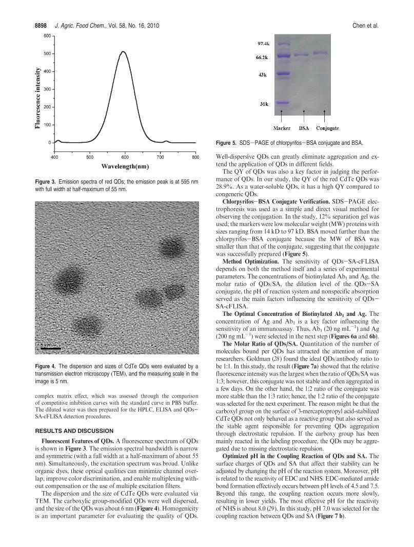
Fluorescent Features of QDs. A fluorescence spectrum of QDsis shown in Figure 3. The emission spectral bandwidth is narrowand symmetric (with a full width at a half-maximum of about 55nm). Simultaneously, the excitation spectrum was broad. Unlikeorganic dyes, these optical qualities can minimize channel over-lap, improve color discrimination, and enable multiplexing with-out compensation or the use of multiple excitation filters.
The dispersion and the size of CdTe QDs were evaluated viaTEM. The carboxylic group-modified QDs were well dispersed,and the size of the QDswas about 6 nm (Figure 4). Homogenicityis an important parameter for evaluating the quality of QDs.
Well-dispersive QDs can greatly eliminate aggregation and ex-tend the application of QDs in different fields.
The QY of QDs was also a key factor in judging the perfor-mance of QDs. In our study, the QY of the red CdTe QDs was28.9%. As a water-soluble QDs, it has a high QY compared tocongeneric QDs.
Chlorpyrifos-BSA Conjugate Verification. SDS-PAGE elec-trophoresis was used as a simple and direct visual method forobserving the conjugation. In the study, 12% separation gel wasused; themarkers were lowmolecular weight (MW) proteins withsizes ranging from 14 kD to 97 kD. BSA moved further than thechlorpyrifos-BSA conjugate because the MW of BSA wassmaller than that of the conjugate, suggesting that the conjugatewas successfully prepared (Figure 5).
Method Optimization. The sensitivity of QDs-SA-cFLISAdepends on both the method itself and a series of experimentalparameters. The concentrations of biotinylated Ab1 and Ag, themolar ratio of QDs/SA, the dilution level of the QDs-SAconjugate, the pH of reaction system and nonspecific absorptionserved as the main factors influencing the sensitivity of QDs-SA-cFLISA.
The Optimal Concentration of Biotinylated Ab1 and Ag. Theconcentration of Ag and Ab1 is a key factor influencing thesensitivity of an immunoassay. Thus, Ab1 (20 ng mL-1) and Ag(200 ng mL-1) were selected in the next step (Figures 6a and 6b).
The Molar Ratio of QDs/SA. Quantitation of the number ofmolecules bound per QDs has attracted the attention of manyresearchers. Goldman (28) found the ideal QDs/antibody ratio tobe 1:1. In this study, the result (Figure 7a) showed that the relativefluorescence intensitywas the largestwhen the ratio ofQDs/SAwas1:3; however, this conjugate was not stable and often aggregated ina few days. On the other hand, the 1:2 ratio of the conjugate wasmore stable than the 1:3 ratio; hence, the 1:2 ratio of the conjugatewas selected for the next experiment. The reason might be that thecarboxyl group on the surface of 3-mercaptopropyl acid-stabilizedCdTe QDs not only behaved as a reactive group but also served asthe stable agent responsible for preventing QDs aggregationthrough electrostatic repulsion. If the carboxy group has beenmainly reacted in the labeling procedure, the QDs may be aggre-gated due to missing electrostatic repulsion.
Optimized pH in the Coupling Reaction of QDs and SA. Thesurface charges of QDs and SA that affect their stability can beadjusted by changing the pH of the reaction system.Moreover, pHis related to the reactivity of EDC andNHS. EDC-mediated amidebond formation effectively occurs between pH levels of 4.5 and 7.5.Beyond this range, the coupling reaction occurs more slowly,resulting in lower yields. The most effective pH for the reactivityof NHS is about 8.0 (29). In this study, pH 7.0 was selected for thecoupling reaction between QDs and SA (Figure 7 b).
Figure 3. Emission spectra of red QDs; the emission peak is at 595 nmwith full width at half-maximum of 55 nm.
Figure 4. The dispersion and sizes of CdTe QDs were evaluated by atransmission electron microscopy (TEM), and the measuring scale in theimage is 5 nm.
Figure 5. SDS-PAGE of chlorpyrifos-BSA conjugate and BSA.

Article J. Agric. Food Chem., Vol. 58, No. 16, 2010 8899
Optimal Dilution Ratio of the QDs-SAConjugate.The bindingof QDs to Ab1 occurs through the interaction between SA andbiotin. Hence, the relative molar ratio of the QDs-SA conjugateto biotinylated-Ab1 is important. Based on the optimized con-centration of biotinylated-Ab1 and Ag, QDs-SA-cFLISA wasthe most sensitive when QDs-SA conjugate was diluted 20-foldand the final concentration was 0.4 mM (Figure 7c).
Sensitivity. In the above optimal conditions, the sensitivitylevels of conventional ELISA and QDs-SA-cFLISA were in-vestigated using the chlorpyrifos standard.
For conventional ELISA, the standard curves (Figure 8a) wereobtained under optimalAg (200 ngmL-1), Ab1 (20 ngmL-1) andAb2 concentration (1:4000). The IC50 and LOD for chlorpyrifoswere 96.8 ng mL-1 and 20.6 ng mL-1, respectively.
For QDs-SA-cFLISA, the standard curves (Figure 8b) wereobtained under optimal Ag (200 ng mL-1), biotinylated-Ab1(20 ng mL-1), and QDs-SA (0.4 mM). The IC50 and LOD forchlorpyrifos were 28.5 ng mL-1 and 3.8 ng mL-1, respectively.The sensitivity increased 5.5-fold compared with conventionalELISA; hence, it can meet current EPA andWHO standards forthe detection of chlorpyrifos in drinking water. For furtherresearch, a higher bioactive and qualitative QDs-SA conjugate
should be prepared and applied in QDs-SA-cFLISA immuno-assay to detect pesticide residues in environment and food.
Figure 6. Optimized concentration of biotinylated-Ab1 and Ag in ELISA.(a) Concentration of biotinylated-Ab1 and Ag was first observed bycheckerboard titration. When OD450nm = 1, the concentration of biotiny-lated-Ab1 and Agwas selected for the next step. (b) Effect of concentrationof biotinylated-Ab1 and Ag in the sensitivity.
Figure 7. (a) Optimize the molar ratio of QDs/SA in QDs-SA conjugate.(b) Effect of pH in formation of QDs-SA conjugate. (c)Optimize dilution levelof QDs-SA conjugate. Three dilution levels QDs-SA conjugate (1:10, 1:20and 1:50) were observed to obtain optimal sensitivity of QDs-SA-cFLISA.

8900 J. Agric. Food Chem., Vol. 58, No. 16, 2010 Chen et al.
Specificity. CR is an important parameter in evaluating thespecificity of an immunoassay. The CRs of seven differentchlorpyrifos structural analogues or intermediates are shown inTable 1. Ab1was highly selective toward chlorpyrifos, and onlychlorpyrifos-methyl was determined to exhibit significant cross-reactivity. Results for this study are consistent with earlierfindings (30). The high affinity of the chlorpyrifos antibody forthe methyl analogue may lead to overestimations of the targetantigen. This, however, should not be problematic since chlor-pyrifos-methyl is no longer registered for use inChina. The CRof3,5,6-trichloro-2-pyridinol (chlorpyrifos intermediates) suggestedthat the antigenic determinant was 3,5,6-trichloro-2-pyridinol.QDs-SA-cFLISA results were similar to those of conventionalELISA, indicating that the Ab1 was very specific for chlorpyrifos.
Accuracy and Precision. Intra-assay precision was tested byconducting three replicate analyses at three concentrations ran-ging from 100 to 20 ng mL-1 in PBS buffer. The recovery wasused to measure the accuracy of the assay, and the percent
coefficients of variation (CV) were used to establish the precisionof antibody response at each concentration. Quality controlestimations for QDs-SA-cFLISA demonstrated that it per-formed within a satisfactory range of variability (Table 2). Reco-veries of 85.9% to 105.3% by QDs-SA-cFLISA and 92.2% to106.2% by conventional ELISA were obtained at all fortificationlevels with CVs of 6.3% to 13.5% and 6.6% to 10.5%, respec-tively (Table 2). Recovery and CV obtained from the QDs-SA-cFLISA were similar to those from conventional ELISA. Toevaluate the reproducibility of QDs-SA-cFLISA, interassayprecision from three independent analyses on three different dayswas investigated. The mean recovery and mean CVs of all spikelevels were 88.5% to 110.2% and 14.3% to 19.2%, respectively(Table 3). Recoveries and variabilities in the intra-assay/inter-assay satisfy current EPA criteria for the assessment of analyticalmethods, indicating that mean recoveries must lie within therange of 70-120%, and that CVs must lie within the range(20%. The results shown in Tables 2 and 3 indicate thatQDs-SA-cFLISA is sufficiently accurate and can be used forfurther application in the detection of pesticide residues.
Matrix Effects Study.QDs-SA-cFLISA is a competitive assayformat, that is, the coating antigen and the target antigen competefor antibody binding sites and are consequently prone to interfer-ence due to nonspecific binding between antibodies and nontargetanalytes that may be present in a particular matrix. In addition,
Figure 8. (a) Calibration curve of conventional ELISA for chlorpyrifos in PBSbuffer. The sigmoid represents the standard curve of chlorpyrifos, and errorbars represent the standard deviation. (b) Calibration curve of QDs-SA-cFLISA for chlorpyrifos in PBS. The sigmoid represents the standardcurve of chlorpyrifos, and error bars represent the standard deviation.
Table 1. Specificity of Ab1 to Chlorpyrifos and Its Analogues
Table 2. Intra-Assay Reproducibility and Accuracy of QDs-SA-cFLISA andELISA Spiked with Chlorpyrifos in PBS at Three Concentrations (n = 3)a
QDs-SA-cFLISA ELISA
spiked (ng mL-1) recovery (%) CV (%) recovery (%) CV (%)
100 85.9 13.5 92.2 6.6
50 95.7 6.3 96.3 9.8
20 105.3 7.8 106.2 10.5
a Every spiked level was replicated three times.

Article J. Agric. Food Chem., Vol. 58, No. 16, 2010 8901
the fluorescence signal detection system is under the matrix influ-ence, given that fluorescence intensity of QDs is affected by thecomplicated matrix (Figure 9). Due to the fact that agriculturalrunoff water is rich in heavy metals (such as Cu2þ and Hgþ) andorganics, it exhibits significant matrix effects, thereby affecting thefluorescent signal system. Additional resources and efforts must bedirected atmitigating such influences to help degradematrix effects.For example, the use of more aggressive preanalysis filteringtechniques, centrifugation, or detergents or binding agents to reducethe amount of suspended solids and other interferences might bebeneficial and should be studied more thoroughly. Overall, QDs-SA-cFLISA has practical applications in monitoring and detectingpesticide residues in food and the environment.
Drinking Water Analysis. In water sample analysis, the com-plex matrix effect should be minimized to attain the necessarysensitivity. In a previous section, drinking water was described asshowing relatively small matrix effects in the assay, as it was dilutedby PBS to degrade matrix effects. The results indicated that thematrix effect could be eliminated after a 3-fold dilution. Chlorpy-rifos could not be detected in drinkingwater byQDs-SA-cFLISA,ELISA and HPLCmethod. Investigating further, serial concentra-tions of chlorpyrifos were added into the 3-fold diluted drinkingwater and the amounts of chlorpyrifos were quantified by QDs-SA-cFLISA and HPLC. There was no significant differencebetween the amounts of chlorpyrifos quantified by the twomethods(Table 4). As a screening method, therefore, the detection ofchlorpyrifos by the QDs-SA-cFLISA was very precise.
Comparison of Conventional ELISA and QDs-SA-cFLISA.
There are many advantages in using QDs-SA-cFLISA com-pared with conventional ELISA (Table 5). First, in QDs-SA-cFLISA, a signal amplification system is applied through the
interaction between the biotinylated Ab1 and the QDs-SAconjugate. The utility of B-S system increases the amount ofQDs linked to the biotinylated Ab1-Ag immunocomplex, am-plifying the fluorescent intensity and improving sensitivity. Inaddition, QDs, a fluorescent signal system in QDs-SA-cFLISA,has strong fluorescent intensity that can increase the sensitivity.Second, conventional ELISA requires labeling of antibodies withexternal reagents, such as enzymes and/or fluorescent dyes. Thislabeling procedure may cause structural changes in the epitopeand suppress the specific recognition ability. Moreover, theenzymatic and fluorescence dye labeling procedures are time-consuming and may cause a high background signal. In contrast,the application of SA-coated QDs greatly simplifies the labelingprocedure without affecting the binding efficiency of the Ab-Agreaction. Third, conventional ELISA requires substrate and stopbuffer reagents that are not only unstable but also harmful tohuman health. In comparison, QDs-SA-cFLISA can avoid thisproblem because QDs have long-term photostability and are verystable at RT. Finally, the detection time for QDs-SA-cFLISA isshorter by 1 h compared with conventional ELISA.
Nevertheless, there are some problems with QDs-SA-cFLISAthat need further investigation, such as QDs aggregation andnonspecific absorption. QDs aggregation resulting from nonopti-mal surface chemistry that, in turn, leads to the loss of colloidalstability in the biolabeling procedure is an important problem forQDs-based biosensors and immunoassay applications (31). In thisstudy, the stability of theQDs-SAconjugatewas directly related totheQDs/SA ratio and the pHof the reaction system, and the degreeof aggregation was positively correlated with the amount of SAwithin a certain range. When the conjugate was dissolved inPBS-BSA solution (1% BSA, m/v), the degree of aggregationand nonspecific absorption greatly decreased. Further investiga-tions involving adjustments in the ionic strength and species of thebuffer used should be conducted to solve this problem.Moreover, itshould extend the application of QDs-based immunoassays andbiosensors to the analysis of pesticide residues.
Nonspecific absorption in the control group often affects thesensitivity of immunoassays. To address this concern, the PBS-BSA solution was used to minimize the background signal in ourexperiment. A previous research has suggested that the sizes of QDprobes affect the labeling efficiency to the microsphere due to sterichindrance (32); the big-sized QDs probes had weaker bindingcapacity onto the surface of polystyrene microspheres than small-sized ones. Therefore, big-sized QDs probes can reduce nonspecific
Table 3. Interassay Reproducibility and Accuracy of QDs-SA-cFLISASpiked with Chlorpyrifos in PBS and Performed on Three Separate Days
spike level (ng mL-1) mean recover y(%) mean CV (%)
100 88.5 18.5
50 92.1 15.2
20 95.6 14.3
5 110.2 19.2
Figure 9. Analysis of matrix effect by several environmental waters (drinkingwater, surface water and agriculture runoff water). The result was comparedwith the competitive inhibition curves with standard curve in PBS.
Table 4. Results from Analysis of Chlorpyrifos in Incurred Drinking Water byQDs-SA-cFLISA and HPLC (n = 3)
determinedconcn of chlorpyrifosin in
drinking water (ng mL-1) QDs-SA-cFLISA HPLC
100 102.2 ( 3.5 99.3 ( 2.7
50 51.2 ( 2.8 50.8 ( 2.5
20 20.6 ( 2.2 21.8 ( 2.0
0 NDa ND
aND = not determined.
Table 5. The Comparison of Conventional ELISA and QDs-SA-cFLISAMethod
method
IC50(ng mL-1)
LOD
(ng mL-1)
working range
(ng mL-1)
detection
time (h)
ELISA 96.8 20.6 56.7-258.8 5
QDs-SA-cFLISA 28.5 3.8 10.5-180.4 4

8902 J. Agric. Food Chem., Vol. 58, No. 16, 2010 Chen et al.
absorption. Bruchez et al. (33) coated the surface ofQDswith silica,and then linked biomolecules onto the silica surface. The size ofthese silica-coated QDs can be varied according to the objective ofexperiment. In further investigations, we should prepare large-sizedsilica-coated QDs and reduce nonspecific absorption maximally toincrease the sensitivity in the optimization condition.Moreover, thelabeling efficiency of QDs-SA coupling is related to the size of theQDs. Labeling efficiency may be increased by using big-sized QDsdue to the big surface area.
As demonstrated in the present study, QDs-SA-cFLISAperforms exceptionally well under optimal conditions, exhibitingexcellent accuracy and low variability in quality control evalua-tions. Its overall performance characteristics in controlled assayswere found to be compliant with the currentU.S. EPA criteria forthe assessment of analytical methods. Compared with conven-tional ELISA, the most significant advantage of this assay wasthat it increased sensitivity 5.5-fold (up to 3.8 ng mL-1); thus, thesensitivity of this assay can satisfy current EPA and WHOstandards for the detection of chlorpyrifos in drinking water.Therefore, QDs-SA-cFLISA could play a significant role indetecting pesticide residues, veterinary drugs residues, environ-ment pollutants, etc.
Further studies should focus on establishing the standardprocedure for labeling different Abs with QDs nanocrystals ofdifferent colors and then performing a reliable multiplexed assayby optimizing the assay conditions. It is believed that QDsnanocrystals could provide powerful fluorescent probes for thedetection of pesticide residues in the future.
ABBREVIATIONS USED
Ab1, monoclonal antibody; biotinylated-Ab1, biotinylatedmonoclonal antibody; QDs-SA-cFLISA, biotin-streptavidin sys-tem competitive fluorescence-linked immunosorbent assay; BSA,bovine serum albumin; CR, cross reactivity; CV, coefficient ofvariation; EDC, 1-ethyl-3-(3-dimethylaminopropyl)carbodiimide;ELISA, enzyme-linked immunosorbent assay; GC-MS, gas chro-matographic-mass spectrometric;HPLC,high-performance liquidchromatography;HPLC-MS, high-performance liquid chromato-graphy-mass spectrometric; IC50, 50% inhibition concentration;NHS, N-hydroxysulfosuccinimide sodium salt; ND, not deter-mined; LOD, limit of detection; PBS, phosphate-buffered saline;PBST, phosphate buffered saline containing 0.05% Tween 20;QDs, quantum dots; QY, quantum yields; SEB, staphylococcalenterotoxin B; SDS-PAGE, sodium dodecyl sulfate-polyacryl-amide gel electrophoresis; TNT, trinitrotoluene; TEM, transmis-sion electron microscopy.
LITERATURE CITED
(1) Jiang, X.; Li, D.; Xu, X.; Ying, Y.; Li, Y.; Ye, Z.; Wang, J.Immunosensors for detection of pesticide residues. Biosens Bioelec-tron. 2008, 23, 1577–1587.
(2) Rial-Otero, R.; Gaspar, E. M.; Moura, I.; Capelo, J. L. Chromato-graphic-based methods for pesticide determination in honey: Anoverview. Talanta 2007, 71, 503–514.
(3) Nunez, O.; Moyano, E.; Galceran, M. T. LC-MS/MS analysis oforganic toxics in food. Trends Anal. Chem. 2005, 24, 683–703.
(4) Bruchez, M., Jr.; Moronne, M.; Gin, P.; Weiss, S.; Alivisatos, A. P.Semiconductor nanocrystals as fluorescent biological labels. Science1998, 28, 2013–2016.
(5) Bailey, R. E.; Smith, A. M.; Nie, S. M. Quantum dots in biology andmedicine. J. Phys. E 2004, 25, 1–12.
(6) Dabbousi, B. O.; Rodriguez-Viejo, J.; Mikulec, F. V.; Heine, J. R.;Mattoussi, H.; Ober, R.; Jensen, K. J.; Bawendi, M. G. CdSe)ZnSCore-Shell Quantum Dots: Synthesis and Characterization of a SizeSeries of Highly Luminescent Nanocrystallites. J. Phys. Chem. B1997, 101, 9463–9475.
(7) Sapsford, K. E.; Pons, T.; Medintz, I. L.; Mattoussi, H. Biosensingwith Luminescent Semiconductor Quantum Dots. Sensors 2006, 6,925–953.
(8) Parak, W. J.; Gerion, D.; Zanchet, D.; Woerz, A. S.; Pellegrino, T.;Micheel, C.; Williams, S. C.; Seitz, M.; Bruehl, R. E.; Bryant, Z.;Bustamante, C.; Bertozzi, C. R.; Alivisatos, A. P. Conjugation ofDNA to silanized colloidal semiconductor nanocrystalline quantumdots. Chem. Mater. 2002, 14, 2113–2119.
(9) Mason, J. N.; Farmer, H.; Tomlinson, I. D.; Schwartz, J. W.;Savchenko, V.; DeFelice, L. J.; Rosenthal, S. J.; Blakely, R. D.Novel fluorescence-based approaches for the study of biogenicamine transporter localization, activity, and regulation. J. Neurosci.Methods 2005, 143, 3–25.
(10) Jin, T.; Fujii, F.; Komai, Y.; Seki, J.; Seiyama, A.; Yoshioka, Y.Preparation and Characterization of Highly Fluorescent, Glu-tathione-coated Near Infrared Quantum Dots for in Vivo Fluores-cence Imaging. Int. J. Mol. Sci. 2008, 9, 2044–2061.
(11) Lingerfelt, B. M.; Mattoussi, H.; Goldman, E. R.; Mauro, J. M.;Anderson, G. P. Preparation of quantum dot-biotin conjugates andtheir use in immunochromatography assays. Anal. Chem. 2003, 75,4043–4049.
(12) Goldman, E. R.; Anderson, G. P.; Tran, P. T.; Mattoussi, H.; Charles,P. T.; Mauro, J. M. Conjugation of luminescent quantum dots withantibodies using an engineered adaptor protein to provide new reagentsfor fluoroimmunoassays. Anal. Chem. 2002, 74, 841–847.
(13) Ding, S. Y.; Jun, X. C.; Hai, Y. J.; Ji, Y. H.; Wei, M. S.; Wen, S. Z.;Jian, Z. S. Application of Quantum Dot-Antibody Conjugates forDetection of Sulfamethazine Residue in Chicken Muscle Tissue.J. Agric. Food Chem. 2006, 54, 6139–6142.
(14) Jun, X. C.; Fei, X.; Hai, Y. J.; Ya, L. H.; Qin, X. R.; Pen, G.. G.;Shuang, Y. D. A novel quantum dot-based fluoroimmunoassaymethod for detection of Enrofloxacin residue in chicken muscletissue. Food Chem. 2009, 113, 1197–1202.
(15) Jian, Z. S.; Fei, X.; Hai, Y. J.; Zhan, H. W.; Jing., T.; Peng, J. G.;Shuang, Y. D. Characterization and application of quantum dotnanocrystal-monoclonal antibody conjugates for the determinationof sulfamethazine in milk by fluoroimmunoassay. Anal. Bioanal.Chem. 2007, 389, 2243–2250.
(16) Huet, A. C.; Charlier, C.; Tittlemier, S. A.; Gurmit, S.; Benrejeb, S.;Delahaut, P. Simultaneous determination of (fluoro)quinolone anti-biotics in kidney, marine products, eggs, and muscle by enzyme-linked immunosorbent assay (ELISA). J. Agric. Food Chem. 2006,54, 2822–2827.
(17) Goldman, E. R.; Medintz, I. L.; Mattoussi, H. D. Luminescentquantum dots in immunoassays. Anal. Bioanal. Chem. 2006, 384,560–563.
(18) Moriarty, G. C.; Unabia, G. Application of the avidin-biotin-perox-idase complex (ABC) method to the light microscopic localization ofpituitary hormones. J. Histochem. Cytochem. 1982, 30, 713–716.
(19) He, J. T.; Shi, Z. H.; Yan, J.; Zhao, M. P.; Guo, Z. Q.; Chang, W. B.Biotin-avidin amplified enzyme-linked immunosorbent assay fordetermination of isoflavone daidzein. Talanta 2005, 65, 621–626.
(20) Manclus, J. J.; Montoya, A. Development of Enzyme-Linked Im-munosorbent Assays for the Insecticide Chlorpyrifos. 2. AssayOptimization and Application to Environmental Waters. J. Agric.Food Chem. 1996, 44, 4063–4070.
(21) Brun, E.M.; Garceas-Garciaa,Marta.; Puchades, Rosa.;Maquieira,Ngel. Highly sensitive Enzyme-Linked Immunosorbent Assay forchlorpyrifos. application to olive oil analysis. J. Agric. Food Chem.2005, 53, 9352–9360.
(22) Guardino, X.; Obiols, J.; Rosell, M. G.; Farran, A.; Serra, C.Determination of chlorpyrifos in air, leaves and soil from a green-house by gas-chromatography with nitrogen-phosphorus detection,high-performance liquid chromatography and capillary electropho-resis. J. Chromatogr., A 1998, 823, 91–96.
(23) Zhang, H..; Yang, B. X-Ray Photoelectron Spectroscopy Studies ofthe Surface Composition of Highly Luminescent CdTe Nanoparti-cles in Multilayer. Thin Solid Films 2002, 418, 169–174.
(24) Zhelev, Z.; Bakalova, R.; Ohba, H.; Jose, R.; Imai, Y.; Baba, Y.Versatile Immunosensor Using CdTe Quantum Dots as Electro-chemical and Fluorescent Labels. Anal. Chem. 2006, 78, 321–330.

Article J. Agric. Food Chem., Vol. 58, No. 16, 2010 8903
(25) Zhang, B. B.; Gong, X. Q.; Hao, L. J.; Cheng, J.; Han, Y.; Chang, J.A novel method to enhance quantum yield of silica-coated quantumdots for biodetection. Nanotechnology 2008, 19, 1–9.
(26) Zhelev, Z.; Ohba, H.; Bakalova, R.; Jose, R.; Fukuoka, S.; Nagase,T.; Ishikawa, M.; Baba, Y. Fabrication of quantum dot-lectinconjugates as novel fluorescent probes for microscopic and flowcytometric identification of leukemia cells from normal lymphocytes.Chem. Commun. 2005, 15, 1980–1982.
(27) Manclus, J. J.; Primo, J.; Montoya, A. Development of an enzyme-linked immunosorbent assay for the insecticide chlorpyrifos.1. Monoclonal antibody production and immunoassay design.J. Agric. Food Chem. 1996, 44, 4052–4062.
(28) Goldman, E. R.; Anderson, G. P.; Tran, P. T.; Mattoussi, H.; Charles,P. T.; Mauro, J. M. Conjugation of luminescent quantum dots withantibodies using an engineered adaptor protein to provide new reagentsfor fluoroimmunoassays. Anal. Chem. 2002, 74, 841–847.
(29) Zhang, F.; Li, C.; Li, X.; Wang, X.; Wan, Q.; Xian, Y.; Jin, L.;Yamamoto, K. ZnS quantum dots derived a reagentless uric acidbiosensor. Talanta 2006, 68, 1353–1358.
(30) Jonathan, J. S.; Ye, G. C.; Kean, S. G. Performance Assessment andValidation of a Paramagnetic Particle-Based Enzyme-Linked Im-munosorbent Assay for Chlorpyrifos in Agricultural RunoffWaters.J. Agric. Food Chem. 2007, 55, 6407–6416.
(31) Seydack, M. Nanoparticle labels in immunosensing using opticaldetection methods. Biosens. Bioelectron. 2005, 20, 2454–2469.
(32) Bailey, E. R.; Smith, A. M.; Nie, S. Quantum dots in biology andmedicine. Physica E 2004, 25, 1–12.
(33) Bruchez, M.; Moronne, M.; Gin, P.; Weiss, S.; Alivisatos, A. P.Semiconductor nanocrystals as fluorescent biological labels. Science1998, 281, 2013–2016.
Received for review February 3, 2010. Revised manuscript received July
12, 2010. Accepted July 12, 2010. The authors gratefully acknowledge
the financial support by National High Technology R&D Program of
China (No. 2007AA06Z419), Natural Science Foundation of China
(No. 30771810) and Science and Technology Program of Tianjin (No.
09ZCKFSH02700).