Embed Size (px)
Citation preview

Cardiovascular Research FoundationCardiovascular Research FoundationNew York City, NYNew York City, NY
A Futuristic Vision of A Futuristic Vision of Next Generation IVUS Next Generation IVUS
Imaging SystemsImaging Systems
Gary S. Mintz, MDGary S. Mintz, MD

DisclosuresDisclosures
•• Boston ScientificBoston Scientific•• VolcanoVolcano

Basics of an IVUS ApparatusBasics of an IVUS Apparatus
•• SystemSystem•• TransducerTransducer•• CatheterCatheter•• Pullback devicePullback device•• ImageImage
PresentationPresentationBorder recognitionBorder recognitionPlaque compositionPlaque compositionLesion Lesion ““pathophysiologypathophysiology””
•• Data storage and retrievalData storage and retrieval

Full system integration Full system integration (BSC (BSC iLabiLab))
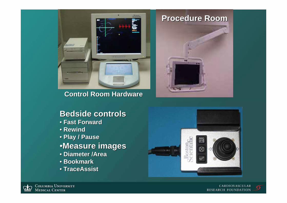
Procedure RoomProcedure Room
Bedside controlsBedside controls•• Fast ForwardFast Forward•• RewindRewind•• Play / PausePlay / Pause••Measure imagesMeasure images•• Diameter /AreaDiameter /Area•• BookmarkBookmark•• TraceAssistTraceAssist
Control Room HardwareControl Room Hardware
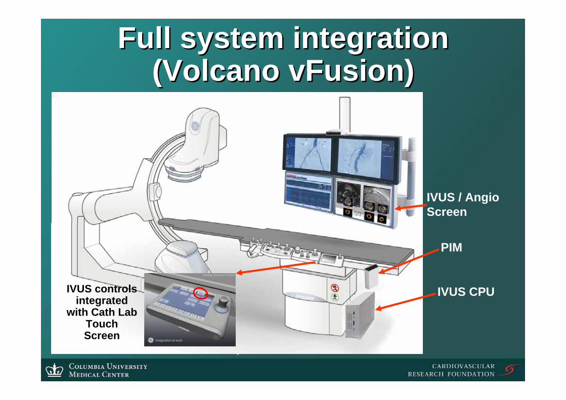
Full system integration Full system integration (Volcano (Volcano vFusionvFusion))
IVUS CPU
IVUS / AngioScreen
PIM
IVUS controls integrated
with Cath Lab Touch Screen

•• IVUS controls at the IVUS controls at the tabletable
•• IVUS controls in the IVUS controls in the control roomcontrol room
•• IVUS controls both at IVUS controls both at the table and in the the table and in the control roomcontrol room
•• Fully integrated network Fully integrated network operating IVUS in operating IVUS in multiple labsmultiple labs
Multiple Multiple configurations configurations
become possiblebecome possiblePIM
EquipmentRoom
GE INNOVALCD
CPU
GE INNOVACPU
Control RoomCath Lab
INNOVACONTROL
DISPLAYCLUSTER
ARCHIVE
CONTROLSTATION
PIM
EquipmentRoom
GE INNOVALCD
CPU
ARCHIVE
GE INNOVACPU
Control RoomCath Lab
INNOVACONTROL
DISPLAYCLUSTER
CONTROLSTATION
CONTROLSTATION

•• Full Full cathlabcathlab integration at a integration at a total total cost comparable to cost comparable to ““standstand--alonealone””instruments (2.5instruments (2.5--3 integrated labs 3 integrated labs should be equivalent to the cost of should be equivalent to the cost of one one ““standstand--alonealone”” unitunit””))

Catheter and TransducerCatheter and Transducer
•• TransducerTransducerHigher frequenciesHigher frequenciesDual frequencies Dual frequencies
•• CatheterCatheterHandling equivalent to a balloon catheterHandling equivalent to a balloon catheterImaging Imaging guidewireguidewireForward looking IVUSForward looking IVUS

Forward looking IVUS catheterForward looking IVUS catheter
•• While prototypes have been suggested While prototypes have been suggested and developed, to date none have been and developed, to date none have been shown to be practical and to yield shown to be practical and to yield diagnostically and therapeutically diagnostically and therapeutically helpful imageshelpful images

Prototype ForwardPrototype Forward--Looking CTO Looking CTO DeviceDevice
•• VisualizationVisualizationForwardForward--looking IVUS using looking IVUS using proprietary proprietary micromanipulator and micromanipulator and shape memory actuationshape memory actuation
•• SteeringSteeringSteering with multiple Steering with multiple degrees of freedom for degrees of freedom for distal tipdistal tip
•• PowerPowerRFRF--enabled tipenabled tip
•• UsabilityUsability0.0140.014”” guidewireguidewire of choiceof choice3 3 –– 3.5F profile3.5F profile
0.014” guidewire
Transducer
RF-enableddistal tip
Steering Mechanism

Forward Looking Navigational Forward Looking Navigational UltrasoundUltrasound
Vessel Lumen
Occlusion
Arterial Wall Tip ofGuidewire
GuidewirePenetratingOcclusion
LumenRecanalized
Click to play

Pullback devicePullback device
•• Resurrect the HResurrect the H--P P ““Fishing ReelFishing Reel”” -- in in my opinion, the best pullback device my opinion, the best pullback device ever devisedever devised
•• Or eliminate the pullback device Or eliminate the pullback device completely while still maintaining completely while still maintaining accurate length measurements and accurate length measurements and good imaging habitsgood imaging habits

Medical Positioning System (MPS)Medical Positioning System (MPS)
3D Coordinate System
MPS sensorMiniature sensors provide Position and Orientation (P&O) projected on 3D imaging model Accuracy < 1mm
sensorsensor transducertransducer

ECG and Position & Orientation (P&O) ECG and Position & Orientation (P&O) data integrationdata integration
T1
T2
Tn
P&O Data
ECG Cycles

Image CoImage Co--Registration (linking the Registration (linking the angiographic roadmap to the 2angiographic roadmap to the 2--D or D or
33--D IVUS images)D IVUS images)

CoCo--Registration of IVUS and Registration of IVUS and angiographic images (angiographic images (MediGuideMediGuide))
Real Time catheter tip tracking on stabilized previously recorded
roadmap
Real Time catheter tip Real Time catheter tip tracking on stabilized tracking on stabilized previously recorded previously recorded
roadmaproadmap


CoCo--registration of IVUS and registration of IVUS and angiographic images (angiographic images (Volcano&PaieonVolcano&Paieon))

Dynamic review Dynamic review ((BostonScientificBostonScientific))
Accurate segmentation Accurate segmentation algorithmsalgorithms
Enhanced border recognitionEnhanced border recognitionEspecially important if transducer frequency increases Especially important if transducer frequency increases
because blood speckle will become more intense and closer because blood speckle will become more intense and closer in appearance to plaque. Therefore, correlation algorithms to in appearance to plaque. Therefore, correlation algorithms to
remove blood speckle may be necessary.remove blood speckle may be necessary.
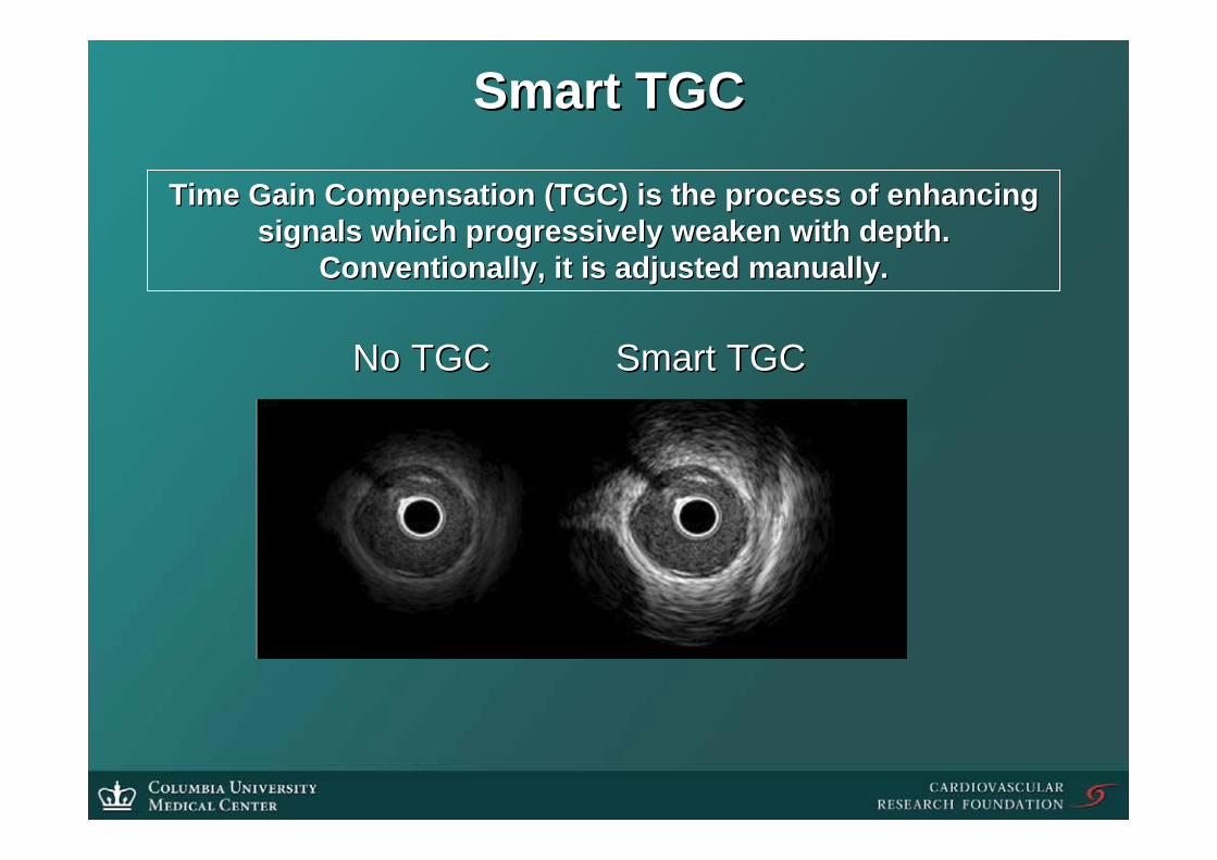
Smart TGCSmart TGC
Time Gain Compensation (TGC) is the process of enhancing Time Gain Compensation (TGC) is the process of enhancing signals which progressively weaken with depth. signals which progressively weaken with depth.
Conventionally, it is adjusted manually.Conventionally, it is adjusted manually.
No TGCNo TGC Smart TGCSmart TGC

Virtual HistologyVirtual Histology™™ IVUSIVUSOnly the envelope amplitude (echo Only the envelope amplitude (echo intensity) is used in formation of the grayintensity) is used in formation of the gray--scale IVUS imagescale IVUS image
Frequency of echo signal can also vary, depending on the tissue
Eight amplitude Eight amplitude ANDAND frequency frequency parameters are parameters are used in Virtual used in Virtual HistologyHistology

Thin plate Thin plate splinespline morphing morphing of distorted of distorted histologichistologic image image
after which the computer after which the computer was taught to recognize four was taught to recognize four
basic tissue typesbasic tissue types
IVUS B scan IVUS B scan
MovatMovat pentachromepentachrome stain stain

Accurate tissue characterizationAccurate tissue characterization
99.3%99.3%99.7%99.7%97.8%97.8%Dense calcium (n=92)Dense calcium (n=92)
94.4%94.4%93.8%93.8%97.1%97.1%Necrotic core (n=69)Necrotic core (n=69)
93.4%93.4%95.1%95.1%86.9%86.9%FibrofattyFibrofatty (n=84)(n=84)
92.8%92.8%98.8%98.8%84.0%84.0%Fibrous tissue (n=162)Fibrous tissue (n=162)
Predictive Predictive AccuracyAccuracySpecificitySpecificitySensitivitySensitivity
Eagle Eye VH Accuracy Eagle Eye VH Accuracy VH IVUS vs histopathology from fresh postVH IVUS vs histopathology from fresh post--mortem coronary arteriesmortem coronary arteries

Plaque Classification Plaque Classification -- IIAdaptive Intimal ThickeningAdaptive Intimal ThickeningPlaque comprised of nearly all fibrous Plaque comprised of nearly all fibrous
tissue. (<5% of fibrofatty, tissue. (<5% of fibrofatty, calcification and/or NC plaque).calcification and/or NC plaque).((Generally not viewed by Dr. Generally not viewed by Dr. Virmani to be acutely dangerous)Virmani to be acutely dangerous)
Pathological Intimal ThickeningPathological Intimal Thickening ––Mainly mixture of fibrous, fibrofatty Mainly mixture of fibrous, fibrofatty (>5%), and necrotic core and some (>5%), and necrotic core and some calcified tissue <5%.calcified tissue <5%.

Plaque Classification Plaque Classification -- IIII
““FibroFibro--AtheromaAtheroma”” –– Fibrotic cap and significant Necrotic Fibrotic cap and significant Necrotic Core Core in in fibroticfibrotic and/or and/or fibrofattyfibrofatty tissuetissue
It is very likely be that the most important goal is to It is very likely be that the most important goal is to differentiate the differentiate the FibroAtheromaFibroAtheroma plaque types from the plaque types from the other three plaque types during assessments of high other three plaque types during assessments of high
risk lesions for rupture.risk lesions for rupture.

Diagnostic accuracy of realDiagnostic accuracy of real--time time IB (integrated Backscatter)IB (integrated Backscatter)--IVUSIVUS
90%90%85%85%92%92%90%90%Lipid pool (n=205)Lipid pool (n=205)
94%94%93%93%93%93%94%94%Fibrosis (n=335) Fibrosis (n=335)
99%99%93%93%99%99%95%95%Calcification (n=144)Calcification (n=144)
NPVNPVPPVPPVSpecificitySpecificitySensitivitySensitivity
(Kawasaki et al. Circulation2002;105:2487(Kawasaki et al. Circulation2002;105:2487--92)92)

Masson Masson TrichromeTrichromeStaining Staining
FibrosisFibrosis
CalcificationCalcification
Lipid pool or Lipid pool or IntimalIntimal HyperplasiaHyperplasia
Dense fibrosisDense fibrosis
Conventional IVUSConventional IVUSIntegrated Backscatter Integrated Backscatter
Intravascular Ultrasound Intravascular Ultrasound (IB(IB--IVUS) ColorIVUS) Color--coded coded
MapMap
(Kawasaki et al. Circulation2002;105:2487(Kawasaki et al. Circulation2002;105:2487--92)92)
(Resolution 0.1 mm)(Resolution 0.1 mm)
(Resolution 0.05 mm)(Resolution 0.05 mm)
**
*
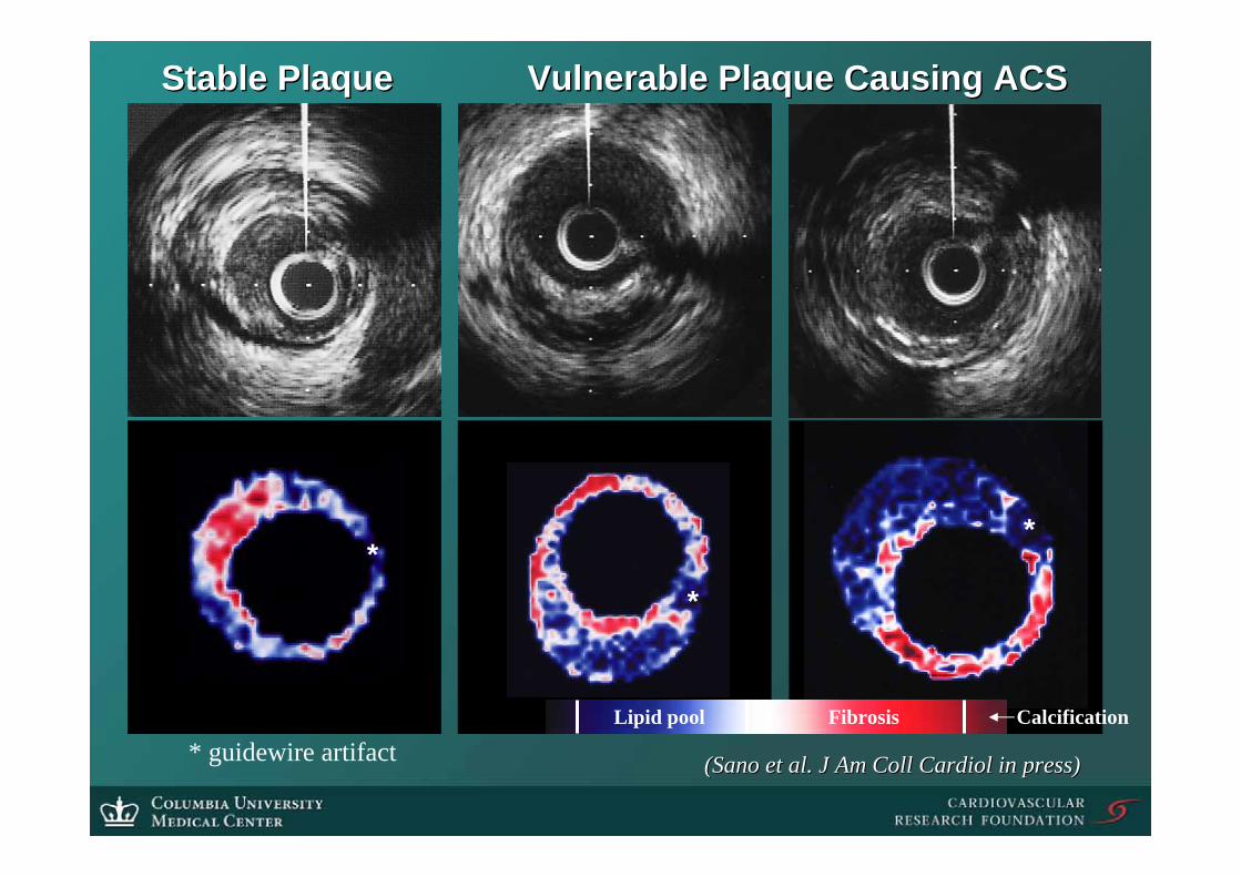
Stable PlaqueStable Plaque Vulnerable Plaque Causing ACSVulnerable Plaque Causing ACS
FibrosisLipid pool Calcification
(Sano et al. J Am (Sano et al. J Am CollColl CardiolCardiol in press)in press)* guidewire artifact

SOFT
HARD
SchaarSchaar et al. Circulation 2003;108:2535et al. Circulation 2003;108:2535--4141
Independent predictors Independent predictors of strain were of strain were
macrophages (p=0.006) macrophages (p=0.006) and smooth muscle and smooth muscle
cells (p=0.0001)cells (p=0.0001)

Baseline images are acquired for 20 seconds, Baseline images are acquired for 20 seconds, and regions of interest are assignedand regions of interest are assigned
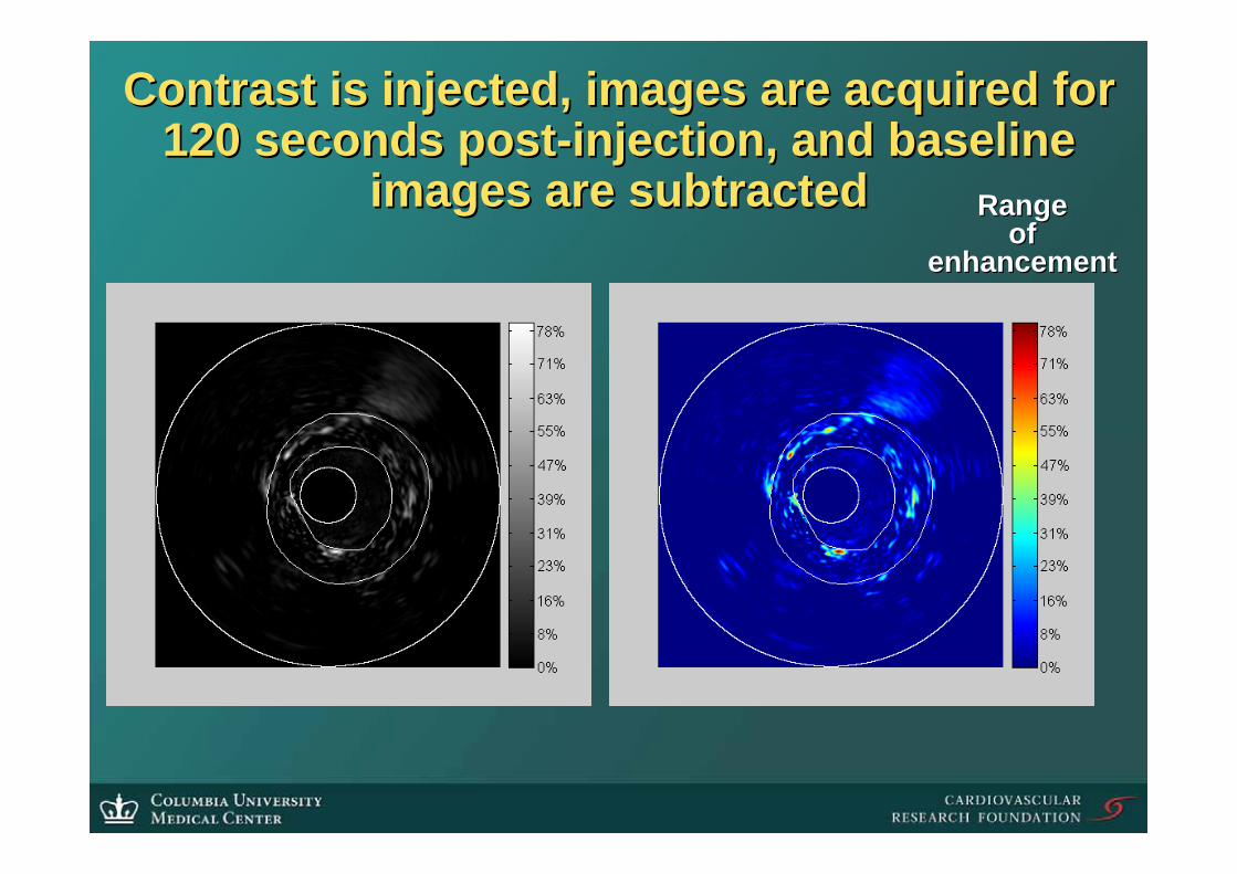
Range Range of of
enhancementenhancement
Contrast is injected, images are acquired for Contrast is injected, images are acquired for 120 seconds post120 seconds post--injection, and baseline injection, and baseline
images are subtractedimages are subtracted

PostPost--injection injection (Frame #800)(Frame #800)
Peak Injection Peak Injection (Frame #600)(Frame #600)
PrePre--injection injection (Frame #200)(Frame #200)
Lumen subtracted Lumen subtracted ((microbubblemicrobubble shadow shadow
effect is not effect is not calculated) calculated)
The enhancement The enhancement lasts for at least 25 lasts for at least 25
seconds.seconds.
Background motions Background motions are cancelledare cancelled
Storage of IVUS with the angiogramsStorage of IVUS with the angiograms
•• Only one set of patient demographicsOnly one set of patient demographics•• Minimizes errorsMinimizes errors•• Minimizes onMinimizes on--screen annotationscreen annotation•• Enhances Enhances ““plugplug--andand--playplay”” conceptconcept
•• The IVUS imaging runs can be viewed as they The IVUS imaging runs can be viewed as they occurred in sequence during the case without occurred in sequence during the case without having to having to ““matchmatch”” IVUS and angiographic IVUS and angiographic runs. The IVUS images can be related to the runs. The IVUS images can be related to the flow of the case.flow of the case.
At the very least, IVUS studies should be At the very least, IVUS studies should be completely DICOM compatiblecompletely DICOM compatible

TimeTime--activity curves with quantitative activity curves with quantitative monitoring of plaque perfusionmonitoring of plaque perfusion
IntimoIntimo--Medial and Plaque AreaMedial and Plaque Area Adventitia AreaAdventitia Area

ConclusionConclusion
•• IVUS should be IVUS should be ““plugplug--andand--playplay””•• With the exception of transducer design and With the exception of transducer design and
catheter improvements, each component of catheter improvements, each component of this this ““futuristic visionfuturistic vision”” is possible (and even is possible (and even available!) now. available!) now.
•• The problem is that these efforts are not The problem is that these efforts are not coordinated, nor are they likely to be. And no coordinated, nor are they likely to be. And no one company has the ability to deliver the one company has the ability to deliver the entire package. This forces the consumer (us) entire package. This forces the consumer (us) to chose between equally desirable, but to chose between equally desirable, but mutually exclusive features.mutually exclusive features.
•• So, the final element of this So, the final element of this ““futuristic visionfuturistic vision””is one of a coordinated or crossis one of a coordinated or cross--licensed licensed effort among the companies. effort among the companies.