Embed Size (px)
Citation preview

Computer Science Publications Computer Science
2013
A Genome-Wide Survey of Highly Expressed Non-Coding RNAs and Biological Validation of SelectedCandidates in Agrobacterium tumefaciensKeunsub LeeIowa State University, [email protected]
Xiaoqiu HuangIowa State University, [email protected]
Chichun YangIowa State University
Danny LeeIllumina, Inc.
Kan NobutaIllumina, Inc.
See next page for additional authors
Follow this and additional works at: http://lib.dr.iastate.edu/cs_pubs
Part of the Agricultural Science Commons, Agronomy and Crop Sciences Commons, OtherComputer Sciences Commons, and the Plant Breeding and Genetics Commons
The complete bibliographic information for this item can be found at http://lib.dr.iastate.edu/cs_pubs/6. For information on how to cite this item, please visit http://lib.dr.iastate.edu/howtocite.html.
This Article is brought to you for free and open access by the Computer Science at Iowa State University Digital Repository. It has been accepted forinclusion in Computer Science Publications by an authorized administrator of Iowa State University Digital Repository. For more information, pleasecontact [email protected].

AuthorsKeunsub Lee, Xiaoqiu Huang, Chichun Yang, Danny Lee, Kan Nobuta, Jian-Bing Fan, and Kan Wang
This article is available at Iowa State University Digital Repository: http://lib.dr.iastate.edu/cs_pubs/6
A Genome-Wide Survey of Highly Expressed Non-CodingRNAs and Biological Validation of Selected Candidates inAgrobacterium tumefaciensKeunsub Lee1,2, Xiaoqiu Huang3, Chichun Yang1,2, Danny Lee4, Vincent Ho4, Kan Nobuta4, Jian-
Bing Fan4, Kan Wang1,2*
1 Center for Plant Transformation, Plant Sciences Institute, Iowa State University, Ames, Iowa, United States of America, 2 Department of Agronomy, Iowa State University,
Ames, Iowa, United States of America, 3 Department of Computer Science, Iowa State University, Ames, Iowa, United States of America, 4 Scientific Research, Illumina Inc.,
San Diego, California, United States of America
Abstract
Agrobacterium tumefaciens is a plant pathogen that has the natural ability of delivering and integrating a piece of its ownDNA into plant genome. Although bacterial non-coding RNAs (ncRNAs) have been shown to regulate various biologicalprocesses including virulence, we have limited knowledge of how Agrobacterium ncRNAs regulate this unique inter-Kingdom gene transfer. Using whole transcriptome sequencing and an ncRNA search algorithm developed for this work, weidentified 475 highly expressed candidate ncRNAs from A. tumefaciens C58, including 101 trans-encoded small RNAs(sRNAs), 354 antisense RNAs (asRNAs), 20 59 untranslated region (UTR) leaders including a RNA thermosensor and 6riboswitches. Moreover, transcription start site (TSS) mapping analysis revealed that about 51% of the mapped mRNAs have59 UTRs longer than 60 nt, suggesting that numerous cis-acting regulatory elements might be encoded in the A. tumefaciensgenome. Eighteen asRNAs were found on the complementary strands of virA, virB, virC, virD, and virE operons. FifteenncRNAs were induced and 7 were suppressed by the Agrobacterium virulence (vir) gene inducer acetosyringone (AS), aphenolic compound secreted by the plants. Interestingly, fourteen of the AS-induced ncRNAs have putative vir boxsequences in the upstream regions. We experimentally validated expression of 36 ncRNAs using Northern blot and RapidAmplification of cDNA Ends analyses. We show functional relevance of two 59 UTR elements: a RNA thermonsensor(C1_109596F) that may regulate translation of the major cold shock protein cspA, and a thi-box riboswitch (C1_2541934R)that may transcriptionally regulate a thiamine biosynthesis operon, thiCOGG. Further studies on ncRNAs functions in thisbacterium may provide insights and strategies that can be used to better manage pathogenic bacteria for plants and toimprove Agrobacterum-mediated plant transformation.
Citation: Lee K, Huang X, Yang C, Lee D, Ho V, et al. (2013) A Genome-Wide Survey of Highly Expressed Non-Coding RNAs and Biological Validation of SelectedCandidates in Agrobacterium tumefaciens. PLoS ONE 8(8): e70720. doi:10.1371/journal.pone.0070720
Editor: Alfredo Herrera-Estrella, Cinvestav, Mexico
Received December 19, 2012; Accepted June 26, 2013; Published August 8, 2013
Copyright: � 2013 Lee et al. This is an open-access article distributed under the terms of the Creative Commons Attribution License, which permits unrestricteduse, distribution, and reproduction in any medium, provided the original author and source are credited.
Funding: This work was supported by Iowa State University Plant Sciences Institute. The funder had no role in study design, data collection and analysis, decisionto publish, or preparation of the manuscript.
Competing Interests: DL, VH, KN and JBF are employees of Illumina, Inc. The study used the RNA-seq products developed at Illumina. However, this does notalter the authors’ adherence to all the PLOS ONE policies on sharing data and materials.
* E-mail: [email protected]
Introduction
Last decade, non-coding RNAs (ncRNAs) have emerged as
crucial regulators of diverse physiological and cellular processes in
both bacteria and eukaryotes. In bacteria, ncRNAs control various
biological processes including, but not limited to, virulence [1],
photosynthesis [2], iron homeostasis [3], pH sensing [4],
temperature sensing [5], plasmid replication [6–9], and toxin
suppressions [10–12]. Bacterial ncRNAs include ribosomal RNAs
(rRNAs), transfer RNAs (tRNAs), cis-encoded antisense RNAs
(asRNAs) and trans-encoded small RNAs (sRNAs). rRNAs and
tRNAs regulate protein translation and some tRNAs have also
been implicated to have regulatory roles [13,14]. AsRNAs regulate
genes encoded on the opposite strand, while sRNAs have their
gene targets encoded elsewhere in the genome and often have
multiple targets [15,16]. Both asRNAs and sRNAs base-pair with
target mRNAs and alter translation and/or mRNA stability. Most
of the time they inhibit translation and lead to co-degradation
[17].
Various strategies can be used to identify ncRNAs in bacteria.
Computational predictions followed by experimental validations
have revealed many sRNAs in the earlier days [18–20].
Sequencing cDNA clones prepared from small-sized RNA
fractions can be useful to identify sRNAs [21]. Most recently,
tiling arrays and deep sequencing analyses have identified tens to
hundreds of ncRNAs from various bacterial species [22–25].
However, it remains difficult to answer how many regulatory
ncRNAs exist in any bacterial genome [17] due to several major
challenges: 1) extremely abundant ribosomal RNAs, which
account for 95–97% of total RNA, reduce detection sensitivity
for transcriptome analysis [26], 2) some ncRNAs are only
expressed under specific conditions, and 3) ncRNAs derived from
mRNAs are hard to be distinguished. The presence of internal
transcriptional start site (TSS) within annotated ORFs also adds
PLOS ONE | www.plosone.org 1 August 2013 | Volume 8 | Issue 8 | e70720

another level of complexity to the transcriptome [27]. In addition,
due to the huge data size generated by deep sequencing, often in
hundreds of gigabytes (GB), differences in the algorithms for data
analysis affect the outcomes [28]. Thus, more robust sequencing
technology and advances in data analysis will likely expand the
inventories of ncRNAs, and ultimately help us to see better insights
into the gene regulatory networks.
A. tumefaciens is a gram-negative bacterium and the causal agent
of crown gall disease. A. tumefaciens recognizes various signals from
plants and convert its life style from a free-living saprobe to a plant
pathogen. The way A. tumefaciens affects infected plants is unique: it
genetically transform host plant by exporting a segment of tumor
inducing (Ti) plasmid (T-DNA) into plant cell where the T-DNA
integrates into host plant genome [29]. Genes encoded by the T-
DNA are expressed to produce plant hormones to induce tumor
formation and enzymes to make novel compounds called opines
that are utilized by A. tumefaciens [29]. The transition from a
saprobe to a pathogen requires expression of virulence genes on Ti
plasmid and some other genes encoded on the chromosome [30].
This process begins with recognition of plant-produced com-
pounds, such as phenolics (e.g., acetosyringone) and sugars, by the
Agrobacterium VirA two-component sensor kinase in accordance
with a periplasmic sugar binding protein ChvE encoded by the
chromosomal virulence gene E (chvE) [31–33]. VirA phosphory-
lates VirG, the two-component response regulator, which activates
transcription of other virulence genes [34,35]. While much is
known about how the T-DNA border sequences, its virulence
genes and some plant genes are involved in this unique inter-
Kingdom gene transfer [29], the importance of regulatory
functions of ncRNAs in that process remains unanswered. In
various bacteria, some ncRNAs have been found to be crucial
virulence factors [1]; similar mechanisms may exist in plant
pathogenic bacteria, such as A. tumefaciens.
Several transcriptome analyses using microarrays have identi-
fied differentially expressed genes in A. tumefaciens in response to
various conditions, such as low pH [36], plant-derived signaling
molecules [37], and the loss of phosphatidylcholine biosynthesis
[38]. A few recent studies showed that A. tumefaciens uses ncRNAs
to regulate important biological processes, such as Ti plasmid
replication [39] and the uptake of a plant-derived signaling
molecule, c-amynobutyric acid (GABA) by an ABC transporter
[40]. The RNA chaperone Hfq was involved in the sRNA-
mediated regulation of the GABA transporter, and the hfq deletion
mutant showed reduced virulence, suggesting that ncRNAs may
play important roles during Agrobacterium-plant interactions [41]. A
recent deep sequencing study by Wilms et al [51] obtained a total
of 348,998 cDNA reads ($18 nt) from four cDNA libraries and
reported 152 sRNAs and 76 asRNAs from A. tumefaciens C58.
Some of these were induced by the phenolic compound (AS),
which is essential for the induction of Agrobacterium virulence genes
[42].
In this study, we present an extensive list of ncRNAs for the
same strain A. tumefaciens C58. We improved RNA-seq detection
sensitivity by using a combination of treatments, including the
MICROBExpressTM kit and 59-phosphate-dependent exonuclease
to deplete ribosomal RNAs. We then performed whole tran-
scriptome sequencing (RNA-seq) on an Illumina GAII platform
and obtained over 48 million uniquely mapped reads ( = 50 bp)
from eight cDNA libraries. Using an ncRNA search algorithm
developed for this work, we identified and generated a list of highly
expressed ncRNAs in Agrobacterium strain C58 grown under 4
different conditions. Selected ncRNAs were experimentally
validated by RACE and Northern analyses.
Results and Discussion
Overview of whole genome transcriptome sequencingTo identify A. tumefaciens ncRNAs expressed under different
conditions, we grew the strain C58 and harvested the cultures at:
1) nutrient rich medium at mid-log phase (YEP-L: OD600 = 0.5),
2) nutrient rich medium at late stationary phase (YEP-S: OD600
= 1.3), 3) modified AB induction medium without AS (AB: OD600
= 0.8) and 4) modified AB induction medium with AS (IND:
OD600 = 0.8). To improve non-rRNA detection sensitivity, we
removed 16S and 23S rRNAs using two commercially available
kits. First, total RNA sample was treated according to the
MICROBExpressTM kit (Ambion, USA), which uses hybridization
oligonucleotides attached to magnetic beads to selectively deplete
16S and 23S rRNAs. Analysis by an Agilent 2100 BioAnalyzer
showed that about 55% of 16S and 23S rRNAs had been
successfully removed using this kit. Because Agrobacterium 23S
rRNA undergoes post-transcriptional fragmentation [43], some
23S rRNA could not be removed by the hybridization oligonu-
cleotides. The remaining rRNAs were further depleted using the
terminator 59-phosphate-dependent exonuclease (TEX), which
selectively digests processed RNA molecules with 59-monophos-
phate [44]. Because rRNAs are processed from the primary
transcript upon transcription, this enzyme is useful to deplete
rRNAs from total RNA samples as well as to enrich primary
transcripts [26]. This dual treatment can increase the non-
ribosomal RNA components from 3–5% to up to 25% in a total
RNA population [26]. Thus, a total of eight cDNA libraries, YEP-
L, YEP-S, AB and IND with/without primary transcript
enrichment (2/+ TEX), were prepared and sequenced in parallel
on the Illumina GAII platform.
We obtained a total of 842.109 million reads from the 8 cDNA
libraries (Table 1; 429.340 million reads from the –TEX samples
and 412.769 million reads from the +TEX samples). These short
sequence reads (50 bp) were aligned to the reference genome
(NC_003062.2, NC_003063.2, NC_003064.2, NC_003065.3)
using the Bowtie 2 program [45]. A total of 490.552 million
reads, 252.825 ( = 18,959+233,866) from the –TEX samples and
237.728 ( = 29,343+208,385) million reads from the +TEX
samples, were mapped to the reference genome (Table 1). Among
these, 92.5% of the reads (233.866 millions) from the –TEX
samples and 87.7% of the reads (208.385 millions) from the +TEX
samples were aligned more than once to the reference genome.
Table 1. Summary of RNA sequencing and alignment*.
Number of reads (61000)
TEX YEP-L YEP-S AB IND Total
Total generated 2 106,658 102,249 112,547 107,886 429,340
+ 115,420 84,435 105,172 107,741 412,769
Total aligned 2 60,046 60,536 67,643 64,600 252,825
+ 61,617 53,185 62,453 60,473 237,727
Aligned 1 time 2 4,033 3,534 5,760 5,632 18,959
+ 5,155 5,411 8,929 9,848 29,343
Aligned .1 time 2 56,013 57,001 61,883 58,969 233,866
+ 56,462 47,774 53,524 50,625 208,385
*Short sequence reads were aligned to the A. tumefaciens C58 referencegenome using the Bowtie 2 program [45].doi:10.1371/journal.pone.0070720.t001
Noncoding RNAs in Agrobacterium tumefaciens
PLOS ONE | www.plosone.org 2 August 2013 | Volume 8 | Issue 8 | e70720

The number of reads that were mapped exactly once to the
reference genome (uniquely mapped reads, UMR) varied among
different cDNA libraries, ranging from 3.534 million (YEP-S, –
TEX) to 9.848 million (IND, +TEX). We used only these UMRs
for gene expression quantification and subsequent analysis. A total
of 18.959 million UMRs in the –TEX samples and 29.343 million
UMRs in the +TEX samples were found, representing 7.5% (of
the 252.825 million) and 12.3% (of the 237.727 million) of total
mapped reads, respectively. Thus, a considerably higher percent-
age of UMRs were obtained in the +TEX libraries than the –TEX
libraries, suggesting improved non-rRNA detection sensitivity.
The increased number of UMRs was likely due to the depletion
of rRNAs and tRNAs by TEX. We computed the total number of
UMRs that were aligned to the rRNAs and tRNAs. In the –TEX
libraries, a total of 3.785 million reads were mapped to rRNAs
and tRNAs, while in the +TEX libraries only 0.662 million reads
were mapped to rRNAs and tRNAs. The percentage of the rRNAs
and tRNAs reads were significantly decreased from 20.9% in the –
TEX samples (YEP-L, 25.0; YEP-S, 26.0; AB, 16.3; IND, 16.3) to
2.3% in the +TEX samples (YEP-L, 3.5; YEP-S, 1.5; AB, 2.3;
IND, 1.9).
The UMRs were then piled up using the SAMtools [46], which
provides various tools to manipulate sequence alignment data.
This pile-up step allowed us to compute the depth of coverage (i.e.,
the number of reads mapped to a nucleotide position on the
forward or reverse strands) throughout the entire genome of A.
tumefaciens C58. For gene expression quantification, we first
computed the average depth of coverage (ADC) of each gene,
and then converted ADC to RPKM (Reads per Kilobase per
Million mapped reads) by a simple formula:
RPKM~ADC|L
l|1000
bp
kilobase7N, where L is the length
of a gene, l is the length of a sequence read ( = 50 bp), and N is the
number of total reads in millions [47].
Primary transcript enrichment using TEX was helpful to obtain
relatively under-represented RNA species (Table 1; Figure S1A&B
in File S2). As shown in Figure S1A and B, the vir genes expression
was higher with TEX treatment than was without TEX treatment.
To determine whether TEX treatment has systematic effects on
gene expression quantification, we generated a scatter plot with
the log-transformed RPKM values of all annotated protein-coding
genes with and without TEX treatment (Figure S1C in File S2).
Some genes had lower expressions with TEX treatment than were
without TEX treatment (e.g., data points on the X-axis in
Figure S1C in File S2). These could represent post-transcription-
ally processed transcripts. However, TEX treatment did not have
systematic biased effects on quantifying gene expression (Pearson’s
product-moment coefficient, r = 0.91; Figure S1C in File S2), and
many genes became detectable after TEX treatments (i.e., data
points on the Y-axis in Figure S1C in File S2). Indeed, among the
5432 protein-coding genes, 3411,3842 genes were detected
(RPKM .0) without TEX treatment (YEP-L, 3603; YEP-S, 3487;
AB, 3842; IND, 3411), while 3957,4361 genes were detected
with TEX enrichment (YEP-L, 3957; YEP-S, 3959; AB, 4361;
IND, 4156). These results suggest that combination of rRNA
depletion kit (MICROBExpressTM kit) and TEX treatment is very
useful to improve overall RNA-seq detection sensitivity.
Data validationFor validation purposes, we visualized vir genes expression on
the Ti plasmid. We plotted the depth of coverage at each
nucleotide position from 180,590 to 211,094 of Ti plasmid
(Figure 1). As shown in Figure 1, there was a large difference in vir
genes expression with/without AS induction. For instance, the two
component sensor kinase virA was expressed at a low level without
induction, 33 RPKM, but was expressed at 22.6 fold higher level
with induction, 734 RPKM. Likewise, the expression levels of the
two component response regulator virG were 219 RPKM without
induction and 1614 RPKM with induction, a 7.4 fold increase. In
a previous study by Winans et al. [48], a lacZ reporter assay
demonstrated about 9-fold increase in virA expression by AS. In
our RNA-seq study, virA transcript level was increased by a 22.6-
fold. Although the fold changes were not equal (9 vs. 22.6), it has
been well-documented that mRNA level is not directly correlated
with protein abundance [49,50]. Other vir operons, such as virB,
virC and virD were only expressed under induction condition.
These vir genes expression patterns were consistent with previous
microarray and RNA-seq studies (Table S1 in File S1) [38,51].
Interestingly, there were some noticeable antisense transcripts
on the complementary strands of virB9, virC, virD and virE. The
existence of some of these cis-antisense transcripts were confirmed
by 59 and 39 RACE (Rapid Amplification of cDNA Ends)
(Figure S2 in File S2). In addition, virB10, virB11 and virD4 had
internal transcripts expressed without AS induction. Especially,
virD4 internal transcript (virD4*; pTi 201529–201869) was
expressed under all four growth conditions (Figure S3 in File S2;
RPKM: YEP-L, 267; YEP-S, 380; AB, 526; IND, 738), as opposed
to the full length transcript, virD4, which is only expressed under
vir gene induction conditions. Therefore, if virD4* has a functional
role, if any, it may not be restricted to pathogenicity. The
functional relevance of these RNAs needs to be determined, but
the presence of these transcripts suggests that the A. tumefaciens
transcriptome could be as complex as those of other bacterial
transcriptomes such as Listeria monocytogenes, Escherichia coli, and
Sinorhizobium meliloti [52], and the full inventory of transcripts, both
protein-coding and non-coding, may be significantly expanded in
the future.
Transcriptional Start Sites (TSS) mappingWe identified TSSs for 705 annotated protein-coding genes
(Table S2 in File S1). Excluding 30 genes whose TSS were
mapped within the coding region, we estimated the 59 UTR
lengths for 675 protein-coding genes. The length of the 59 UTR
varied from 0 to 521 nt, averaging 88 nt with a median of 61 nt
(Figure 2). About 39% (253) of the protein-coding genes had short
59 UTRs (#50 nt), while 30% (203) of them had long 59 UTRs
(.100 nt). About 51% (345) had 59 UTRs longer than 60 nt,
which is long enough to contain cis-regulatory element [53]. There
were 12 genes with 59 UTR length no longer than 10 nt (Table S2
in File S1), suggesting that leaderless mRNAs exist in this
bacterium, which may require special ribosomes for translation
[54]. These results were comparable to those obtained by Wilms
et al. [51]: the estimated length of 59 UTRs reported in their study
varied from 0 to 544 nt averaging 87 nt and about 40% (145/356)
were short (#50 nt).
We also found that at least 27 genes, 20 of them encoding
hypothetical proteins (marked by * in Table S2 in File S1), had
TSSs mapped within annotated coding sequences, suggesting that
they might be incorrectly annotated. Indeed, BLAST searches
against the GenBank database using the predicted amino acid
sequences as queries showed that 19 of those 27 genes have longer
N-termini than their homologs (Table S2 in File S1). Further
investigation is required to verify these sequences.
Identification of non-coding RNAsTo identify highly expressed ncRNA transcripts, we calculated
depth of coverage at each nucleotide position on both forward and
reverse strands of all four replicons of A. tumefaciens. Then, using
Noncoding RNAs in Agrobacterium tumefaciens
PLOS ONE | www.plosone.org 3 August 2013 | Volume 8 | Issue 8 | e70720

already annotated gene features [55–57], we searched for non-
gene-coding genomic regions that have at least 10 times higher
depth of coverage than adjacent regions. This was done to avoid
erroneous annotations due to pervasive transcription [58,59]. This
approach yielded a total of 475 candidate ncRNAs, 101 trans-
encoded small RNAs (sRNAs), 354 antisense RNAs (asRNAs) and
20 59 UTR elements.Some of these were differentially expressed
under different growth conditions (Table 2; Table S3 in File S1).
Candidate ncRNAs were distributed across all four replicons: 221
on the circular chromosome, 164 on the linear chromosome, 43
on the pAt plasmid and 47 on the Ti plasmid. The vast majority of
the sRNAs (89/101) were found on the two chromosomes and
only 12 of them were found on the two plasmids. In addition, 87%
of ncRNAs (78/90) found on the two plasmids were asRNAs; 18 of
them were encoded on the opposite strand of virA, virB, virD, virE,
virF and virK.
Figure 1. Induction of vir genes with AS. Expression of 24 vir genes with and without AS was visualized for data validation. Depth of coverage ateach nucleotide position from 180,590 to 211,094 of Ti plasmid was plotted for (A) forward strand and (B) reverse strand. A total of 24 vir genes wereincluded: virA, virB (B1,B11), virG, virC (C1, C2), virD (D1,D5) and virE (E0,E3).doi:10.1371/journal.pone.0070720.g001
Figure 2. Variation of 59 UTR length. The distance between TSS andstart codon (59 UTR) varied substantially from 0 to 521 nt, averaging88 nt and median of 61 nt. Among the 675 protein coding genes, 1.8%(12) were leaderless (#10 nt), 39% (253) were short (11,50 nt), while30% (203) were long (.100 nt). About 51% (345) of 59 UTRs werelonger than 60 nt.doi:10.1371/journal.pone.0070720.g002
Table 2. Distribution of ncRNAs on four replicons.
Replicon sRNA asRNA 59 UTR Total %
Circular chromosome 56 154 11 221 46.5
Linear chromosome 33 125 6 164 34.5
At plasmid 8 33 2 43 9.1
Ti plasmid 4 42 1 47 9.9
Total 101 354 20 475 100
% 21.3 74.5 4.2 100.0
doi:10.1371/journal.pone.0070720.t002
Noncoding RNAs in Agrobacterium tumefaciens
PLOS ONE | www.plosone.org 4 August 2013 | Volume 8 | Issue 8 | e70720

We searched Rfam database (http://rfam.sanger.ac.uk/) and
previously reported ncRNAs, and found that 91 of the 475
candidate ncRNAs (37 sRNAs and 44 asRNAs, and 10 59 UTR
elements) had been identified previously [39,40,51]. Those 91
ncRNAs correspond to 92 previously identified ncRNAs including
recently identified A. tumefaciens sRNAs, repE [39], AbcR1 and
AbcR2 [40]. Some well conserved sRNAs were also identified,
such as 6S RNA, the signal recognition particle (SRP) RNA (4.5S
RNA), tmRNA (SsrA, Atu2049), RNase P, and counter-tran-
scribed RNA (ctRNA_p42d, Atu8080), which binds to repB
mRNA to inhibit translation (Table S3 in File S1) [60]. The
discrepancy (91 vs. 92) was because an ncRNA identified by our
study (C1_1533961R) overlapped with 2 ncRNAs identified by
Wilms et al. [51], 1533826–1533764 and 1533957–1533833.
Thus, a total of 384 novel ncRNAs were identified in this study,
including 64 sRNAs, 310 asRNAs, and 10 59 UTR leaders.
A previous study by Wilms et al [51] used the Roche 454
platform to sequence the A. tumefaciens transcriptome and identified
228 candidate ncRNAs. They obtained a total of 348,998 cDNA
reads ($18 bp) mapped to the reference genomes from four
libraries, representing two growth conditions (2Vir and +Vir). We
used Illumina GAII platform and obtained a total of 2415
megabases (Mb) sequences from more than 48.3 million UMRs
( = 50 bp). In addition, we sequenced four more cDNA libraries
representing two more growth conditions including stationary
phase in a nutrient rich medium, under which many stress-related
ncRNAs accumulate [17]. As summarized in Table 3, we
categorized the candidate ncRNAs into three groups: sRNAs,
asRNAs, and 59 UTR leaders. Wilms et al. [51] originally
reported 152 sRNAs and 76 asRNAs, but our study suggested
that three sRNAs reported by Wilms et al were likely to be 59
UTR leaders (Table 3). From our data set, we identified 101
sRNAs, 354 asRNAs and 20 59 UTR leaders. Among those, 36
sRNAs, 44 asRNAs and three 59 UTR leaders were identified by
both studies (Table 3; Common). A total of 145 ncRNAs were
identified only by Wilms et al. [51] and 393 ncRNAs were
identified only by our study. Therefore, 621 ncRNA candidates
were identified in A. tumefaciens C58 by two RNA-seq studies: 215
sRNAs, 386 asRNAs and 20 59 UTR leaders (Table 3).
Interestingly, Wilms et al. [51] identified more sRNAs (149)
than our study (101), while we identified many more asRNAs (354)
than Wilms et al. [51] (76). This might be due to the differences in
RNA-seq technology and ncRNA search algorithm. We treated
the RNA samples consecutively with two methods to deplete
rRNAs using hybridization oligos (MICROBExpressTM kit, Am-
bion, USA) and TEX, while Wilms et al only treated their samples
with TEX (e.g., Figure 1A&B Figure 2B in Wilms et al. [51]). The
dual treatment in our study could help to obtain a higher overall
coverage. In addition, we developed an ncRNA search algorithm,
which identified genomic regions that did not overlap with any
annotated genes and had at least ten times higher expression levels
than neighboring regions (see Experimental procedures for detail).
On the one hand, this algorithm has the strength to quickly
identify highly expressed asRNAs, and indeed we did identify 354
asRNAs (6.6% of the 5,355 protein-coding genes, Table 3). On the
other hand, some intergenic sRNAs may not be identified by this
algorithm if adjacent genes are highly expressed at the same time.
For example, the sRNAs C3 and Ti2 from the Wilms et al. [51]
were not reported as a sRNA by our study because the immediate
downstream genes (dnaA and Atu6155) were also highly expressed.
However, it is also possible that some of the sRNAs identified by
Wilms et al. [51] might be part of 59 UTRs of protein coding
genes. As shown in Figure S4 in File S2, for instance, our data
suggested that C3 could be part of the 369 nt 59 UTR of dnaA
(Figure S4A in File S2) and Ti2 could be part of the 207 nt 59
UTR of Atu6155 (Figure S4B in File S2). Thirty-two sRNAs
identified by Wilms et al. [51] appeared to be part of the long 59
UTRs in our TSS mapping analysis (marked by { in Table S2 in
File S1). In fact, the 59 ends of 11 of those 32 sRNAs (including
C3) were also identified as TSSs of protein-coding genes by Wilms
et al. [51] (Table S2 in File S1). Another explanation could be that
the bacterial growth conditions used for each RNA-seq study were
different. Validation of all identified ncRNAs is needed for future
studies.
Differentially expressed ncRNAsWe identified differentially expressed ncRNAs by using the
Bioconductor DESeq package [61]. Briefly, the number of reads
mapped to each gene was calculated using a simple formula (Read
count ~ADC|L
l, where L is the length of a gene and l is the
length of a sequence read, 50), and normalized by effective cDNA
library sizes. Differentially expressed ncRNAs were identified by
comparing the full generalized linear model (GLM: , treatment +TEX) against the null model (GLM: , TEX).
We first identified differentially expressed ncRNAs (P,0.05)
under induction conditions by AS (IND vs. AB) (Table S4-A, B in
File S1). Fifteen ncRNAs were induced (Table S4-A in File S1),
while 7 ncRNAs were suppressed (Table S4-B in File S1) by AS.
Fourteen of the 15 AS induced ncRNAs have putative vir box
sequences [62] in the upstream region (Table S4-A in File S1). It
will be worthwhile to determine if some of these ncRNAs have
regulatory roles during Agrobacterium-plant interactions.
We then identified differentially expressed ncRNAs during the
stationary phase and the mid-log phase (YEP-S vs. YEP-L).
Sixteen ncRNAs were accumulated during the stationary phase
(Table S4-C in File S1) and 8 ncRNAs were suppressed
(Table S4–D in File S1). Those ncRNAs accumulated during
the stationary phase might be involved in stress-related responses
[17].
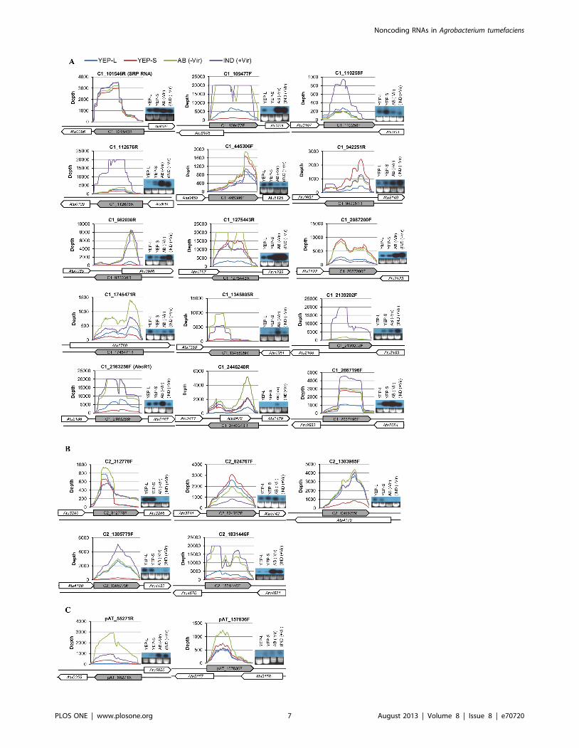
Validation of selected ncRNAsTo confirm the expression of the identified ncRNAs, we
employed two independent techniques: Northern blot analysis and
RACE. We validated a total of 36 ncRNAs. Northern blot analysis
confirmed the expression of 24 of 28 ncRNAs (Table 4). Twenty-
two representative ncRNAs are presented in Figure 3. RACE
Table 3. Comparison of two A. tumefaciens RNA-seq studies.
Number of ncRNAs
Category Wilms et al. [51] Our study CommonbGrandtotal
Total Uniquea Total Uniquea
sRNA 149c 113 101 66d 36 215
asRNA 76 32 354 310 44 386
59 UTRleader
3c 0 20 17 3 20
Total 228 145 475 393 83 621
aUnique ncRNAs were identified by one study but not by the other study.bCommon ncRNAs were identified by both RNA-seq studies.cThree sRNA identified by Wilms et al. [51] were found to be 59 UTR leaders inour study.dOne sRNA identified by our study overlaps with two sRNAs identified by Wilmset al. [51].doi:10.1371/journal.pone.0070720.t003
Noncoding RNAs in Agrobacterium tumefaciens
PLOS ONE | www.plosone.org 5 August 2013 | Volume 8 | Issue 8 | e70720

independently confirmed the expression of 16 of 18 ncRNAs
(Table S5 in File S1) and we present the results for 9 ncRNAs
found on the Ti plasmid (Figure S2 in File S2). Four ncRNAs were
validated by both methods. Fourteen of the 36 validated ncRNAs,
9 by Northern blot analysis and 5 by RACE, were identified for
the first time by this study.
Among the 24 ncRNAs validated with Northern blot analysis,
three were 59 UTR elements, 14 were sRNAs and 7 were asRNAs.
In most cases, the ncRNA sizes predicted by RNA sequencing
were consistent with Northern blot analysis results with an
exception of C1_10956F (thermosensor). This is because this
ncRNA was not transcribed as an independent transcript
(,227 nt) but was transcribed as part of downstream gene in all
four growth conditions (see below for detail). Two ncRNAs,
C1_112676R and C1_1345805R had two bands (Table 4;
Figure 3), suggesting that they might be transcribed from different
promoters or they might be processed to become mature
transcripts.
Analysis of cis-antisense RNAsInterestingly, while the expression level of all seven validated
asRNAs varied considerably under different growth conditions
(Table 4), the putative target mRNAs encoded on the comple-
mentary strand were not expressed at detectable levels or only
expressed at a very low level (,10 RPKM). For example, the
expression level of C1_109477F varied from 413 RPKM (YEP-S:
2TEX) to 37950 RPKM (IND: +TEX) as shown in Table 4, but
its putative target Atu0105 (hypothetical protein; Ref 56 & 57)
mRNA was not detectable in all eight cDNA libraries. Similarly,
the expression level of C1_982034R varied from 57 RPKM (YEP-
S: 2TEX) to 5579 RPKM (AB: +TEX), but its putative target
Atu0986 (hypothetical protein; Ref 56 & 57) was not expressed at
all.
To investigate whether there was a general trend between the
transcriptional levels of asRNAs and genes encoded on the
complementary strands, we performed a Pearson product-moment
correlation test. In a recent study, it has been shown that pervasive
asRNAs play an important role for degradation of sense mRNAs
by base-paring with them to form double stranded substrates of
RNase III [63]. Furthermore, the presence of promoters on the
opposite strands can affect expression of genes on the sense strand
via transcription interference [64,65]. The RPKM values of each
asRNA and its putative target gene on the complementary strand
were log-transformed before plotted. A Pearson product-moment
test (SPSS 17; SPSS Inc., USA) showed that there was no evident
correlation between the two (r2 = 0.02; Figure 4). Clearly, there
were many asRNAs with varying expression levels while their
putative target genes on the opposite strands were not expressed at
all. The lack of correlation might be attributed to the fact that
some asRNAs may have positive effects while others have negative
effects on target gene expression at the transcriptional level [52].
Alternatively, some of these so-called asRNAs may have their real
targets encoded somewhere else in the genome; thus they might be
trans-acting sRNAs. Because candidate asRNAs were named so
solely due to the presence of annotated genes on the opposite
strand, it is still possible that these ncRNAs may interact with other
mRNAs that have sufficient sequence complementarity, especially
when the genes encoded on the opposite strand are not expressed.
A third possibility is that some candidate asRNAs might be
protein-coding genes. We found that eight putative asRNAs
contained a putative open reading frame (ORF; indicated by 1 in
Table S3 in File S1). Because some of the annotated genes on the
opposite strand of these candidate asRNAs were not detectable in
all eight libraries, it is possible that the candidate asRNAs could be
the protein-coding genes and the annotated genes on the opposite
strand might represent pseudo genes.
As A. tumefaciens virulence is of great interest, it was intriguing to
find that some asRNAs were encoded on the opposite strands of
known virulence genes, such as virC2, virB9, virB10, virD3, virD4,
virE2 and virE3. To test if some of these asRNAs affect A. tumefaciens
virulence, we chose two asRNAs: pAt_157836F is antisense to
atsD, which might be important for bacterial attachment to plant
cells [66], and pTi_191667R is antisense to virB10 (Atu6176), an
essential component of the Type IV secretion system that
transports T-DNA into plant cells along with other effector
proteins [67]. We generated a knock-out mutant strain, DatsD, in
which the gene atsD and its antisense RNA pAt_157836F was
deleted. We also generated overexpression strains of A. tumefaciens
C58 that harbored replicating plasmid vectors carrying either the
sense or antisense strands of the asRNA pAt_157836F driven by a
constitutive promoter. Similarly, we made overexpression con-
structs for the sense and antisense sequences of pTi_191667R and
introduced them into the wild type C58.
Tobacco leaf disk assay, Arabidopsis root segment assay and
maize immature embryo transformation were performed as
previously described [68–70]. Overexpression of pTi_191667R
or its complementary sequence (anti-pTi_191667R) did not show
detectable effects on A. tumefaciens virulence (Figure S5A in File
S2). One explanation could be the limitation of the tobacco leaf
disc assays for the quantitative virulence measurement. It has been
suggested that bacterial small RNAs often have quantitative effects
on the target gene expression [71]. Tobacco leaf disk assay may
not be sensitive enough for measuring low level changes of A.
tumefaciens virulence. Another explanation could be that the real
target gene for pTi_191667R might not be its sense strand virB10
gene, but rather a gene elsewhere in the genome.
Overexpression or knockout mutation of pAt_157836F also did
not have significant effects on A. tumefaciens virulence measured by
Arabidopsis root segment assay (Figure S5B in File S2). However,
we observed marginally significant effects of the knockout
mutation of atsD and pAt_157836F (DatsD) on maize immature
embryo transformation frequency (Figure S5C in File S2; paired
sample t-test, P = 0.017). Future work is needed to determine
whether these ncRNAs have regulatory functions on other target
genes that may affect bacterial phenotypes other than T-DNA
delivery to plants.
Two 59 UTR elements function as a thermosensor and athi-box riboswitch
The two 59 UTR elements (C1_109596F and C1_2541934R)
were predicted to be trans-encoded sRNAs after initial screening,
but C1_109596F was located immediate upstream of a cold shock
protein (Atu0106: cspA) and C1_2541934R was found at the
upstream of a thiamine biosynthesis operon (thiCOGG). A RNA
family database search (Rfam: http://rfam.sanger.ac.uk/) suggest-
ed that they were homologous to a thermosensor (C1_109596F:
http://rfam.sanger.ac.uk/family/cspA) and a thiamine riboswitch
(C1_2541934R: http://rfam.sanger.ac.uk/family/TPP), respec-
tively. A thermosensor is a 59 UTR element of mRNAs and
regulates translation of downstream coding sequence [72]. The
secondary structure of a thermosensor changes depending on
ambient temperature, and regulates the accessibility of the mRNA
to ribosomes, thus affecting translation. One of the best studied
thermosensors is located at the 59 UTR of the global virulence
regulator of Listeria monocytogenes, prfA [73]. Our Northern blot
analysis suggested that C1_109596F is not expressed by itself
(,227 nt), but was transcribed as a 59 UTR of cspA (Figure 5).
Thus, the corresponding transcript of C1_109596F from Northern
Noncoding RNAs in Agrobacterium tumefaciens
PLOS ONE | www.plosone.org 6 August 2013 | Volume 8 | Issue 8 | e70720
Noncoding RNAs in Agrobacterium tumefaciens
PLOS ONE | www.plosone.org 7 August 2013 | Volume 8 | Issue 8 | e70720

blot analysis was about 503 nt, including the 227 nt 59 UTR,
210 nt coding sequence and 66 nt 39 UTR (Figure 5). These
results suggest that the thermosensor (C1_109596F) may post-
transcriptionally regulate cspA expression like its homolog in
Escherichia coli [74].
Riboswitches are located at the 59 UTRs of many bacterial
mRNAs and affect expression of downstream protein-coding
regions upon binding of metabolites [75]. When there are
sufficient metabolites, riboswitch-metabolite binding results in
conformational changes in the RNA secondary structure leading
to transcription termination by forming rho-independent termina-
Figure 3. Validation of selected ncRNAs by Northern blot analysis. Depth of coverage profiles and Northern hybridization images of 22Agrobacterium ncRNAs under four growth conditions: YEP medium until mid-log phase (YEP-L), YEP medium until late stationary phase (YEP-S), ABinduction medium without AS (AB), AB induction medium with AS (IND). (A) Fifteen ncRNAs encoded on the circular chromosome (C1), (B) fivencRNAs encoded on the linear chromosome (C2), and (C) two ncRNAs encoded on the pAt plasmid (pAt).doi:10.1371/journal.pone.0070720.g003
Table 4. Validated ncRNAs with Northern blot analysis.
RPKM
Position Size (nt) (2TEX) (+TEX)
ncRNA tag 59 end 39 endRNAseq
Northernblot YEP-L YEP-S AB IND YEP-L YEP-S AB IND antisense to
Circular chromosome
1 C1_101545R{
(SRP RNA)101545 101446 100 ,100 10697 14477 13806 9254 6999 5692 3723 3674 Small SRP
2 C1_109477F{ 109477 109594 118 ,120 1503 413 15354 24677 16287 3009 37950 34389 Atu0105
3 C1_109596F(thermosensor)
109596 109822 227 ,500 719 377 868 802 1346 1098 1443 1275 thermosensor
4 C1_110258F* 110258 110380 123 ,180 5420 5444 2955 3636 559 381 220 983 intergenic
5 C1_112676R{ 112676 112535 142 ,140 541 402 819 8686 5028 3404 3819 23172 intergenic
,100
6 C1_445306F 445306 445498 193 ,200 307 577 538 313 1377 2210 1519 776 intergenic
7 C1_942251R{ 942251 942016 236 ,220 233 1171 830 1080 636 3179 1046 1109 intergenic
8 C1_982030R{ 982030 981727 304 ,310 337 57 3986 5579 962 229 5155 4584 Atu0986
9 C1_1275443R{ 1275443 1275297 147 ,150 2713 12965 25899 11016 8574 36917 30473 16440 Atu1287
10 C1_1345805R{ 1345805 1345651 155 ,200 471 1602 3667 1634 454 1593 7684 4123 intergenic
,140
11 C1_1745471R{ 1745471 1745262 210 ,220 213 108 677 646 966 403 1676 851 Atu1760
12 C1_2087200F{ 2087200 2087384 185 ,200 1661 8580 6413 1243 6545 20617 11237 2333 intergenic
13 C1_2139202F{ 2139202 2139332 131 ,150 818 1387 1597 7564 644 902 2166 21501 intergenic
14 C1_2163256F{ 2163256 2163370 115 ,110 5043 1273 28677 25589 17172 4036 27964 27289 suhB ( = AbcR1)
15 C1_2446240R{ 2446240 2445919 322 ,330 476 1551 2583 1261 1032 3072 4164 1903 Atu2478
16 C1_2541934R*
(TPP RS)2541935 2541832 103 ,110 6422 13871 12566 5053 753 909 849 674 TPP riboswitch
(Atu2569, thiC)
17 C1_2667196F*{ 2667196 2667281 86 ,90 977 3379 1448 2276 2537 6206 4108 5534 Intergenic
Linear chromosome
1 C2_312778F(TPP RS)
312778 312932 155 ,150 337 566 510 281 582 481 636 463 TPP riboswitch(Atu3286)
2 C2_824767F 824767 824863 97 ,100 595 1432 642 259 5276 6043 2292 914 Intergenic
3 C2_1303985F 1303985 1304143 159 ,160 1597 430 1382 1800 7403 1648 4913 3853 Atu4179
4 C2_1305779F 1305779 1305881 103 ,120 1726 543 2406 3228 5564 580 3553 5343 Intergenic
5 C2_1831446F{ 1831446 1831607 162 ,160 689 272 3458 3970 10541 2865 25085 25357 Intergenic
At plasmid
1 pAT_55271R*{ 55271 55154 118 ,140 45 866 3597 877 105 717 3728 1042 Intergenic
2 pAT_157836F*{ 157836 158083 248 ,270 288 656 622 259 966 1279 1243 506 Atu5157 (atsD);Atu5158
*ncRNAs have been validated with 59 and 39 RACE.{ncRNAs have been previously identified or detected by Wilms et al. [51].F and R at the end of each ncRNA tag denote strand information: Forward and Reverse.doi:10.1371/journal.pone.0070720.t004
Noncoding RNAs in Agrobacterium tumefaciens
PLOS ONE | www.plosone.org 8 August 2013 | Volume 8 | Issue 8 | e70720

tor or to translation inhibition by masking the ribosomal binding
site [76,77]. Thiamine is an essential enzyme co-factor for carbon
metabolism in all living organisms. Bacteria, fungi and plants can
synthesize thiamine. The thi-box riboswitch, also known as TPP
(thiamine pyrophosphate) riboswitch (RF00059), directly binds to
TPP and regulates downstream gene expression by means of
premature transcription termination (attenuation) or translation
inhibition [78].
According to the Rfam database, there were three TPP
riboswitches in the A. tumefaciens C58 genome. Two TPP
riboswitches were identified as candidate ncRNAs in our data
set (C1_2541934R and C2_312778F) and the third one was also
represented in our data set when we manually examined the
predicted region in our files (Circular chromosome, 2700230–
2700340, reverse strand). C1_2541934R was located in the 59
UTR of an operon encoding proteins required for thiamine
biosynthesis, thiCOGG (Figure 6A; Table 5). To determine whether
this riboswitch is regulated by thiamine, as its homolog located at
the 59 UTR of thiCOGE in Rhizobium etli [78], we added thiamine
to modified AB induction medium without AS (AB) to a
concentration of 100 mg/mL. As can be seen in Figure 6, no thiC
expression was observed in lanes YEP-L and YEP-S (Figure 6B&C)
because YEP medium contains thiamine. Only the riboswitch
(,110 nt) was transcribed (Figure 6B), suggesting transcriptional
regulation of the thiCOGG operon. However, thiC was expressed in
the minimal medium (Figure 6B&C, lanes AB, IND and *AB) due
to the absence of thiamine in the medium. Addition of thiamine
clearly shut down transcription of downstream genes (Figure 6B&
C, *AB+Thi), suggesting that this leader element works as a thi-
box riboswitch. We also note that treating samples with the
RNAprotect Bacteria reagent (Qiagen, USA) before RNA
isolation can be important for stabilizing RNA molecules. Smaller
bands observed in lane *AB (not treated) and AB (treated) in
Figure 6C may represent degradation products of thiCOGG
mRNA, demonstrating fast turnover of bacterial mRNAs [79].
Notably, the riboswitch transcript (,110 nt) accumulated
during the stationary phase (Figure 6B, YEP-S; Table S3A in File
S1, C1_2541934R). The short transcript could be the truncated
by-product caused by transcriptional attenuation [80]. But given
that two S-adenosylmethionine (SAM) riboswitches, SreA and
SreB, act as trans-acting sRNAs in L. monocytogenes [81], it would be
worthwhile to examine if this thi-box riboswitch has additional
targets in trans.
Conclusion
We have generated a large date set consisting of over
840 million reads from 8 cDNA library representing four bacterial
growth conditions and two treatments for enhancing RNA-seq
quality (NCBI accession number, SRR747854). Depleting abun-
dant rRNAs improved RNA-seq detection sensitivity, leading to
the discovery of 384 novel ncRNAs. Our results show that
numerous ncRNAs are transcribed from the opposite strands of
many protein-coding genes as well as from the intergenic regions
of the A. tumefaciens genome. Intriguingly, many asRNAs were
discovered on the complementary strand of important virulence
genes and operons, such as virA, virB, virC, virD, and virE.
Furthermore, some candidate ncRNAs were differentially ex-
pressed when the cells are incubated with the vir gene inducer AS,
suggesting that the identified ncRNAs may play a role in virulence
regulation in A. tumefaciens. Whether these ncRNAs play crucial
roles for physiological and cellular responses has yet to be
elucidated, but their high abundance in the transcriptome suggests
that they may have functional roles. Accumulating evidence
Figure 4. Expression correlation between cis-antisense RNAsand putative target genes. Log-transformed RPKM data for 354Agrobacterium asRNAs were plotted against log-transformed RPKM dataof genes encoded on the complementary strand. Pearson product-moment coefficient was given (r2 = 0.02).doi:10.1371/journal.pone.0070720.g004
Figure 5. Expression profiling of a thermosensor, C1_109596Fand a major cold shock protein, cspA. The depth of coverage dataof the nucleotide positions of 109596–110198 on the Circularchromosome was plotted (+TEX). Northern blot analysis using a probespecific to the 59 UTR showed that cspA is transcribed as anapproximately 500 nt transcript, which was consistent with the RNA-seq results, 503 nt including 227 nt 59 UTR (109596–109822), 210 ntcspA (Atu0106) coding region (109823–110032), and 66 nt 39 UTR(110033–110098). YEP-L, YEP medium until mid-log phase, YEP-S, YEPmedium until late stationary phase, AB, AB induction medium withoutAS, IND, AB induction medium with AS.doi:10.1371/journal.pone.0070720.g005
Noncoding RNAs in Agrobacterium tumefaciens
PLOS ONE | www.plosone.org 9 August 2013 | Volume 8 | Issue 8 | e70720

strongly suggests that even tRNAs and protein-coding mRNAs can
have regulatory functions [14,82–85]. We speculate that future
studies on ncRNAs functions during Agrobacterium-plant interac-
tions will provide valuable tools to improve plant transformation
efficiency as well as better understanding of fundamental plant-
pathogen interactions.
Experimental Procedures
Media and bacterial growth conditionsA. tumefaciens C58 was grown at 28uC in YEP (10 g yeast extract,
10 g Bacto peptone, and 5 g NaCl per L, pH 7.0) or modified AB
induction medium [1 g NH4Cl, 0.3 g MgSO4?7H2O, 0.15 g KCl,
0.01 g CaCl2, 2.5 mg FeSO4?7H2O, 2 mM phosphate buffer (pH
5.6), 50 mM 2-(4-morpholinoo)-ethane sulfonic acid (MES), 0.5%
glucose per L, pH 5.6] with or without the vir gene inducer AS
(100 mM) [86]. Cultures for strains carrying plasmid vectors were
amended with appropriate antibiotics at the following concentra-
tions: kanamycin, 50 mg/ml; spectinomycin, 100 mg/ml. Viru-
lence gene induction was performed as described previously [86].
Briefly, A. tumefaciens cells were grown overnight in YEP medium
containing appropriate antibiotics, if carrying plasmid vectors, and
0.5 mL culture was transferred to 50 mL AB-sucrose minimal
medium containing appropriate antibiotics in a 250 mL flask. The
culture was incubated at 28uC on a shaker-incubator (250 rpm) for
about 16 hours. The bacterial densities were measured at OD600.
The cultures were centrifuged at 40006g for 10 min at room
temperature, resuspended in two volumes of induction medium
without AS (AB) and then divided equally (50 mL each) into two
sterile 250 mL flasks. For virulence induction, AS was added to a
final concentration of 100 mg/ml (IND) and incubated for
20 hours at 25uC (150 rpm).
Figure 6. Transcriptional regulation of thiCOGG operon by a TPP riboswitch (C1_2541934R). A putative riboswitch at the 59 UTR ofthiamine biosynthesis operon, thiCOGG, transcriptionally regulates gene expression. A) Secondary structure predicted by mFold web server [94]: DG= 235.08 kcal/mol. (B) Northern blot analysis with a probe specific to the riboswitch and (C) a probe specific to the downstream gene, thiC (C). TotalRNA was isolated from A. tumefaciens strain C58 grown in YEP medium until mid-log phase (YEP-L), YEP medium until late stationary phase (YEP-S),AB induction medium without AS (AB), AB induction medium with AS (IND), and AB with 100 mg/mL of thiamine (AB+Thi). +RP, RNA samples weretreated with RNAprotect Bacteria reagent (Qiagen, USA). *AB and *AB+Thi, RNA samples were not treated. Ethidiumbromide stained 16S rRNA bandswere included as loading control.doi:10.1371/journal.pone.0070720.g006
Table 5. A thi-box riboswitch and thiamine biosynthesis gene operon.
RPKM (2TEX) RPKM (+TEX)
Gene ID 59 end 39 endGenename Product YEP-L YEP-S AB IND YEP-L YEP-S AB IND
Atu2566 2538732 2537959 thiG thiazole synthase 0 0 28 117 0 0 47 114
Atu2567 2538934 2538737 thiG sulfur carrier proteinThiS
0 0 14 92 0 0 36 112
Atu2568 2539905 2538931 thiO thiamine biosynthesisoxidoreductase
0 0 28 107 4 0 65 179
Atu2569 2541730 2539907 thiC thiamine biosynthesisprotein ThiC
0 0 139 362 0 0 143 380
C1_2541934R 2541934 2541832 TPP riboswitch 6422 13871 12566 5053 753 909 849 674
doi:10.1371/journal.pone.0070720.t005
Noncoding RNAs in Agrobacterium tumefaciens
PLOS ONE | www.plosone.org 10 August 2013 | Volume 8 | Issue 8 | e70720

Whole transcriptome analysisRNA sample preparation. Total RNA was isolated from A.
tumefaciens cells using RNeasy Protect Bacteria mini kit (Qiagen,
USA) according to the manufacturer’s manual. Briefly, total RNA
was extracted from A. tumefaciens strain C58 grown under four
different growth conditions: YEP medium until mid-log phase
(YEP-L: OD600 = 0.5), YEP medium until late stationary phase
(YEP-S: OD600 = 1.3), modified AB induction medium without
AS (AB: OD600 = 0.8), AB induction medium with AS (IND:
OD600 = 0.8). One volume of A. tumefaciens culture (0.5,2.5 mL)
was mixed with two volumes of RNAprotect Bacteria reagent
(Qiagen, USA), vortexed vigorously for 5 seconds, let sit for 5 min,
and centrifuged for 10 min at 4000 x g at room temperature. Cell
pellet was suspended in 200 mL of lysing buffer (10 mM Tris,
1 mM EDTA, pH 8.0) containing 15 mg/mL of lysozyme (Sigma)
and incubated at 25uC for 10 min. Seven hundred mL of Buffer
RLT containing 10 mL/mL of beta-mercaptoethanol was added,
vortexed vigorously for 10 sec and centrifuged for 2 min at
150006g. Only the supernatant was carefully transferred to a new
tube and 500 mL of 100% ethanol was added and mixed by
pipetting. Sample was applied to a spin column, centrifuged for
15 sec at 60006g and the column was washed once with buffer
RW1 and twice with buffer RPE (Qiagen, USA). To elute RNA
from the column, 50 mL of RNase-free water was directly applied
to the center of the membrane and centrifuged for 1 min at
210006g. Contaminating DNA was removed by treating total
RNA with DNase I (Invitrogen, USA) and checked by PCR.
Because the extreme abundance of rRNAs, which often account
for 95–97% of total RNA in bacteria [26], is a major challenge for
transcriptome analysis, we used two commercial kits to deplete
rRNAs and tRNAs. A previous study demonstrated that rRNA
content can be reduced by up to 19% by treating total RNA with
MICROBExpressTM kit (Ambion, USA), and then with 59-
phosphate-dependent exonuclease (TEX; Epicentre, USA) [26].
That is, mRNA content was increased from 5% to 25%, a 5-fold
enrichment, which can improve mRNA detection sensitivity by up
to 230% [26]. The MICROBExpressTM kit uses hybridization
oligos which specifically capture 16S and 23S rRNAs. TEX
selectively digests processed RNA molecules with 59 mono-
phosphate, such as rRNAs, and is useful to enrich primary
transcripts. About 10 mg of total RNA was treated MICROBE-
xpressTM kit (Ambion, USA) as recommended by the kit manual,
and RNA integrity was checked by an Agilent 2100 BioAnalyzer.
RNA samples were then further treated with TEX to remove
remaining rRNAs and to enrich primary transcripts.
cDNA library preparation. cDNA libraries were prepared
and analyzed at Illumina (San Diego, CA). The detailed protocol
describes the steps for total RNA fragmentation, adapter ligation,
reverse transcription, PCR amplification, purification, cluster
generation and sequencing, and can be found in Illumina TruSeq
Small RNA SamplePrep Guide (#15004197).
All purified total-RNA samples were started out with 100 ng in
total volume of 16 ml. Each sample was treated with 2 ml of 5X
fragmentation buffer (EPF#15016648, Illumina) and was incu-
bated at 94uC for 4 minutes. Then the samples were cooled on ice.
The sample was mixed with a 7 ml master mix as following: 1 ml
of RNAseOUT (40 U/ml) from Epicentre or RNAse Inhibitor
(part#15003548, Illumina), 2 ml of T4 Polynucleotide Kinase
(PNK) (part#M0201S, NEB), 2 ml of 10X PNK Buffer
(part#M0201S, NEB), and 2 ml of 10 mM ATP (part#R109AT,
R109AT, Epicentre/Illumina). A 25 ml reaction mixture was
incubated at 37uC for 1 hour on a pre-heated thermal cycler. The
fragmented total-RNA samples (small RNA) were purified with
procedure 1 of the RNA clean & concentrator-5 (part#R1015,
Zymo Research), and were then eluted with 6 ml of RNase-free
water.
Five microliter of purified fragmented RNAs were ligated with
1 ml RNA 39 Adapter (RA3) (part# 15013207, Illumina); reactions
were heated at 70uC for 2 minutes, then immediately cooled on
ice. Next, a master mix of 4 ml was prepared as following before
adding to the 6 ml reaction: 2 ml Ligation Buffer (HML)
(part#15013206, Illumina), 1 ml RNase Inhibitor
(part#15003548, Illumina), and 1 ml T4 RNA Ligase 2 Deletion
Mutant (part# M0242S, NEB). The 10 ml reaction was incubated
on the pre-heated thermal cycler at 28uC for 1 hour. With the
reaction tube remaining on the thermal cycler, 1 ml Stop Solution
(STP) (part#15016304, Illumina) was added to the reaction tube
and mixed thoroughly by pipetting. Then the reaction mixture was
incubated at 28uC for additional 15 minutes.
One microliter of RNA 59 Adapter (RA5) (part#15013205,
Illumina) was added into the 11 ml of the 39 adapter ligation
reaction mixture, and the sample was denatured at 70uC for
2 minutes and immediately cooled on ice. One microliter of
10 mM ATP (part#15007432, Illumina) and 1 ml of 10U T4
RNA ligase (part#1000587, Illumina) were added into the
reaction to bring the final volume of 14 ml that was incubated at
28uC for another hour and then placed on ice.
For first strand cDNA synthesis, 6 ml of the 14 ml of 39 and 59
adapter ligated RNA samples was mixed with 1 ml RNA RT
Primer (RTP) (part#15013981, Illumina), denatured at 70uC for
2 minutes, and then immediately cooled on ice. A 5.5 ml of master
mix containing 2 ml 5X First Strand Buffer (part#18064-014,
Invitrogen), 0.5 ml 12.5 mM dNTP mix (dilute from 25 mM
dNTP mix, part #11318102, Illumina), 1 ml 100 mM DTT
(part#18064-014, Invitrogen), 1 ml RNAse Inhibitor
(part#15003548, Illumina), and 1 ml SuperScript II Reverse
Transcriptase (part#18064-014, Invitrogen) was added and
incubated at 50uC for 1 hour on a pre-heated thermal cycler.
A 50 ml PCR reaction was set up by adding 8.5 ml Ultra-Pure
Water (part#1001913, Illumina), 25 ml PCR Mix (PML)
(part#15022681, Illumina), 2 ml RNA PCR Primer (RP1)
(part#15013198, Illumina), and 2 ml RNA PCR Primer Index
(RPI1) (part#15013181, Illumina) into the 12.5 ml of the first
strand cDNA reaction. PCR reaction was carried out in a thermal
cycler with following profile: 98uC for 30 sec followed by 11 cycles
of 98uC for 10 sec, 60uC for 30 sec, 72uC for 15 sec, and a final
extension at 72uC for 10 min. The PCR product was held at 4uCuntil purification.
PCR products were purified using the Agencourt AMPure XP
beads (part#A63881, Beckman Coulter Genomics) and verified
with Agilent High Sensitivity DNA-1000 chip (part#5067-1504,
Agilent), and the molar concentration for each sample was
obtained. 10 pM of each cDNA library was used for clusters
generation on cBot and sequencing was performed on Illumina
sequencers (GAiix) with paired-end mode (2 x 50 bp). FASTQ files
were generated using bcl2fastq script from CASAVA pipeline
(Illumina).
Sequence alignment. For each of the 8 library data sets of
Illumina RNA reads, the Bowtie 2 program [45] was used to map
short reads in the data set onto the reference genome. Then the
SAMtools program [46] was used to pile mapped reads with a
mapping error rate of less than 1 in 10,000 along the reference
genome. The pileup step allowed us to compute, for each position
of each strand of the reference genome, its depth of coverage,
which is the number of correctly mapped reads in sense
orientation that cover the position.
Noncoding RNAs in Agrobacterium tumefaciens
PLOS ONE | www.plosone.org 11 August 2013 | Volume 8 | Issue 8 | e70720

TSS Mapping for protein-coding genesTranscriptional start sites (TSS) were identified as follows: 1)
sequence reads were mapped to the A. tumefaciens reference
genome (NC_003062.2, NC_003063.2, NC_003064.2,
NC_003065.3) using the Bowtie 2 program [45], 2) depth of
coverage (number of reads per nucleotide) for each nucleotide
position on all four replicons were computed using SAMtools [46],
3) RPKM for all annotated protein-coding genes were computed,
and 4) upstream regions of protein-coding genes that had
expression levels greater than 50 RPKM were inspected to
minimize erroneous annotations and a TSS was identified as a
nucleotide position where the depth starts to steeply increase with
a minimum value of 10.
Identification of non-coding RNAsNon-coding transcripts were identified as follows: 1) To find
regions of much higher coverage depths by Illumina RNA reads,
we selected the following regions sizes in bp: 800, 400, 200, 100,
and 50. 2) For each of the above region sizes and each strand of
the reference genome, a region of the size in the strand was
reported to a file as having much higher coverage depths by
Illumina RNA reads if the region has no overlap with any known
protein coding region and the total sum of coverage depths of the
region is at least 10 times higher than those of the non-overlapping
regions of the same size right before and after the region,
respectively. 3) Candidate ncRNAs were identified by manually
examining the file of reported regions of much higher coverage
depths and the file of all positions along with their coverage
depths. 4) Transcriptional start and stop sites for each candidate
ncRNA were determined based on the depth of coverage of
upstream and downstream region of the search window with a
minimum value of six.
Identification of differentially expressed ncRNAsDifferentially expressed ncRNAs were identified using the
Bioconductor DESeq package [61]. Firstly, the sequence reads
were mapped against the reference genome using Bowtie 2 [45].
Secondly, the SAMtools program [46] was used to pile mapped
reads and calculate depth of coverage for each nucleotide position.
Thirdly, read counts per gene (annotated genes and identified
ncRNAs) were computed for each sample (read count
~ADC|L
l; ADC, average depth of coverage = the number
of reads that mapped to a nucleotide position on a given
orientation, forward or reverse strand; L, length of a gene, l,
length of sequence read = 50). Lastly, read counts per gene were
normalized by effective library sizes using DESeq package and
differentially expressed ncRNAs were identified by comparing the
full generalized model (GLM: , treatment + TEX) against the
null model (GLM: , TEX) with a cut-off P-value of 0.05.
RACE39 RACE (Rapid Amplification of cDNA Ends) were conducted
as described previously [87]. Briefly, 10 mg of total RNA was
ligated with 500 pmol of RNA oligonucleotide E1 using T4 RNA
ligase (New England BioLabs Inc., USA) for 1 hr at 37uC and
purified with phenol-chloroform extraction. First strand cDNA
was synthesized using an oligonucleotide primer (59-
CATGCGGCCGCTAAGAAC-39) specific to E1 and Thermo-
Script First strand synthesis kit (Invitrogen, USA), and PCR
amplification was performed using E1-specific primer and a gene
specific primer. As a control, duplicate samples were set up
without reverse transcriptase (-RT). PCR products that were only
obtained with +RT treatment were cloned into TA-cloning vector
(5 PRIME Inc., USA) and their sequence was determined.
59 RACE was also carried out similarly as described above,
except that total RNA was treated with tobacco acid pyrophos-
phatase (TAP; Epicentre, USA) prior to 59 RNA adapter ligation
[88]. First strand cDNA was synthesized with either random
hexamers or a gene-specific primer and PCR amplification was
carried out with an adapter-specific primer and a gene-specific
primer. PCR products obtained only after TAP treatment
represent intact 59 ends, thus they were cloned and sequenced to
identify transcriptional start sites.
Northern blot analysisNorthern blot analysis was carried out using NorthernMaxH-
Gly kit (Ambion, USA) according to the manufacturer’s instruc-
tion. About 10 mg of total RNA was mixed with an equal volume
of Glyoxal loading dye and incubated at 50uC for 30 min before
loading. RNA MillenniumTM markers and RNA CenturyTM
markers (Ambion, USA) were loaded next to samples as size
references. After electrophoresis, RNA was transferred to
positively-charged membrane, UV cross linked, and hybridized
overnight at 37–42uC with oligonucleotide probes end-labeled
with 32P. Membranes were washed three times with washing
buffers, and then exposed to X-ray films for 1–4 days at 280uC.
Knock-out mutant generationAgrobacterium knock-out mutant was generated as previously
described [40]. Briefly, upstream and downstream flanking
sequences of atsD were PCR amplified (upstream: atsD-UP-F1-
SphI, atsD-UP-R1-SacII; downstream: atsD-DN-F1-SacII, atsD-
DN-R1-EcoRI; Table S6 in File S1), cloned into cloning vector (5
PRIME, USA), and sequenced. Flanking sequences without point
mutations were digested by restriction enzymes (upstream, SphI &
SacII; downstream, SacII & EcoRI), separated on an agarose gel,
and DNA bands were recovered from the gel and ligated to a
suicide vector pK19mobsacB (ATCC 87098) [89] digested with
SphI and EcoRI. After subcloning, atsD knockout plasmid was
introduced into A. tumefaciens C58 by electroporation. Kanamycin
resistant colonies were tested for sucrose sensitivity on LB medium
(10 g tryptone, 5 g yeast extract, and 10 g NaCl per L) containing
10% sucrose. Two sucrose-sensitive colonies were picked and
resuspended in 500 mL of LB broth, and 100 mL was spread on LB
plate with 10% sucrose and incubated at 28uC for two days.
Twenty to forty sucrose-resistant colonies were picked and tested
for kanamycin susceptibility. Finally, sucrose-resistant and kana-
mycin-susceptible colonies were PCR screened using the upstream
forward primer (atsD-UP-F1-SphI) and downstream reverse
primer (atsD-DN-R1-EcoRI), and the fragment was cloned and
sequenced to verify the deletion of atsD.
Overexpression plasmid vector constructionThe expression vector pTF505 (Figure S6 in File S2) was
constructed as follows. Firstly, two replication origins (pBR322 and
pVS1) were obtained from a binary vector pTF101.1 [90] by using
restriction enzymes BssHII and SphI. Secondly, the selectable
marker aadA (SpR) was PCR-amplified from pL3 [91] (PaadAT-F2
and TpsbANT-R2; Table S6 in File S1) and digested with BssHII
and KpnI. Thirdly, promotor-Multiple Cloning Sites (MCS)-
terminator cassette was prepared as follows. A constitutive
promoter PrrnC was predicted by a BLAST search using
Sinorhizobium meliloti PrrnC sequence (AF252864) [92]. The
146 bp PrrnC promoter (NC_003305.1, from 1041328 to
1041473) was PCR amplified (AtuPrrnC-F1-KpnI and
AtuPrrnC1-R-BglII; Table S6 in File S1), cloned into pPCV
Noncoding RNAs in Agrobacterium tumefaciens
PLOS ONE | www.plosone.org 12 August 2013 | Volume 8 | Issue 8 | e70720

cloning vector (5 PRIME, USA), and sequenced at ISU DNA
facility. Promoter activity was tested using mCherry [93] as a
reporter gene, and the transcription start site was confirmed by 59
RACE and sequencing. Oligonucleotides for multiple cloning sites
(MCS) (MCS-BEF1 and MCS-BER1; Table S6 in File S1) were
synthesized at ISU DNA facility, annealed, and treated with T4
PNK. Transcriptional terminator (TpsbA) was PCR-amplified
from pL3 using TpsbANT-F-EcoRI and TpsbANT-R-SphI
(Table S6 in File S1), cloned and sequenced. PrrnC and TpsbANT
were digested with restriction enzymes and ligated with MCS
using the T4 DNA ligase (Promega, USA). Then PrrnC-MCS-
TpsbANT cassette was PCR-amplified using AtuPrrnC-F1-KpnI
and TpsbANT-R-SphI, cloned into pPCV cloning vector and
sequenced. After KpnI and SphI digestion, PrrnC-MCS-TpsbANT
cassette was recovered and ligated with the replication origin
(pBR322 and pVS1) and aadA cassette using T4 DNA ligase
(Promega, USA). To overexpress atsD and sense or antisense
strands of pAt_157836F and pTi_191667R, PCR was carried out
using the primers listed in Table S6 in File S1, and the fragments
were cloned, sequenced, digested with restriction enzymes, and
ligated to pTF505.
Plant tumorigenicity assayTobacco leaf disk assay. Tobacco leaf disk assay was
performed using A. tumefaciens C58 strains overexpressing an
antisense RNA (pTi_191667R, antisense to virB10) or its
complementary sequence (anti-pTi_191667R) with/without its
native promoter (S/L) as previously described [69] with some
modifications. Shortly, Nicotiana tobacum (Petit Havana) seeds were
surface-sterilized using bleach and ethanol, and germinated on MS
medium [69]. One to two weeks after germination, seedlings were
transferred to magenta boxes containing germination medium and
incubated up to 1 month, under 18-h light at 24–28uC.
A. tumefaciens strains were inoculated into 5 mL YEP medium
and grown for 16 hours at 28uC (250 rpm). Two milliliters of
overnight culture was transferred to 50 mL of YEP medium in a
250 mL flask and allowed to grow for 6 to 8 hours until OD600
reached 0.8. Cells were harvested by centrifugation at 40006g,
and resuspended to an OD660 of 0.5 in liquid MS medium.
Inoculum was placed on ice until used.
For inoculation, tobacco leaf discs were prepared and inocu-
lated as previously described [69]. After inoculation, eighteen leaf
disks were carefully transferred onto MS medium and incubated
for 48 hours. Leaf disks were then transferred onto a fresh MS
medium containing 100 mg/L of cefotaxime. Tumor develop-
ments were monitored and the numbers of tumors on each leaf
disk were recorded three weeks after inoculation.Arabidopsis root segment transformation
assay. Arabidopsis root segment transformation assay was con-
ducted as described [68]. A. tumefaciens strains were inoculated into
5 mL YEP medium and grown overnight at 28uC. Two milliliter
of overnight culture was transferred to 50 mL of YEP medium and
allowed to grow for 6 to 8 hours until reached OD600 of 0.8 (109
cells/mL). Cells were harvested by centrifugation at 40006g,
washed once in 0.9% NaCl, and then resuspended in 0.9% NaCl
at a concentration of 108 cells/mL. Inoculum was placed on ice
until used.
For inoculation, Arabidopsis roots were cut into 0.3,0.5 cm
segments and transferred onto a Petri plate containing MS
medium. Two to three drops of bacterial inoculum was placed
onto root segments and left for 10 min. Bacterial suspension was
removed by a pipette and the Petri plates were sealed and
incubated for 48 hours in a growth chamber at 20uC. After
cocultivation, root segments were rinsed with sterile water
containing Timentin (100 mg/L). Root segments were transferred
onto Petri plates containing fresh MS medium. Sixty root
segments were used for each strain and the numbers of root
segments with tumors were recorded three weeks after inoculation.
Experiments were repeated 2–6 times for each strain.
Maize immature embryo transformation. Maize imma-
ture embryo transformation was conducted using disarmed
Agrobacterium strain EHA105 and atsD/pAt_157836F knockout
strain carrying a binary vector pTF101.1 [90] according to the
published protocol [70]. For each infection experiment, 5 to 8
maize ears were used and 31 to 40 immature embryos were
harvested from each ear; thus about 200 maize immature embryos
were used for both strains. A total of three independent infection
experiments were performed for this side-by-side comparison.
Supporting Information
File S1 Table S1, Comparision of expression fold change of vir
genes in a microarray study and two RNA-seq studies; Table S2,
TSS-mapping; Table S3, List of identified candidate ncRNAs on
all four replicons; Table S4, Differentially expressed candidate
ncRNAs; Table S5, Selected ncRNAs for 59 and 39 RACE;
Table S6, Oligonucleotides used in this study.
(PDF)
File S2 Figure S1, Effects of primary transcript enrichment by
terminator 59-phosphate-dependent exonuclease; Figure S2, 59
and 39 RACE for ncRNAs on Ti plasmid; Figure S3, Expression
profiling of virD4* internal transcript with primary transcript
enrichment (+TEX); Figure S4, Expression profiling of C3 and
Ti2; Figure S5, Effects of two antisense RNAs (pTi_191667R and
pAt_157836F) on Agrobacterium virulence; Figure S6, Map of the
expression vector pTF505.
(PDF) Chi
Acknowledgments
KL, CY and KW thank Q. Gan and S. Martin-Ortigosa for their assistance
in tumorigenesis assays of tobacco and Arabidopsis, B. Frame for maize
immature embryo transformation experiments, C. Eisley for technical
assistance during data analysis, and H-H. Chou and H. Shou for helpful
discussions. Authors also thank I. Farran at the Agrobiotechnology Institute
(UPNA-CSIC-Gobierno de Navarra) for providing the plasmid pL3.
Author Contributions
Conceived and designed the experiments: KW KL. Performed the
experiments: KL CY DL VH KN. Analyzed the data: KL XH.
Contributed reagents/materials/analysis tools: KW JBF XH. Wrote the
paper: KL KW. Wrote script used in analysis: XH.
References
1. Gripenland J, Netterling S, Loh E, Tiensuu T, Toledo-Arana A, et al. (2010)
RNAs: regulators of bacterial virulence. Nat Rev Micro 8: 857–866.
2. Duhring U, Axmann IM, Hess WR, Wilde A (2006) An internal antisense RNA
regulates expression of the photosynthesis gene isiA. Proc Natl Acad Sci USA
103: 7054.
3. Vecerek B, Moll I, Afonyushkin T, Kaberdin V, Blasi U (2003) Interaction of the
RNA chaperone Hfq with mRNAs: direct and indirect roles of Hfq in iron
metabolism of Escherichia coli. Mol Microbiol 50: 897–909.
4. Krulwich TA, Sachs G, Padan E (2011) Molecular aspects of bacterial pH
sensing and homeostasis. Nat Rev Micro 9: 330–343.
Noncoding RNAs in Agrobacterium tumefaciens
PLOS ONE | www.plosone.org 13 August 2013 | Volume 8 | Issue 8 | e70720
5. Johansson J, Mandin P, Renzoni A, Chiaruttini C, Springer M, et al. (2002) An
RNA Thermosensor Controls Expression of Virulence Genes in Listeria
monocytogenes. Cell 110: 551–561.
6. Figurski D, Helinski D (1979) Replication of an origin-containing derivative of
plasmid RK2 dependent on a plasmid function provided in trans. Proc Natl Acad
Sci USA 76: 1648–1652.
7. Tomizawa J (1984) Control of ColE1 plasmid replication: the process of binding
of RNA I to the primer transcript. Cell 38: 861.
8. Tomizawa J, Som T (1984) Control of ColE1 plasmid replication: enhancement
of binding of RNA I to the primer transcript by the Rom protein. Cell 38: 871.
9. Malmgren C, Wagner EGH, Ehresmann C, Ehresmann B, Romby P (1997)
Antisense RNA control of plasmid R1 replication. The dominant product of the
antisense RNA-mRNA binding is not a full RNA duplex. J Biol Chem 272:
12508.
10. Ji Y, Marra A, Rosenberg M, Woodnutt G (1999) Regulated antisense RNA
eliminates alpha-toxin virulence in Staphylococcus aureus infection. J Bacteriol 181:
6585.
11. Kawano M, Aravind L, Storz G (2007) An antisense RNA controls synthesis of
an SOS-induced toxin evolved from an antitoxin. Mol Microbiol 64: 738.
12. Fozo EM, Kawano M, Fontaine F, Kaya Y, Mendieta KS (2008) Repression of
small toxic protein synthesis by the Sib and OhsC small RNAs. Mol Microbiol
70: 1076.
13. Saad NY, Schiel B, Braye M, Heap JT, Minton NP, et al. (2012) Riboswitch (T-
Box)-mediated control of tRNA-dependent amidation in Clostridium acetobutylicum
rationalizes gene and pathway redundancy for asparagine and asparaginyl-
tRNAAsn synthesis. J Biol Chem.
14. Green NJ, Grundy FJ, Henkin TM (2010) The T box mechanism: tRNA as a
regulatory molecule. FEBS Letters 584: 318–324.
15. Masse E, Salvail H, Desnoyers G, Arguin M (2007) Small RNAs controlling iron
metabolism. Curr Opin Microbiol 10: 140.
16. Guillier M, Gottesman S (2008) The 59 end of two redundant sRNAs is involved
in the regulation of multiple targets, including their own regulator. Nucleic Acids
Research 36: 6781–6794.
17. Gottesman S, Storz G (2010) Bacterial small RNA regulators: versatile roles and
rapidly evolving variations. Cold Spring Harb Perspect Biol.
18. Valverde C, Livny J, Schluter JP, Reinkensmeier J, Becker A, et al. (2008)
Prediction of Sinorhizobium meliloti sRNA genes and experimental detection in
strain 2011. BMC Genomics 9: 416.
19. Alkan C, Karakoc E, Nadeau JH, Sahinalp SC, Zhang K (2006) RNA-RNA
interaction prediction and antisense RNA target search. J Comput Biol 13: 267–
282.
20. Livny J, Brencic A, Lory S, Waldor MK (2006) Identification of 17 Pseudomonas
aeruginosa sRNAs and prediction of sRNA-encoding genes in 10 diverse
pathogens using the bioinformatic tool sRNAPredict2. Nucleic Acids Res 34:
3484–3493.
21. Sharma CM, Vogel J (2009) Experimental approaches for the discovery and
characterization of regulatory small RNA. Current Opinion in Microbiology 12:
536–546.
22. Sharma CM, Hoffmann S, Darfeuille F, Reignier J, Findeiss S, et al. (2010) The
primary transcriptome of the major human pathogen Helicobacter pylori.
Nature 464: 250–255.
23. Mitschke J, Georg J, Scholz I, Sharma CM, Dienst D, et al. (2011) An
experimentally anchored map of transcriptional start sites in the model
cyanobacterium Synechocystis sp. PCC6803. Proc Natl Acad Sci U S A 108:
2124–2129.
24. Schluter JP, Reinkensmeier J, Daschkey S, Evguenieva-Hackenberg E, Janssen S
(2010) A genome-wide survey of sRNAs in the symbiotic nitrogen-fixing alpha-
proteobacterium Sinorhizobium meliloti. BMC Genomics 11: 245.
25. Vijayan V, Jain I, O’Shea E (2011) A high resolution map of a cyanobacterial
transcriptome. Genome Biology 12: R47.
26. He S, Wurtzel O, Singh K, Froula JL, Yilmaz S, et al. (2010) Validation of two
ribosomal RNA removal methods for microbial metatranscriptomics. Nat Meth
7: 807–812.
27. Guell M, Yus E, Lluch-Senar M, Serrano L (2011) Bacterial transcriptomics:
what is beyond the RNA horiz-ome? Nat Rev Micro 9: 658–669.
28. Li H, Homer N (2010) A survey of sequence alignment algorithms for next-
generation sequencing. Briefings in Bioinformatics 11: 473–483.
29. Tzfira T, Citovsky V, editors (2008) Agrobacterium: From Biology to Biotechnol-
ogy. New York: Springer Science+Business Media, LLC. 735 p.
30. Pitzschke A, Hirt H (2010) New insights into an old story: Agrobacterium-induced
tumour formation in plants by plant transformation. EMBO J 29: 1021–1032.
31. Hu X, Zhao J, DeGrado WF, Binns AN (2013) Agrobacterium tumefaciens
recognizes its host environment using ChvE to bind diverse plant sugars as
virulence signals. Proceedings of the National Academy of Sciences 110: 678–
683.
32. Banta LM, Joerger RD, Howitz VR, Campbell AM, Binns AN (1994) Glu-255
outside the predicted ChvE binding-site in VirA is crucial for sugar
enhancement of acetosyringone perception by Agrobacterium tumefaciens. Journal
of Bacteriology 176: 3242–3249.
33. He F, Nair GR, Soto CS, Chang Y, Hsu L, et al. (2009) Molecular basis of ChvE
function in sugar binding, sugar utilization, and virulence in Agrobacterium
tumefaciens. Journal of Bacteriology 191: 5802–5813.
34. Jin S, Roitsch T, Ankenbauer RG, Gordon MP, Nester EW (1990) The VirA
protein of Agrobacterium tumefaciens is autophosphorylated and is essential for vir
gene regulation. Journal of Bacteriology 172: 525–530.
35. Pazour GJ, Das A (1991) Characterization of the VirG binding site of
Agrobacterium tumefaciens. Nucleic Acids Research 19: 1358–1358.
36. Yuan Z-C, Liu P, Saenkham P, Kerr K, Nester EW (2008) Transcriptome
profiling and functional analysis of Agrobacterium tumefaciens reveals a general
conserved response to acidic conditions (pH 5.5) and a complex acid-mediated
signaling involved in Agrobacterium-plant Interactions. Journal of Bacteriology
190: 494–507.
37. Yuan Z-C, Haudecoeur E, Faure D, Kerr KF, Nester EW (2008) Comparative
transcriptome analysis of Agrobacterium tumefaciens in response to plant signal
salicylic acid, indole-3-acetic acid and c-amino butyric acid reveals signalling
cross-talk and Agrobacterium–plant co-evolution. Cellular Microbiology 10: 2339–
2354.
38. Klusener S, Hacker S, Tsai Y-L, Bandow J, Gust R, et al. (2010) Proteomic and
transcriptomic characterization of a virulence-deficient phosphatidylcholine-
negative Agrobacterium tumefaciens mutant. Molecular Genetics and Genomics 283:
575–589.
39. Chai Y, Winans SC (2005) A small antisense RNA downregulates expression of
an essential replicase protein of an Agrobacterium tumefaciens Ti plasmid. Mol
Microbiol 56: 1574–1585.
40. Wilms I, Voss B, Hess WR, Leichert LI, Narberhaus F (2011) Small RNA-
mediated control of the Agrobacterium tumefaciens GABA binding protein.
Molecular Microbiology 80: 492–506.
41. Wilms I, Moller P, Stock AM, Gurski R, Lai EM, et al. (2012) Hfq influences
multiple transport systems and virulence in the plant pathogen Agrobacterium
tumefaciens. J Bacteriol 194: 5209–5217.
42. Stachel SE, Messens E, Vanmontagu M, Zambryski P (1985) Identification of
the signal molecules produced by wounded plant-cells that activate T-DNA
transfer in Agrobacterium tumefaciens. Nature 318: 624–629.
43. Evguenieva-Hackenberg E (2005) Bacterial ribosomal RNA in pieces. Molecular
Microbiology 57: 318–325.
44. Croucher NJ, Fookes MC, Perkins TT, Turner DJ, Marguerat SB, et al. (2009)
A simple method for directional transcriptome sequencing using Illumina
technology. Nucleic Acids Research 37: e148.
45. Langmead B, Salzberg SL (2012) Fast gapped-read alignment with Bowtie 2.
Nat Meth 9: 357–359.
46. Li H, Handsaker B, Wysoker A, Fennell T, Ruan J, et al. (2009) The sequence
alignment/map format and SAMtools. Bioinformatics 25: 2078–2079.
47. Bullard J, Purdom E, Hansen K, Dudoit S (2010) Evaluation of statistical
methods for normalization and differential expression in mRNA-Seq experi-
ments. BMC Bioinformatics 11: 94.
48. Winans SC, Kerstetter RA, Nester EW (1988) Transcriptional regulation of the
virA and virG genes of Agrobacterium tumefaciens. Journal of Bacteriology 170: 4047–
4054.
49. Gygi SP, Rochon Y, Franza BR, Aebersold R (1999) Correlation between
protein and mRNA abundance in yeast. Molecular and Cellular Biology 19:
1720–1730.
50. Greenbaum D, Colangelo C, Williams K, Gerstein M (2003) Comparing protein
abundance and mRNA expression levels on a genomic scale. Genome Biology 4:
117.
51. Wilms I, Overloper A, Nowrousian M, Sharma CM, Narberhaus F (2012) Deep
sequencing uncovers numerous small RNAs on all four replicons of the plant
pathogen Agrobacterium tumefaciens. RNA Biology 9: 446–457.
52. Georg J, Hess WR (2011) cis-antisense RNA, another level of gene regulation in
bacteria. Microbiol Mol Biol Rev 75: 286–300.
53. Weinberg Z, Barrick JE, Yao Z, Roth A, Kim JN, et al. (2007) Identification of
22 candidate structured RNAs in bacteria using the CMfinder comparative
genomics pipeline. Nucleic Acids Research 35: 4809–4819.
54. Vesper O, Amitai S, Belitsky M, Byrgazov K, Kaberdina Anna C, et al. (2011)
Selective translation of leaderless mRNAs by specialized ribosomes generated by
MazF in Escherichia coli. Cell 147: 147–157.
55. Slater SC, Goldman BS, Goodner B, Setubal JC, Farrand SK, et al. (2009)
Genome sequences of three Agrobacterium biovars help elucidate the evolution of
multichromosome genomes in bacteria. Journal of Bacteriology 191: 2501–2511.
56. Wood DW, Setubal JC, Kaul R, Monks DE, Kitajima JP, et al. (2001) The
genome of the natural genetic engineer Agrobacterium tumefaciens C58. Science 294:
2317–2323.
57. Goodner B, Hinkle G, Gattung S, Miller N, Blanchard M, et al. (2001) Genome
sequence of the plant pathogen and biotechnology agent Agrobacterium tumefaciens
C58. Science 294: 2323–2328.
58. Clark MB, Amaral PP, Schlesinger FJ, Dinger ME, Taft RJ, et al. (2011) The
reality of pervasive transcription. PLoS Biol 9: e1000625.
59. Dornenburg JE, DeVita AM, Palumbo MJ, Wade JT (2010) Widespread
antisense transcription in Escherichia coli. MBio 1: e00024.
60. Venkova-Canova T, Patek M, Nesvera J (2003) Control of rep gene expression in
plasmid pGA1 from Corynebacterium glutamicum. Journal of Bacteriology 185:
2402–2409.
61. Anders S, Huber W (2010) Differential expression analysis for sequence count
data. Genome Biology 11: R106.
62. Pazour GJ, Das A (1990) Characterization of the VirG binding site of
Agrobacterium tumefaciens. Nucleic Acids Research 18: 6909–6913.
Noncoding RNAs in Agrobacterium tumefaciens
PLOS ONE | www.plosone.org 14 August 2013 | Volume 8 | Issue 8 | e70720

63. Lasa I, Toledo-Arana A, Dobin A, Villanueva M, de los Mozos IR, et al. (2011)
Genome-wide antisense transcription drives mRNA processing in bacteria.Proceedings of the National Academy of Sciences 108: 20172–20177.
64. Gullerova M, Proudfoot NJ (2010) Transcriptional interference and gene
orientation in yeast: noncoding RNA connections. Cold Spring Harb SympQuant Biol 75: 299–311.
65. Shearwin KE, Callen BP, Egan JB (2005) Transcriptional interference: a crashcourse. Trends Genet 21: 339.
66. Matthysse AG, Yarnall H, Boles SB, McMahan S (2000) A region of the
Agrobacterium tumefaciens chromosome containing genes required for virulence andattachment to host cells. Biochimica et Biophysica Acta (BBA) – Gene Structure
and Expression 1490: 208–212.67. Cascales E, Christie PJ (2004) Agrobacterium VirB10, an ATP energy sensor
required for type IV secretion. Proceedings of the National Academy of Sciencesof the United States of America 101: 17228–17233.
68. Gelvin SB (2006) Agrobacterium transformation of Arabidopsis thaliana roots: A
quantitative assay. In: Wang K, editor. Agrobacterium protocols. Second ed. NewJersey, USA: Humana Press Inc. 105–113.
69. Clemente T (2006) Nicotiana (Nicotiana tobaccum, Nicotiana benthamiana). In: WangK, editor. Agrobacterium protocols. Second ed. New Jersey, USA: Humana Press
Inc. 143–154.
70. Frame BR, Paque T, Wang K (2006) Maize (Zea mays L.). In: Wang K, editor.Agrobacterium protocols. 2 ed. Totowa, New Jersey: Humana Press Inc. 185–199.
71. Levine E, Zhang Z, Kuhlman T, Hwa T (2007) Quantitative characteristics ofgene regulation by small RNA. PLoS Biol 5: e229.
72. Narberhaus F, Waldminghaus T, Chowdhury S (2006) RNA thermometers.FEMS Microbiology Reviews 30: 3–16.
73. Johansson J, Mandin P, Renzoni A, Chiaruttini C, Springer M, et al. (2002) An
RNA thermosensor controls expression of virulence genes in Listeria monocytogenes.
Cell 110: 551–561.
74. Giuliodori AM, Di Pietro F, Marzi S, Masquida B, Wagner R, et al. (2010) ThecspA mRNA is a thermosensor that modulates translation of the cold-shock
protein CspA. Molecular Cell 37: 21–33.
75. Henkin TM (2008) Riboswitch RNAs: using RNA to sense cellular metabolism.Genes Dev 22: 3383–3390.
76. Toledo-Arana A, Dussurget O, Nikitas G, Sesto N, Guet-Revillet H (2009) TheListeria transcriptional landscape from saprophytism to virulence. Nature 459:
950.77. Hollands K, Proshkin S, Sklyarova S, Epshtein V, Mironov A, et al. (2012)
Riboswitch control of Rho-dependent transcription termination. Proceedings of
the National Academy of Sciences 109: 5376–5381.78. Miranda-Rıos J, Navarro M, Soberon M (2001) A conserved RNA structure (thi
box) is involved in regulation of thiamin biosynthetic gene expression in bacteria.Proceedings of the National Academy of Sciences 98: 9736–9741.
79. Belasco JG, Biggins CF (1988) Mechanisms of mRNA decay in bacteria: a
perspective. Gene 72: 15–23.
80. Naville M, Gautheret D (2010) Transcription attenuation in bacteria: theme and
variations. Briefings in Functional Genomics 9: 178–189.
81. Loh E, Dussurget O, Gripenland J, Vaitkevicius K, Tiensuu T, et al. (2009) A
trans-acting riboswitch controls expression of the virulence regulator PrfA in
Listeria monocytogenes. Cell 139: 770–779.
82. Gimpel M, Heidrich N, Mader U, Krugel H, Brantl S (2010) A dual-function
sRNA from B. subtilis: SR1 acts as a peptide encoding mRNA on the gapA
operon. Mol Microbiol 76: 990–1009.
83. Wadler CS, Vanderpool CK (2007) A dual function for a bacterial small RNA:
SgrS performs base pairing-dependent regulation and encodes a functional
polypeptide. Proc Natl Acad Sci U S A 104: 20454–20459.
84. Liu Y, Wu N, Dong J, Gao Y, Zhang X, et al. (2010) SsrA (tmRNA) acts as an
antisense RNA to regulate Staphylococcus aureus pigment synthesis by base pairing
with crtMN mRNA. FEBS Lett 584: 4325–4329.
85. Dorazi R (2003) Can tRNAs act as antisense RNA? The case of mutA and
dnaQ. J Theor Biol 225: 383–388.
86. Gelvin SB (2006) Agrobacterium virulence gene induction. In: Wang K, editor.
Agrobacterium Protocols. Second ed. New Jersey, USA: Humana Press Inc. 77–84.
87. Argaman L, Hershberg R, Vogel J, Bejerano G, Wagner EGH (2001) Novel
small RNA-encoding genes in the intergenic regions of Escherichia coli. Curr
Microbiol 11: 941.
88. Gerhart E, Wagner H, Vogel J (2005) Approaches to identify novel non-
messenger RNAs in bacteria and to investigate their biological functions:
functional analysis of identified non-mRNAs: Wiley-VCH Verlag GmbH. 614–
642 p.
89. Schafer A, Tauch A, Jager W, Kalinowski J, Thierbach G, et al. (1994) Small
mobilizable multi-purpose cloning vectors derived from the Escherichia coli
plasmids pK18 and pK19: selection of defined deletions in the chromosome of
Corynebacterium glutamicum. Gene 145: 69–73.
90. Paz M, Shou H, Guo Z, Zhang Z, Banerjee A, et al. (2004) Assessment of
conditions affecting Agrobacterium -mediated soybean transformation using the
cotyledonary node explant. Euphytica 136: 167–179.
91. Farran I, Rıo-Manterola F, Iniguez M, Garate S, Prieto J, et al. (2008) High-
density seedling expression system for the production of bioactive human
cardiotrophin-1, a potential therapeutic cytokine, in transgenic tobacco
chloroplasts. Plant Biotechnology Journal 6: 516–527.
92. Gustafson AM, O’Connell KP, Thomashow MF (2002) Regulation of
Sinorhizobium meliloti 1021 rrnA-reporter gene fusions in response to cold shock.
Can J Microbiol 48: 821–830.
93. Nelson BK, Cai X, Nebenfuhr A (2007) A multicolored set of in vivo organelle
markers for co-localization studies in Arabidopsis and other plants. The Plant
Journal 51: 1126–1136.
94. Zuker M (2003) Mfold web server for nucleic acid folding and hybridization
prediction. Nucleic Acids Research 31: 3406–3415.
Noncoding RNAs in Agrobacterium tumefaciens
PLOS ONE | www.plosone.org 15 August 2013 | Volume 8 | Issue 8 | e70720