Embed Size (px)
Citation preview
Communication 1
A head and neck squamous cell carcinoma model to 2
study and target HPV replication and transcription 3
Michael R. Evans 1, Christian T. Fontan1, Claire D. James1, Xu Wang1, Iain M. Morgan1,2, and 4 Molly L. Bristol1,* 5
1 VCU Philips Institute for Oral Health Research, Department of Oral and Craniofacial Molecular Biology, 6 Virginia Commonwealth University School of Dentistry, Richmond, VA 23298, USA 7
2 VCU Massey Cancer Center, Richmond, VA 23298, USA 8 * Correspondence: [email protected]; Tel.: +1-804-828-5702 9
Abstract: The incidence of human papillomavirus-related head and neck squamous cell carcinoma 10 (HPV+HNSCC) has reached epidemic levels in the last decade. While prophylactic vaccines will 11 prevent future HPV infections, there are currently no HPV-specific antiviral drugs to treat current 12 HPV infections or HPV+HNSCC. HPV replication and transcription are promising targets for 13 anti-HPV therapeutics, as modulation of these processes can alter expression levels of HPV E6 and 14 E7, which are required for maintenance of the transformed phenotype. This is a particularly 15 attractive target in in HPV+HNSCC where the majority of tumors have episomal genomes 16 replicating in an E1-E2 dependent manner. Here, we describe a model system to study HPV16 17 E1-E2 mediated DNA replication and HPV16 E2-mediated transcriptional activation and 18 repression in multiple HNSCC cell lines. Our results demonstrate that low levels of IFIT1 are 19 required for HPV16 replication in HNSCC cell lines and HPV16 E1 interacts with IFIT1. Restoration 20 of IFIT1 expression in HNSCC cell lines partially inhibits HPV16 E1-E2 mediated replication. This 21 system can be used to study replication and transcription by HPV16 E1 and E2 in HNSCC as well 22 as be utilized to screen potential anti-HPV therapeutics that target HPV16 replication and 23 transcription. 24
Keywords: papillomaviruses; HPV; replication; transcription; E1; E2; IFIT1; cancer; HNSCC 25 26
1. Introduction 27
Human papillomaviruses (HPVs) are double-stranded DNA viruses that cause a variety of diseases 28 ranging from warts to cancer [1]. HPV is the most common sexually transmitted infection in the 29 United States, with an estimated 80 percent of sexually active adults acquiring a HPV-related 30 infection in their lifetime [2]. Of the high-risk HPVs known to be causative in the development of 31 cancer, HPV16 is the most prevalent genotype [3]. HPV16 is causative in around 50% of cervical 32 cancers and nearly 90% of HPV-related head and neck squamous cell carcinomas (HPV+HNSCCs) 33 [1,3]. The prevalence of HPV+ HNSCCs has been rapidly increasing in the last decade, with over half 34 a million new cases per year worldwide [4]. While prophylactic vaccines are available that will 35 reduce future HPV16 disease burden by preventing future HPV16 infections, current vaccines are 36 not therapeutic and do not benefit patients with current HPV infections. Additionally, overall 37 vaccine coverage is low, with less than half of American adolescents receiving all of the 38 recommended doses of HPV vaccines [5]. Further understanding of HPV16 and its life cycle in the 39 head and neck region is needed in order to develop novel anti-viral therapies against HPV16. 40
In HPV infected cells, the E6 and E7 oncoproteins degrade p53 and pRb leading to cellular 41 proliferation, prevention of apoptosis and, over time, cellular transformation [6-14]. Reducing E6 42
Preprints (www.preprints.org) | NOT PEER-REVIEWED | Posted: 30 January 2019
© 2019 by the author(s). Distributed under a Creative Commons CC BY license.
Preprints (www.preprints.org) | NOT PEER-REVIEWED | Posted: 30 January 2019 doi:10.20944/preprints201901.0298.v1
© 2019 by the author(s). Distributed under a Creative Commons CC BY license.

2 of 18
and E7 levels induces apoptosis or senescence of HPV-transformed cells via restoration of p53 and 43 pRb, making these viral oncoproteins a viable target for anti-HPV therapeutics [15-22]. One way to 44 reduce E6 and E7 levels, particularly in HPV+HNSCC, is targeting viral replication. Contrary to 45 HPV+ cervical cancers, recent evidence suggests that the HPV genome remains episomal in a 46 majority of HPV+ HNSCCs, and is replicated in a E1-E2 dependent manner [23-26]. Reducing E1-E2 47 dependent replication in HPV+HNSCC would reduce viral genome levels, thereby reducing 48 transcript levels and subsequently the protein levels of E6 and E7. To our knowledge, no 49 characterized system exists to study HPV16 E1-E2 mediated replication in HNSCC. In addition to 50 targeting replication, directly targeting transcription of E6 and E7 is also a viable approach to 51 reducing the levels of these oncoproteins in HPV+HNSCC. Numerous studies have shown that 52 adding exogenous E2 (a negative regulator of E6/E7 transcription when expressed at high levels) to 53 HPV-transformed cervical cancer cells leads to their apoptosis or senescence, presumably due to a 54 decrease in E6 and E7 levels [27-31]. This is supported by work demonstrating that silencing E6 or E7 55 in HPV-transformed cervical cancer cell lines leads to similar effects [15-18, 20]. Further 56 investigation of HPV16 transcription in HNSCC is needed to determine the efficacy of similar 57 approaches in HNSCC as well to determine potential novel strategies for targeting E6 and E7 58 transcription in these cancers. 59
Here, we describe a model system to investigate HPV16 E2-mediated transcription and HPV16 60 E1-E2 mediated DNA replication in multiple HNSCC cell lines. We observe E2-dependent 61 transcriptional activation and repression in HN-22 and HN-31 HNSCC cell lines, the pattern of 62 which may be unique to HNSCC. We also observe detectable, reproducible amounts of HPV16 63 E1-E2 mediated replication in these lines. Performing the E1-E2 mediated replication assay in 64 multiple HNSCC cell lines, we discovered that high levels of endogenous interferon induced protein 65 with tetratricopeptide repeats 1 (IFIT1) prevents measurable E1-E2 DNA replication (observed in 66 nu61, SCC-61, HN-11, and HN-30). Adding exogenous IFIT1 to HN-22 (no detectable endogenous 67 IFIT1) and performing immunoprecipitation experiments revealed an interaction between IFIT1 and 68 HPV16 E1. This E1-IFIT1 interaction reduced E1-E2 mediated replication. Overall, this model system 69 can be used to study HPV16 E1-E2 mediated replication and E2-dependent transcription in HNSCC 70 cells and could be used as a tool to screen replication and transcription targeting anti-HPV agents. 71
2. Materials and Methods 72
2.1 HNSCC cell lines and cell culture 73
The HN-4, HN-8, HN-11, HN-22, HN-30, and HN-31 cell lines have been described previously [32]. 74 SCC-61 and radioresistant nu61 cell lines have also been previously described [33]. All cell lines were 75 grown in Dulbecco’s Modified Eagle’s Medium (Invitrogen) supplemented with 10% fetal bovine 76 serum (VWR) and passaged every 3 days. All cell lines were routinely tested for the presence of 77 mycoplasma. 78 79 2.2 Plasmids 80 All plasmids utilized in this study have been previously described: HPV16 pOri (pOri) [34], HPV16 81 E1-HA (hemagglutinin, HA) (E1) [35], HPV16 E2 (E2) [36], pcDNA3.1 3xFlag IFIT1 (IFIT1) [37], 82 pcDNA3.1 (Invitrogen), pLX302 [38], pBABE-hygro [39], pGL3 [34], pTK6E2 [40]. pGL3-16LCR was 83 generated by PCR amplification of the HPV16 LCR from W12 cells, introducing KpnI and BglIII 84 restriction sites, and cloned into a pGL3 backbone. 85
Preprints (www.preprints.org) | NOT PEER-REVIEWED | Posted: 30 January 2019 Preprints (www.preprints.org) | NOT PEER-REVIEWED | Posted: 30 January 2019 doi:10.20944/preprints201901.0298.v1
3 of 18
86 2.3 Transient E1-E2 mediated DNA replication assay 87 Transient E1-E2 mediated DNA replication assays were performed as described previously 88 [34,41,42]. Briefly, 5 x 10⁵ HNSCC cells were plated onto 100mm2 cell culture dishes. The next day, 89 plasmid DNA was transfected into HNSCC cells using the calcium phosphate method [43]. Three 90 days after transfection, low molecular weight DNA was extracted using the HIRT method as 91 described previously [42]. Sample DNA was extracted twice with phenol:chloroform:isoamyl 92 alchohol (25:24:1) and precipitated with ethanol. Samples were then centrifuged, and the resulting 93 DNA pellet was washed with 70% ethanol, dried, and resuspended in 100µL of nuclease free water. 94 30µL of sample DNA were digested with DpnI (New England Biolabs) overnight to remove input 95 HPV16 pOri or with MboI (New England Biolabs) to remove replicated HPV16 pOri. Quantity of 96 replicated HPV16 pOri DNA relative to input was determined by real-time PCR as described 97 previously [34]. 98 99 2.4 E2 transcriptional activation and repression assays 100 HN-22 and HN-31 cells were plated at 5 x 10⁵ in 100-mm dishes. The following day, the calcium 101 phosphate method [43] was used to transfect cells with 1µg pGL3 basic, pTK6E2 or pGL3-16LCR 102 plasmids and varying concentrations of HPV16 E2 plasmid (1-1000ng). 24-hours post transfection 103 cells were washed and media replaced. 48-hours post transfection, cells were harvested utilizing 104 Promega Reporter Lysis Buffer and analyzed for luciferase using the Promega Luciferase Assay 105 System. Concentrations were normalized to protein levels, as measured by the BioRad Protein Assay 106 Dye, and relative luciferase units were measured using the BioTek Synergy H1 Hybrid Reader. 107 108 2.5 Western blots 109 HNSCC cells were transfected using calcium phosphate method [43]. Cells were harvested with 110 trypsin, washed with PBS, and resuspended in 50µL lysis buffer (0.5% Nonidet P-40, 50mM Tris-HCl 111 pH 8, 150mM NaCl) supplemented with protease inhibitor (Roche Molecular Biochemicals) and 112 0.1% phosphatase inhibitor cocktail (Sigma). Cells were left in lysis buffer mixture on ice for 30 113 minutes, centrifuged at 18,000 RCF at 4°C, and supernatant was collected. Total protein levels were 114 determined utilizing the Bio-Rad protein estimation assay and equal amounts of protein were then 115 boiled in 2x Laemmli sample buffer (Bio-Rad). Samples were loaded onto a Novex 4-12% gradient 116 Tris-Glycine gel (Invitrogen), run at 100V, and transferred onto nitrocellulose membranes (Bio-Rad) 117 via the wet blot method overnight at 30V. Membranes were blocked in Odyssey blocking buffer 118 (LI-COR) diluted 1:1 in PBS and probed with relevant antibody diluted in the same blocking buffer 119 overnight at 4°C. Membranes were then washed with PBS with 0.1% Tween (PBS-Tween) and 120 probed with Odyssey secondary antibody (goat anti-mouse IRdye800cw or goat anti-rabbit 121 IRdye680cw, LI-COR) diluted 1:10,000 for 1 hour at 4°C. Membranes were then washed with 122 PBS-Tween before infrared scanning with the Odyssey CLx LI-COR imaging system. The following 123 antibodies were used at the indicated concentrations: E1(HA) rabbit (1:1000, Abcam, ab9110), 124 E2-TVG261 mouse (1:1000, Abcam, ab17185), IFIT1 rabbit (1:1000, Cell Signaling Technologies, 125 D2X9Z), β-actin mouse (1:1000, Santa Cruz Biotechnology, sc-81178), β-actin rabbit (1:1000, Santa 126 Cruz Biotechnology, sc-130656) GAPDH (1:2000, Santa Cruz Biotechnology, sc-47724). 127 128
Preprints (www.preprints.org) | NOT PEER-REVIEWED | Posted: 30 January 2019 Preprints (www.preprints.org) | NOT PEER-REVIEWED | Posted: 30 January 2019 doi:10.20944/preprints201901.0298.v1
4 of 18
129 130 2.6 Immunofluorescence 131 HN-22 and HN-31 cells were grown on coverslips to 40% confluency and transfected with 1µg of the 132 corresponding plasmids, listed in 2.2, using Lipofectamine 2000 (Life Technologies). 48 hours 133 post-transfection, cells were fixed with chilled methanol and incubated at -20°C for 5 minutes and 134 then washed 3 times with PBS. Cells were then permeabilized with 0.2% Triton X-100 in PBS for 15 135 minutes and washed with PBS. Coverslips were blocked with 10% normal goat serum (Life 136 Technologies) for 1 hour then incubated with the following primary antibodies diluted in 10% 137 normal goat serum overnight at 4°C: E1(HA) rabbit (1:1000, Abcam, ab9110), E2 mouse (unpurified 138 TVG261, 1:20). Coverslips were then washed with PBS and incubated for 1 hour with a 1:1000 139 dilution of AlexaFluor 488 goat anti-rabbit secondary and AlexaFluor 594 goat anti-mouse 140 secondary (Life Technologies). Coverslips were washed with PBS and cellular DNA was stained 141 with 0.1µg/mL DAPI (Santa Cruz sc-3598) for 15 minutes. Coverslips were again washed with PBS 142 and mounted to using Vectashield medium (ThermoFisher) and sealed. Images were acquired using 143 a Zeiss LSM 700 confocal laser scanning microscope configured around an AxioImager (Carl Zeiss). 144 Images were analyzed using Zen lite software and Adobe Photoshop. 145 146 2.7 Immunoprecipitation 147 HN-22 cells were transfected using the calcium phosphate method [43] with 1µg of varying 148 combinations of the plasmids listed in 2.2. Cells were harvested with trypsin and lysed according to 149 the protocol described for western blotting in 2.4. 250µg of protein lysate was taken for each 150 transfection condition and additional lysis buffer was added so that all samples totaled 500µL. 2µL 151 of DYKDDDDK(FLAG) tag rabbit antibody (Invitrogen, PA1-984B) was added to each sample and 152 samples were then rotated overnight at 4°C. The next day, Protein A-Sepharose 4B Fast Flow beads 153 (Sigma) were washed five times with lysis buffer and then equilibrated with lysis buffer so that an 154 equal volume of beads could be added to each sample. The lysate-bead mixture was rotated at 4°C 155 for 6 hours and beads were washed five more times with lysis buffer to reduce nonspecific binding. 156 Beads were then boiled in 4x Laemmli sample buffer (Bio-Rad) and the supernatant of this mixture 157 was run on a gel, transferred to a membrane, and visualized as described in section 2.5. 158
3. Results 159
3.1 Characteristics of the head and neck cancer cell lines used in this study 160 The HNSCC cell lines used in this study varied in tissue of origin, sex of patient, and p53 status. 161 (Table 1). In order to determine transfectability of the selected cell lines, 1µg YFP was transfected via 162 the calcium phosphate method. Ability to transfect was determined as greater than 5% of cells with 163 visible YFP, although all cell lines deemed transfectable demonstrated >50% transfection efficiency. 164 (not shown). 165 166 167 168
Preprints (www.preprints.org) | NOT PEER-REVIEWED | Posted: 30 January 2019 Preprints (www.preprints.org) | NOT PEER-REVIEWED | Posted: 30 January 2019 doi:10.20944/preprints201901.0298.v1
5 of 18
169
Table 1. Head and neck squamous cell carcinoma lines utilized. Matching symbols next to cell line 170 names indicate lines isolated from the same patient. 171
3.2 HN-22 and HN-31 HNSCC cell lines allow E1-E2 mediated DNA replication 172
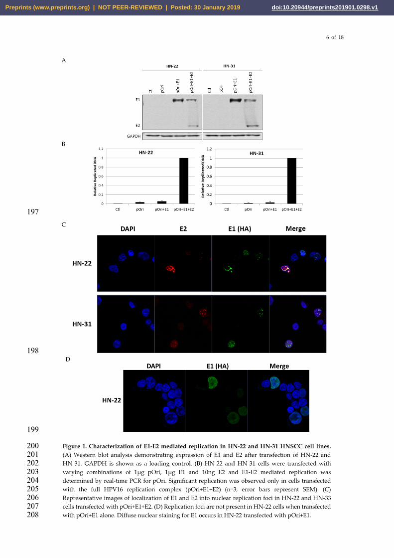
Our lab has previously described a real-time PCR-based assay to detect transient E1-E2 mediated 173 DNA replication that is more sensitive, less labor intensive, and less hazardous than southern 174 blotting while also being highly quantitative [34]. This assay has primarily been performed in 175 non-HNSCC cell lines such as c33a and 293T. In order to create a robust system to study and target 176 HPV16 E1-E2 mediated DNA replication in HNSCC, we transfected nu61, SCC-61, HN-11, HN-22, 177 HN-30, and HN-31 with varying combinations of the components of the HPV16 replication complex 178 (pOri, E1, E2). Nu61, SCC-61, HN-11, and HN-30 did not allow for measurable amounts of E1-E2 179 mediated DNA replication in cells (not shown). Conversely HN-22 and HN-31 lines allowed for 180 quantifiable E1-E2 mediated DNA replication (Figure 1 A-C). 72 hours post-transfection with pOri, 181 E1 and E2 HN-22 and HN-31 lines expressed appreciable amounts of the E1 and E2 proteins 182 detectable by western blot (Figure 1 A). HN-22 and HN-31 cells cotransfected with the complete 183 HPV16 replication complex (pOri+ E1+ E2) demonstrated a detectable DNA replication signal, while 184 untransfected control cells (Ctl), or cells transfected with pOri or pOri+E1 demonstrated little to no 185 DNA replication signal (Figure 1 B). Previous studies have demonstrated that E1 and E2 form 186 distinct nuclear replication foci in the presence of the viral origin of replication [41,44]. To determine 187 if this was the case in HNSCC cells, HN-22 and HN-31 were grown on coverslips and transfected 188 with pOri+E1+E2. Coverslips were fixed and stained for E1(HA) and E2. As expected, E1 and E2 189 formed distinct replication foci in both HN-22 and HN-31 cells only when transfected with the 190 complete replication complex (pOri+E1+E2) (Figure 1 C). These replication foci did not form unless 191 cells were transfected with the complete replication complex. HN-22 cells transfected with pOri+E1 192 display diffuse nuclear staining for E1 (Figure 1 D). Overall, the results in Figure 1 demonstrate that 193 we have created a system to consistently observe and quantify E1-E2 mediated DNA replication in 194 the HNSCC lines HN-22 and HN-31. 195
196
Preprints (www.preprints.org) | NOT PEER-REVIEWED | Posted: 30 January 2019 Preprints (www.preprints.org) | NOT PEER-REVIEWED | Posted: 30 January 2019 doi:10.20944/preprints201901.0298.v1
6 of 18
197
198
199
Figure 1. Characterization of E1-E2 mediated replication in HN-22 and HN-31 HNSCC cell lines. 200 (A) Western blot analysis demonstrating expression of E1 and E2 after transfection of HN-22 and 201 HN-31. GAPDH is shown as a loading control. (B) HN-22 and HN-31 cells were transfected with 202 varying combinations of 1µg pOri, 1µg E1 and 10ng E2 and E1-E2 mediated replication was 203 determined by real-time PCR for pOri. Significant replication was observed only in cells transfected 204 with the full HPV16 replication complex (pOri+E1+E2) (n=3, error bars represent SEM). (C) 205 Representative images of localization of E1 and E2 into nuclear replication foci in HN-22 and HN-33 206 cells transfected with pOri+E1+E2. (D) Replication foci are not present in HN-22 cells when transfected 207 with pOri+E1 alone. Diffuse nuclear staining for E1 occurs in HN-22 transfected with pOri+E1. 208
A
B
C
D
Preprints (www.preprints.org) | NOT PEER-REVIEWED | Posted: 30 January 2019 Preprints (www.preprints.org) | NOT PEER-REVIEWED | Posted: 30 January 2019 doi:10.20944/preprints201901.0298.v1
7 of 18
3.3 HN-22 and HN-31 HNSCC cell lines allow for E2-mediated transcriptional activation and repression 209
Because HN-22 and HN-31 lines allowed reproducible E1-E2 mediated replication, these lines were 210 selected for study of E2-mediated transcription. To study transcriptional activation by E2, HN-22 211 and HN-31 cells were transfected with 1µg pTK6E2 and varying amounts of E2. pTK6E2 contains a 212 thymidine kinase promoter with 6 HPV E2 binding sites which allow transactivation of downstream 213 luciferase reporter upon binding of E2. pGL3, which has no E2 binding sites, serves as a negative 214 control. Figure 2 A summarizes the results of the transcriptional activation assay in HN-22 and 215 HN-31. HN-22 or HN-31 cells transfected with pGL3 basic or pTK6E2 alone demonstrated low, 216 background levels of luciferase signal. Addition of 1ng of HPV16 E2 did little to change this basal 217 level of signal. Upon addition of 10ng E2, a substantial increase in luciferase signal was measured in 218 both HN-22 and HN-31 and this signal increased stepwise upon addition of 100ng or 1000ng E2. 219 These results clearly demonstrate that E2 activates transcription from pTK6E2 in a dose dependent 220 manner, signifying that E2-mediated transcription is occurring in HN-22 and HN-31 HNSCC cell 221 lines 222 223 Perhaps more relevant to the HPV life cycle is the ability of E2 to repress transcription, particularly 224 the transcription of E6 and E7. E2 transcriptionally represses E6 and E7 expression by binding to 225 E2-binding sites in the LCR region of the HPV genome, thereby sterically hindering the action of 226 cellular transcription factors on the HPV p97 early promoter and other proximal elements [45-49]. To 227 study transcriptional repression by E2, HN-22 and HN-31 cells were transfected with 1µg 228 pGL3-16LCR and varying amounts of E2. pGL3-16LCR contains the HPV16 LCR region from W12 229 cells upstream of a luciferase reporter. pGL3 serves as a negative control. Figure 2B demonstrates 230 that there is low, background luciferase signal when HN-22 or HN-31 are transfected with pGL3, but 231 measurable transcription is observed in cells transfected with pGL3-16LCR. Addition of 1ng of E2 232 does little to reduce the transcription of pGL3-16LCR. However, addition of 10ng E2 appears to 233 reduce transcription by about 30% and addition of 100ng E2 appears to reduce transcription by 40%. 234 Interestingly, when 1000ng E2 is added an increase in transcription is observed in both HN-22 and 235 HN-31. This is distinct from cervical cancer lines where E2 is a strong transcriptional repressor on 236 HPV LCRs and remains so at high levels of E2 expression [50]. This may represent novel regulation 237 of HPV E2-mediated transcriptional repression in HNSCC and this is currently being investigated in 238 our lab. 239
240
241 242
243
244
245
246
247
Preprints (www.preprints.org) | NOT PEER-REVIEWED | Posted: 30 January 2019 Preprints (www.preprints.org) | NOT PEER-REVIEWED | Posted: 30 January 2019 doi:10.20944/preprints201901.0298.v1

8 of 18
248
249
250
Figure 2. E2-mediated transcriptional activation and repression in HN-22 and HN-31 HNSCC 251 cells. (A) Luciferase transcription activation assay in HN-22 and HN-31 utilizing pTK6E2. Relative 252 luciferase units increase with increasing amounts of E2 protein present, indicating E2 mediated 253 transcriptional activation of pTK6E2. pGL3 and pTK6E2 (no E2) lanes are shown as controls. (B) 254 Luciferase transcription repression assay in HN-22 and HN-31 utilizing pGL3-16LCR (16LCR). 255 Relative luciferase units decrease upon addition of E2 indicating transcriptional repression. At 256 1000ng E2 transcription is increased past baseline. pGL3 is shown as a negative control while 257 pGL3-16LCR (no E2) represents basal, unrepressed transcription levels. (n=3, error bars represent 258 SEM). 259
260
261
262
B
B
Preprints (www.preprints.org) | NOT PEER-REVIEWED | Posted: 30 January 2019 Preprints (www.preprints.org) | NOT PEER-REVIEWED | Posted: 30 January 2019 doi:10.20944/preprints201901.0298.v1
9 of 18
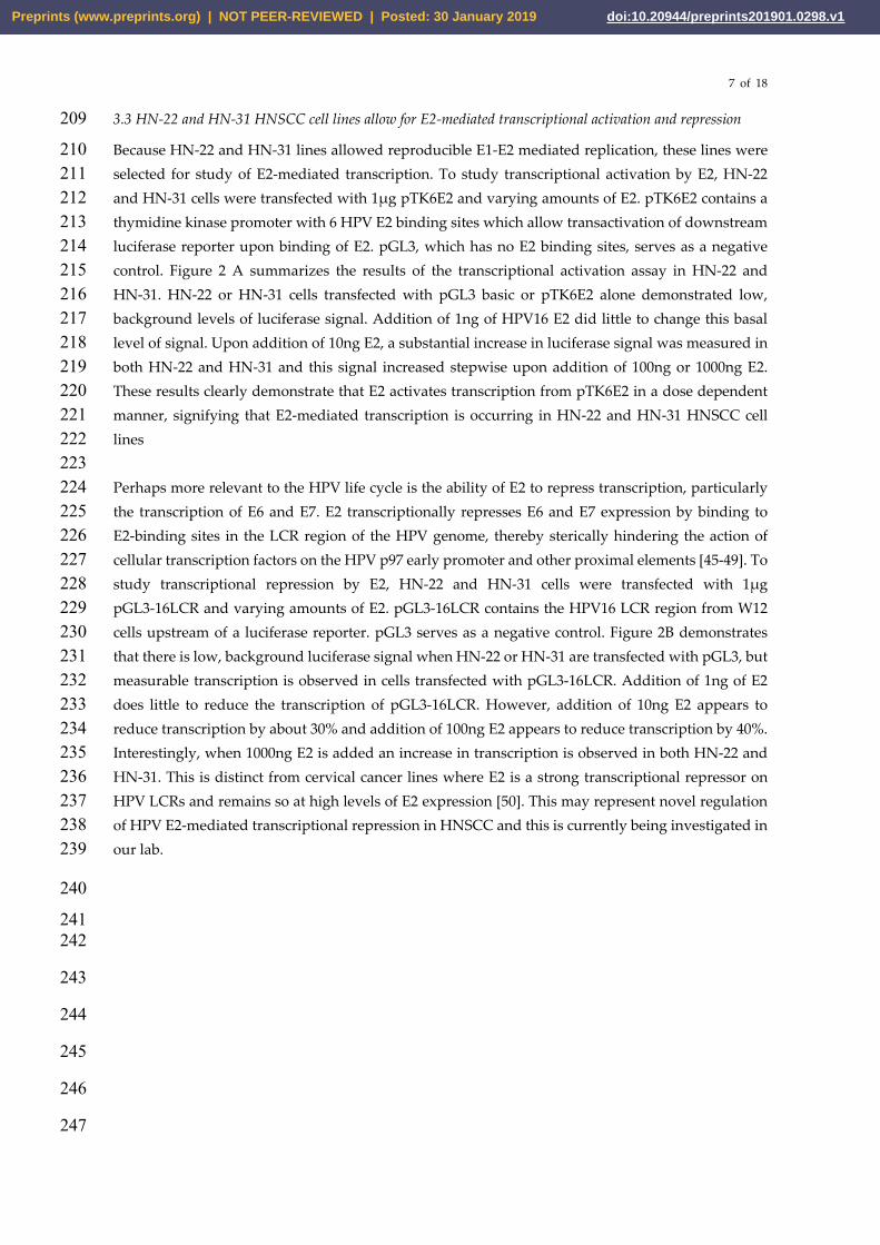
3.4 Endogenous levels of IFIT1 determine the viability of E1-E2 mediated DNA replication in HNSCC cells 263
While HN-22 and HN-31 cells allowed for E1-E2 mediated DNA replication and for E2-mediated 264 transcriptional activation and repression, the other transfectable HNSCC lines tested did not allow 265 for detectable DNA replication when transfected with pOri+E1+E2. Others have previously shown 266 that the innate immune response protein, IFIT1, binds the HPV18 E1 protein and inhibits HPV18 267 replication [51, 52]. In many cell types, IFIT1 is expressed at low levels and is induced in response 268 by interferon in the presence of pathogens [53]. Our previous work in oral keratinocytes 269 demonstrated that NOKs (TERT-immortalized normal oral keratinocytes) express high levels of 270 endogenous IFIT1, which is greatly reduced upon introduction of the HPV16 genome [54]. We 271 hypothesized that IFIT1 may also bind the HPV16 E1 protein and that high levels of endogenous 272 IFIT1 in certain HNSCC cell lines may explain the discrepancy in E1-E2 mediated replication 273 observed among the HNSCC cells tested. Western blotting for IFIT1 in HNSCC cell lines revealed 274 that HN-22 and HN-31 lines had no detectable endogenous IFIT1 protein (Figure 3). All other cells 275 tested, with detectable IFIT1 via western blot analysis, did not allow for HPV16 E1-E2 mediated 276 DNA replication. HN-8 cells also had low levels of IFIT1 but had poor transfectability (Table 1) and 277 thus could not be used for our replication assay. This result suggests that higher levels of 278 endogenous IFIT1 may interfere with HPV16 E1-E2 mediated DNA replication in HNSCC cells. 279
280
Figure 3. Low levels of IFIT1 are required for E1-E2 replication. Western blot analysis demonstrating 281 levels of endogenous IFIT1 in HNSCC cell lines. β-actin is shown as a loading control. Of cell lines that 282 were transfectable, only HN-22 and HN-31 (no detectable IFIT1) allowed for robust, measurable E1-E2 283 mediated DNA replication. 284
3.5 Exogenous IFIT1 binds HPV16 E1 in HN-22 cells and significantly attenuates E1-E2 mediated DNA 285 replication 286
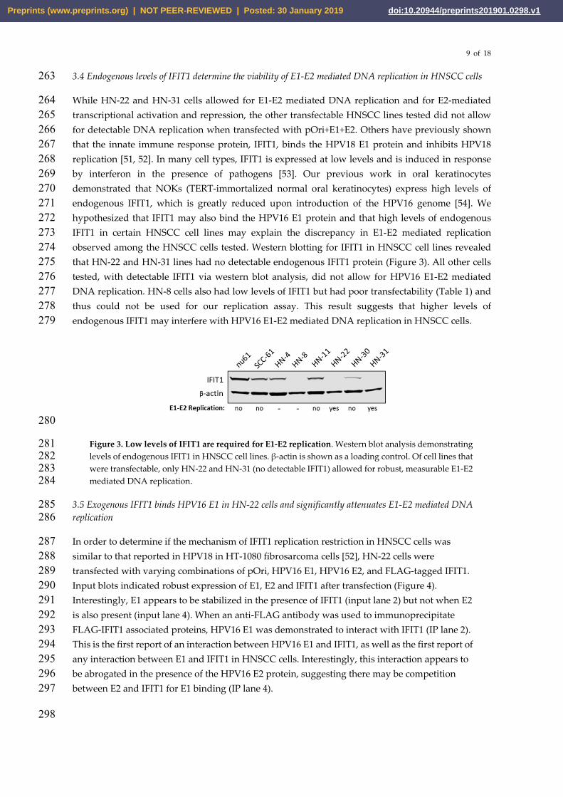
In order to determine if the mechanism of IFIT1 replication restriction in HNSCC cells was 287 similar to that reported in HPV18 in HT-1080 fibrosarcoma cells [52], HN-22 cells were 288 transfected with varying combinations of pOri, HPV16 E1, HPV16 E2, and FLAG-tagged IFIT1. 289 Input blots indicated robust expression of E1, E2 and IFIT1 after transfection (Figure 4). 290 Interestingly, E1 appears to be stabilized in the presence of IFIT1 (input lane 2) but not when E2 291 is also present (input lane 4). When an anti-FLAG antibody was used to immunoprecipitate 292 FLAG-IFIT1 associated proteins, HPV16 E1 was demonstrated to interact with IFIT1 (IP lane 2). 293 This is the first report of an interaction between HPV16 E1 and IFIT1, as well as the first report of 294 any interaction between E1 and IFIT1 in HNSCC cells. Interestingly, this interaction appears to 295 be abrogated in the presence of the HPV16 E2 protein, suggesting there may be competition 296 between E2 and IFIT1 for E1 binding (IP lane 4). 297
298
Preprints (www.preprints.org) | NOT PEER-REVIEWED | Posted: 30 January 2019 Preprints (www.preprints.org) | NOT PEER-REVIEWED | Posted: 30 January 2019 doi:10.20944/preprints201901.0298.v1
10 of 18
299
Figure 4. IFIT1 binds HPV16 E1 in HN-22 cells. Immunoprecipitation experiment demonstrating an 300 interaction between HPV16 E1 and exogenous FLAG-IFIT1 in HN-22 cells (IP lane 2). This interaction 301 is abrogated in the presence of E2 (IP lane 4). β-actin is shown as a loading control for the input blot. 302
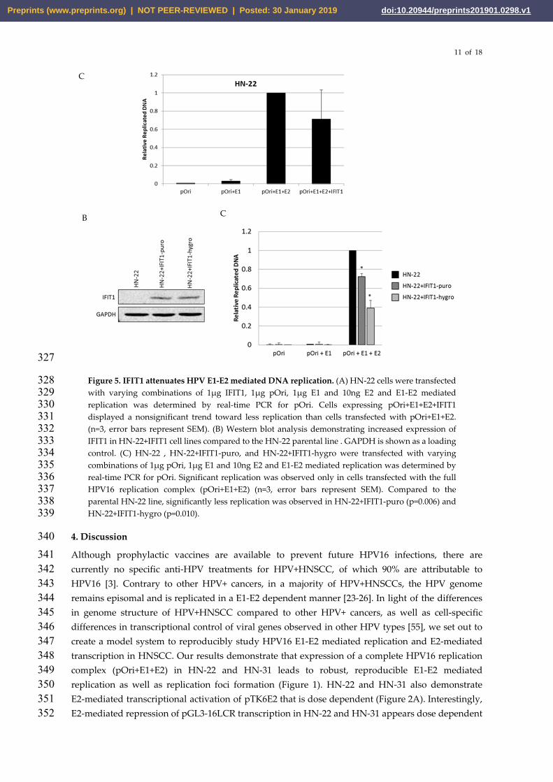
To determine if addition of exogenous IFIT1 attenuated E1-E2 mediated DNA replication in 303 HNSCC cells, pOri, E1, E2, were cotransfected into HN-22 cells as performed in Figure 1. To 304 directly compare levels of E1-E2 mediated DNA replication, HN-22 cells were also transfected 305 with pOri+E1+E2+IFIT1. HN-22 cells transfected with IFIT1 alongside the complete replication 306 complex (pOri+E1+E2) demonstrated a trend towards less E1-E2 mediated replication compared 307 to pOri+E1+E2 cells but the effect was variable in magnitude (Figure 5 A). These results 308 suggested there may be some temporality to the interaction between IFIT1 and E1. Because 309 IFIT1 is primarily cytoplasmic, IFIT1 may need to be expressed before E1 is translocated to the 310 nucleus in order to bind E1 and attenuate E1-E2 mediated replication. To test this, HN-22 cells 311 were cotransfected with IFIT1 and one of two resistance plasmids (pLX302-puromycin or 312 pBABE-hygromycin) and selected with the appropriate antibiotic to create two cell lines stably 313 overexpressing IFIT1 (HN-22+IFIT1-puro and HN-22+IFIT1-hygro). Increased levels of IFIT1 are 314 observed in HN-22+IFIT1-puro and HN-22+IFIT1-hygro lines compared to the parental HN22 315 cell line (Figure 5 B). To determine if overexpression of IFIT1 in these lines attenuated 316 replication, HN-22+IFIT1-puro and HN-22+IFIT1-hygro lines were transfected with pOri, 317 pOri+E1 or pOri+E1+E2 alongside the parental HN22 line. As expected, only cells transfected 318 with the full replication complex (pOri+E1+E2) demonstrated significant amounts of replication. 319 Significantly less replication was observed in HN-22+IFIT1-puro and HN-22+IFIT1-hygro 320 transfected with the full replication complex compared to the parental HN-22 line (Figure 5 C). 321 These results suggest that high levels of IFIT1, present before the expression of HPV proteins E1 322 and E2, leads to less E1-E2 mediated DNA replication. This reduction of replication in 323 HN-22+IFIT1 lines is presumably due to the previously described interactions between IFIT1 324 and E1 (Figure 4). 325
326
Preprints (www.preprints.org) | NOT PEER-REVIEWED | Posted: 30 January 2019 Preprints (www.preprints.org) | NOT PEER-REVIEWED | Posted: 30 January 2019 doi:10.20944/preprints201901.0298.v1
11 of 18
327
Figure 5. IFIT1 attenuates HPV E1-E2 mediated DNA replication. (A) HN-22 cells were transfected 328 with varying combinations of 1µg IFIT1, 1µg pOri, 1µg E1 and 10ng E2 and E1-E2 mediated 329 replication was determined by real-time PCR for pOri. Cells expressing pOri+E1+E2+IFIT1 330 displayed a nonsignificant trend toward less replication than cells transfected with pOri+E1+E2. 331 (n=3, error bars represent SEM). (B) Western blot analysis demonstrating increased expression of 332 IFIT1 in HN-22+IFIT1 cell lines compared to the HN-22 parental line . GAPDH is shown as a loading 333 control. (C) HN-22 , HN-22+IFIT1-puro, and HN-22+IFIT1-hygro were transfected with varying 334 combinations of 1µg pOri, 1µg E1 and 10ng E2 and E1-E2 mediated replication was determined by 335 real-time PCR for pOri. Significant replication was observed only in cells transfected with the full 336 HPV16 replication complex (pOri+E1+E2) (n=3, error bars represent SEM). Compared to the 337 parental HN-22 line, significantly less replication was observed in HN-22+IFIT1-puro (p=0.006) and 338 HN-22+IFIT1-hygro (p=0.010). 339
4. Discussion 340 Although prophylactic vaccines are available to prevent future HPV16 infections, there are 341 currently no specific anti-HPV treatments for HPV+HNSCC, of which 90% are attributable to 342 HPV16 [3]. Contrary to other HPV+ cancers, in a majority of HPV+HNSCCs, the HPV genome 343 remains episomal and is replicated in a E1-E2 dependent manner [23-26]. In light of the differences 344 in genome structure of HPV+HNSCC compared to other HPV+ cancers, as well as cell-specific 345 differences in transcriptional control of viral genes observed in other HPV types [55], we set out to 346 create a model system to reproducibly study HPV16 E1-E2 mediated replication and E2-mediated 347 transcription in HNSCC. Our results demonstrate that expression of a complete HPV16 replication 348 complex (pOri+E1+E2) in HN-22 and HN-31 leads to robust, reproducible E1-E2 mediated 349 replication as well as replication foci formation (Figure 1). HN-22 and HN-31 also demonstrate 350 E2-mediated transcriptional activation of pTK6E2 that is dose dependent (Figure 2A). Interestingly, 351 E2-mediated repression of pGL3-16LCR transcription in HN-22 and HN-31 appears dose dependent 352
C
B C
Preprints (www.preprints.org) | NOT PEER-REVIEWED | Posted: 30 January 2019 Preprints (www.preprints.org) | NOT PEER-REVIEWED | Posted: 30 January 2019 doi:10.20944/preprints201901.0298.v1

12 of 18
up to a certain level of E2, however this repression appears to be lost when higher amounts of E2 353 (1000ng) are expressed alongside pGL3-16LCR (Figure 2 B). This phenomenon is not observed in 354 cells from other anatomical regions [56-59] and highlights the potential for cell-type or tissue-specific 355 differences in HPV16 transcription in HNSCC. Overall, our model is well-suited to study and better 356 characterize E2-mediated transcription and E1-E2 mediated replication in HNSCC. 357
In developing our real-time PCR assay to measure E1-E2 mediated replication in HNSCC cells, we 358 discovered that low endogenous levels of IFIT1 are required for measurable replication to occur 359 (Figure 3). This is supported by our previous findings that IFIT1 is significantly downregulated by 360 HPV16 in oral keratinocytes [54]. IFIT1 is an interferon induced protein that exerts numerous known 361 antiviral functions [53]. IFIT1 has been shown to have nonspecific antiviral functions, such as 362 binding eukaryotic initiation factor 3 (eIF3) and inhibiting cellular and viral translation, as well as by 363 binding directly viral RNAs that lack 2’-O-methylation [60, 61]. Because HPVs utilize cellular 364 transcriptional machinery, HPV transcripts should contain 2’-O-methylation, so this latter 365 mechanism is likely not relevant in HPV infections. However, specifically relating to HPV, IFIT1 has 366 been shown to bind the HPV18 E1 protein in 1080T fibrosarcoma cells [51]. We observe a similar 367 binding interaction between HPV16 E1 and IFIT1 in HN-22 cells (Figure 4) and hypothesize that this 368 E1-IFIT1 interaction is the primary anti-HPV action of IFIT1. This is supported by our results 369 demonstrating that overexpression of IFIT1 in HN-22 cells leads to less E1-E2 mediated replication 370 than in parental HN-22 cells that express no detectable IFIT1 (Figure 5 C). 371
Our results demonstrate that the effect of IFIT1 on HPV E1-E2 mediated replication in our system is 372 more significant when IFIT1 is expressed before transfection with E1, E2, and pOri. It is possible that 373 the role of IFIT1 in restricting HPV replication is most important during the initial establishment 374 stage of the HPV life cycle. Cells with high endogenous IFIT1 expression may be more resistant to 375 initial HPV infection, giving possible insight as to why not all HPV infections result persistent 376 disease. Further studies are necessary to determine if there is such temporality to the IFIT1-E1 377 interaction or if it is simply a matter of IFIT1 levels being sufficient to out-compete E2 for E1 binding, 378 as the presence of E2 appears to reduce the IFIT1-E1 interaction (Figure 4). It is of note that 379 overexpression of IFIT1 in HN-22 only reduces replication by about 50% (Figure 5 C). There are 380 likely other factors that negatively regulate HPV replication, as numerous innate immune factors 381 like IFIT1, have been shown to be downregulated by HPV16 in cells of the head and neck region and 382 in HPV+HNSCC clinical samples [54, 62]. While further characterization of the HPV16 IFIT1-E1 383 interaction should be investigated to inform future anti-viral therapies, there is reason to believe that 384 other interactions, pertaining to both HPV16 replication and transcription, can be discovered and 385 tested utilizing this HNSCC model system. Overall, this model system can be used to study HPV16 386 E1-E2 mediated replication and E2-dependent transcription in HNSCC cells and could be used as a 387 tool to screen replication and transcription targeting anti-HPV agents. 388
389
390
391
392
Preprints (www.preprints.org) | NOT PEER-REVIEWED | Posted: 30 January 2019 Preprints (www.preprints.org) | NOT PEER-REVIEWED | Posted: 30 January 2019 doi:10.20944/preprints201901.0298.v1

13 of 18
Acknowledgments: I.M.M received funding from the Virginia Commonwealth University Philips Institute for 393 Oral Health Research and the Virginia Commonwealth University National Cancer Institute Designated 394 Massey Cancer Center, NIH P30 CA016059. Microscopy was performed at the VCU Microscopy Facility, 395 supported in part, by funding from National Institutes of Health-National Cancer Institute Cancer Center 396 Support Grant P30 CA016059. 397 Author Contributions: M.R.E., M.L.B. and I.M.M conceived and designed experiments. M.R.E., C.T.F., M.L.B. 398 performed and analyzed most experiments. C.D.J. and M.L.B. performed immunofluorescence experiments. 399 C.D.J. performed cloning. X.W. performed real-time PCR. M.R.E. wrote the manuscript and C.D.J. edited the 400 manuscript. 401 Conflicts of Interest: The authors declare no conflict of interest. 402
Preprints (www.preprints.org) | NOT PEER-REVIEWED | Posted: 30 January 2019 Preprints (www.preprints.org) | NOT PEER-REVIEWED | Posted: 30 January 2019 doi:10.20944/preprints201901.0298.v1
14 of 18
References 403
1. zur Hausen, H. Papillomaviruses in the Causation of Human Cancers — a Brief Historical 404 Account. Virology. 2009, 384, 260-265. 405 406
2. Chesson, H.W.; Dunne, E.F.; Hariri, S.; Markowitz, L.E. The Estimated Lifetime Probability of 407 Acquiring Human Papillomavirus in the United States. Sex. Transm. Dis. 2014, 41, 660-664. 408 409
3. Psyrri, A.; DiMaio, D. Human Papillomavirus in Cervical and Head-and-Neck Cancer. Nat. Clin. 410 Pract. Oncol. 2008, 5, 24-31. 411 412
4. Marur, S.; D'Souza, G.; Westra, W.H.; Forastiere, A.A. HPV-Associated Head and Neck Cancer: A 413 Virus-Related Cancer Epidemic. Lancet Oncol. 2010, 11, 781-789. 414 415
5. Walker, T.Y.; Elam-Evans, L.D.; Yankey, D.; et al. National, Regional, State, and Selected Local 416 Area Vaccination Coverage Among Adolescents Aged 13–17 Years — United States, 2017. 417 MMWR Morb Mortal Wkly Rep. 2018, 67, 909–917. 418 419
6. Munger, K.; Phelps, W.C.; Bubb, V.; Howley, P.M.; Schlegel, R. The E6 and E7 Genes of the 420 Human Papillomavirus Type 16 Together are Necessary and Sufficient for Transformation of 421 Primary Human Keratinocytes. J. Virol. 1989, 63, 4417-4421. 422 423
7. Munger, K.; Werness, B.A.; Dyson, N.; Phelps, W.C.; Harlow, E.; Howley, P.M. Complex formation 424 of human papillomavirus E7 proteins with the retinoblastoma tumor suppressor gene product. 425 EMBO J. 1989, 8,4099–4105. 426 427
8. Dyson, N.; Howley, P.M.; Munger, K.; Harlow, E. The human papillomavirus-16 E7 oncoprotein is 428 able to bind to the retinoblastoma gene product. Science. 1989, 243, 934-937. 429 430
9. Scheffner, M.; Werness, B.A.; Huibregtse, J.M.; Levine, A.J.; Howley, P.M. The E6 oncoprotein 431 encoded by human papillomavirus types 16 and 18 promotes the degradation of p53. Cell. 1990, 63, 432 1129–1136. 433 434
10. Sedman, S.A.; Barbosa, M.S.; Vass, W.C.; Hubbert, N.L.; Haas, J.A.; Lowy, D.R.; Schiller, J.T. The 435 Full-Length E6 Protein of Human Papillomavirus Type 16 has Transforming and 436 Trans-Activating Activities and Cooperates with E7 to Immortalize Keratinocytes in Culture. J. 437 Virol. 1991, 65, 4860-4866. 438
439 11. Werness, B.A.; Munger, K.; Howley, P.M.. Role of the human papillomavirus oncoproteins in 440
transformation and carcinogenic progression. Important Adv. Oncol. 1991, 3–18. 441 442
Preprints (www.preprints.org) | NOT PEER-REVIEWED | Posted: 30 January 2019 Preprints (www.preprints.org) | NOT PEER-REVIEWED | Posted: 30 January 2019 doi:10.20944/preprints201901.0298.v1
15 of 18
12. Huibregtse, J.M.; Scheffner, M.; Howley, P.M. Cloning and expression of the cDNA for E6AP, a 443 protein that mediates the interaction of human papillomavirus E6 oncoprotein with p53. 444 Molecular and Cellular Biology. 1993, 13, 775-784. 445 446
13. Riley, R.R.; Duensing, S.; Brake, T.; Munger, K.; Lambert, P.F.; Arbeit, J.M. Dissection of Human 447 Papillomavirus E6 and E7 Function in Transgenic Mouse Models of Cervical Carcinogenesis. 448 Cancer Res. 2003, 63, 4862-4871. 449
450 14. Huh, K.; Zhou, X; Hayakawa, H.; Cho, J.Y.; Libermann, T.A.; Jin, J.; Harper, J.W.; Munger, K. 451
Human papillomavirus type 16 E7 oncoprotein associates with the cullin 2 ubiquitin ligase 452 complex, which contributes to degradation of the retinoblastoma tumor suppressor. Journal of 453 Virology. 2007, 81, 9737-9747. 454
455 15. Jiang, M.; Milner, J. Selective silencing of viral gene expression in HPV-positive human cervical 456
carcinoma cells treated with siRNA, a primer of RNA interference. Oncogene. 2002, 21, 604-6048. 457 458 16. Butz, K.; Ristriani, T.; Hengstermann, A.; Denk, C.; Scheffner, M.; Hoppe-Seyler, F. SiRNA 459
Targeting of the Viral E6 Oncogene Efficiently Kills Human Papillomavirus-Positive Cancer 460 Cells. Oncogene. 2003, 22, 5938-5945. 461
462 17. Hall, A.H.; Alexander, K.A. RNA Interference of Human Papillomavirus Type 18 E6 and E7 463
Induces Senescence in HeLa Cells. J. Virol. 2003, 77, 6066-6069. 464 465 18. Yoshinouchi, M.; Yamada, T.; Kizaki, M.; Fen, J.; Koseki, T.; Ikeda, Y.; et al. In vitro and in vivo 466
growth suppression of human papillomavirus16-positive cervical cancer cells by E6 siRNA. Mol 467 Ther. 2003, 8, 762-768. 468
469 19. Gu, W.; Putral, L.; Hengst, K.; Minto, K.; Saunders, N.A. Leggatt, G.; et al. Inhibition of cervical 470
cancer cell growth in vitro and in vivo with lentiviral-vector delivered short hairpin RNA 471 targeting human papillomavirus E6 and E7 oncogenes. Cancer Gene Ther. 2006, 13, 1023–1032. 472
473 20. Yamato, K.; Yamada, T.; Kizaki, M.; Ui-Tei. K.; Natori, Y.; Fujino, M.; Nishihara, T.; Ikeda, Y.; 474
Nasu, Y.; Saigo, K.; Yoshinouchi, M. New highly potent and specific E6 and E7 siRNAs for 475 treatment of HPV16 positive cervical cancer. Cancer Gene Therapy. 2008, 15, 140-153. 476
477 21. Jabbar, S.F.; Abrams, L.; Glick, A.; Lambert, P.F. Persistence of high-grade cervical dysplasia and 478
cervical cancer requires the continuous expression of the human papillomavirus type 16 E7 479 oncogene. Cancer Research. 2009, 69, 4407-4414 480
481 22. Hoppe-Seyler, K.; Bossler, F; Braun, J.A.; Herrmann, A.L.; Hoppe-Seyler, F. The HPV E6/E7 482
Oncogenes: Key Factors for Viral Carcinogenesis and Therapeutic Targets. Trends in Microbiology. 483 2018, 26, 158-168. 484
485 486
Preprints (www.preprints.org) | NOT PEER-REVIEWED | Posted: 30 January 2019 Preprints (www.preprints.org) | NOT PEER-REVIEWED | Posted: 30 January 2019 doi:10.20944/preprints201901.0298.v1
16 of 18
23. Parfenov, M.; Pedamallu, C.S.; Gehlenborg, N.; Freeman, S.S.; Danilova, L.; Bristow, C.A.; Lee, S.; 487 Hadjipanayis, A.G.; Ivanova, E.V.; Wilkerson, M.D.; et al. Characterization of HPV and Host 488 Genome Interactions in Primary Head and Neck Cancers. Proc. Natl. Acad. Sci. U. S. A. 2014, 111, 489 15544-15549. 490
491 24. Cancer Genome Atlas Network. Comprehensive Genomic Characterization of Head and Neck 492
Squamous Cell Carcinomas. Nature. 2015, 517, 576-582. 493 494 25. Ramqvist, T.; Mints, M.; Tertipis, N.; Nasman, A.; Romanitan, M.; Dalianis, T. Studies on Human 495
Papillomavirus (HPV) 16 E2, E5 and E7 mRNA in HPV-Positive Tonsillar and Base of Tongue 496 Cancer in Relation to Clinical Outcome and Immunological Parameters. Oral Oncol. 2015, 51, 497 1126-1131. 498
499 26. Nulton, T.J.; Olex, A.L.; Dozmorov, M.; Morgan, I.M.; Windle, B. Analysis of the Cancer Genome 500
Atlas Sequencing Data Reveals Novel Properties of the Human Papillomavirus 16 Genome in 501 Head and Neck Squamous Cell Carcinoma. Oncotarget. 2017, 8, 17684-17699. 502
503 27. Steger, G.; Corbach, S. Dose-dependent regulation of the early promoter of human 504
papillomavirus type 18 by the viral E2 protein. J. Virol. 1997, 71, 50–58. 505 506
28. Desaintes, C.; Demeret, C.; Goyat, S.; Yaniv, M.; Thierry, F. Expression of the papillomavirus E2 507 protein in HeLa cells leads to apoptosis. EMBO J. 1997, 16, 504-514. 508 509
29. Goodwin, E.C.; Naeger, L. K.; Breiding, D. E.; Androphy, E. J.; DiMaio, D. 510 Transactivation-competent bovine papillomavirus E2 protein is specifically required for efficient 511 repression of human papillomavirus oncogene expression and for acute growth inhibition of 512 cervical carcinoma cell lines. J. Virol. 1998, 72, 3925-3934. 513 514
30. Goodwin, E.C.; Yang, E.; Lee, C.J.; Lee, H.W.; DiMaio, D.; Hwang, E.S. Rapid induction of 515 senescence in human cervical carcinoma cells. Proc. Natl. Acad. Sci. USA. 2000, 97, 10978-10983. 516 517
31. Francis, D.A.; Schmid, S.I.; Howley, P.M. Repression of the integrated papillomavirus E6/E7 518 promoter is required for growth suppression of cervical cancer cells. J. Virol. 2000, 74, 2679-2686. 519 520
32. Cardinali, M.; Pietraszkiewicz, H.; Ensley, J.F.; Robbins, K.C. Tyrosine phosphorylation as a 521 marker for aberrantly regulated growth-promoting pathways in cell lines derived from head and 522 neck malignancies. International Journal of Cancer. 1995, 61, 98-103. 523 524
33. Khodarev, N.N.; Beckett, M.; Labay, E.; Darga, T.; Roizman, B.; Weichselbaum, R.R. STAT1 is 525 overexpressed in tumors selected for radioresistance and confers protection from radiation in 526 transduced sensitive cells. Proc Natl Acad Sci USA. 2004, 101, 1714-1719. 527 528
34. Taylor, E.R.; Morgan, I.M. A Novel Technique with Enhanced Detection and Quantitation of 529 HPV-16 E1- and E2-Mediated DNA Replication. Virology. 2003, 315, 103-109. 530 531
35. Kadaja, M.; Sumerina, A.; Verst, T.; Ojarand, M.; Ustav, E.; Ustav, M. Genomic Instability of the 532 Host Cell Induced by the Human Papillomavirus Replication Machinery. EMBO J. 2007, 26, 533 2180-2191. 534 535
36. Bouvard, V.; Storey, A.; Pim, D.; Banks, L. Characterization of the Human Papillomavirus E2 536 Protein: Evidence of Trans-Activation and Trans-Repression in Cervical Keratinocytes. EMBO J. 537 1994, 13, 5451-5459. 538
Preprints (www.preprints.org) | NOT PEER-REVIEWED | Posted: 30 January 2019 Preprints (www.preprints.org) | NOT PEER-REVIEWED | Posted: 30 January 2019 doi:10.20944/preprints201901.0298.v1
17 of 18
539 37. Katibah, G.E.; Lee, H.J.; Huizar, J.P.; Vogan, J.M.; Alber, T.; Collins, K. TRNA Binding, Structure, 540
and Localization of the Human Interferon-Induced Protein IFIT5. Mol. Cell. 2013, 49, 743-750. 541 542
38. Yang, X.; Boehm, J.S.; Yang, X.; Salehi-Ashtiani, K.; Hao, T.; Shen, Y.; Lubonja, R.; Thomas, S.R.; 543 Alkan, O.; Bhimdi, T.; Green, T.M.; Johannessen, C.M.; Silver, S.J.; Nguyen, C.; Murray, R.R.; 544 Hieronymus, H.; Balcha, D.; Fan, C.; Lin, C.; Ghamsari, L.; Vidal, M.; Hahn, W.C.; Hill, D.E.; Root, 545 D.E. A public genome-scale lentiviral expression library of human ORFs. Nat Methods. 2011, 8, 546 659-661. 547 548
39. Morganstern, J.P.; Land, H. Advanced mammalian gene transfer: high titre retroviral vectors with 549 multiple drug selection markers and a complementary helper-free packaging cell line. Nucleic 550 Acids Res. 1990, 18, 3587-3596. 551 552
40. Vance, K.W.; Campo, M.S.; Morgan, I.M. An enhanced epithelial response of a papillomavirus 553 promoter to transcriptional activators. J. Biol. Chem. 1999, 274, 27839-27844. 554
555 41. Bristol, M.L.; Wang, X.; Smith, N.W.; Son, M.P.; Evans, M.R.; Morgan, I.M. DNA Damage 556
Reduces the Quality, but Not the Quantity of Human Papillomavirus 16 E1 and E2 DNA 557 Replication. Viruses 2016, 8, 10.3390/v8060175. 558 559
42. Boner, W.; Taylor, E.R.; Tsirimonaki, E.; Yamane, K.; Campo, M.S.; Morgan, I.M. A Functional 560 Interaction between the Human Papillomavirus 16 transcription/replication Factor E2 and the 561 DNA Damage Response Protein TopBP1. J. Biol. Chem. 2002, 277, 22297-22303. 562 563
43. Kingston, R.E.; Chen, C.A.; Rose, J.K. Calcium Phosphate Transfection. Curr. Protoc. Mol. Biol. 564 2003, Chapter 9, Unit 9.1. 565 566
44. Swindle, C.S.; Zou, N.; Van Tine, B.A.; Shaw, G.M.; Engler, J.A.; Chow, L.T. Human 567 Papillomavirus DNA Replication Compartments in a Transient DNA Replication System. J. Virol. 568 1999, 73, 1001-1009. 569 570
45. Tan, S.H.; Gloss, B.; Bernard H.U. During negative regulation of the human papillomavirus-16E6 571 promoter, the viral E2 protein can displace Sp1 from a proximal promoter element. Nucleic Acids 572 Res. 1992, 20, 251-256. 573 574
46. Tan, S.H.; Leong, L.E.; Walker, P.A.; Bernard, H.U. The human papillomavirus type 16 E2 575 transcription factor binds with low cooperativity to two flanking sites and represses the E6 576 promoter through displacement of Sp1 and TFIID. J. Virol. 1994, 68, 6411-6420. 577 578
47. Dong, G.; Broker, T.R.; Chow, L.T. Human papillomavirus type 11 E2 proteins repress the 579 homologous E6 promoter by interfering with the binding of host transcription factors to adjacent 580 elements. J. Virol. 1994, 68, 1115-1127. 581 582
48. Soeda, E.; Ferrari, M.C.; Baker, C.C.; McBride, A.A. Repression of HPV16 early region 583 transcription by the E2 protein. Virology. 2006, 351, 29-41. 584 585
49. Smith, J.A.; Haberstroh, F.S.; White, E.A.; Livingston, D.M.; DeCaprio, J.A., Howley, P.M. SMCX 586 and components of the TIP60 complex contribute to E2 regulation of the HPV E6/E7 promoter. 587 Virology. 2014, 468-470:311-321. 588 589
Preprints (www.preprints.org) | NOT PEER-REVIEWED | Posted: 30 January 2019 Preprints (www.preprints.org) | NOT PEER-REVIEWED | Posted: 30 January 2019 doi:10.20944/preprints201901.0298.v1
18 of 18
50. Gauson, E.J.; Donaldson, M.M.; Dornan. E.S.; Wang, X.; Bristol, M.L.; Bodily, J.M.; Morgan, I.M. 590 Evidence supporting a role for TopBP1 and Brd4 in the initiation but not continuation of human 591 papillomavirus 16 E1/E2-mediated DNA replication. J Virol. 2015, 89, 4980-4991. 592
593 51. Terenzi, F.; Saikia, P.; Sen, G.C. Interferon-Inducible Protein, P56, Inhibits HPV DNA Replication 594
by Binding to the Viral Protein E1. EMBO J. 2008, 27, 3311-3321. 595 596
52. Saikia, P.; Fensterl, V.; Sen, G.C. The Inhibitory Action of P56 on Select Functions of E1 Mediates 597 Interferon's Effect on Human Papillomavirus DNA Replication. J. Virol. 2010, 84, 13036-13039. 598 599
53. Fensterl, V.; Sen, G.C. Interferon-Induced Ifit Proteins: Their Role in Viral Pathogenesis. J. Virol. 600 2015, 89, 2462-2468. 601 602
54. Evans, M.R.; James, C.D.; Loughran, O.; Nulton, T.J.; Wang, X.; Bristol, M.L.; Windle, B.; Morgan, 603 I.M. An Oral Keratinocyte Life Cycle Model Identifies Novel Host Genome Regulation by Human 604 Papillomavirus 16 Relevant to HPV Positive Head and Neck Cancer. Oncotarget 2017, 47, 605 81892-81909. 606 607
55. Rapp, B.; Pawellek, A.; Kraetzer, F.; Schaefer, M.; May, C.; Purdie, K.; Grassman, K.; Iftner, T. 608 Cell-type-specific separate regulation of the E6 and E7 promoters of human papillomavirus type 609 6a by the viral transcription factor E2. J. Virol. 1997, 71, 6956-6966. 610 611
56. Thierry, F.; Yaniv, M. The BPV1 E2 trans-acting protein can be either a repressor or activator of 612 the HPV18 regulatory region. EMBO J. 1987, 6, 3391–3397. 613 614
57. Bernard, B.A.; Bailly, C.; Lenoir, M.C.; Darmon, M.; Thierry, F.; Yaniv M. The human 615 papillomavirus type 18 (HPV18) E2 gene product is a repressor of the HPV18 regulatory region in 616 human keratinocytes. J. Virol. 1989, 63, 4317–4324 617 618
58. Stenlund, A.; Botchan, M.R. The E2 trans-activator can act as a repressor by interfering with a 619 cellular transcription factor. Genes Dev. 1990, 4, 123–136. 620 621
59. Nishimura, A.; Ono, T.; Ishimoto, A.; Dowhanick, J.J.; Frizzell, M.A.; Howley, P.M.; Sakai, H. 622 Mechanisms of human papillomavirus E2-mediated repression of viral oncogene expression and 623 cervical cancer cell growth inhibition. J. Virol. 2000, 8, 3752-3760. 624
625 60. Hui, D.J.; Bhasker, C.R.; Merrick, W.C.; Sen, G.C. Viral Stress-Inducible Protein p56 Inhibits 626
Translation by Blocking the Interaction of eIF3 with the Ternary Complex eIF2.GTP.Met-tRNAi. J. 627 Biol. Chem. 2003, 278, 39477-39482. 628 629
61. Habjan, M.; Hubel, P.; Lacerda, L.; Benda, C.; Holze, C.; Eberl, C.H.; Mann, A.; Kindler, E.; 630 Gil-Cruz, C.; Ziebuhr, J.; et al. Sequestration by IFIT1 Impairs Translation of 2'O-Unmethylated 631 Capped RNA. PLoS Pathog. 2013, 9, e1003663. 632 633
62. Evans, M.R.; James, C.D.; Bristol, M.L.; Wang, X.; Kaur, N.; White, E.A.; Windle, B.; Morgan, I.M. 634 Human papillomavirus 16 E2 regulates keratinocyte gene expression relevant to cancer and the 635 viral life cycle. J. Virol. 2018, doi: 10.1128/JVI.01941-18. 636
Preprints (www.preprints.org) | NOT PEER-REVIEWED | Posted: 30 January 2019 Preprints (www.preprints.org) | NOT PEER-REVIEWED | Posted: 30 January 2019 doi:10.20944/preprints201901.0298.v1


![ARID1A prevents squamous cell carcinoma initiation and ...SCCs include the skin, head and neck, esophagus, lung, and cervix [2]. Cutaneous squamous cell carcinoma (cSCC) is a nonmelanoma](https://img.pdfslide.net/doc/110x75/6012df67f7a82c062d6f1b92/arid1a-prevents-squamous-cell-carcinoma-initiation-and-sccs-include-the-skin.jpg)