Embed Size (px)
Citation preview

ORIGINAL ARTICLE
A highly specific q-RT-PCR assay to address the relevanceof the JAK2WT and JAK2V617F expression levels and controlgenes in Ph -negative myeloproliferative neoplasms
Francesca Fantasia & Emma Nora Di Capua & Natalia Cenfra & Gloria Pessina &
Sergio Mecarocci & Angela Rago & Ettore Cotroneo & Anna Busanello &
Francesco Equitani & Francesco Lo-Coco & Clara Nervi & Giuseppe Cimino
Received: 20 September 2013 /Accepted: 1 October 2013 /Published online: 31 October 2013# The Author(s) 2013. This article is published with open access at Springerlink.com
Abstract In Ph− myeloproliferative neoplasms, the quantifi-cation of the JAK2V617F transcripts may provide some advan-tages over the DNA allele burden determination. We developeda q-RT-PCR to assess the JAK2WT and JAK2V617F mRNAexpression in 105 cases (23 donors, 13 secondary polycythe-mia, 22 polycythemia vera (PV), 38 essential thrombocythemia
(ET), and 9 primary myelofibrosis (PMF)). Compared with thestandard allele-specific oligonucleotide (ASO)-PCR technique,our assay showed a 100 % concordance rate detecting theJAK2V617F mutation in 22/22 PV (100 %), 29/38 (76.3 %)ET, and 5/9 (55.5 %) PMF cases, respectively. The sensitivityof the assay was 0.01 %. Comparing DNA and RNA samples,we found that the JAK2V617F mutational ratios were signifi-cantly higher at the RNA level both in PV (p =0.005) and ET(p =0.001) samples. In PV patients, JAK2WT expression levelspositively correlated with the platelets (PLTs) (p =0.003)whereas a trend to negative correlation was observed with theHb levels (p =0.051). JAK2V617F-positive cases showed thelowest JAK2WT and ABL1 mRNA expression levels. In all thesamples, the expression pattern of beta-glucoronidase (GUSB)wasmore homogeneous than that ofABL1 orβ2microglobulin(B2M). Using GUSB as normalizator gene, a significant in-crease of the JAK2V617F mRNA levels was seen in two ETpatients at time of progression to PV. In conclusion, the pro-posed q-RT-PCR is a sensitive and accurate method to quantifythe JAK2 mutational status that can also show clinical correla-tions suggesting the impact of the residual amount of theJAK2WT allele on the Ph− MPN disease phenotype. Ourobservations also preclude the use of ABL1 as a housekeepinggene for these neoplasms.
Keywords Ph− myeloproliferative neoplasms . JAK2WTlevel . JAK2V617F level . Housekeeping gene . q-RT-PCR
Introduction
Philadelphia chromosome-negative myeloproliferative neo-plasms (Ph− MPNs) are clonal myeloid disorders characterized
Electronic supplementary material The online version of this article(doi:10.1007/s00277-013-1920-0) contains supplementary material,which is available to authorized users.
F. Fantasia : E. N. Di Capua :A. Busanello : C. NerviDepartment of Medico-Surgical Sciences and Biotechnologies,University of Rome “Sapienza” Polo Pontino, , Latina, Italy
N. Cenfra :G. Cimino (*)Department of Cellular Biotechnology and Hematology,University of Rome “Sapienza”–Polo Pontino, via Benevento 6,00161 Rome, Italye-mail: [email protected]
G. PessinaMolecular Biology Laboratory, AUSL-VT,Belcolle Hospital, Viterbo, Italy
S. Mecarocci :A. Rago :G. CiminoHematology unit (2U), S. Maria Goretti Hospital/ICOT, AUSLLatina, Latina, Italy
E. Cotroneo : F. Lo-CocoDepartment of Biomedicine and Prevention, University“Tor Vergata”,, Rome, Italy
F. Lo-CocoLaboratory of Neuro-Oncohematology, Santa Lucia Foundation,Rome, Italy
F. EquitaniTransfusion Medicine and Immuno-Hematology Unit,Santa Maria Goretti Hospital, Latina, Italy
Ann Hematol (2014) 93:609–616DOI 10.1007/s00277-013-1920-0
by an increased production of terminally differentiated cells.Themechanisms ofMPN initiation and progression have beenextensively studied. A number of genetic and epigenetic ab-normalities associated with Ph− MPNs have been reported[1–5]. The somatic mutation of JAK2V617F is the mostfrequent genetic alteration in these diseases and is presentlyconsidered as a major diagnostic criterion for Ph− MPNs. Thefrequency of this mutation is greater than 95% in patients withpolycythemia vera (PV) and around 50 % in patients withessential thrombocythemia (ET) or primary myelofibrosis(PMF).
JAK2V617F-positive Ph− MPNs show a biological con-tinuum with clinical presentation, which is in part influencedby the JAK2V617F mutational load [6]. A relationship be-tween JAK2V617F mutational burden and disease phenotypehas been reported [7].
Although the clinical relevance of the correct quantificationof the JAK2 allele burden is still not clearly stated, the JAK2tyrosine kinase activity is now a therapeutic target for inno-vative and more specific treatment of these diseases [8]. Thestill debated pathogenesis of MPNs forces to seek methodswhich are increasingly specific and sensitive for the quantifi-cation of the JAK2 allele burden at diagnosis and duringtreatment. To date, the detection and quantification of theJAK2WT and JAK2V617F alleles are usually assessed usinggenomic DNA. However, the quantification of JAK2V617FmRNA transcripts by a real-time quantitative RT-PCR (q-RT-PCR) may provide some advantages over the DNA alleleburden [9]. In PV mononuclear cell samples, Zhao et al. [10]demonstrated that the ratio JAK2V617F/JAK2WT is higher incDNA than in genomic DNA. Using an ARMS assay oncDNA originated from granulocyte mRNAs, Vannucchiet al. [11] identified more JAK2-mutated transcripts in MPNpatients (9 %), as compared to conventional allele-specificPCR. Moreover, the mRNA (but not DNA) can be extractedfrom platelets present in buffy coats, and this may increase theJAK2V617F assay sensitivity in ET patient samples. Finally,in transgenic mice models, the JAK2V617F transcript levelswere found to be strictly correlated with the Ph− MPNphenotypes [12]. By contrast, q-RT-PCR requires a parallelamplification of a housekeeping gene (HKG) as control gene(CG) to correct variations in RNA quality and quantity and tocalculate the sensitivity of each measurement. Therefore, tobetter investigate the pathogenesis complexity of the Ph−MPNs and to provide a useful assay to monitor minimalresidual disease (MRD), we set up an absolute q-RT-PCRmethod for the quantification of JAK2WT and JAK2V617FmRNA. The data herein reported show that this method ishighly specific and sensitive. Moreover, the observation of asignificant variability within the sample groups of the expres-sion levels of ABL1 , one of the most commonly used HKGs,leads us to test other genes as more appropriate CGs in thisclinical setting.
Materials and methods
Patients and samples
Peripheral blood buffy coat specimens were collected from105 individuals. Twenty-two of them were diagnosed as hav-ing PV, 38 ET, and 9 PMF according to the 2008 WHOdiagnostic criteria [13]. Thirteen patients with secondary poly-cythemia (SP) and 23 healthy blood donors were included ascontrols. For three patients with initial diagnosis of ET whoprogressed to PV, paired samples of both disease phases wereavailable. All specimens were collected after patients hadsigned an informed consent. The study was approved by ourIRB at AUSL Latina (no. 6315/A001/2012).
DNA, RNA extraction, and cDNA synthesis
Cell pellets were either processed for DNA purification usingthe Wizard Genomic DNA Purification Kit (Promega Corpo-ration, Madison,WI), according to the manufacturer’s instruc-tions, or resuspended in guanidine isothiocyanate for RNAextraction [14, 15]. Equal amounts of RNA (1 μg) werereverse transcribed into cDNAs with random hexamers andMuLV reverse transcriptase (Applied Biosystems, Monza,Italy).
JAK2V617F mutation analysis and allele burden assay
JAK2V617F mutation was detected in DNA samples usingthe allele-specific PCR method previously described by Bax-ter et al. [16] The quantification of JAK2V617F allele burdenon genomic DNA was performed using the JAK2V617F
the manufacturer’s instructions.
Allele-specific q-RT-PCR
To obtain reference curves, standard plasmids weremanufactured to contain JAK2WT or JAK2V617F cDNA se-quences. cDNAs were obtained from RNA extracted from hu-manK562 cells (homozygous for JAK2WT allele) andHEL cells(homozygous for JAK2V617F allele). We designed specificprimers (forward: TTCTGGATAAAGCACACAGAAA; re-verse: CCAAATTTTACAAACTCCTGAACC), in order to am-plify 150 bp including theV617F codon (NM_004972.3:c.1745-1895), using the Primer 3 software (http://www.bioinformatics.nl/cgi-bin/primer3plus/primer3plus.cgi). One-step cloning strate-
(Invitrogen, Monza, Italy). Each plasmid vector was checkedby sequencing and includes five 10-fold serial dilutions, rang-ing from 105 to 10 targets per well.
JAK2WT and JAK2V617F mRNAs were measured asdescribed by Merker et al. [17]. Primers and probe
610 Ann Hematol (2014) 93:609–616
MutaQuant® Allele kit (Qiagen, Milano, Italy), according to
gy was performed by using the TOPO TA Cloning® kit
concentrations were optimized at concentrations of 300 and200 nM, respectively, in a final volume of 25 μl. q-RT-PCRwas performed in the ABI 7900 (Applied Biosystems) with aninitial 10-min incubation at 95 °C, followed by 40 cycles of15 s at 95 °C and 1 min at 60 °C. All samples and no-templatecontrols were run in triplicates.
For each sample, the ABL1 , B2M , and GUSB copy num-bers were determined by using standard endogenous plasmid
ing to the manufacturer’s instructions and in accordance withthe Europe Against Cancer (EAC) network [18].
JAK2 mutational burden evaluation
The mutational burden ratio of JAK2 was calculated using theDNA and RNA copy numbers according to the followingformula: JAK2V617F /(JAK2V617F +JAK2WT).
Statistical analysis
Statistical analyses and graphs were generated using SPSS-10software. Further, targeted pairwise comparisons to evaluatethe significance between individual diseases were performedby the two-sided Student’s t test. Results were expressed asmeans ± SEM. The level of significance from t test was p <0.05.
Results
Using the standard qualitative allele-specific oligonucleotide(ASO)-PCR method [17], the JAK2V617F was detected in22/22 PV (100 %), in 29/38 (76.3 %) ET, and in 5/9 (55.5 %)PMF cases, respectively (Table 1). Our q-RT-PCR methodconfirmed the presence/absence of the JAK2V617F mutationin all the cases, resulting in a 100 % concordance rate with thestandard assay. In addition, we did not observe nonspecificamplification in either JAK2WT or JAK2V617F plasmids orin positive or negative controls and JAK2V617F-positive andnegative samples (Fig. S1 A, B). cDNA carrying apredetermined JAK2V617F copy number was diluted withcDNA from a healthy control to establish the sensitivity ofour q-RT-PCR method. Serial dilution experiments demon-strated a detection limit of 0.01 % (Fig. S2 A, B).
The main demographic, clinical, and laboratory character-istics of donors and patients, grouped according to initialdiagnosis, together with their JAK2WT, JAK2V617F, ABL1 ,B2M , and GUSB median absolute copy numbers are reportedin Table 1. Several lines of evidence demonstrate that thetransition between PV, ET, and PMF is frequent inJAK2V617F-positive Ph− MPN patients, suggesting for the-se disease entities a biologic continuum [6]. Therefore, theanalysis of the expression levels of the JAK2WT, ABL1 , B2M ,
and GUSB genes was done, grouping Ph− MPN cases ac-cording to the presence/absence of the JAK2V617F mutation.
The median of absolute JAK2WT mRNA copy numbersdetected in the donor, SP, and JAK2V617F -positive andJAK2V617F -negative Ph− MPN groups is depicted inFig. 1a. JAK2V617F-positive patients presented a significant-ly lower JAK2WT mRNA expression compared with donorsand SP and JAK2V617F -negative patients (p =0.002; p<0.001; p< 0.001, respectively). Significantly higherJAK2V617F mRNA expression levels were detectable inPMF patients when compared with ET cases; differencesbetween PV and ET samples were not statistically significant(Fig. 1b).
In a group of 12 PV, 17 ET, and 3 PMF, we matched theJAK2V617F mutational ratios, in patient-derived DNA andRNA samples. As shown in Fig. 2, with respect to DNA,ratios were significantly higher when assessed at the RNAlevel both in PV (p =0.005) and ET (p =0.001) samples andnot statistically significant in PMFs (three cases). The com-parisons of the DNA or RNA JAK2V617F mutational burdenratios showed significant differences whether they were cal-culated at the DNA or RNA level. At the DNA level, ETpatients showed significantly lower JAK2V617F allele burdenratios as compared to both PV and PMF (22 % vs. 45 % andvs. 47 %, respectively [ET vs. PV p =0.01; ET vs. PMF p =0.02]) whereas, at the RNA level, significant differences wererecorded only between ET and PMF patient groups (56 vs.80 %; p =0.03) (Fig. 2).
We did not observe significant associations correlating theJAK2WT or JAK2V617F expression levels and Hb, PLTs,WBC, splenomegaly, or occurrence of thrombosis in the entirepatient group. Instead, considering the sole PV group, weobserved a positive statistically significant correlation be-tween the JAK2WT expression levels and PLT count (R2=0.39; p =0.003) and a trend to negative correlation betweenthe JAK2WT expression levels and the Hb levels (R2=0.10;p =0.051) (Fig. 3a, b).
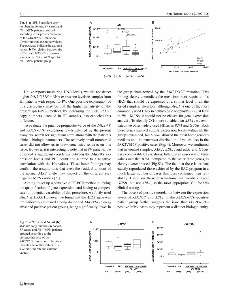
We also found that the ABL1 gene was not uniformlyexpressed among healthy donors and patient groups, beingexpressed at higher levels in donors and SP and JAK2V617F-negative patients compared to JAK2V617F -positive cases(Fig. 4a). A significant positive correlation between the copynumbers of ABL1 and JAK2WT was measured inJAK2V617F-positive patients (Fig. 4b). These observationsraise several doubts about the appropriateness of using ABL1as CG in Ph− MPN patients. Therefore, to identify CGs moresuitable than ABL1 , using the q-RT-PCR protocols optimizedby the EAC program, we evaluated the B2M and GUSBexpression levels in a group of 68 samples (donors=11, SP=5, PV=16, ET=31, and PMF=5) with remaining availableRNA. Both B2M andGUSB showed similar expression levelswithin all the groups examined. However, GUSB showed themost homogeneous medians and the narrowest distribution of
Ann Hematol (2014) 93:609–616 611
controls (FusionQuant® Standards Ipsogen, Qiagen), accord-

values also in the JAJ2V617F -positive cases (Fig. 5a, b)where the expression levels of ABL1 were significantly af-fected. In 11 control samples, we assessed the Ct values ofABL1 , B2M , GUSB , and JAK2 genes. In all cases, the vari-ations in Ct values turned out to be comparable, and all fellwithin three Ct values. However, the Ct values of B2M werelower than those of ABL1 , GUSB , and JAK2 , thus indicatingthe higher level of expression of B2M in these samples(Fig. S3).
In three ET patients, who progressed to PV during thisstudy, we could serially assess at ET diagnosis and PV pro-gression the DNA and RNA JAK2V617F mutational ratiosand the JAK2V617F expression levels normalized with GUS .The clinical–biologic parameters of these three patients arereported in Table 2. As illustrated in Fig. 6, we observed asignificant increase of the JAK2V617F expression levels atPV progression in two of the three cases (case 1 and 3, Fig. 6c)and a trend toward statistical significance in the remaining
Table 1 The diagnostic main clinical–hematologic characteristics of the 105 individuals included in the present study grouped according to diagnosis
Characteristics Donors(n =23)
SP(n =13)
PV (n =22) ET (n =38) PMF (n =9)
JAK2WT/V617F(n =0) (n =22; 100 %)
JAK2WT/V617F(n =9; 19.5 %) (n=29; 80.5 %)
JAK2WT/V617F(n=4) (n =5)
Age, median (years)(range)
45(26–61)
59(27–90)
65(52–88)
63(35–80)
62(38–81)
75(70–80)
68(47–83)
Gender, male/female 16/7 13/0 17/5 4/5 20/29 2/4 3/5
Hb (g/dl),median (range)
15(11.5–16.9)
17(16–18.2)
18(17.6–20.2)
12.5(11.6–14)
15(12–19)
9.6(8.9–10)
15.4(13.8–17.3)
WBC (1×10⁹/l),median (range)
6.6(4.7–9.3)
7.1(4.8–11.4)
10.6(4.4–15)
8.5(6–10)
9.9(7.1–18)
8(1.3–16.8)
20.6(20–10.8)
PLTs (1×10⁹/l),median (range)
232(192–349)
200.5(91–274)
370(75–712)
792(631–1,420)
718(256–1,318)
364(129–489)
555(197–946)
Neutrophils (1×10⁹/l),median (range)
4.4(1.4–7)
4.6(1.4–7.5)
7.9(2.7–13)
4.6(2.8–7.6)
6(1.4–13.5)
5.1(1–9.4)
15.9(8.4–19.5)
JAK2WT copy numbers (1×10³),median (range)
7.7(1.2–38.5)
8.7(2.5–30.9)
6.9(0.02–22.6)
15.6(6.2–34.5)
6.0(0.9–16.7)
11.2(3.8–18.5)
3.0(0.3–4.5)
JAK2V617F copy numbers (1×10³),median (range)
– – 26.9(0.2–203.6)
– 6.9(0.7–52.8)
– 29.4(1.4–78.7)
ABL1 copy number (1×10³),median (range)
4.6(1.6–11.9)
5.0(2.4–20.6)
2.9(0.9–12.7)
7.4(1.8–17.7)
3.8(0.3–8.7)
8.6(4.6–14.9)
3.3(1.7–4.6)
B2M copy number (1×10⁶),median (range)a
8.0(2.8–10.1)
4.8(2.5–13.0)
8.8(2.4–17.7)
6.1(2.4–7.9)
8.1(2.4–17.3)
6.0 7.0(2.3–13.4)
GUSB copy number (1×10³),median (range)a
37.6(7.4–53.2)
34.5(3.2–58.7)
37.8(0.2–56.7)
39.8(8.6–71.6)
33.2(5.8–59.1)
23.4 41.0(19.3–93.5)
aB2M andGUSB copy numbers were detected in donor (n =11), SP (n =5), PV (n =16), ET (WT n =7; V617F n =24), and PMF (WT n =1;V617F n =4)samples
A B
0
AB
SO
LU
TE
CO
PY
NU
MB
ER
JAK2V617F
p=.005
PV ET PMF(n=22) (n=29) (n=5)
AB
SO
LU
TE
CO
PY
NU
MB
ER
DONORS SP JAK2WT JAK2V617FPh-MPN
5x10⁴
JAK2WT
(n=23) (n=13) (n=13) (n=56)
0
p< .001p< .001
p=.002
4x10⁴
3x10⁴
2x10⁴
1x10⁴
5x10⁴
1x10⁵
Fig. 1 JAK2WT andJAK2V617F mRNA expressionlevels detected by q-RT-PCRmethod. a JAK2WT absolutecopy numbers, in donors, SPcases, and Ph− MPN patients. bJAK2V617F absolute copynumbers in JAK2V617F-positivePh− MPN patients. Circlesindicate the outlier values.Asterisks indicate the extremevalues
612 Ann Hematol (2014) 93:609–616

case. By contrast, the JAK2 mutational ratios at DNA andRNAwere increased only in case 1 (Fig. 6a, b and Table 2).
Discussion
Our study shows that, in the diagnostic approach toPh− MPNpatients, a q-RT-PCR assay to detect JAK2V617F expressionlevels offers comparable specificity than ASO-PCR and alleleburden quantitative assays. Moreover, this method is probablymore sensitive than these “standard”methods, as supported bythe fact that the JAK2V617F mutational ratios were steadilysuperior at the RNA than at the DNA level in all the groupsstudied. These latter findings, showing an impact of the typeof nucleic acid on the determination of the JAK2V617F bur-den, are in apparent contrast to those reported by Vannucchiet al. [11] who did not see differences with respect to the use ofRNA or DNA. A likely explanation of these discrepanciesmay rely on the fact that we used buffy-coat preparations,which include platelets instead of the isolated leukocytes.These findings might also suggest that, using RNA, it ispossible to avoid the cost-effective and time-consumingmethods for leukocyte isolation.
The higher sensitivity to detect the JAK2V617F mutation atthe RNA level was firstly demonstrated by the above
mentioned study of Vannucchi et al. [11], who reported anincreased percentage (9 %) of JAK2-mutated ET patients byusing RNA instead of DNA. Thus, the mRNA template shouldbe recommended for the diagnosis of ET patients, usuallypresenting a lower JAK2V617F allele burden, or in the moni-toring of Ph− MPN treatment response.
The present q-RT-PCR method was derived from thatrecently proposed by Merker et al. [17] for the quantificationof the JAK2WT and JAK2V617F transcript levels in Ph−MPNs. However, after testing more than 100 samples ofpatients with and without Ph− MPNs, these authors reporteda low level of nonspecific amplifications in samples contain-ing a high copy number of standard plasmids and in bloodspecimens from patients without Ph− MPNs. To eliminatethese undesired amplifications, Merker et al. [17] established amutant to wild type cutoff of <0.0005.We optimizedMerker’samplification reaction by reducing the amount of final con-centrations of primers from 800 to 300 nM and of probes to400 to 200 nM, respectively. We did not observe nonspecificamplifications in JAK2WT or JAK2V617F reaction by usingplasmid standards, K562 and HEL cell lines, or JAK2V617F-positive and negative samples, tested as positive or negativecontrols (Fig. S1 A, B). The lack of nonspecific amplificationsjustifies the use of this test for diagnostic purposes.
Although the analysis of DNA is in principle technicallysimpler than that of RNA, discrepant results are often reportedeven after DNA-based techniques, so that Lippert et al. [19]have emphasized the need of using positive and negativequality controls, and calibration to a reference standard toimprove reproducibility. More recently, the European Leuke-mia Net/MPN&MPNr-EuroNet group, to avoid that the vari-ations in the performance of the plethora of qPCR assaysroutinely used to detect JAK2V617F could potentially impacton their clinical utility, selected the most sensitive andperforming of nine DNA-based quantitative PCR assays asthe optimal quantitative-polymerase chain reactionmethod forroutine diagnosis and tracking of minimal residual disease inJAK2V617F-associated myeloproliferative neoplasms [20].
DNA
RNA
JAK2V
617F
MU
TAT
ION
AL
BU
RD
EN
RA
TIO
PV ET PMF (n= 12) (n= 17) (n= 3)
Fig. 2 JAK2V617F mutational burden ratio, calculated at the DNA andRNA levels. The circle indicates the outlier values
p= .003R2 = .39n=22
0
30x103
JA2W
T A
BS
OL
UT
E C
OP
Y N
UM
BE
R
0
20x103
20x103
200 400 600 800 1000 1200
Platelets (1x10⁹/L)
30x103
JA2W
T A
BS
OL
UT
E C
OP
Y N
UM
BE
R
0
20x103
20x103
Hemoglobin (gr/dl)
12 14 16 18 20 22
p= .051R2 = .10n=22
BAFig. 3 Correlations between theJAK2WT expression levels andPLT count (a) and Hb levels (b)in the PV patient group
Ann Hematol (2014) 93:609–616 613
Unlike reports measuring DNA levels, we did not detecthigher JAK2V617F mRNA expression levels in samples fromET patients with respect to PV. One possible explanation ofthis discrepancy may be that the higher sensitivity of thepresent q-RT-PCR method, by increasing the JAK2V617Fcopy numbers detected in ET samples, has canceled thisdifference.
To evaluate the putative prognostic value of the JAK2WTand JAK2V617F expression levels detected by the presentassay, we search for significant correlations with the patient’sclinical–biologic parameters. The relatively small number ofcases did not allow us to draw conclusive remarks on thisissue. However, it is interesting to note that in PV patients, weobserved a significant correlation between the JAK2WT ex-pression levels and PLT count and a trend to a negativecorrelation with the Hb values. These latter findings mayconfirm the assumptions that even the residual amount ofthe normal JAK2 allele may impact on the different Ph -negative MPN entities [21].
Aiming to set up a sensitive q-RT-PCR method allowingthe quantification of gene expression, and having to compen-sate for potential variability of this procedure, we firstly usedABL1 as HKG. However, we found that the ABL1 gene wasnot uniformly expressed among donor and JAK2V617F-neg-ative and positive patient groups, being significantly lower in
the group characterized by the JAK2V617F mutation. Thisfinding clearly contradicts the most important requisite of aHKG that should be expressed at a similar level in all thetested samples. Therefore, although ABL1 is one of the mostcommonly used HKG in hematologic neoplasms [22], at leastin Ph− MPNs, it should not be chosen for gene expressionanalysis. To identify CGs more suitable than ABL1 , we eval-uated two other widely used HKGs as B2M and GUSB . Boththese genes showed similar expression levels within all thegroups examined, but GUSB showed the most homogeneousmedians and the narrowest distribution of values also in theJAK2V617F-positive cases (Fig. 4). Moreover, we confirmedthat in control samples, JAK2 , ABL1 , and B2M and GUSBhave comparable Ct variations, falling in all cases within threevalues and that B2M , compared to the other three genes, isclearly overexpressed (Fig.S3). The fact that these latter dataexactly reproduced those achieved by the EAC program in amuch larger number of cases than ours confirmed their reli-ability. Based on these observations, we would suggestGUSB , but not ABL1 , as the most appropriate GC for thisclinical setting.
The observed positive correlation between the expressionlevels of JAK2WT and ABL1 in the JAK2V617F -positivepatient group further suggests the issue that JAK2V617F-positive MPN cases may represent a distinct biologic entity.
A B
AB
SO
LU
TE
CO
PY
NU
MB
ER
3x10⁴
DONORS SP JAK2WT JAK2V617FPh -MPN
2x10⁴
1x10⁴
0
ABLp= .003
p= .001p< .001
(n=23) (n=13) (n=13) (n=56)
ABL ABSOLUTE COPY NUMBER
JA2W
T A
BS
OL
UT
E C
OP
Y N
UM
BE
R
n=
0 15x10310x1035x103
20x103
10x103
0
R2=.52P<.0001
56
Fig. 4 a ABL-1 absolute copynumbers in donors, SP cases, andPh− MPN patients groupedaccording to the presence/absenceof the JAK2V617F mutation.Circles indicate the outlier values.The asterisks indicate the extremevalues. b Correlation between theABL-1 and JAK2WT expressionlevels in the JAK2V617F-positivePh− MPN patient group
DONORS SP JAK2WT JAK2V617FPh- MPN
(n= 11) (n=5) (n=8) (n=44)
2x10⁷
1x10⁷
0
AB
SO
LU
TE
CO
PY
NU
MB
ER
B2M
DONORS SP JAK2WT JAK2V617FPh- MPN
(n= 11) (n=5) (n=8) (n=44)
AB
SO
LU
TE
CO
PY
NU
MB
ER
4x10⁵
3x10⁵
2x10⁵
1x10⁵
0
GUSBBAFig. 5 B2M (a) and GUSB (b)
absolute copy numbers in donors,SP cases, and Ph− MPN patientsgrouped according to thepresence/absence of theJAK2V617F mutation. The circleindicates the outlier values. Theasterisks indicate the extremevalues
614 Ann Hematol (2014) 93:609–616

In addition, this finding may support the model recentlyproposed by Irino et al. [23]. These authors, focusing on genesinvolved in the JAK-STAT signaling pathway, identified twoupregulated genes in MPN patients: SOCS3 , a known targetof the JAK-STAT axis and a potentially novel target, andSPI1 , encoding PU.1 . The latter gene is a regulator of prolif-eration and differentiation of hematopoietic cells [24]. Thepathogenic effect of JAK2 mutation appears mediated, at leastin part, through the upregulation of PU.1. In addition, theyshowed that PU.1 is regulated by both JAK2 and ABL1 , andsuggested that SOCS3 and PU.1 are common downstreamtargets of both JAK2 and ABL1 . Together, these observationsand our results link the constitutive activation of JAK2 to thedownregulation of the expression of ABL1 .
The potential usefulness of GUSB as CG might also besupported by the additional findings observed in the three ETpatients who progressed to PV. The serial monitoring of theDNA and RNA JAK2V617F mutational ratios and theJAK2V617F expression levels normalized with GUSBallowed to observe a significant increase of the JAK2V617Fexpression levels at PV progression in two of the three cases(case 1 and 3, Fig. 6c) and a trend toward statistical
significance in the remaining case. By contrast, the JAK2mutational ratios at DNA and RNA were increased only incase 1 (Fig. 6a, b). These data support in humans the findingsrecently demonstrated in transgenic mice showing that thelevels of JAK2V617F expression influence the Ph− MPNphenotype: higher levels favor erythrocytosis whereas lowerlevels favor thrombocytosis [12]. Similarly, Barosi et al. ob-served a progression toward JAK2V617F homozygosity inserial DNA samples collected from 64 patients with PMF[25].
In conclusion, the q-RT-PCR assay hereby reported is asensitive and accurate method to quantify the JAK2 mutation-al status that can also show clinical correlations suggesting theimpact of the residual amount of the JAK2WT allele on the Ph− MPN disease phenotype. In addition, for the first time, weprovided evidences that ABL1 is not a useful CG to obtainreliable JAK2V617F quantifications in Ph− MPN patientswhereas GUSB turned out be more appropriate for this pur-pose. These findings might become clinically relevant in lightof the availability of several new and effective targeted ther-apies which require sensitive and precise assessment of thepatient’s response to treatments.
Table 2 The hematologic characteristics of the three ET patients who progressed to PV referred at the time of ET diagnosis and at PV progression (pPV)
Characteristics Case 1 Case 2 Case 3
ET pPV ET pPV ET pPV
WBC (1×10⁹/l), median (range) 9.9 26.2 7.1 12 9.2 10.69
PLTs (1×10⁹/l), median (range) 696 1,003 1,184 1,500 519 516
Neutrophils (1×10⁹/l), median (range) 7.5 21 5.6 7.6 5.4 6.73
JAK2WT copy number (1×10³), mean ± SEM 5.8±0.7 1±0.01 5.4±0.25 3.7±0.2 3.2±0.08 7.1±1
JAK2V617F copy number (1×10³), mean ± SEM 1.3±0.3 5.7±0.5 2.6±0.1 3.6±0.6 4.1±0.5 10.2±0.3
DNA mutational burden ratio 22 69 29.4 28 24 21
mRNA mutational burden ratio 18 85 32 49 56 59
B2M copy number (1×10³), mean ± SEM 1,751±27 2,256±500 1,974±75 1,953±71 1,376±142 1,749±163
GUSB copy number (1×10³), mean ± SEM 15.5±0.6 10.4±1.1 15.6±0.8 16.1±0.3 7.0±0.06 6.9±0.2
PE
RC
EN
TAG
E
A B
1
3
5
7
9
11
13
15
C
p<.0001
p<.0001
p=.06
CASE 1
ET PV
CASE 2
ET PV
CASE 3
ET PV
CASE 1
ET PVCASE 2
ET PVCASE 3
ET PVCASE 1
ET PVCASE 2
ET PVCASE 3
ET PV
1
3
5
7
9
11
13
15
1
3
5
7
9
11
13
15
Fig. 6 The JAK2V617Fmutational burden ratio in thethree ET patients who progressedto PVevaluated by the followingthree different methods: a theabsolute allele-specific PCR, bthe present absolute allele-specific q-RT-PCR, and c usingGUSB to normalize the valuesachieved by the present q-RT-PCR assay
Ann Hematol (2014) 93:609–616 615
Acknowledgments This work was partially supported by researchfunding from the University of Roma “La Sapienza” (to C.N. e G.C.),Fondazione Roma (to C.N. e G.C.), Ministero dell’Istruzione, dell’Universitàe della Ricerca (PRIN), and the Italian Association for Cancer Research (IG-11949 to C.N.).
Conflict of interest The authors report no potential conflicts of interest.
Open Access This article is distributed under the terms of the CreativeCommons Attribution License which permits any use, distribution, andreproduction in any medium, provided the original author(s) and thesource are credited.
References
1. Lacout C, Pisani DF, Tulliez M, Gachelin FM, Vainchenker W,Villeval JL (2006) JAK2V617F expression in murine hematopoieticcells leads to MPD mimicking human PV with secondary myelofi-brosis. Blood 108:1652–1660
2. Abdel-Wahab O (2011) Genetics of the myeloproliferative neo-plasms. Curr Opin Hematol 18:117–123
3. Klampfl T, Harutyunyan A, Berg T, Gisslinger B, Schalling M,Bagienski K, Olcaydu D, Passamonti F, Rumi E, Pietra D, Jäger R,Pieri L, Guglielmelli P, Iacobucci I, Martinelli G, Cazzola M,Vannucchi AM, Gisslinger H, Kralovics R (2011) Genome integrityof myeloproliferative neoplasms in chronic phase and during diseaseprogression. Blood 118:167–176
4. Vainchenker W, Delhommeau F, Constantinescu SN, Bernard OA(2011) New mutations and pathogenesis of myeloproliferative neo-plasms. Blood 118:1723–1735
5. Zhan H, Cardozo C, YuW,Wang A, Moliterno AR, Dang CV, SpivakJL (2013) MicroRNA deregulation in polycythemia vera and essentialthrombocythemia patients. Blood Cells Mol Dis 50:190–195
6. Shafer AI (2006) Molecular basis of the diagnosis and treatment ofpolycythemia vera and essential thrombocythemia. Blood 107:4214–4222
7. Passamonti F, Rumi E (2009) Clinical relevance of JAK2 (V617F)mutant allele burden. Haematologica 94:7–10
8. Vannucchi AM, Pieri L, Susini MC, Guglielmelli P (2012) BCR-ABL1-negative chronic myeloid neoplasms: an update on manage-ment techniques. Future Oncol 8:575–593
9. Kim HR, Choi HJ, Kim YK, Kim HJ, Shin JH, Suh SP, Ryang DW,Shin MG (2013) Allelic expression imbalance of JAK2 V617Fmutation in BCR-ABL negative myeloproliferative neoplasms.PLoS ONE 8:512–518
10. Zhao R, Xing S, Li Z, Fu X, Li Q, Krantz SB, Zhao ZJ (2005)Identification of an acquired JAK2 mutation in polycythemia vera. JBiol Chem 280:22788–22792
11. Vannucchi AM, Pancrazzi A, Bogani C, Antonioli E, Guglielmelli P(2006) A quantitative assay for JAK2(V617F) mutation in myelopro-liferative disorders by ARMS-PCR and capillary electrophoresis.Leukemia 20:1055–1060
12. Skoda RC, Tiedt R, Hao-Shen H, Sobas M, Looser R, Dirnhofer S,Schwaller J (2008) Ratio of mutant JAK2-V617F to wild-type Jak2determines the MPD and phenotypes in transgenic mice. Blood 111:3931–3940
13. Thiele J, Kvasnicka HM (2009) The 2008 WHO diagnostic criteriafor polycythemia vera, essential thrombocythemia, and primary my-elofibrosis. Curr Hematol Malignancy Rep 4:33–40
14. Chomczynski P, Sacchi N (1987) Single-step method of RNA isola-tion by acid guanidinium thiocyanate-phenol-chloroform extraction.Anal Biochem 162:156–159
15. Elia L, Gottardi E, Floriddia G, Grillo R, Ciambelli F, Luciani M,Chiusolo P, Invernizzi R, Meloni G, Foà R, Saglio G, Cimino G
(2004) Retrospective comparison of qualitative and quantitative re-verse transcriptase polymerase chain reaction in diagnosing andmonitoring the ALL1-AF4 fusion transcript in patients with acutelymphoblastic leukaemia. Leukemia 18:1824–1830
16. Baxter EJ, Scott LM, Campbell P, East C, Fourouclas N, Swanton S,Vassiliou GS, Bench AJ, Boyd EM, Curtin N, Scott MA, Erber WN,Green AR (2005) Acquired mutation of the tyrosine kinase JAK2 inhuman myeloproliferative disorders. Lancet 365:1054–1061
17. Merker JD, Jones CD, Oh ST, Schrijver I, Gotlib J, Zehnder L (2010)Design and evaluation of a real-time PCR assay for quantification ofJAK2 V617F and wild-type JAK2 transcript levels in the clinicallaboratory. J Mol Diagn 12:58–64
18. Beillard E, Pallisgaard N, van der Velden VHJ, Bi W, Dee R, van derSchoot E, Delabesse E, Macintyre E, Gottardi E, Saglio G,WatzingerF, Lion T, van Dongen JJ, Hokland P, Gabert J (2003) Evaluation ofcandidate control genes for diagnosis and residual disease detectionin leukemic patients using ‘real-time’ quantitative reverse-transcriptase polymerase chain reaction (RQ-PCR)—a EuropeAgainst Cancer program. Leukemia 17:2474–2486
19. Lippert E, Girodon F, Hammond E, Jelinek J, Reading NS, Fehse B,Hanlon K, Hermans M, Richard C, Swierczek S, Ugo V, Carillo S,Harrivel V, Marzac C, Pietra D, Sobas M, Mounier M, Migeon M,Ellard S, Kröger N, Herrmann R, Prchal JT, Skoda RC, Hermouet S(2009) Concordance of assays designed for the quantification ofJAK2V617F: a multicenter study. Haematologica 94:38–45
20 . Jovanov ic JV, Ivey A, Vannucch i AM, Lippe r t E ,OppligerLeibundgut E, Cassinat B, Pallisgaard N, Maroc N,Hermouet S, Nickless G, Guglielmelli P, van der Reijden BA,Jansen JH, Alpermann T, Schnittger S, Bench A, Tobal K, WilkinsB, Cuthill K, McLornan D, Yeoman K, Akiki S, Bryon J, Jeffries S,Jones A, Percy MJ, Schwemmers S, Gruender A, Kelley TW,Reading S, Pancrazzi A, McMullin MF, Pahl HL, Cross NC,Harrison CN, Prchal JT, Chomienne C, Kiladjian JJ, Barbui T,Grimwade D (2013) Establishing optimal quantitative-polymerasechain reaction assays for routine diagnosis and tracking of minimalresidual disease in JAK2-V617F-associated myeloproliferative neo-plasms: a joint European Leukemia Net/MPN&MPNr-EuroNet(COST action BM0902) study. Leukemia 27:2032–2039
21. Campbell PJ, Scott LM, Buck G, Wheatley K, East CL, Marsden JT,Duffy A, Boyd EM, Bench AJ, Scott MA, Vassiliou GS, MilliganDW, Smith SR, Erber WN, Bareford D, Wilkins BS, Reilly JT,Harrison CN, Green AR (2005) Definition of subtypes of essentialthrombocythaemia and relation to polycythaemia vera based onJAK2 V617F mutation status: a prospective study. Lancet 366:1945–1953
22. Gabert J, Beillard E, van der Velden VHJ, Bi W, Grimwade D,Pallisgaard N, Barbany G, Cazzaniga G, Cayuela JM, Cavé H,Pane F, Aerts JL, De Micheli D, Thirion X, Pradel V, González M,Viehmann S, Malec M, Saglio G, van Dongen JJ (2003)Standardization and quality control studies of ‘real-time’ quantitativereverse transcriptase polymerase chain reaction of fusion gene tran-scripts for residual disease detection in leukemia—a Europe AgainstCancer Program. Leukemia 17:2318–2357
23. Irino T, Uemura M, Yamane H, Umemura S, Utsumi T, Kakazu N,Shirakawa T, Ito M, Suzuki T, Kinoshita K (2011) JAK2 V617F-dependent upregulation of PU.1 expression in the peripheral blood ofmyeloproliferative neoplasm patients. PloS ONE 6:2214822
24. Koschmieder S, Rosenbauer F, Steidl U, Owens BM, Tenen DG(2005) Role of transcription factors C/EBPalpha and PU.1 in normalhematopoiesis and leukemia. Int J Hematol 81:368–377
25. Barosi G, Bergamaschi G, Marchetti M, Vannucchi AM,Guglielmelli P, Antonioli E, Massa M, Rosti V, Campanelli R,Villani L, Viarengo G, Gattoni E, Gerli G, Specchia G, Tinelli C,Rambaldi A, Barbui T (2007) JAK2 V617F mutational status pre-dicts progression to large splenomegaly and leukemic transformationin primary myelofibrosis. Blood 110:4030–4036
616 Ann Hematol (2014) 93:609–616