Embed Size (px)
Citation preview
A Laboratory Class Exploring and Classifying Anoxygenic PhototrophicBacteria Using Culture-Based Approaches, Microscopy, and Pigment Analysis
Resource Type: Curriculum: Laboratory
Publication Date: 11/17/2005
Authors
Sarah BoomerNatural Sciences and MathWestern Oregon UniversityMonmouth, Oregon 97361USAEmail: [email protected]
Kelly ShipleyWestern Oregon UniversityMonmouth, Oregon 97361USAEmail: [email protected]
Abstract
In this exercise, students set up bottle enrichments for phototrophic microbes using river sediment and then, usingrepresentative controls, they view and describe wet mounts of representative oxygenic algae and cyanobacteria andanoxygenic purple bacteria. Following 2 to 3 weeks of incubation, students characterize pigmented enrichments using basicmicroscopy and pigment extraction and analysis, the latter of which requires a spectrophotometer. Using provided tables andtheir textbook, they classify enriched phototrophs using pigment absorption data, cell shape and organization, and growthconditions such as the presence or absence of sulfur.
Activity
Invitation for User Feedback. If you have used the activity and would like to provide feedback, please send an e-mail [email protected]. Feedback can include ideas which complement the activity and new approaches forimplementing the activity. Your comments will be added to the activity under a separate section labeled "Feedback."Comments may be edited.
INTRODUCTION
Learning Objectives.
Upon completion of this activity, students will be able to(1) better understand anoxygenic phototrophic bacteria based on direct enrichment and observation from local soil or riverenvironments;(2) compare and contrast anoxygenic phototrophs with more familiar oxygenic phototrophs (e.g., plants, algae);(3) generate and use molecular pigment spectrum data; and(4) combine a variety of microbial phenotypes (pigment profiles, microscopy, growth conditions) to classify unknownphototrophs from natural environments.
Background.This laboratory exercise has been carried out at Western Oregon University. It was included as one component of our posterentitled "A General Microbiology Lab Curriculum Featuring Culture-Dependent and -Independent Approaches andComputer-Based Project Presentations" at the 2004 American Society for Microbiology General Meeting. Here, it receivedconsiderable attention from undergraduate microbiology instructors interested in environmental enrichment lab exercises.Although comparable undergraduate pigment-based exercises exist, most involve oxygenic plants or algae (3, 4, 5). We havebeen unable to find examples of undergraduate labs that combine bacterial culture-driven procedures with pigmentassessment for the purpose of classification. Although these approaches are less exact for genus-level identification than,say, solving "unknown" enteric isolates using media-based tests and phenotypic charts, they provide valuable models forenriching from a natural mixed population, for exposure to relevant culture-independent phenotypic assessment (e.g.,spectrophotometry and microscopy), and for studying environmentally important and beneficial microbes.
This curriculum was first implemented in 1997 for the laboratory component of General Microbiology (Biology 331), a coursethat all Biology majors are required to take. Students complete this laboratory exercise during the first month of the10-week course, concurrent with extensive lectures about microbial diversity, ecology, and metabolism (with an entirelecture devoted to microbial phototrophs and diversity). The course requires Brock Biology of Microorganisms(2), an
MicrobeLibrary http://archive.microbelibrary.org/edzine/details_print.asp?id=2016&lang=
1 of 5 3/13/2012 3:11 PM
invaluable resource for bacterial diversity, particularly during this exercise. To take this course, students have also had topass 1 year of introductory Principles of Biology (Biology 211-213), the laboratory component of which includes pigmentassessment of spinach extracts using a spectrophotometer (3).
The same instructor (Boomer) who has developed and delivered lectures also runs the laboratory; first-person portions ofthis report refer to Boomer. Students receive all instruction materials at the beginning of the term. During the firstlaboratory session, students collect local mud and/or river samples and set up replicate bottle enrichments using both sulfurand nonsulfur media. Two to three weeks later, the typical time it takes bottles to develop visible color (either green orpurple-red), students perform pigment isolation and microscopy on positive enrichments. Prelab lecturing is limited to anoverview of specific methods and a review of microbial phototrophs, emphasizing key phenotypic traits that will be used toidentify enriched isolates to the phylum level.
PROCEDURE
Materials.
This exercise can be run with two to four people working together. Each team needs:
Session One
4 g of freshwater mud or soil (sandy mud or soil should be avoided)4 clear glass bottles with screw caps or tight-fitting corks (ours hold 200 ml of liquid)Microscopy controls: Spirogyra, Anabaena, Rhodospirillum (for microscopic viewing)aBasic light microscopy supplies: microscope, standard slides, and coverslips1 L of each of the following liquid media (protocol written for 200-ml bottles, some extra for spillage):
Nonsulfur phototroph media Sulfur phototroph media5.0 g of NaHCO3 1.0 g of NH4Cl2.0 g of NaCl 1.0 g of KH2PO4
1.0 g of (NH4)2SO4 0.5 g of MgCl20.5 g of K2HPO4 2.0 g of NaHCO3
0.1 g of MgSO4 1.0 g of Na2S-9H2O2.0 g of yeast extract 1.0 g of NaCl0.1 g of Na2S-9H2O
aThese samples are all available fresh from biological supply companies (e.g., Ward's). Alternatively, prepared slides may beused, although these have been stained during preservation and colors do not accurately reflect the natural color of livingcells.
Session Two
Basic light microscopy supplies: microscope, standard slides, and coverslipsMicrocentrifuge (capable of 10,000 rpm) that holds 1- to 2-ml size microcentrifuge tubesPipettes that fit into 200-ml bottles (disposable plastic pipettes are adequate)Microcentrifuge tubes (this procedure is written for 1- to 2-ml size)Methanol (with gloves, waste beakers, and hood for evaporation and disposal)Spectrophotometersb and appropriate cuvettes
bGiven the variety of available spectrophotometers, a complete discussion of this piece of equipment is beyond the scope ofthis curriculum. The least expensive spectrophotometers, used by freshmen in our introductory biology courses, requiremanual setting, repetitious zeroing between data collection, and data recording and graphing by hand; Morgan and Carterprovide a thorough explanation of basic spectrophotometer operation (3). More expensive spectrophotometers, whichadvanced students use in this course lab, are fully digital, allow simultaneous analysis of the experimental sample and theblank, and provide automatic graphical output of the data. Instructors should carefully research their options if they do notcurrently own a spectrophotometer and cuvettes.
Student Version.Lab Session OneAppendix 1. Collection and EnrichmentLab Session TwoAppendix 2. Pigment Assessment
Instructor Version.This class extends over two sessions that are separated by a mandatory 2- to 3-week enrichment wait period. Each sessionis 2 to 3 hours in duration. For this course, students have previous experience using centrifuges, spectrophotometers, andmicroscopes. Although these students typically take 2 hours to complete these exercises, students lacking comparableexperience should be provided 3 hours. If students have not been exposed to information about photosynthetic microbialdiversity, an additional hour of lecture time should be developed to address these topics.
Session OneIn session one, students set up bottle enrichments for anoxygenic phototrophs (both sulfur and nonsulfur), and review and
MicrobeLibrary http://archive.microbelibrary.org/edzine/details_print.asp?id=2016&lang=
2 of 5 3/13/2012 3:11 PM
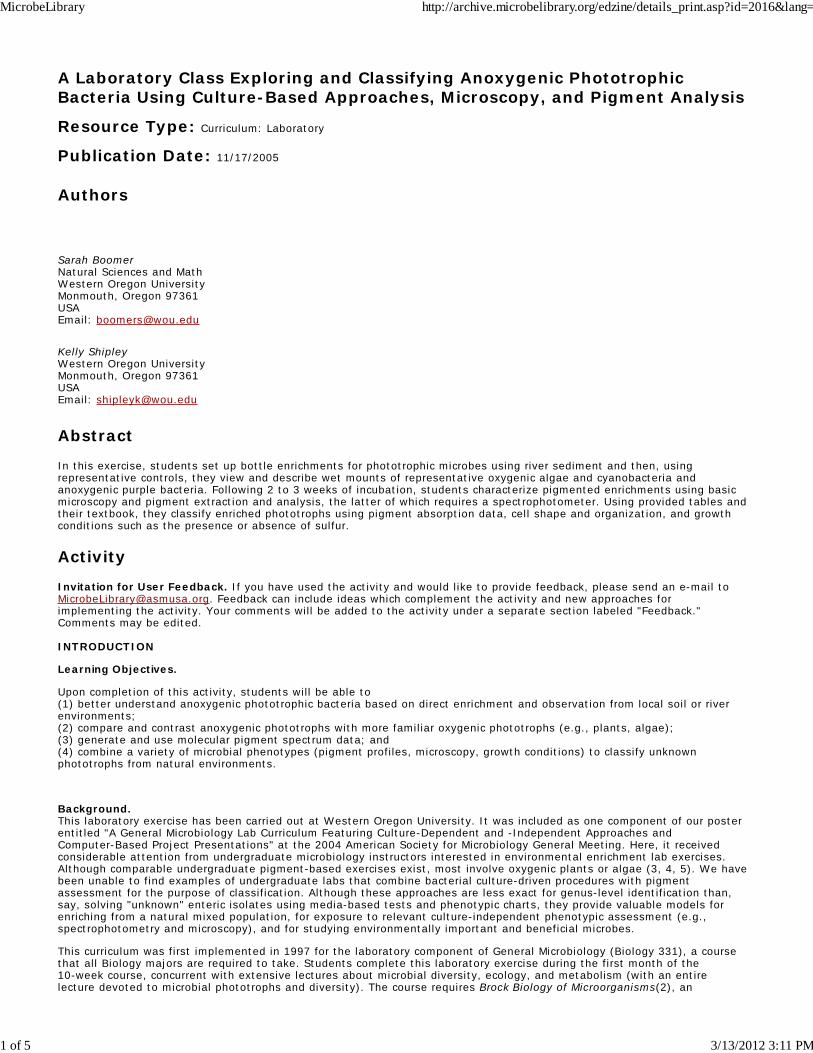
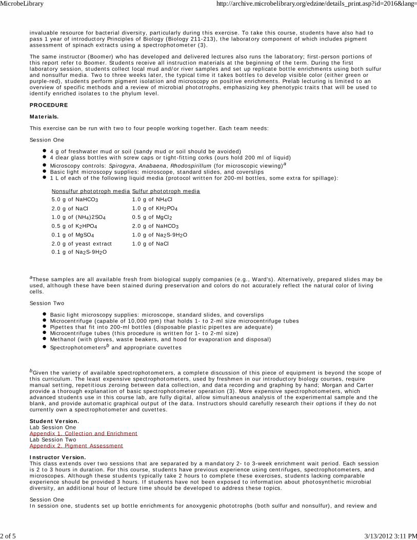
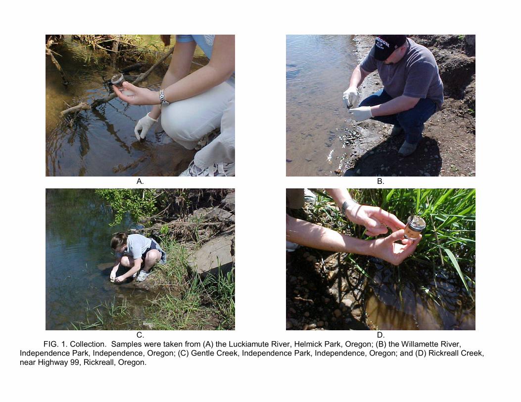
view control phototrophs: oxygenic algae (Spirogyra) and cyanobacteria (Anabaena), and anoxygenic purple nonsulfurbacteria (Rhodospirillum). I provide interested students with the option of collecting river sediment and mud, a 2- to 3-miledriving excursion to local rivers an hour before lab begins. The three to five interested volunteer participants typically enjoythis optional field trip. Instructors who do not have access to nearby rivers can alternatively collect from any moist soil area.This exercise is messy, and I try to provide student teams with large plastic containers over which they can pour andinoculate materials. I also recommend that students wear gloves during the collection and inoculation steps given that,minimally, local rivers harbor agricultural run-off from many farms, both dairy and heavily-fertilized grass seed operations. Ialso carry antibacterial wipes to collection sites for students to wash off. Representative student-derived photographs ofmud collection at the four local rivers or creeks (Luckiamute, Willamette, Rickreall, and Gentle) we sample, taken with digitalcameras provided for class use and intended only for simple presentations (i.e., low-resolution), are shown in Fig. 1. Arepresentative student-derived photograph of bottle enrichments is shown in the top panel of Fig. 2.
FIG. 1. Collection
FIG. 2. Bottle enrichments
After enrichments are set up, students view and draw the three aforementioned control phototrophs. Although students haveperformed basic microscopy before, it is often necessary to review good wet mount technique and the kinds of informationthese preparations provide. Students accurately draw the entire field of view for a given sample, emphasizing the relativesize, shape, and organization of the microbial samples. Comparing large, filamentous eukaryotic Spirogyra (with its visiblechloroplasts) with smaller, filamentous prokaryotic Anabaena at the same magnification typically provides meaningfulevidence about the differences between these two cell types. Observant students will notice abundant heterocysts in theAnabaena sample, reminding them about nitrogen fixation (previously covered in lecture and lab). Viewing spiral, unicellular,and motile Rhodospirilla is frustrating for some students. Although liquid cultures of this microbe appear macroscopicallypurple-red, it is challenging to see this color microscopically. Its size and unicellular organization, however, will be evident,contrasting with both the algae and cyanobacteria. Given previous lectures, I avoid assisting students with the "ConceptReview Check" summary table in favor of making them synthesize information from their notes and text. A sample key isprovided for this worksheet in Appendix 3.
Appendix 3. Concept Review Check key
Session TwoOver the 2 to 3 weeks between sessions, students should have made at least one weekly record of their enrichments in theirterm-long informal notebooks. In general, most retrieved cultures (60%) are purple, with a 50:50 split between sulfur andnonsulfur. Our most frequent failures have involved sandy inoculum, prevalent at one local river site that I now avoid. Manybottles change dramatically over time, turning black, then slightly green, and then fully purple-red. Occasionally, we havealso retrieved green bottles that, upon closer inspection (in our case, under ultraviolet light), reveal fluorescent chlorophylls(i.e., cyanobacteria or algae). This finding indicates one of two possibilities: (i) that bottles were not capped properly,allowing oxygen into the system; or (ii) that oxygenic phototrophs were performing photosynthesis using only PSII (whichdoesn't require oxygen).
In general, one to two students should analyze one enrichment sample. In most cases, team enrichments adequatelyprovide two to three samples, allowing larger teams to divide into smaller groups for pigment and microscopy exercises.Given that bottle phototrophs often form biofilms on the glass, students will be challenged by the retrieval process, whichinvolves careful pipette scraping and extracting. Many students incorrectly assume that they can simply place a pipette inthe center of the bottle, squeeze the bulb, and withdraw the same color they see through the glass. Students also need toallow solid material to settle at the end of the pipette before adding it to the microcentrifuge tube or their final collectionwill be too dilute. In the event that their first sample attempt is too dilute, students can centrifuge the sample (using thesame settings as those for methanol extraction), discard the supernatant, and add more sample. In any case, collection ishighly variable, requiring manual skill and patience. Thus, students should be encouraged to show the instructor their finalsample before proceeding.
Although students have few problems with methanol extraction procedures, they often have questions about their wetmounts, which typically appear less uniform as compared with previously-viewed controls. Initially, I steer them towardmaking certain they are focused on a visibly colored mass, as opposed to an open and dispersed area where color is not asdiscernable. Once students concentrate on these colored regions, emphasizing cells along their peripheries, they havealways reported unicellular rods or ovals. To date, we have yet to isolate any filaments or spirilla. If students seemextremely frustrated and confused about shape, I have them prepare and view heat-fixed smears and simple stains of theirsamples.
Instructors will have to make necessary time amendments for spectrophotometer analysis. If students have never used aspectrophotometer and if only manual spectrophotometers are available, instructors should add 45 to 60 minutes to thesecond lab session. It is also possible to freeze methanol extracts for a few days (so long as they are wrapped in foil andnot exposed to light), allowing instructors to add a third lab session if need be. If this option is used, extracts should beanalyzed within 1 week.
Beyond the Lab Homework ActivitiesOnce student teams have completed both lab sessions and worksheets, instructors may wish to provide or assign additionalindividual student activities to encourage a broader understanding of these organisms in the environment, in an evolutionarycontext, and in current research. Three recommended activities include: (i) describing specific ecological role(s) their isolatesperform in the environment, using both text information and internet resources they search and cite; (ii) speculating on theevolutionary significance of their team isolates, using both text information and internet resources they search and cite; and(iii) locating and summarizing a current primary research article that involves one specific genus that is related to one oftheir isolates. For the third activity, students should be specifically encouraged to use htttp://www.asm.org, selectingpublications/journals. For environmental and general research papers, I typically recommend searching titles or abstracts ofApplied and Environmental Microbiology and Journal of Bacteriology using a specific genus name. Students should berequired to print and attach selected articles and, minimally, briefly summarize the questions being addressed, majorfindings, and what they think is significant about the research.
MicrobeLibrary http://archive.microbelibrary.org/edzine/details_print.asp?id=2016&lang=
3 of 5 3/13/2012 3:11 PM
Safety Issues. Pathogenic organismsAs stated, local river beaches we collect from contain, minimally, coliforms from untreated agricultural and dairy run-off; weknow this because we collect and enumerate coliforms from adjacent river water during another lab exercise performedearlier in the term (typically, we observe coliforms on the order of 102-3/L). Consequently, students are required to weargloves (we use nonlatex given allergy issues) and clean hands immediately (either using antibacterial wipes in the field orwashing hands in the lab) while handling these potential pathogens. All materials contaminated with mud inoculum arebagged and autoclaved immediately. A key reason we favor bottle enrichments over Winogradsky columns (described later inthe Possible Modications section) is because the latter involve substantially more inoculum and potential exposure topathogens. Although moist soil provides an alternative inoculum, this probably also contains potential pathgens (e.g., inmanure, animal feces, etc.) and the same precautions need to be exercised during collection, inoculation, and disposal. MethanolPrior to using methanol, I review safety issues concerning this toxic solvent. In particular, I stress that methanol can befatal if ingested (because it readily converts to formaldehyde, a highly toxic tissue fixative that most students have heardabout) and can cause skin and eye irritations. Given these safety issues, students, according to Material Safety Data Sheetrecommendations, are required to wear gloves (again, we use nonlatex) when handling this solvent. Each table is providedwith transfer pipettes and only a small amount of methanol (3 ml) that I have previously aliquoted. As recommended for allmaterials, students are strongly cautioned to keep all microfuge tubes capped as they work to avoid unnecessary exposureand contamination. Each table is also provided a labeled waste beaker and required to place all methanol waste in thiscontainer during the lab. At the end of the lab, teams move their waste to our lab fume hood for methanol evaporation anddisposal.
ML Safety Statement regarding Environmental Isolates
The Curriculum Resources Committee recognizes that isolated organisms can be a powerful learning tool as well as apotential biological hazard. We strongly recommend that:
· Environmental enrichment laboratories should only be performed in classes in which students have been trained towork at a BSL2.
· Direct environmental samples (eg. soil, water) which are known to contain infectious organisms should be handledaccording to the biosafety level of that infectious agent.
· Cultures of enriched microorganisms, derived from environmental samples, should be handled using Biosafety Level2 precautions.
· Mixed, enriched or pure cultures of microorganisms from environmental samples with a significant probability ofcontaining infectious agents should be manipulated in a biosafety cabinet if available.
· Where possible, media used for the enrichment of environmental isolates should contain an appropriate anti-fungalagent.
· Instructors should be aware if they are teaching in regions with endemic fungi capable of causing systemicinfections, and should avoid environmental isolations.
ASSESSMENT and OUTCOMES
Suggestions for Assessment.Individual students each turn in completed copies of the Microscopy and Concept Check Worksheet (15 points). Studentteams turn in completed Phenotypic Charts with attached pigment data print-outs (15 points). The combined value of thisassignment is 10% of the lab assignment grade (30/300 total points). Additionally, 10% of the lab exam (15/150 totalpoints) covers this lab exercise.
Field Testing.Since being developed in 1997, approximately 160 junior- or senior-level undergraduate Biology majors have completed thiscurriculum. Most students (50 to 60%) were pursuing careers in the health sciences. The remaining students sought careersin secondary education and research (academic, government, or biotechnology).
Student Data.Since 2004, we have completed assessment of lab curricula in General Microbiology, which serves a maximum of 16 studentsper term. Twenty-two students rated this curriculum on a 10-point scale in Fall 2003 and Spring 2004, as summarized below:Please rate the statement: This lab... Average ratingmade connections beyond microbiology. 8.4improved my awareness of microbial diversity. 9improved my interest in microbial diversity. 7.7enhanced my interest in scientific research. 8.1enhanced my ability to use computers. 7.5exposed me to new technology. 8.1enhanced my organizational skills. 8.2enhanced my writing skills. 7.7My overall rating of this lab is 9
SUPPLEMENTARY MATERIALS
Possible Modifications.This exercise can be modified in many ways, some based on different methods, equipment, and available time, and somebased on analyzing research specimens taken from thermal photosynthetic mats collected in Yellowstone National Park (1).
MicrobeLibrary http://archive.microbelibrary.org/edzine/details_print.asp?id=2016&lang=
4 of 5 3/13/2012 3:11 PM
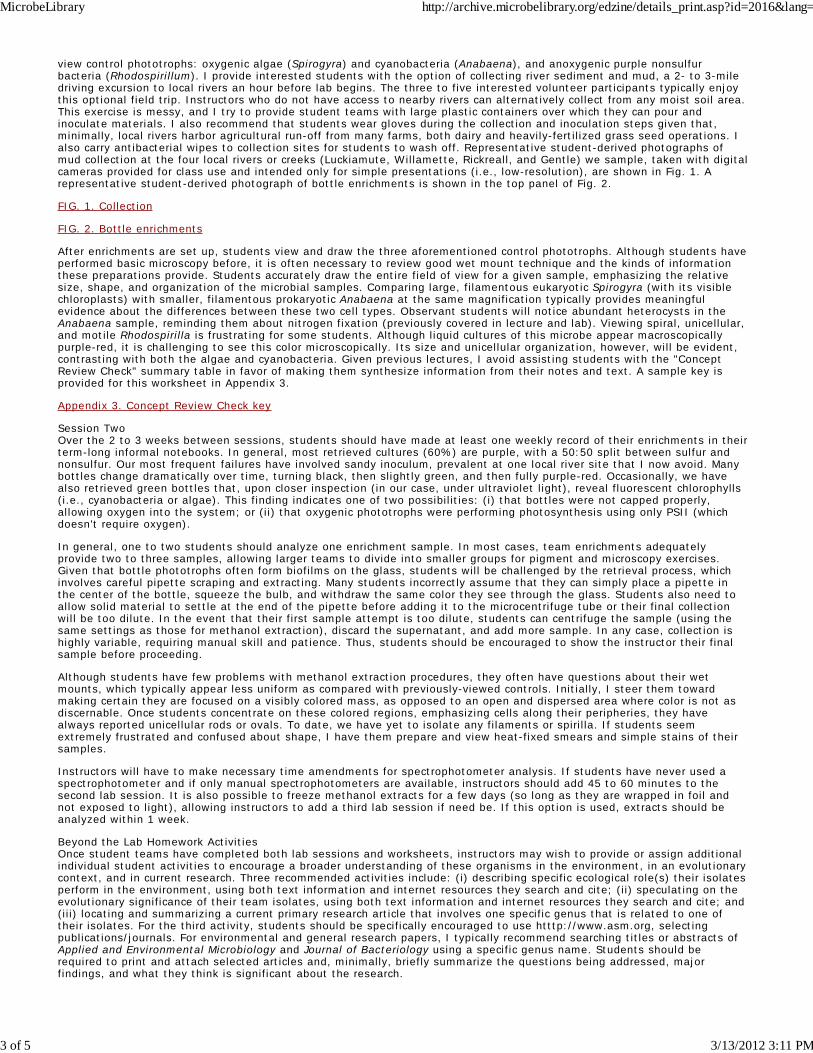
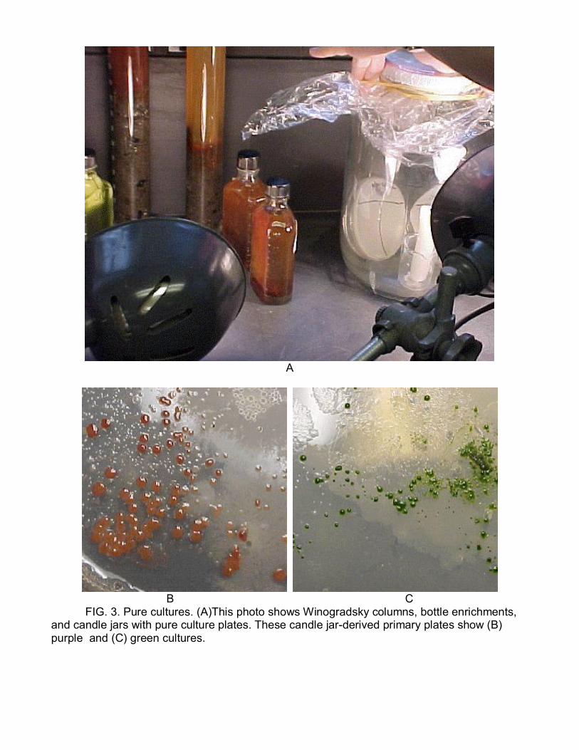
Modification One. The Winogradsky columnDeveloped by S. Winogradsky in the late 1800s to study sulfur cycling bacteria, the Winogradsky column provides a simplemicrobial ecosystem for observing and enriching both oxygenic and anoxygenic phototrophs. Winogradsky columns arewell-described in most textbooks and set-up procedures are included in many microbiology lab manuals (2, 6). Although werecommend it for set-up and observational purposes (based on actively carrying it out from 1997-2000, Fig. 3, top panel), wehave found that it is difficult to work with in terms of performing postenrichment sampling for microscopy and pigmentanalysis. In general, we have found that columns typically develop two zones of activity over the course of 2 to 3 weeks: anupper oxic region that is green (cyanobacteria and/or algae) and a lower anoxic region that is green or purple (anoxygenicgreen or purple bacteria that usually, but not always, represent sulfur groups). This modification would add 30 to 60 minutesto each session.
FIG. 3. Pure cultures
Modification Two. Pure culturesIt is also possible to use streak-plating methods to purify colonies of bottle-enriched phototrophs. For this modification,agar plates (20 g of agar per liter) corresponding to each kind of media (sulfur and nonsulfur) must be prepared in advanceof session two. Using loops, students streak colored material from bottles onto appropriate agar plates (three to fourreplicates are recommended per student). Because these are anoxygenic phototrophs, they must be grown in the absence ofoxygen. We have successfully used both inexpensive candle jars as well as anaerobic chambers (e.g., GasPak Jar Systemsavailable from Fisher Scientific). When we have done this lab modification, most teams require at least two week-longsubculturing times to fully purify phototrophs. A candle jar set-up with two resulting primary plates is shown in Fig. 3.
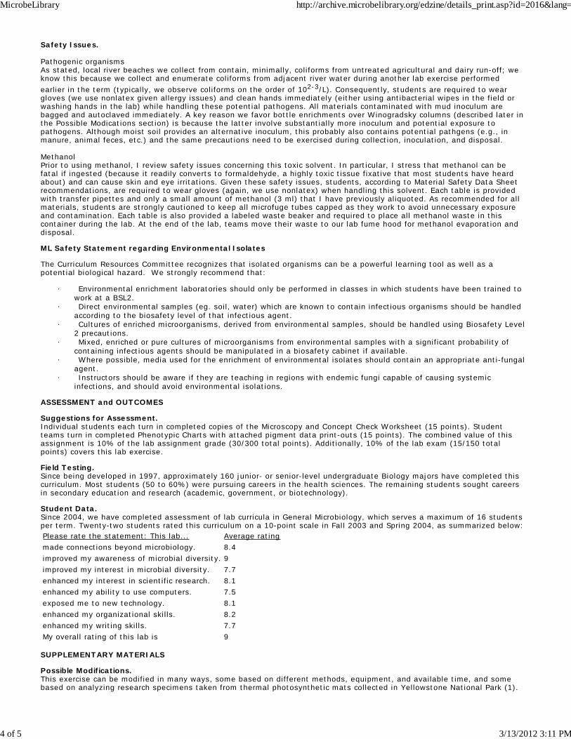
Modification Three. Alternative phototroph sourcesBecause we are involved in a long-term research project to identify new thermophilic phototrophs from nonsulfur bacterialmat communities in Yellowstone National Park, we also have students analyze original research samples using pigmentanalysis and microscopy, both light and UV-fluorescence (chlorophyll fluoresce red under UV, bacteriochlorophyll emit greento no fluorescence). The particular communities we study are composed of two distinct layers of phototrophs: top or surfacecyanobacteria (typically unicellular Synechococcus) and lower or deeper red filaments (new members of the Chloroflexiphylum). This ecosystem layering mirrors similar structures found in deep lakes, sediments, and Winogradsky columns. Somerepresentative images and data are shown in Fig. 4.
FIG. 4. Yellowstone National Park phototroph images and data
Acknowledgments.This work was supported, in part, by a National Science Foundation Microbial Observatories/Research at UndergraduateInstitute grant (NSF-MO/RUI 0237167). I wish to thank Bryan Dutton for helping to edit this manuscript, and Jim Staley andJohn Leigh for introducing me to bottle enrichment methods and media via a graduate-level microbial ecology course at theUniversity of Washington in 1989.
References.
Boomer, S. M., D. P. Lodge, B. E. Dutton, and B. Pierson. 2002. Molecular characterization of novel red greennonsulfur bacteria from five distinct hot spring communities in Yellowstone National Park. Appl. Environ. Microbiol.68:346-355.
1.
Madigan, M. T., J. M. Martinko, and J. Parker. 2003. Brock biology of microorganisms, 10th ed. Prentice Hall, UpperSaddle River, N.J.
2.
Morgan, J. G., and M. E. B. Carter. 2002. Investigating biology, 4th ed. Addison Wesley Longman, Inc., Menlo Park,Calif.
3.
Motten, A. F. 1995. Diversity of photosynthetic pigments. Proceedings of the 16th Workshop/Conference of theAssociation for Biology Laboratory Education 16:81-98.
4.
Taras, L. B. 2003. Promoting student involvement with environmental laboratory experiments in a generalmicrobiology course. Microbiol. Educ. 4:23-29.
5.
Wistreich, G. A. 2003. Microbiology laboratory fundamentals and applications, 2/E. Prentice Hall, Upper Saddle River,N.J.
6.
MicrobeLibrary http://archive.microbelibrary.org/edzine/details_print.asp?id=2016&lang=
5 of 5 3/13/2012 3:11 PM
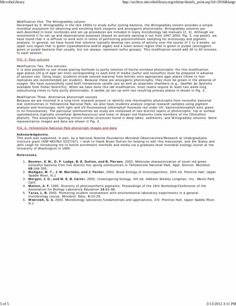
APPENDIX 1. Anoxygenic Phototrophs Week One—Collection and Enrichment
Introduction to Anoxygenic Phototrophs Anoxygenic phototrophs include members of the following bacterial phyla: Proteobacteria (both purple sulfur and nonsulfur), Chloroflexi (green nonsulfur), Chlorobi (green sulfur), and Heliobacteria (gram positives). Review your text and lecture notes about phototrophs, contrasting the major features of oxygenic photosynthesis (as performed by plants, algae, and cyanobacteria) with those of anoxygenic photosynthesis (as performed by the bacteria listed above). Today, you will set up enrichments for anoxygenic phototrophs. In 2 to 3 weeks, you will use the following phenotypic traits to classify them: sulfur utilization, pigment absorption data, and cell morphology.
ACTIVITIES—ENRICHMENTS USING MUD
Bottle Enrichments 4 g of mud from one of four local rivers will be collected per team. There is an optional field trip before lab. Add 1 g of mud to each of two bottles, one labeled S (sulfur) and the other NS (nonsulfur). Put your initials on the bottles. Fill the "S" bottle to the top with sulfur media. Cap or cork tightly. Fill the “NS” bottle to the top with nonsulfur media. Cap or cork tightly. Place bottles in the light (can use a 40W bulb placed 10 to 15 cm from the bottles) and grow for 2 to 3 weeks. During enrichment, take time each lab session to note and record color changes.
ACTIVITIES—MICROSCOPY and DIVERSITY
Phototroph Wet Mounts Wet mounts are good for viewing pigmented microbes such as Spirogyra, Anabaena, and Rhodospirillum. Adding too much material or water to the slide will result in the inability to focus, because everything will be moving too much. Using a pipette or tweezers, withdraw a small amount of material making sure the sample contains colored filaments. Place filaments in the middle of the slide, add half a drop of sourcewater, and cover with a coverslip. View Spirogyra and Anabaena under the microscope using 40x magnification; note color, shape, cell organization, and motility. View Rhodospirillum under the microscope using 40x magnification; you may be able to view at 100x without oil. Complete colored drawings of each organism in the space provided, depicting the entire field of view and showing relative sizes. When finished, complete concept check information on the next page.
Microscopy and Concept Check Name: _____________________________
Spirogyra, 40x objective Spirogyra Concept Check
Microbial domain
Microbial phylum
Primary pigment(s)
Specialized membranes?
Oxygenic or anoxygenic?
Number of photosystems
Typical habitat(s)
Anabaena, 40x objective Anabaena Concept Check
Microbial domain
Microbial phylum
Primary pigment(s)
Specialized membranes?
Oxygenic or anoxygenic?
Number of photosystems
Typical habitat(s)
Rhodospirillum, 40x objective Rhodospirillum Concept Check
Microbial domain
Microbial phylum
Primary pigment(s)
Specialized membranes?
Oxygenic or anoxygenic?
Number of photosystems
Typical habitat(s)
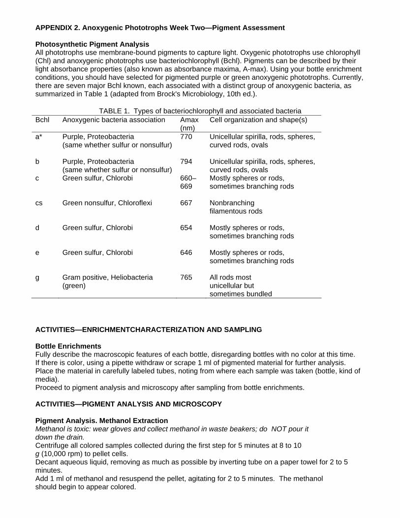
APPENDIX 2. Anoxygenic Phototrophs Week Two—Pigment Assessment Photosynthetic Pigment Analysis All phototrophs use membrane-bound pigments to capture light. Oxygenic phototrophs use chlorophyll (Chl) and anoxygenic phototrophs use bacteriochlorophyll (Bchl). Pigments can be described by their light absorbance properties (also known as absorbance maxima, A-max). Using your bottle enrichment conditions, you should have selected for pigmented purple or green anoxygenic phototrophs. Currently, there are seven major Bchl known, each associated with a distinct group of anoxygenic bacteria, as summarized in Table 1 (adapted from Brock's Microbiology, 10th ed.).
TABLE 1. Types of bacteriochlorophyll and associated bacteria Bchl Anoxygenic bacteria association Amax
(nm) Cell organization and shape(s)
a* Purple, Proteobacteria (same whether sulfur or nonsulfur)
770 Unicellular spirilla, rods, spheres, curved rods, ovals
b Purple, Proteobacteria (same whether sulfur or nonsulfur)
794 Unicellular spirilla, rods, spheres, curved rods, ovals
c Green sulfur, Chlorobi 660–669
Mostly spheres or rods, sometimes branching rods
cs
Green nonsulfur, Chloroflexi 667 Nonbranching filamentous rods
d Green sulfur, Chlorobi 654 Mostly spheres or rods, sometimes branching rods
e Green sulfur, Chlorobi 646 Mostly spheres or rods, sometimes branching rods
g Gram positive, Heliobacteria (green)
765 All rods most unicellular but sometimes bundled
ACTIVITIES—ENRICHMENTCHARACTERIZATION AND SAMPLING Bottle Enrichments Fully describe the macroscopic features of each bottle, disregarding bottles with no color at this time. If there is color, using a pipette withdraw or scrape 1 ml of pigmented material for further analysis. Place the material in carefully labeled tubes, noting from where each sample was taken (bottle, kind of media). Proceed to pigment analysis and microscopy after sampling from bottle enrichments. ACTIVITIES—PIGMENT ANALYSIS AND MICROSCOPY Pigment Analysis. Methanol Extraction Methanol is toxic: wear gloves and collect methanol in waste beakers; do NOT pour it down the drain. Centrifuge all colored samples collected during the first step for 5 minutes at 8 to 10 g (10,000 rpm) to pellet cells. Decant aqueous liquid, removing as much as possible by inverting tube on a paper towel for 2 to 5 minutes. Add 1 ml of methanol and resuspend the pellet, agitating for 2 to 5 minutes. The methanol should begin to appear colored.
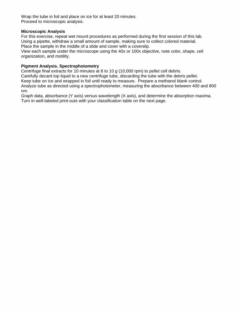
Wrap the tube in foil and place on ice for at least 20 minutes. Proceed to microscopic analysis. Microscopic Analysis For this exercise, repeat wet mount procedures as performed during the first session of this lab. Using a pipette, withdraw a small amount of sample, making sure to collect colored material. Place the sample in the middle of a slide and cover with a coverslip. View each sample under the microscope using the 40x or 100x objective, note color, shape, cell organization, and motility. Pigment Analysis. Spectrophotometry Centrifuge final extracts for 10 minutes at 8 to 10 g (10,000 rpm) to pellet cell debris. Carefully decant top liquid to a new centrifuge tube, discarding the tube with the debris pellet. Keep tube on ice and wrapped in foil until ready to measure. Prepare a methanol blank control. Analyze tube as directed using a spectrophotometer, measuring the absorbance between 400 and 800 nm. Graph data, absorbance (Y axis) versus wavelength (X axis), and determine the absorption maxima. Turn in well-labeled print-outs with your classification table on the next page.
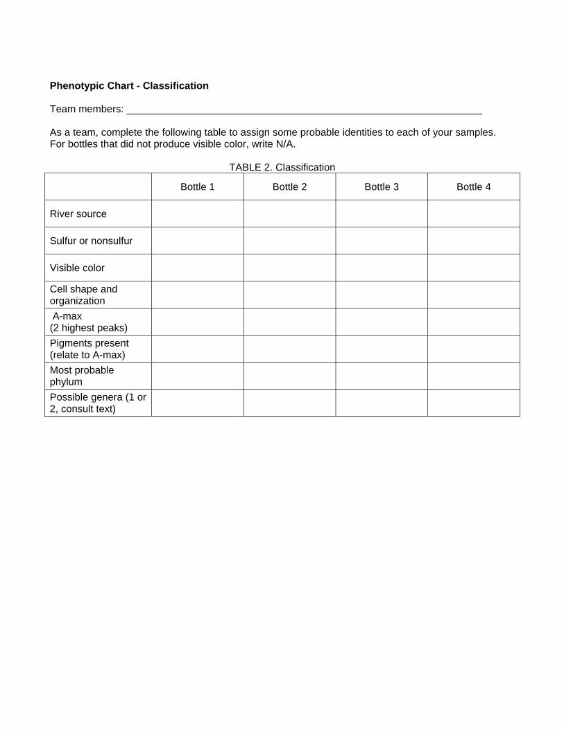
Phenotypic Chart - Classification Team members: _______________________________________________________________ As a team, complete the following table to assign some probable identities to each of your samples. For bottles that did not produce visible color, write N/A.
TABLE 2. Classification
Bottle 1 Bottle 2 Bottle 3 Bottle 4
River source
Sulfur or nonsulfur
Visible color
Cell shape and organization
A-max (2 highest peaks)
Pigments present (relate to A-max)
Most probable phylum
Possible genera (1 or 2, consult text)
A. B.
C. D. FIG. 1. Collection. Samples were taken from (A) the Luckiamute River, Helmick Park, Oregon; (B) the Willamette River,
Independence Park, Independence, Oregon; (C) Gentle Creek, Independence Park, Independence, Oregon; and (D) Rickreall Creek, near Highway 99, Rickreall, Oregon.
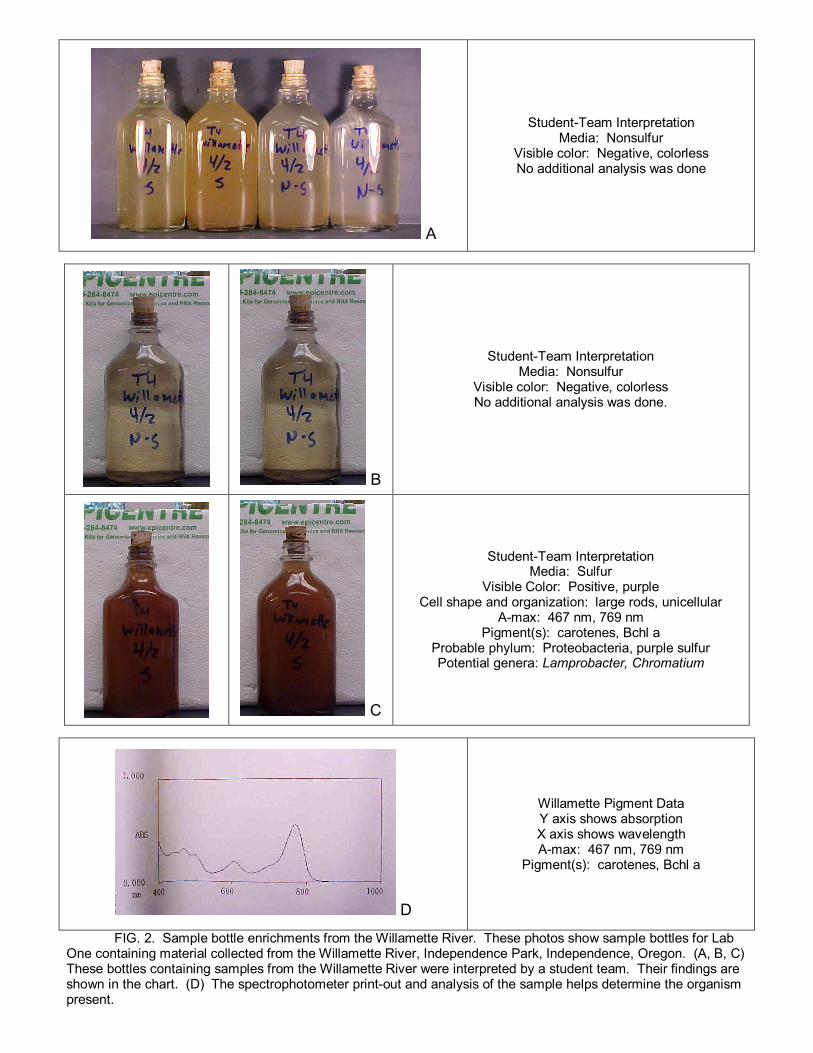
A
StudentTeam Interpretation Media: Nonsulfur
Visible color: Negative, colorless No additional analysis was done
B
StudentTeam Interpretation Media: Nonsulfur
Visible color: Negative, colorless No additional analysis was done.
C
StudentTeam Interpretation Media: Sulfur
Visible Color: Positive, purple Cell shape and organization: large rods, unicellular
Amax: 467 nm, 769 nm Pigment(s): carotenes, Bchl a
Probable phylum: Proteobacteria, purple sulfur Potential genera: Lamprobacter, Chromatium
D
Willamette Pigment Data Y axis shows absorption X axis shows wavelength Amax: 467 nm, 769 nm
Pigment(s): carotenes, Bchl a
FIG. 2. Sample bottle enrichments from the Willamette River. These photos show sample bottles for Lab One containing material collected from the Willamette River, Independence Park, Independence, Oregon. (A, B, C) These bottles containing samples from the Willamette River were interpreted by a student team. Their findings are shown in the chart. (D) The spectrophotometer printout and analysis of the sample helps determine the organism present.
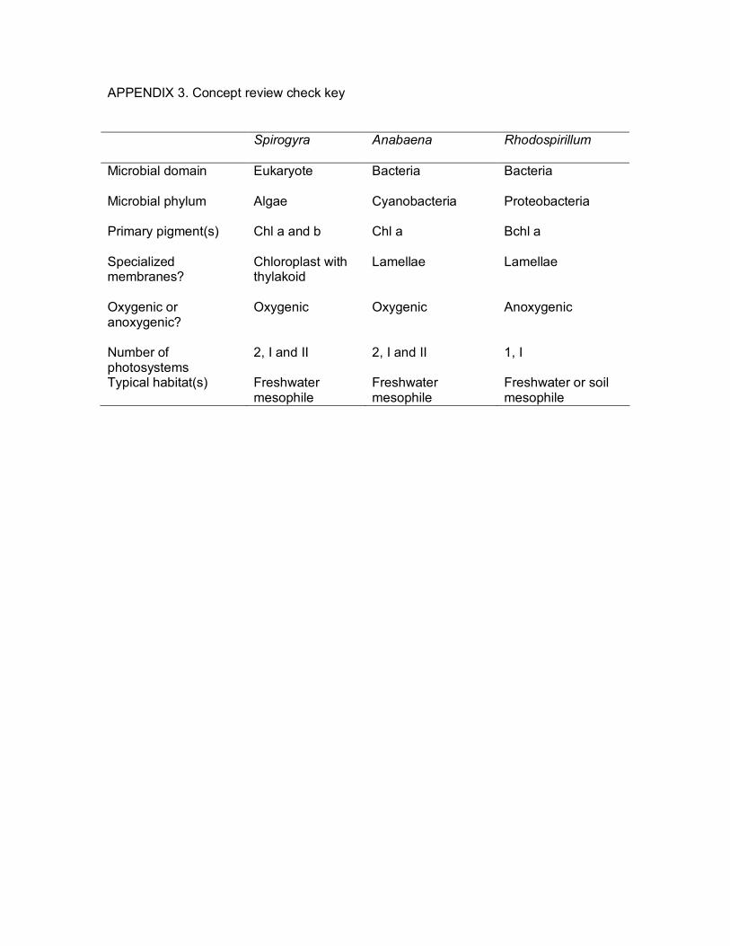
APPENDIX 3. Concept review check key
Spirogyra Anabaena Rhodospirillum
Microbial domain Eukaryote Bacteria Bacteria
Microbial phylum Algae Cyanobacteria Proteobacteria
Primary pigment(s) Chl a and b Chl a Bchl a
Specialized membranes?
Chloroplast with thylakoid
Lamellae Lamellae
Oxygenic or anoxygenic?
Oxygenic Oxygenic Anoxygenic
Number of photosystems
2, I and II 2, I and II 1, I
Typical habitat(s) Freshwater mesophile
Freshwater mesophile
Freshwater or soil mesophile
A
B C FIG. 3. Pure cultures. (A)This photo shows Winogradsky columns, bottle enrichments,
and candle jars with pure culture plates. These candle jarderived primary plates show (B) purple and (C) green cultures.

Green Layer Light Microscopy Green Layer UV/Fluorescence Green Layer Major Pigments
A
440480 nm 660665 nm
Red Layer Light Microscopy Red Layer UV/Fluorescence Red Layer Major Pigments
B C
440480 nm 760790 nm
FIG. 4. Yellowstone phototroph images and data. (A) This photo shows sampling at a Yellowstone site. (B) A whole mat core with green upper layer and red lower layer is shown. (C) This chart shows the microscopy images and spectrophotometer information for each layer.