Embed Size (px)
Citation preview

A Malignant Cystosarcoma Phyllodes With Positive Estrogen Receptor and its Heterotransplantation Into Nude Mice
YUTAKA TOKUDA, MD, MlTSUHlRO KUBOTA, MD, YOSHITO UEYAMA, MD,*t KOJl MARUO,? JUN-CHI HATA, MD,* NORIKAZU TAMAOKI, MD,* TOMOO TAJIMA, MD, AND TOSHIO MITOMI, MD
We detected cytosol estrogen receptors in a malignant cystosarcoma phyllodes and succeeded in serial transplantations into athymic nude mice. The tumors of this transplantable strain (MC-3-JCK) have the same histologic features as the original tumors, and retain significant amounts of cytosol estrogen receptors. This strain appears to provide a useful experimental model for the study of biologic and therapeutic aspects of human cystosarcoma phyllodes of the breast.
Cancer 55:370-374, 1985.
YSTOSARCOMA PHYLLODES of the breast was first C described and established as a disease entity in 1838 by Miiller, who believed the lesion was entirely benign in character.l Since the first malignant case was reported by Lee and Pack in 1931; numerous subse- quent reports have proved the malignant potential of this tumor. Metastases have been reported to occur in 6.2% to 17% of all cases of cystosarcoma phyl l~des .~ ,~ These metastasizing cases usually run a fatal course, and effective treatment has not yet been established.
In 1977, Rao and Meyer demonstrated the presence of progesterone receptors in a cystosarcoma phyllodes and suggested the possibility of effective endocrine the rap^.^ However, the presence of specific steroid hormone re- ceptors and the hormone dependency of this disease re- main controversial.6-8
Recently, we detected estrogen receptors in a malig- nant cystosarcoma phyllodes and succeeded in trans- plantation into athymic nude mice together with signifi- cant amounts of estrogen receptors. The current report deals with details of the establishment and properties of this serially transplantable human cystosarcoma phyl- lodes in nude mice.
Case Report
A 27-year-old, unmamed, nulliparous woman noted a tumor in the left breast 1 year before admission. Since then, it had grown very rapidly. On her admission to Tokai University Hospital on January 10, 1982, there was a large, tender mass occupying the entire left breast. The overlying skin was adher- ent and reddened. A mastectomy with removal of the axillary contents and the major pectoral muscle was performed on January 18, 1982. The original tumor measured 22X 19 X 12 cm in diameter. The bulging cut surface was grayish white and lobulated, without cystic change.
The tumor recurred beneath the skin graft immediately after the mastectomy. Palliative extended tumorectomy with left internal mammary dissection and pectoral myocutaneous flap was performed on February 16, 1982. The tumor soon re- curred and grew large. A chest roentgenogram showed proba- ble infiltration of both the pleura and the lungs. Radiation therapy was applied to the recurrent tumor of the chest wall, and the antiestrogen tamoxifen was administered systemati- cally with no response. The disease progressed, and the patient died on March 23, 1982.
At autopsy, metastases were found in both lungs with in- volvement of the pleura; bilateral adrenal glands; thoracic and lumbar vertebrae; and mediastinal, perigastric, and peripan- creatic lymph nodes.
From the Department of Surgery I1 and *Pathology, School of Medi- cine, Tokai University, Kanagawa, and 'Central Institute for Experi- mental Animals, Kawasaki, Japan.
Supported by a Tokai medical research grant. Address for reprints: Yutaka Tokuda, MD, Department of Surgery
11, School of Medicine, Tokai University, Bohseidai, Isehara, Kana- gawa 259- 1 1, Japan.
The authors thank Mr. Kazuo Hayashi of Teijin Bio-Science Labo- ratories for the receptor analyses, Miss Kumiko Ishikawa for her chro- mosomal analysis, and Miss Kayoko Mochizuki for preparation ofthis manuscript.
Accepted for publication December 3 I , 1983.
Materials and Methods
Mice
Male and female nude mice, 4 to 6 weeks of age, with a C57BL/6N genetic background, were obtained from the Central Institute for Experimental Animals, Kawasalu, Japan. Mice were maintained under specific-pathogen- free conditions.
370

No. 2 CYSTOSARCOMA PHYLLODES IN NUDE MICE Tokuda et al. 37 1
Heterotransplantation Into Nude Mice
Solid fragments of both primary and recurrent tumors obtained in the operations were injected subcutaneously into nude mice. When the transplanted tumor reached I cm or more at its greatest width, serial transfer was performed.
Examination cf Transplunted Tumors
Three dimensions of the tumors were measured by calipers after inoculation. The tumor volume was calcu- lated by the formula of Battelle Columbus Laboratorie~,~ and the growth curve was drawn.
Both original and transplanted tumor tissues were processed for routine light and electron microscopy, and chromosomal analysis of the transplanted tumors was performed to identify their origin.
Estrogen Receptors
Estrogen receptor content of the tumor was analyzed using sucrose density gradientdo and dextran-coated charcoal.” The tumor was frozen in liquid nitrogen and pulverized using a Spex homogenizer. The tumor powder was homogenized in four volumes of cold 5 mmol/l phosphate buffer, pH 7.5, containing 0.5 mmol/l dithiothreitol and 10% glycerol, using a Teflon glass homogenizer. Cytosol was prepared from the ho- mogenate by centrifugation at 2000 X g for 20 minutes followed by 65,000 X gfor 1 hour. The supernatant pro- tein content was measured by the Coomassie dye method. l 2
For the sucrose gradient analysis of estrogen receptors, aliquots of 500 pl of supernatant were incubated for 20 hours at 4°C with 1.5 nmol/l (2, 4, 6, 7-3H) estradiol (specific activity; 85 Ci/mmol; New England Nuclear, Boston, Massachusetts). Parallel control incubations also contained 200-fold excess of cold hormone in addi- tion to the radioactive hormone. Three hundred micro- liters were layered on 5% to 20% sucrose gradient and centrifuged at 4°C for 17 hours at 2 16,000 X g. Frac- tions, five drops per fraction, were collected from the bottoms, and radioactivity was determined by a liquid scintillation counter. Sedimentation coefficients were determined using carbon 14 ( 14C) labeled bovine serum albumin and gamma-globulin standards.
For the saturation analysis of estrogen receptors with dextran-coated charcoal, 250 pl of the cytosol were in- cubated with radioactive estradiol in increasing concen- trations (0.12--6.4 rimol/l) for 20 hours at 4°C. Parallel control incubations at each concentration contained 200-fold excess of dimethylstilbestrol added immedi-
FIG. I . Primary tumor showing subepithelial cellulanty (H & E, X 150).
ately before the radioactive hormone. Non-protein- bound free steroid was removed by 0.5% Norit A (dex- tran-treated), and bound radioactivity was determined. Calculations of the dissociation constant and binding capacity were performed by the method of Scatchard.I3
Results
Multiple sections of the original tumor demonstrated the pattern of an intracanalicular fibroma with many compressed and distorted ducts lined by uniform epithe- lial cells in moderately cellular portions that were quite
FIG. 2. Primary tumor: a dense stromal portion with an obviously malignant pleomorphic pattern (H & E, X300).
372 CANCER January 15 1985 Vol. 55
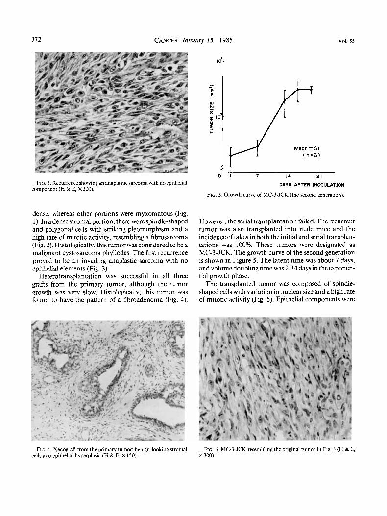
FIG. 3. Recurrence showing an anaplastic sarcoma with no epithelial component (H & E, X 300).
dense, whereas other portions were myxomatous (Fig. 1). In a dense stromal portion, there were spindle-shaped and polygonal cells with striking pleomorphism and a high rate of mitotic activity, resembling a fibrosarcoma (Fig. 2). Histologically, this tumor was considered to be a malignant cystosarcoma phyllodes. The first recurrence proved to be an invading anaplastic sarcoma with no epithelial elements (Fig. 3).
Heterotransplantation was successful in all three grafts from the primary tumor, although the tumor growth was very slow. Histologically, this tumor was found to have the pattern of a fibroadenoma (Fig. 4).
M e a n f S E ( n . 6 )
1 . 0 1 7 14 21
DAYS AFTER INOCULATION
FIG. 5 . Growth curve of MC-3-JCK (the second generation).
However, the serial transplantation failed. The recurrent tumor was also transplanted into nude mice and the incidence of takes in both the initial and serial transplan- tations was 100%. These tumors were designated as MC-3-JCK. The growth curve of the second generation is shown in Figure 5. The latent time was about 7 days, and volume doubling time was 2.34 days in the exponen- tial growth phase.
The transplanted tumor was composed of spindle- shaped cells with variation in nuclear size and a high rate of mitotic activity (Fig. 6). Epithelial components were
FIG. 4. Xenograft from the primary tumor: benign-looking stromal FIG. 6. MC-3-JCK resembling the original tumor in Fig. 3 (H & E, cells and epithelial hyperplasia (H & E, X 150). X 300).

No. 2 CYSTOSARCOMA PHYLLODES IN NUDE MICE Tokudu et ul.
lo4-
373
TABLE I . Estrogen Receptor Analysis
Binding capacity* Sedimentation Dissociation (fmiol/mg coefficient? constant* protein) (Svedberg) (nmol/l)
_ _ ~ . -
Original tumor 24 4 alone 0.05 Xenograft,
1st passage (female mice) 22 4 alone 2
2nd passage (female mice) 8 4 alone 0.7 (male mice) 15 4 alone 0.8
* Charcoal was iused to separate the bound from unbound steroid. Binding capacity and dissociation constant values were calculated from Scatchard plots.
t Sedimentation coefficiient values were calculated from the result of sucrose gradient centrifugation analysis.
Xenograft,
~ _ _ _ _ . -
not found in any of the sections. These histologic fea- tures were similar to those of the original tumor.
A chromosome study of the transplanted tumor showed a human female chromosome constitution and marked variation in chromosome numbers.
Cytosol estrogen receptor analysis of both recurrent and transplanted tumors was performed. The results are shown in Table 1. Both original and xenografted tumors contained specific cytosol receptors for estradiol. Estra- diol binding capacity was 24 fmol/mg of cytosol protein for the original tumor, and 22 and 8 fmol for the first and second passage of xenografts, respectively. The value of the dissociation constant calculated from the Scatchard plot for these tumors was less than 1 nmol/l except for the first passage tumor. Sucrose gradient centrifugation analysis showed that all of the tumor had only a 4s binding component (Fig. 7).
There is no significant difference in growth rate of the transplanted tumorts in male and female mice (Fig. 8).
Discussion
Currently, the management of malignant cystosar- coma phyllodes has discouraging results irrespective of therapeutic efforts, and it is difficult to evaluate a new therapeutic approach requiring critical analysis of a large number of cases since the incidence of this disease is quite low.
Histologically, this transplantable tumor is very simi- lar to the original tumor. Biologically, the recurrent tumor on the (chest ,wall of the patient grew rapidly, with a calculated volume doubling time of about 2 days, which is almost coincident with that of xenografted
8 s
1 4.6 S
4
5 10 15 20 25
TOP FRACTION NUMBER BOTTOM
FIG. 7. Estrogen receptor analysis by sucrose density gradient tech- nique. This pattern was found for all tumors. There is peak (4s) specific binding (open circles), which is inhibited by excess of cold hormone (closed circles).
Meanf SE Female ( n . 6 )
0 Male ( n . 6 ) 1 1
0 1 7 14 21
DAYS AFTER INOCULATION
FIG. 8. Growth curves of MC-3-JCK in male and female mice (the second generation).

374 CANCER January I5 1985 VOl. 55
tumors. Furthermore, our serially transplantable strain retains cytosol estrogen receptors. Thus, this tumor will be a good experimental model for studying the therapeu- tic aspects of this disease.
In 1977, Rao and Meyer demonstrated cytosol pro- gesterone receptors in cystosarcoma phyllodes for the first time.5 They suggested that the progesterone recep- tors are localized in the stromal cells and estrogen recep- tors are localized in epithelial rather than stromal cells.8 However, Palshof and colleagues demonstrated estradiol binding proteins in two patients with this disease and suggested that they are located in the stromal compo- nents.' Porton and Poortman also supported this sugges- tion in their recent report.14 We detected estrogen recep- tors in the original and transplanted tumors that contained no epithelial components.
On the other hand, Kesterson and colleagues evalu- ated six cases of this tumor by steroid receptor analysis and indicated the presence of a nonsaturable estrogen and progesterone 4s binding protein rather than a spe- cific receptor.6 It was a specific 4s estrogen binder with a high affinity for estradiol which we detected, however, although its binding capacity was low, and it is probably different from a type I1 site, which has a low affinity and a high binding capa~ity. '~ However, our preliminary ex- periment with this serially transplantable strain did not show any evidence of its hormone responsiveness. This discrepancy between the presence of specific receptors and hormone responsiveness should be resolved to de- termine the therapeutic implications of these receptors. For solving these problems, our strain will provide us with a potential preclinical model.
REFERENCES 1. Muller J. Uber den feinern Ban und die Formen der krankhafter
Geschwiilste, Lfg. I. Berlin: Reimer, 1838. 2. Lee BJ, Pack CT. Giant intracanalicular fibroadenomyxoma of
the breast: The so-called cystosarcoma phyllodes mammae of Jo- hannes Miiller. Ann Surg 1931; 93:250-268.
3. Hagensen CD. Disease of the Breast, ed. 2. Philadelphia: WB Saunders, 197 1 ; 227 -249.
4. Noms HJ, Taylor HB. Relationship of histologic features to be- havior of cystosarcoma phyllodes: Analysis of ninety-four cases. Cancer 1967; 20:2090-2099.
5. Rao BR, Meyer JS. Progesterone receptor in cystosarcoma phyl- lodes. Arch Surg 1977; 112:620-622.
6. Kesterson GHD, Georgeade N, Seigler HF, Barton TK, McCarty KS Sr, McCarty KS Jr. Cystosarcoma phyllodes: A steroid receptor and ultrastructure analysis. Ann Surg 1979; 190:640-647.
7. PalshofT, Blichert-Toft M, Daehnfeldt JL et al. Estradiol binding protein in cystosarcoma phyllodes of the breast. Eur J Cancer 1980;
..~ 8: Rao BR, Meyer JS, Fry CG. Most cysiosarcoma phyllodes and fibroadenomas have progesterone receptor but lack estrogen receptor: Stromal localization of progesterone receptor. Cancer 198 1; 47:2016- 202 1.
9. Ovejera A& Houchens DP. Selection of potential anticancer agents using human tumor xenografts in athymic nude mice. Contract
10. Gore-Langton RE. Ashwood-Smith MT, Algard FT, Van Net- ten JP. A thin-layer gel filtration assay of cytoplasmic oestrogen recep- tors: A possible screening method for hormone dependent tumors. BrJ Cancer 1973; 28:310-315.
1 1. McGuire WL, DeLaGarza M. Improved sensitivity in the mea- surement of estrogen receptor in human breast cancer. J Clin Endo- crinol Metab 1973; 37:986-989.
12. Bradford MM. A rapid and sensitive method for the quantita- tion of microgram quantities of protein utilizing the principle of pro- tein-dye binding. Anal Biochem 1976; 72:248-254.
13. Scatchard G. The attraction of proteins for small molecules and ions. Ann NYAcad Sci 1949; 51:660-672.
14. Porton WM, Poortman J. Estrogen receptors in cystosarcoma phyllodesofthe breast. EurJCancer 1981; 17:1147-1149.
15. Panko WB, Watson CS, Clark JH. The presence of a second, specific estrogen binding site in human breast cancer. J Steroid Bio- chern 1981; 14:1311-1316.
16:591-593.
NO1 XM-67099-NCI.