Embed Size (px)
Citation preview
1
A MECHANISM OF INTRACELLULAR P2X RECEPTOR ACTIVATION
Venketesh Sivaramakrishnan, Samuel J. Fountain
School of Biological Sciences, University of East Anglia
Running head: Intracellular P2X receptor activation
Address correspondence to: Dr Samuel J. Fountain, School of Biological Sciences, University of East
Anglia, Norwich Research Park, NR4 7TJ, UK. Tel: +44 (0)1603597326; E-mail:
Background: P2X receptors are localised both
to the cell surface and within intracellular
vacuoles. Mechanisms activating intracellular
receptors are unclear.
Results: P2X receptor ATP binding site faces
into the vacuole lumen. ATP translocation
triggers P2X receptor dependent release of
calcium.
Conclusion: Vacuolar P2X receptors are luminal
ATP sensors releasing stored calcium in response
to luminal ATP accumulation.
Significance: Intracellular P2X receptors are
calcium release channels
Summary
P2X receptors (P2XRs) are ATP-activated
calcium permeable ligand-gated ion channels
traditionally viewed as sensors of extracellular
ATP during diverse physiological processes
including pain, inflammation and taste.
However, in addition to a cell surface
residency P2XRs also populate the
membranes of intracellular compartments
including mammalian lysosomes, phagosomes
and the contractile vacuole (CV) of the
amoeba Dictyostelium. The function of
intracellular P2XRs is unclear and represents
a major gap in our understanding of ATP
signalling. Here, we exploit the genetic
versatility of Dictyostelium to investigate the
effects of physiological concentrations of ATP
on calcium signalling in isolated CVs. Within
the CV, an acidic calcium store, P2XRs are
orientated to sense luminal ATP. Application
of ATP to isolated vacuoles leads to luminal
translocation of ATP and release of calcium.
Mechanisms of luminal ATP translocation
and ATP-evoked calcium release share
common pharmacology suggesting they are
linked processes. The ability of ATP to
mobilise stored calcium is reduced in vacuoles
isolated from P2XAR knockout amoeba and
ablated in cells devoid of P2XRs.
Pharmacological inhibition of luminal ATP
translocation or depletion of CV calcium
attenuates CV function in vivo, manifesting as
a loss of regulatory cell volume decrease
following osmotic swelling. We propose that
intracellular P2XRs regulate vacuole activity
by acting as calcium release channels,
activated by translocation of ATP into the
vacuole lumen.
P2X receptors (P2XRs) comprise a family of
calcium permeable cation channels directly
activated by adenosine 5’-triphosphate (ATP).
P2XRs are evolutionarily conserved between
mammals (1), trematode (2), amoeba (3) and
single celled algae (4), yet absent in higher
plants, yeast, fruit flies and nematodes (5). A
traditional view of P2XRs is that of cell surface
sensors for extracellular ATP, sensing ATP
released in physiological processes including
pain, inflammation, taste, mechanical stress and
tissue necrosis (6). However, P2XRs also
populate membranes of intracellular
compartments in mammalian and other
eukaryotic cells including lysosomes (7-9),
phagosomes (10) and the contractile vacuole
(CV) of the amoeba Dictyostelium discoideum
(3,11). The best evidence that intracellular
P2XRs are functional within the cell comes from
studying P2XRs in Dictyostelium amoeba. In
Dictyostelium, the genome encodes five
receptors (P2XA-P2XE) of which P2XA, P2XB
and P2XE form functional receptors when
heterologously expressed in mammalian cells
(3,11). Contrary to mammalian cells,
Dictyostelium P2XRs are exclusively
intracellular, populating the membranes of the
contractile vacuole (CV), the major
http://www.jbc.org/cgi/doi/10.1074/jbc.M112.372565The latest version is at JBC Papers in Press. Published on June 26, 2012 as Manuscript M112.372565
Copyright 2012 by The American Society for Biochemistry and Molecular Biology, Inc.
by guest on May 22, 2018
http://ww
w.jbc.org/
Dow
nloaded from
2
osmoregulatory organelle in protists (3,11).
Genetic disruption of P2XRs in Dictyostelium
causes an aberration in regulatory cell volume
decrease (RCVD) following osmotic swelling
(3,11), though the severity of this phenotype
shows some strain variance. Genetic disruption
of P2XA in AX4 strain amoeba causes a loss of
RCVD, described previously in our own study
(3). However, disruption of all five receptors in
AX2 strain amoeba causes a significant delay in
RCVD but not a loss (11). The reason for this
difference in phenotype between strains is
unknown, though aberration in RCVD observed
in both strains suggests P2XR knockout does
impair CV function though the underlying
mechanism is not described (3,11). Genetic
variation in laboratory strains of Dictyostelium is
widespread (12) and phenotypic differences
between AX2 and AX4 strains are apparent from
previous studies (13-15). Despite this difference,
AX4 Dictyostelium remains the best genetically
amenable model organism with which to
investigate intracellular P2XR function and their
mechanism of activation.
The CV is an acidic calcium store closely related
to acidocalcisomes found in animal cells,
possessing both bafilomycin-sensitive vacuolar
proton pumps (V-H+-ATPase) and vanadate-
sensitive Ca2+
pumps (Ca2+
-ATPase) (16). The
CV is decorated with calcium-sensitive
signalling proteins, including calmodulin (3,17),
and there is some evidence that the CV
participates in receptor-mediated calcium influx
(18). Voiding of the CV is not via a
conventional exocytosis, rather the CV
membranes are recycled after emptying (19).
During voiding the CV and plasma membrane
are transiently connected by a channel permitting
ejection of the CV content without exchange of
plasma and CV membranes (19-21). This has
important implications for potential mechanisms
of intracellular P2XR activation in
Dictyostelium, suggesting receptors are not
exposed to the extracellular face of the cell and
extracellular ATP is not a source of agonist. In
addition, Ludlow et al (2009) provide some
evidence that the postulated ATP binding site is
orientated within the CV lumen and not the
cytoplasm. We here use Dictytostelium as a
model to test the hypothesis that intracellular
P2XRs operate as calcium release channels and
investigate how intracellular P2X receptors are
activated.
Experimental Procedures
Cell culture and transformation – AX4 strain
amoebae were cultures in shaking flasks
containing HL5 medium with glucose at 22ºC.
Amoeba were transformed with plasmids
encoding P2XA-GFP, dajumin-GFP (Professor
Gunther Gerisch, Max-Plank Institute for
Biochemistry) or calreticulin-GFP (Dictybase
depository), and selected with G418. P2XA
knockout amoeba were generated as previously
described (3) using a targeting vector conferring
blasticidin resistance (Dr Steve Ennion,
University of Leicester). Quintuple P2X
receptor knockout AX2 strain was generously
provided by Dr Steve Ennion.
Fluorescence microscopy and protease
protection assay – Amoeba expressing a P2XA
C-terminal GFP fusion protein were seeded on
glass and washed with phosphate buffered saline
(PBS). Endosomes were labelled with TRITC-
dextran (1mg/mL, 30mins; Invitrogen) and
lysosomes labelled with Lysotracker Red DND-
99 (500nM, 45mins; Invitrogen). When
performing the protease protection assay,
amoeba expressing either P2XA-GFP or
calreticulin-GFP were seeded on glass and
washed with PBS containing 2mM MgCl2.
Selective permeabilisation of the plasma
membrane was achieved by incubating cells with
20µM digitonin for 2mins at room temperature.
Cells were washed with PBS then exposed to
5mM trypsin (22). Fluorescence intensity was
captured at 15s intervals using a time-lapse IX71
Olympus microscope equipped with a
Hamamatsu digital camera. Images were
captured using Simple PCI software (Digital
Pixel, UK).
Isolation of vacuoles – For isolation of calcium
loaded vacuoles, 2.5x10⁹ amoeba were
sedimented, washed with PBS and resuspended
in ice cold lysis buffer (mM): 125 sucrose, 50
KCl, 0.5 EDTA, 5 dithiothreitol and 20 HEPES,
pH 7.2 and protease inhibitor cocktail. For
isolation of calcium depleted vacuoles, amoebae
were shaken in Sorenson’s phosphate buffer
containing 5mM EGTA for 5hrs prior to
sedimentation, washing and lysis. All cells were
lysed by repetitive vortexing with glass beads
(Sigma) followed by clarification of lysate at
1,500xg for 10mins at 4oC. Pellets were
resuspended in lysis buffer and homogenised
using a 22 gauge needle. The homogenate was
by guest on May 22, 2018
http://ww
w.jbc.org/
Dow
nloaded from
3
fractionated on a discontinuous iodixanol
gradient (Optiprep, Sigma). The discontinuous
gradient was centrifuged at 50,000xg for 1 hour
at 4oC, followed by collection of 40 1ml
fractions. Estimations of protein concentration
in fractions and whole cell lysates were by
Bradford assay.
Estimation of fraction purity - The enzymatic
activity of marker enzymes and the distribution
of GFP-tagged organelle markers were used to
establish successful purification of intact
contractile vacuole. Marker enzyme assays for
acid phosphatase (AcP, lysosome), alkaline
phosphatase (ALP, contractile vacuole) and
succinate dehydrogenase (SDH, mitochondria)
were carried out on all fractions. Reaction
volumes were 250µl and initiated by addition of
12.5µl fraction. AcP and ALP reactions
contained 5mM p-nitrophenol phosphate in
250mM glycine-HCl, pH3.0 for AcP assay or in
(mM) 100 Tris, 100 NaCl, 1 MgCl2, pH9.5 for
ALP assay. Reactions were incubated at 25C
for 1 hour or 15 min for ALP and AcP assay,
respectively, and terminated by addition of 1mL
1M NaOH. Liberation of p-nitrophenol was
measured at 405nm. SDH assay reaction buffer
contained (mM) 100 Na-phosphate, pH 9.5 and
20 Na-succinate and 0.6 nitro blue tetrazolium
(NBT). Reactions were incubated at 25C for
30mins and terminated by addition of 1mL 2%
SDS. Assays were measured at 630nm.
Enzymatic activity was expressed as a
percentage of total cellular activity, as
determined from whole cell lysates. Latent
enzymatic activity, determined in the presence of
Triton X-100, was calculated in an effort to
demonstrate the intactness of the contractile
vacuole and other organelles.
ATP translocation assay - Fractions 1-3 were
pooled and diluted 1:3 with transport buffer
(50mM KCl, 2mM MgCl2, 3% sucrose, 6g/mL
antimycin A, 6g/ml oligomycin, 100g/mL
NaN3 and 10mM HEPES, pH 7.2), centrifuged at
18,500xg for 30mins at 4C and pellet
resuspended in transport buffer. 1mL reactions
contained 200g/mL protein fraction and were
incubated at 23C for 15mins. Reactions were
initiated by addition of 4mM ATP and quenched
at stipulated time intervals by the addition of
1mL ice cold transport buffer followed by
immediate centrifugation at 18,500xg for 10
mins at 4C. Pellets were washed trice in 2mL
ice cold transport buffer. 100µl supernatant from
the final spin was collected to estimate
background ATP and pellet resuspended in
100l transport buffer. Samples were briefly
heated at 105C for 2 mins immediately prior to
assay. ATP was quantified using the luciferase-
luciferin assay (Roche) and protein estimated by
Bradford assay. Background ATP readings were
subtracted from pellets and measurements at time
zero subtracted from all time points. For
experiments including ATP regeneration,
4g/mL creatine kinase (CK) and 5mM creatine
phosphate (CP) (Roche) was added and reactions
initiated by the addition of ATP.
Identification of vacuolar nucleotides -
Nucleotide extraction was performed following
the methodology of Nedden et al (2009) (23)
using fractions 1-3. 200l fractions were diluted
1:3 with ice cold transport buffer and centrifuged
at 18,500xg for 30mins at 4C. Pellets were
resuspended in either luciferase buffer for
quantitation by the luciferase-luciferin assay
(Roche) or in homogenisation buffer for HPLC
analysis. Perchloric acid (5% final
concentration) was added and thoroughly mixed,
followed by centrifugation at 16,060xg for 2mins
at 4C. Supernatants were precipitated by
addition of 400l tri-n-octylamine in 1,1,2-
trichlorotrifluoroethane (1:1 v/v) and incubating
on ice for 10mins. After centrifugation at
12,100xg for 2mins at 4C, organic extraction
was repeated twice with the upper aqueous
phase. Aqueous phase was mixed 1:1 with
Buffer A and incubated on ice for 15 mins. For
quantification of ATP, 10l of nucleotide extract
was added to 90l luciferase assay buffer and
100l luciferase reagent (Roche).
Bioluminescence for measured in a Modulus
luminometer (Turner Biosystems). For ion-pair
reverse phase HPLC, the mobile phase consisted
of Buffer A (mM: 65 K-phosphate, 39 K2H-
phosphate, 26 KH2-phosphate and 4
tetrabutylammonium hydrogen sulphate, pH6)
and Buffer B (mM: 65 K-phosphate, 39 K2H-
phosphate, 26 KH2-phosphate and 25%
methanol, pH6). Buffers were prepared in
deionised water and filtered through a 0.4m
filter. A Supercosil LC-18-T column (Sigma)
was equilibrated with 10 column volumes of
Buffer B and 30 column volumes of Buffer A at
a flow rate of 1mL/min. Analytical samples
were injected after two blank injections and
compared to nucleotide standards.
by guest on May 22, 2018
http://ww
w.jbc.org/
Dow
nloaded from
4
Measurement of vacuole calcium release - For
preparation of calcium depleted vacuoles,
amoeba where cultured in PBS containing 5mM
EGTA for 4 hours prior to fractionation.
Fractions were diluted 1:3 with transport buffer,
centrifuged at 18,500xg for 30mins at 4C and
pellets resuspended in transport buffer.
200g/mL protein fractions were assayed for
calcium release in transport buffer contained
0.5M fluo-3 (Invitrogen). 1.6mL reactions
were incubated for 15 mins at 23C in a quartz
cuvette with constant mixing in a fluorescence
spectrophotometer (Hitachi F-2000), 505nm
excitation and 526nm emission. Reactions were
initiated by injection of 2mM ATP. Competing
nucleotides were added at 1mM.
Real-time measurement of cell size - Changes in
cell size were monitored by right-angled light
scattering using a Hitachi F-2000
spectrophotometer. 1x10⁶ cells/mL were
exposed to hypotonic stress by complete
replacement of HL5 medium with distilled water.
Scattered light was collected at 600nm. For
depletion of CV calcium, cells were preincubated
in Sorenson’s medium containing 10mM EGTA
for 4 hours prior to experimentation.
Statistical Analysis - Average results are
expressed as the mean ± s.e.m from the number
experiments indicated (minimum of 3 for all).
Data are analysed by an unpaired two-tailed
Student’s t-test to determine significant
differences between data groups.
RESULTS
Intracellular P2XRs are orientated to sense
changes in luminal ATP. We previously reported
that P2XA localises to the CV in Dictyostelium.
However, whether this is an exclusive
localisation or a dynamic association remains
unclear. The nature of P2XA localisation is
important when considering its involvement in
regulating CV function, as some proteins
associate with both CV and endosomal
compartments in Dictyostelium including V-H+-
ATPase (20) and rab4-like GTPase (24). To this
end, we examined the association of P2XA-GFP
with lysosomes and endosomes using fluorescent
marker dyes. The use of GFP as a tag has been
used extensively to localise proteins in
Dictyostelium (3,11,19,21). P2XA-GFP labelled
both the bladder and tubular network of the CV,
showing no overlap with lysotracker dye
(lysosomes) or TRITC-dextran (endosome)
labelled compartments (Fig. 1A). P2XA
remained associated with the CV network during
both filling and voiding of the vacuole (Fig. 1B).
A video of P2XA-GFP movement is given as
supplementary data. Cell surface P2XRs are
orientated to sense extracellular ATP as the
ATP-binding ectodomain faces outward (25),
with the receptor N- and C-termini residing
within the cytoplasm (Fig. 2A). We used two
independent techniques to determine whether
P2XA is orientated to sense cytoplasmic or
luminal ATP. First, we sought to determine the
localisation of the receptor C-terminus by
performing a fluorescence protease protection
assay (22) with amoeba expressing a C-terminal
GFP fusion of P2XA (3). P2XA-GFP or
calreticulin-GFP (a membrane bound ER
marker) expressing cells were briefly exposed to
digitonin to enable selective permeabilisation of
the plasma membrane, followed by addition of
trypsin. Calreticulin-GFP fluorescence showed
no significant depreciation with upon trypsin
addition (Fig. 2B). In stark contrast P2XA-GFP
fluorescence decayed rapidly (Fig. 2C).
Calreticulin-GFP became trypsin sensitive only
after prolonged permeabilisation (data not
shown). The susceptibility of the P2XA receptor
C-terminus to proteolytic degradation by trypsin
demonstrates that the C-terminus resides within
the cytoplasm and is not membrane bound within
the cell. Second, the P2XA receptor has 6
predicted glycan acceptor sites (5 N-linked and 1
O-linked) all of which are within the ectodomain
(Fig. 2D). Immunodetection of P2XA-GFP from
enriched vacuole fractions revealed 2 distinct
bands. A lighter band (~70KDa) corresponding
to the predicted mass of unmodified P2XA-GFP,
and a heavier more diffuse band sensitive to
degradation by PNGase F endoglycosylidase
(Fig. 2E). Anti-GFP immunoreactivity was not
detected in parental AX4 amoeba (Fig. 2E).
These data suggest that the P2XA receptor is
modified by N-linked glycosylation. As the only
N-linked glycan sites are predicted to be within
the ectodomain (Fig. 2A) this indirectly suggests
that the receptor ectodomain is exposed to the
golgi lumen during maturation, placing the
ectodomain with the CV lumen by definition of
the biosynthetic pathway of membrane proteins
in eukaryotes. These data are in good agreement
with the receptor orientation as determined by
Ludlow et al (2009) (11) and suggest the receptor
may served to sense vacuolar ATP.
by guest on May 22, 2018
http://ww
w.jbc.org/
Dow
nloaded from
5
Identification of receptor ligand within vacuole
lumen. If intracellular P2XRs are activated by
luminal ATP, then ATP must be present within
the CV lumen. To test this we adopted a strategy
to isolate intact CV from cultures, and ensure
freedom from contaminated intact mitochondria,
ER and lysosomes. Only the latent activity of
biochemical markers was taken into account as
to determine isolation of intact organelles.
Importantly, as ATP is a major cytoplasmic
component we opted to liberate the water soluble
components of CVs by organic extraction (23)
only after extensive washing to remove any trace
of contaminating bound cytoplasmic ATP. The
distribution of intact CVs was determined by
assaying alkaline phosphatase (ALP) activity,
which is present with the CV (26). Over 40
fractions peak latent ALP activity was detectable
in the most buyont fractions, with ~50% total
cellular latent ALP activity recovered in fractions
1-3 (Fig. 3A). Peak succinate dehydrogenase
activity peaked at fraction 21 and represented
less than 1.5% total cellular activity in fractions
1-3 (Fig. 3B). No significant latent acid
phosphatase activity (lysosomes) was detectable
in fractions 1-16, though non-latent activity did
show a non-uniform distribution across fractions
(Fig. S1). Non-latent acid phosphatase activity
was ~4% total activity cellular activity in
fractions 1-3. These data reveals that lysosomes
isolated using this protocol are not intact and
therefore negated as a source of ATP in fractions
1-3.
As the purity of the CV preparation is critical to
determination of luminal ATP, we further
demonstrate the purity of isolated CVs by
determining the distribution of several GFP-
tagged organelle markers across isolated
fractions. The distribution of CV markers P2XA-
GFP and dajumin-GFP (19) correlated well with
the distribution of latent ALP activity (Fig. 3B).
The peak ER fraction, as labelled by calreticulin-
GFP, was distinct from that of the buyont CV-
enriched fractions peaking at fraction 18 (Fig.
3B). These data demonstrated that fractions 1-3
do not contain intact mitochondria, lysosomes or
ER, and intact CVs are successfully purified.
Following pooling, sedimentation and extensive
washing of fractions 1-3, organic extraction
liberated ATP detectable by both HPLC (Fig. 3C
and 3D) and luciferase-luciferin assay (Fig. 2E).
Importantly, nominal amounts of ATP were
detectable in the supernatant following
sedimentation of vacuoles (Fig. 3E), suggesting
washing had successfully removed any
cytoplasmic ATP, suggesting that any liberated
ATP is derived from inside isolated vacuoles.
ATP translocation triggers ATP-evoked calcium
release in isolated vacuoles. Millimolar
quantities of free cytoplasmic ATP establish a
large chemical gradient across the membranes of
organelles, yet ATP is a strong anion at
physiological pH and unable to passively to
diffuse. The detection of ATP within the CV
lumen suggests the presence of a mechanism to
facilitate ATP transport (27) or a mechanism for
ATP synthesis de novo (10). HPLC analysis of
vacuole nucleotides failed to detect any
significant amounts of AMP or ADP (Fig. 3D)
suggesting that substrates necessary for ATP
synthesis are not present, or present at levels
below the detection limit of HPLC. To this end,
we tested for the presence of an ATP
translocating mechanism. Using 4mM ATP to
mimic cytoplasmic conditions, purified vacuoles
accumulated ATP in a time-dependent manner at
an initial rate of 1.8±0.4 nmol/mg/min (N=3)
(Fig. 4A). The translocation of ATP was
initially very rapid reaching a plateau after 2
mins (Fig. 4A). ATP regeneration using a
creatine kinase-creatine phosphate (CK-CP)
system significantly increased both the initial
rate of ATP translocation (3.2±0.1 nmol/mg
protein/min; N=5, P<0.01) and the total amount
of ATP transported at steady-state with respect to
ATP alone (Fig. 4A). These data suggest that
ATP may be hydrolysed to ADP upon addition to
isolated vacuoles and that CK-CP increases the
amount of free ATP for transport. Not
surprisingly ATP is rapidly hydrolysed upon
addition to vacuoles (Fig. S2). Translocation of
ATP was attenuated by the addition of other
nucleotides to varying degree, with ADP causing
the greatest inhibition (Fig. 4B). HPLC analysis
of luminal ATP revealed transport of ADP,
though GTP and UTP were not transported (Fig.
S3). These data are indicative of adenine
nucleotide selective transport and suggest CK-
CP addition may facilitate ATP transport (Fig.
4A) by depleting the pool of ADP, derived from
ATP hydrolysis, available to attenuate ATP
transport. ATP transport was inhibited by Evans
blue, quercetin and vanadate, though insensitive
to the mitochondrial ATP/ADP exchanger
antagonist atractyloside (Fig. 4B).
The Dictyostelium P2XA receptor, like other
P2XRs, is highly permeable to calcium (3).
by guest on May 22, 2018
http://ww
w.jbc.org/
Dow
nloaded from
6
Moreover, the combination of the receptor
orientation and the presence of an ATP
translocating mechanism suggest ATP
translocation into the vacuole lumen may be a
means to activate vacuolar P2XRs and trigger
release of stored calcium. To test this hypothesis
we mimicked the experimental conditions used
to observe ATP translocation (Fig. 4A) but
instead measured extra-vacuolar calcium using
the membrane impermeable indicator Fluo-3.
Strikingly addition of ATP to vacuoles evoked a
release in calcium which progressed over several
minutes (Fig. 4C). Subsequent addition of CK-
CP caused a rapid rise in calcium release which
slowed but progressed over several minutes (Fig.
4C). Addition of ATP or CK-CP caused no rise
in Fluo-3 fluorescence in the absence of
vacuoles, nor could creatine or inorganic
phosphate substitute for CP in evoking a calcium
release in the presence of vacuoles (data not
shown). ATP-evoked calcium release was
inhibited by ADP (76.4±2.4%; N=4), GTP
(44.2±1.2%; N=3) and UTP (15.4±1.2%; N=3)
(Fig. 4D). Vanadate ablated ATP-evoked
calcium release (95±5.2%; N=4) but
atractyloside was ineffective (1.2±3.2%; N=3)
(Fig. 4D). These data demonstrate common
pharmacology between the ATP translocation
and ATP stimulated calcium release. We found
that Evans blue and quercetin directly quenched
Fluo-3 fluorescence at concentrations that inhibit
ATP translocation (Fig 4B) and therefore could
not be used to probe calcium release. ADP,
AMP nor GTP, which do not activate
recombinant Dictyostelium P2XRs (3,11), did
not evoke calcium release suggesting an effect
specific to ATP (Fig. 4E). β,γ-imido-ATP also
could not evoke calcium release (Fig 4E). To
rule out the possibility that ATP-evoked calcium
release is not specific to CV we tested the ability
of ATP to mobilise calcium in fractions enriched
with other organelles. ATP evoked a calcium
release in crude whole cell lystates (Fig. 4F)
though the magnitude of the response was
greater in CV-enriched fractions (Fig 4F; Fig.
3A & Fig. 3B). ATP did not mobilise calcium in
fractions enriched in ER and mitochondria
(Fraction 21) or denser fractions (Fraction 38)
(Fig. 4F), indeed addition of ATP caused a
decrease in Fluo-3 fluorescence in these fractions
indicative of ATPase-dependent calcium loading
(16).
The ability of ATP to mobilise calcium is
dependent on the calcium state of the store.
Detergent solubilisation of vacuoles used in this
study releases calcium ~30-fold that of the
magnitude of the ATP stimulated release,
demonstrating that the vacuoles are pre-loaded
with calcium (Fig. 5A). Previous studies
examining movement of calcium in isolated CVs
from Dictyostelium have used CVs isolated
following deletion of cellular calcium for several
hours with EGTA (18,28) to allow Ca2+
-ATPase
dependent loading stimulated by ATP addition.
We confirm that the effect of ATP addition on
vacuolar calcium is opposite in CVs from
cultures following EGTA-buffering. Similar to
previous studies (16,18) robust vanadate-
sensitive uptake of calcium is observed upon
ATP addition in calcium depleted vacuoles (Fig.
5B). Interestingly these date suggest the net
movement of calcium across the vacuole
membrane evoked by ATP is dependent upon the
calcium state of the vacuole. Although ADP
alone could not evoke calcium (Fig. 4E), ADP in
the presence of CK-CP could (Fig. 5C). In these
experiments creatine could not substitute CP
(data not shown) suggesting it is the conversion
of ADP to ATP that stimulates release (Fig. 5C).
The initial rapid calcium release evoked by ADP
plus CK-CP was significantly less than that
evoked by ATP plus CK-CP (Fig. 5C) but the
magnitude of calcium where similar after 10
mins (Fig. 5C). In good correlation, the
magnitude of luminal ATP translocation at
steady state was not significantly different if
ATP or ADP were the ATP source, yet the initial
rate of ATP translocation were significantly less
if ADP was the source and not ATP (Fig. 5D).
An explanation for this data is that the processes
of luminal ATP translocation and ATP-evoked
calcium release are coupled. A direct comparison
of ATP translocation in vacuoles isolated from
wild-type and P2XA knockout cells revealed no
significant difference (Fig. 5E).
ATP-evoked calcium release is dependent upon
P2XRs. To test for an involvement of P2XRs in
mediating ATP-evoked calcium release, we
compared the magnitude of release in wild-type
AX4 and P2XA knockout amoeba (3). The
magnitude of calcium release stimulated by ATP
was reduced by 72% (F/F0 0.17±0.02 vs
0.05±0.01 AX4 wild-type vs P2XA KO cells;
N=6-7, P<0.01) in knockout cells (Fig. 6A &
Fig. 6B). ATP plus CK-CP could also evoke a
calcium release in P2XA knockout cells but with
a 42% reduced response (F/F0 2.76±0.18 vs
1.5±0.12 wildtype vs KO; N=7, P<0.01) (Fig. 6A
by guest on May 22, 2018
http://ww
w.jbc.org/
Dow
nloaded from
7
& 6B). The latent activity of ALP in CV
enriched fractions (pooled fraction 1-3) was not
significantly different in wild-type versus P2XA
knockout cells (Fig. S4A). Moreover, the
specific activity of ALP was not significantly
different between wild-type and knockout cells
(Fig. S4B), demonstrating that the amount of
vacuole per mass of protein used to assay
calcium release was not significantly different
between the two cell types and cannot account
for the decreased magnitude in P2XA knockout
cells. One possible reason underlying the
reduced ATP stimulated calcium release in P2XA
knockout cells is that ATP translocation is also
reduced in these cells versus parental cells.
These data can be explained if the P2XA receptor
mediates some of the ATP stimulated calcium
release. To test whether the residual ATP-
evoked calcium release was dependent upon
other P2X receptor subtypes present in the
vacuole (Ludlow et al 2009), we used a quintuple
knockout strain devoid of P2XA-E. Strikingly,
ATP mobilisation of CV calcium was ablated in
the quintuple knockout strain (F/F0 0.06±0.001
vs -0.02±0.01 wild-type vs KO cells; N=5,
P<0.01) (Fig. 6A). Indeed, a significant
decrease in extravesicular calcium is observed
upon addition of ATP in P2XR null cells,
indicative of calcium loading (Fig. 6A). Release
of calcium evoked by addition of ATP plus
CK/CP was inhibited by 78% (F/F0 0.491±0.08
vs 0.107±0.05 wildtype vs KO; N=5, P<0.01) in
P2XR null cells (Fig. 6B). ATP evoked calcium
mobilisation was significantly smaller in AX2
versus AX4 strains.
Antagonists of luminal ATP translocation and
ATP-evoked calcium release impair
osmoregulation. In Dictyostelium RCVD
following osmotic swelling is achieved by the
expulsion of water from the CV. In an effort to
link the information gained from purified
vacuoles with cellular function, we sought to test
the requirement of normal osmoregulation for
ATP-evoked calcium release from the CV using
antagonists that block luminal ATP translocation
and ATP-evoked calcium release. Exposure of
Dictyostelium to hypotonic stress caused a robust
cell swelling followed by RCVD of ~50% in
control experiments (Fig. 7A & 7C). Culturing
cells in the presence of EGTA in order to deplete
cellular CV calcium (Fig. 5B) abolished RCVD
(Fig. 7A & 7C). The effect of EGTA was not
likely to be due to a perturbation of general
intracellular calcium signalling as inhibition of
the ER by thapsigargin had no effect on RCVD
(Fig. 7A). Moreover, cells treated with Evans
blue, quercetin or vanadate all attenuated RCVD
in response to osmotic swelling (Fig. 7B & 7C).
RCVD was insensitive to atractyloside (Fig. 7B).
These data suggest common antagonist
pharmacology is shared by luminal ATP
translocation, P2X receptor-dependent CV
calcium release and RCVD.
DISCUSSION
In this study we demonstrate that translocation of
ATP into the vacuole lumen is a mechanism by
which intracellular P2XRs can be activated. This
mode of activation is atypical amongst other
calcium release channels which are activated by
ligands including nucleotides. Activation of the
inositol 1,4,5-triphosphate receptor, ADP-ribose
activated TRPM2 channels and presumably
NAADP-activation TPC channels is via
interaction with ligand on the cytoplasmic face
of calcium stores (29-31). Our evidence for the
orientation of intracellular P2XRs in
Dictyostelium with the ATP-binding site facing
in the CV lumen is consistent with previous work
(11), and also consistent with the postulated
orientation of the P2X4 receptor in mammalian
lysosomes (7). Unlike the ATP-binding site of
P2X4 which can become exposed to the
extracellular environment following lysosome
exocytosis (7-8), this study and the study of
others (19,32) argues that the membranes of CV
and plasma membrane do not mix upon
exocytosis and hence that intracellular P2XRs in
Dictyostelium would never be exposed to the
extracellular space. The P2XA receptor would be
a rather ineffective sensor of cytoplasmic ATP as
one would predict the receptor to exist in a
permanently desensitised state owing to the
millimolar amounts of ATP in the cytoplasm (3).
Despite the detection of ATP we have not
determined the absolute concentration of ATP
within the lumen of a single vacuole, nor can we
estimate such from our assays carried out on
populations of vacuoles. This is for a number of
reasons (i) the number of vacuoles varies per cell
and the absolute number of vacuoles assayed is
not known and (ii) the volume of CV is dynamic
and changes with time due to swelling by water
by guest on May 22, 2018
http://ww
w.jbc.org/
Dow
nloaded from
8
accumulation. Whatever the absolute
concentration of ATP, translocation of ATP in
isolated vacuoles is capable of raising the
luminal ATP concentration high enough to
activate P2XA (EC50 ~170µM) (3). The
observation that ATP stimulated calcium release
is significantly attenuated but not abolished in
P2XA knockout cells is suggestive of a level of
redundancy between vacuolar P2XRs. Indeed,
P2XB and P2XE have recently been shown to
form functional receptors, though ATP is 3-5
fold less potent at activating B and E receptor
subtypes compared to P2XA (3,11). It is
therefore interesting to observe that the calcium
release evoked during ATP regeneration is only
reduced by 42% in P2XA KO cells compared to
the 72% reduced in calcium released by ATP
alone. Our data suggests that the rate and steady-
state amount of ATP translocated is significantly
greater with ATP regeneration than without, and
that during these conditions there is less
dependency on P2XA for calcium release. One
possible explanation for this observation is that
different receptor subtypes, with higher ATP
EC50 e.g. P2XB and P2XE (11), contribute to
calcium release at higher initial and steady-state
rates of luminal ATP translocation. This may
allow the vacuole to fine-tune a response to ATP,
as observed for the complex repertoire of cell
surface P2XRs in mammalian cells (1).
Experiments with P2XR null cells clearly
demonstrates that ATP evoked calcium
mobilisation is entirely dependent upon P2XRs.
In combination with experiments with P2XA,
these data supports a role for multiple receptor
subtypes in releasing calcium in response to
ATP. Intriguingly, the response to ATP plus
CK/CP is heavily suppressed (78%) though not
abolished in P2XR null cells. It should be noted
that the quasi-instantaneous release observed
upon addition of ATP plus CK/CP is only
observed in the presence of vacuoles, and
therefore not an artifactual effect of CK/CP on
Fluo-3 fluorescence. The molecular basis for the
residual rapid component is unclear at present
though P2XR activation represents the major
component.
Our data shows that luminal ATP translocation
increases ~25% in the presence of an ATP
regenerating system than without, yet ATP
regeneration triggers an immediate calcium
release of some ~500%, though this data does
demonstrate that increased ATP translocation
increases the magnitude of calcium release. It is
more difficult to explain the differences observed
in the magnitudes of responses without knowing
the absolute change in luminal ATP
concentration which results from a 25%
enhancement of ATP translocation. The
relationship between ATP concentration and
receptor activation is not linear and steep for
Dictyostelium P2XRs (3,11) and it is therefore
possible that small changes in luminal ATP
could result in large real-term changes in
receptor activity. The rapid release of calcium
stimulated by ATP regeneration is not observed
in the absence of vacuoles or if creatine
phosphate is substituted for creatine, suggest
vacuoles and ATP regeneration are required.
More importantly the magnitude of calcium
release stimulated by ATP regeneration is
dependent upon P2XA.
One outstanding issue is the molecular correlate
of the vacuolar ATP translocator. Vesicular
adenine nucleotide transporters have been
identified in diverse organisms (33-34), and are
found in secretory vesicles of neurons (35),
chromaffin cells (27) and pancreas (36). The
ATP translocator identified in this study was
inhibited by micromolar Evans blue in common
with some mammalian ATP transporters
(27,35,36). One interesting observation of this
current study is that the translocation of ATP
may be dependent upon ATP hydrolysis.
Vanadate sensitivity suggests ATP hydrolysis
may be required. In addition, the non-
hydrolysable ATP analogue β,γ-imido-ATP is
10-fold more potent that ATP at activating P2XA
(3) and ~3-fold more potent at activating P2XB
(11). Despite this β,γ-imido-ATP does not evoke
calcium release. Our data demonstrates that this
transporter is capable of transporting ATP and
ADP, though not GTP or UTP. Recently,
putative ABC transporters have been identified
in a proteomic analysis of CV from Trypansoma
Cruzi (37). Indeed MRP-type ABC transporters
are responsible for the accumulation of ADP in
secretory granules of platelets (38). The
Dictyostelium genome encodes a plethora of
ABC transporters including some homologous to
mammalian MRP-type transporters (39). The
by guest on May 22, 2018
http://ww
w.jbc.org/
Dow
nloaded from
9
subcellular localisation of ABC transporters in
Dictyostelium is not well defined but they remain
potential candidates underlying the vacuolar
transportation of ATP described here. We have
revealed that ATP controls the net movement of
calcium across the vacuole membrane by two
counteracting mechanisms (i) ATP hydrolysis to
drive calcium uptake and (ii) ATP activation of
P2XRs to release calcium. Although this may
initially appear counterintuitive, it should be
noted we have studied this phenomenon on a
macroscopic scale. Signalling events on a local
level may be different and indeed Ca2+
-ATPases
and the ATP translocator may utilise different
pools of ATP (40). A more refined view will
come from further investigation into the
modulation of this signalling system.
In this study we tested the hypothesis that
intracellular P2XRs can act as vacuolar calcium
release channels in vitro in an attempt to
understand why genetic disruption of P2XA
impairs CV activity in AX4 Dictyostelium in
vivo (3). Although we have not presented direct
evidence that hypotonic stress triggers a calcium
release from the CV in vivo there are some
excellent examples from other studies which
hypothesise that calcium signalling is important
for normal CV function. These include (i) the
CV is an acidic calcium store (16,18), (ii) the CV
is decorated with calcium responsive signalling
proteins (3,17), (iii) calcium permeable ion
channels are exclusive CV residents (3,11) and
(iv) CV calcium release and signalling
necessitates osmoprotection and CV function in
other protists (41-42). In this study we suggest
that P2XR-dependent release of calcium
triggered by vacuolar ATP translocation is
necessary for normal CV function and this may
explain the impairment of CV function following
disruption of the P2XA gene (3). We demonstrate
that conditions that deplete CV calcium lead to
loss of RCVD, which is not mimicked by
depletion of ER calcium by thapsigargin. We
have provided evidence that the mechanisms we
have described in purified vacuoles are important
for cellular function, and that pharmacological
inhibitors of vacuolar ATP translocation and
ATP-evoked release phenocopy the osmotic
swelling observed in P2XA knockout cells (3). It
is difficult to ascertain whether the compounds
that we have used to inhibit RCVD are selective
for ATP transport and ATP-evoked release, for
example vanadate is a generic inhibitor of
ATPase function. However, the observation that
thapsigargin and atractyloside, inhibitors of other
major ER and mitochondrial calcium pools,
respectively, do not impair RCVD gives some
degree of confidence of specificity. A
pharmacological approach remains the only
option until the molecular correlate of the CV
ATP transporter is elucidated, allowing a genetic
approach to be taken in future studies.
The identification of intracellular P2XRs as
calcium release channels in the model eukaryote
Dictyostelium it is tempting to speculate that a
similar role exists for P2XRs that reside in
intracellular compartments of mammalian cells.
A potential candidate for this is the mammalian
P2X4 receptor which targets to lysosomes (7-9).
The receptor is protected from degradation in
this compartment which is suggestive of a
requirement for function. P2X4 receptors are
regulated by pH (43) and lysosomes can
accumulate ATP (44). Our observations further
suggest that the compartmentalisation of ATP by
translocation allows ATP to act as a discrete
signalling molecule, either by its release into the
extracellular space or shuttling into an
intracellular compartment as described here. The
evolutionary conservation of the intracellular
residency of P2XRs between amoeba and
mammals is highly suggestive of a conserved
function. This study also suggests that cell
signalling by the compartmentalisation of ATP
has early evolutionary beginnings (5), and may
have evolved to include plasma membrane
events. Further work is required to elucidate any
functional role for intracellular P2X receptors in
mammalian cells.
FOOTNOTES
We thank Dr Steve Ennion (University of
Leicester) for the provision of P2X knockout
constructs and AX2 cells, and Professor Antony
Galione (University of Oxford) for useful
comments and constructive feedback on the
manuscript. We thank Dr Iain Manfield (Centre
for Biomolecular Interactions, University of
Leeds) and Dr Charles Brealey (UEA) for
technical and scientific assistance with
nucleotide analysis by HPLC. This work was
supported by the Biotechnology & Biological
Sciences Research Council (BBSRC) through a
BBSRC David Phillips Fellowship personal
award to SJF.
by guest on May 22, 2018
http://ww
w.jbc.org/
Dow
nloaded from
10
REFERENCES
1. North, R. A. (2002) Physiol Rev 82(4):1013-67.
2. Agboh, K. C., Webb, T. E., Evans, R. J., and Ennion, S.J. (2004) J Biol Chem 279(40):41650-
7.
3. Fountain, S.J., Parkinson, K., Young, M.T., Cao, L., Thompson, C.R. and North, R.A. (2007)
Nature 448(7150):200-3.
4. Fountain, S. J., Cao, L., Young, M. T., and North, R. A. (2008) J Biol Chem 283(22):15122-
6.
5. Fountain, S. J. and Burnstock, G. (2009) Purinergic Signal 5(3):269-72.
6. Khakh, B. S. and North, R. A. (2006) Nature. 442(7102):527-32.
7. Qureshi, O. S., Paramasivam, A., Yu, J. C. and Murrell-Lagnado, R. D. (2007) J Cell Sci
120(Pt 21):3838-49.
8. Stokes, L. and Surprenant, A. (2009) Eur J Immunol 39(4):986-95.
9. Toulme, E., Garcia, A., Samways, D., Egan, T. M., Carson, M. J. and Khakh, B .S. (2010) J
Gen Physiol 135(4):333-53.
10. Kuehnel, M. P., Rybin, V., Anand, P. K., Anes. E. and Griffiths, G. (2009) J Cell Sci. 122(Pt
4):499-504.
11. Ludlow, M.J., Durai, L., Ennion, S. J. (2009) J Biol Chem 284(50):35227-39.
12. Bloomfield, G., Tanaka, Y., Skelton, J., Ivens, A. and Kay, R. R. (2008) Genome Biol
9(4):R75.
13. May, T., Blusch, J., Sachse, A. and Nellen, W. (1991) Mech Dev 33(2):147-55.
14. Jain, R. and Gomer, R. H. (1994) J Biol Chem 269(12):9128-36.
15. Deery, W. J., Gao, T., Ammann, R. and Gomer, R. H. (2002) J Biol Chem 277(35):31972-9.
16. Rooney, E. K., Gross, J.D. and Satre, M. (1994) Cell Calcium 16(6):509-22.
17. Zhu, Q. and Clarke, M. (1992) J Cell Biol 118(2):347-58.
18. Malchow, D., Lusche, D.F., Schlatterer, C., De Lozanne, A. and Müller-Taubenberger, A.
(2006) BMC Dev Biol 6:31.
19. Gabriel, D., Hacker, U., Köhler, J., Müller-Taubenberger, A., Schwartz, J. M., Westphal, M.
and Gerisch, G. (1999) J Cell Sci 112(Pt 22):3995-4005.
20. Heuser, J., Zhu, Q. and Clarke, M. (1993) J Cell Biol 121(6):1311-27.
21. Becker, M., Matzner, M. and Gerisch, G. (1999) EMBO J 18(12):3305-16.
22. Lorenz, H., Hailey, D.W., Wunder, C. and Lippincott-Schwartz, J. (2006) Nat Protoc
1(1):276-9.
23. Zur Nedden, S., Eason, R., Doney AS, Frenguelli BG (2009) Anal Biochem 388(1):108-14.
24. Bush, J., Temesvari, L., Rodriguez-Paris, J., Buczynski, G. and Cardelli, J. (1996) Mol Biol
Cell 7(10):1623-38.
25. Roberts, J.A. and Evans, R.J. (2007) J Neurosci 27(15):4072-82.
26. Nolta, K. V. and Steck, T. L. (1994) J Biol Chem 269(3):2225-33.
27. Sawada, K., Echigo, N., Juge, N., Miyaji, T., Otsuka, M., Omote, H., Yamamoto, A. and
Moriyama, Y. (2008) Proc Natl Acad Sci U S A 105(15):5683-6.
28. Malchow, D., Lusche, D.F., De Lozanne, A. and Schlatterer, C. (2008) Cell Calcium
43(6):521-30.
29. Berridge, M. J., Bootman, M.D., Roderick, H.L. (2003) Calcium signalling: dynamics,
homeostasis and remodelling. Nat Rev Mol Cell Biol 4(7):517-29.
30. Calcraft, P. J., Ruas, M., Pan, Z., Cheng, X., Arredouani, A., Hao, X., Tang, J., Rietdorf, K.,
Teboul, L., Chuang, K. T., Lin, P., Xiao, R., Wang, C., Zhu, Y., Lin, Y., Wyatt, C. N.,
by guest on May 22, 2018
http://ww
w.jbc.org/
Dow
nloaded from

11
Parrington, J., Ma, J., Evans, A. M., Galione, A., Zhu, M. X. (2009) Nature 459(7246):596-
600.
31. Lange, I., Yamamoto, S., Partida-Sanchez, S., Mori, Y., Fleig, A., Penner, R. (2009) Sci
Signal. 2(71):ra23.
32. Heuser, J. (2006) Eur J Cell Biol 85(9-10):859-71.
33. Palmieri, L., Rottensteiner, H., Girzalsky, W., Scarcia, P., Palmieri, F. and Erdmann, R.
(2001) EMBO J 20(18):5049-59.
34. Leroch, M., Neuhaus, H. E., Kirchberger, S., Zimmermann, S., Melzer, M., Gerhold, J.,
Tjaden, J. (2008) Plant Cell 20(2):438-51.
35. Gualix, J., Alvarez, A. M., Pintor, J., Miras-Portugal, M. T. (1999) Receptors Channels
6(6):449-61.
36. Haanes, K. A., Novak I. (2010) Biochem J 429(2):303-11.
37. Ulrich, P. N., Jimenez, V., Park, M., Martins, V. P., Atwood, J., Moles, K., Collins, D.,
Rohloff, P., Tarleton, R., Moreno, S. N., Orlando, R. and Docampo, R. (2011) PLoS One
6(3):e18013.
38. Jedlitschky, G., Tirschmann, K., Lubenow, L. E., Nieuwenhuis, H. K., Akkerman, J.W.,
Greinacher, A., Kroemer, H.K. (2004) Blood 104(12):3603-10.
39. Anjard, C., Loomis, W. F; Dictyostelium Sequencing Consortium. Eukaryot Cell 1(4):643-52.
40. Yegutkin, G. G., Mikhailov, A., Samburski, S. S., Jalkanen, S (2006) Mol Biol Cell
17(8):3378-85.
41. Bergquist, B. L. (1989) Trans Am Microsc Soc 108(4):369-379.
42. Loukin, S., Zhou, X., Kung, C. and Saimi, Y. (2008) FASEB J 22(7):2405-15.
43. Clarke, C. E., Benham, C. D., Bridges, A., George, A. R., Meadows, H. J. (2000) J Physiol.
523(Pt 3):697-703.
44. Zhang, Z., Chen, G., Zhou, W., Song, A., Xu, T., Luo, Q., Wang, W., Gu, X. S. and Duan, S
(2007). Nat Cell Biol 9(8):945-53.
FIGURE LEGENDS
Fig. 1. P2XA is a permanent contractile vacuole resident. (A) Absence of P2XA from lysosomes and
endosomes. Live P2XA-GFP expressing AX4 amoeba labelled with TRITC-dextran (endosomes;
upper panels) or Lysotracker (lysosomes; lower panels). Arrow indicates contractile vacuole
localisation. Scale 5µm. (B) Association of P2XA with the contractile vacuole during voiding and
filling. Overlapping differential inference contrast (DIC) and fluorescence (GFP) images taken from a
time-lapse movie showing P2XA-GFP association with the contractile vacuole during both voiding
(top panels) and filling (bottom panels) phases. Time in seconds (top right). Scale 5 µm. P2XA-GFP
localisation is shown in exploded view (inset). Arrow indicates contractile vacuole.
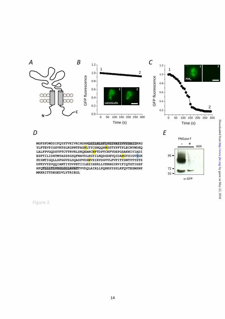
Fig. 2. P2X receptor is orientated to sense luminal ATP. (A) Schematic representation of P2XR
topology. (B, C) Fluorescence protease protection performed using cells expressing calreticulin or
P2XA both with C-terminal GFP tags. Relative fluorescence from representative experiments captured
following trypsin addition to permeabilised cells. Trypsin added a time 0. Inset, representative images
captured at time points 1 and 2. Scale 10µm. (D) Peptide sequence of Dictyostelium P2XA showing
predicted N-linked (yellow) and O-linked (blue) glycan acceptor sites. Transmembrane are
highlighted in grey. (E) Immunoblot for P2XA-GFP showing anti-GFP immunoreactivity probed in
lysates treated with PNGase F (+) or control lysates (-). No anti-GFP immunoreactivity is detected in
parental AX4 cells (AX4). Molecular weights are kDa.
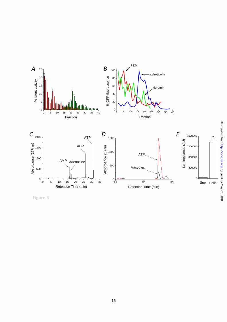
Fig. 3. Detection of P2X receptor ligand in vacuole lumen. (A) purification of intact contractile
vacuoles by subcellular fractionation. Latent activity of alkaline phosphatase (contractile vacuole,
red) and succinate dehydrogenase (mitochondria, green). N = 4 fractionations. Latent activity is
expressed as % total cellular activity. (B) Distribution of GFP tagged organelle markers across
different fractions. Markers are for contractile vacuole (P2XA and dajumin) and endoplasmic
reticulum (calreticulin). GFP fluorescence is expressed as % peak fluorescence. Representative traces
of 4 independent fractions. (C,D) Ion-pair reverse phase HPLC analysis of ATP in isolated vacuoles.
by guest on May 22, 2018
http://ww
w.jbc.org/
Dow
nloaded from
12
Separation of adenine nucleotide standards (1µM each) (C) or water soluble contents liberated from
isolated vacuoles (D). ATP standard is given as reference. Representative traces of 4 independent
experiments. (E) Detection of ATP liberated from vacuole pellet vs supernatant as determined by
luciferase-luciferin assay. N=4; *P<0.01.
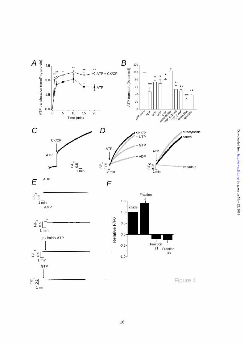
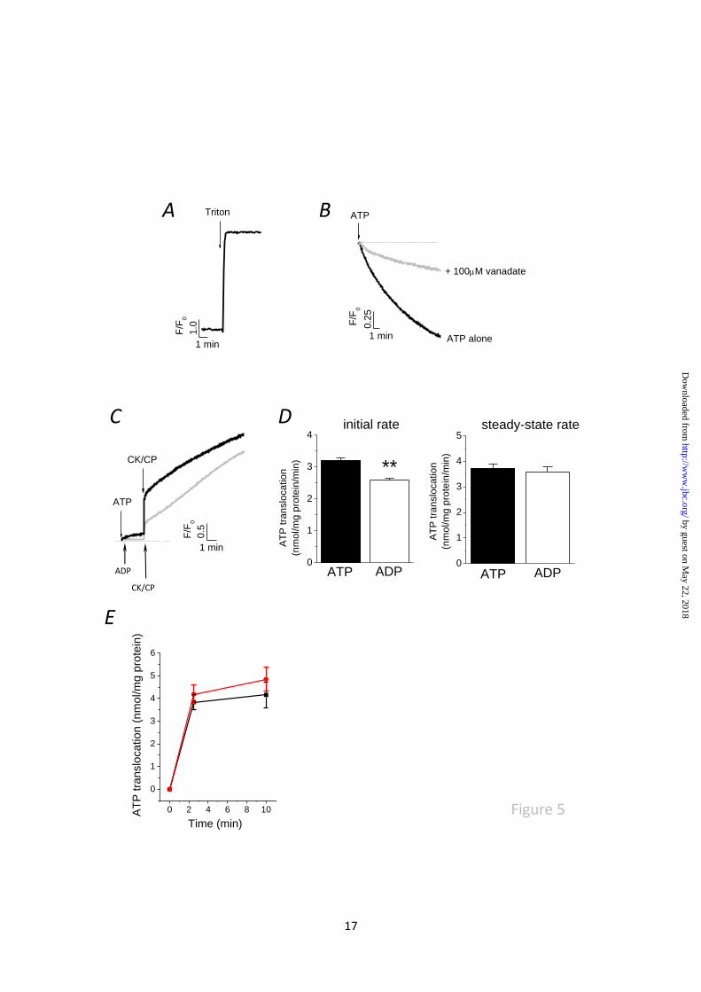
Fig. 4. Luminal ATP translocation triggers release of stored calcium. (A) Time-dependent luminal
ATP translocation in the presence of 4mM ATP alone or ATP with creatine kinase-creatine phosphate
(CK-CP). N=4; **P<0.01. (B) Effect of nucleotides (2mM each), vanadate, atractyloside (100µM),
Evans blue (1µM) and quercetin (100µM) on luminal ATP translocation. N=3-4; *P<0.05, **P<0.01.
(C) ATP evoked calcium release in isolated vacuoles. Representative traces of 6-7 independent
experiments showing calcium release in response to ATP alone with subsequent (CK-CP) addition.
(D) Effect of various inhibitors of luminal ATP translocation on ATP-evoked calcium release.
Concentrations as in (B). (E) β,γ-imido-ATP, ADP, AMP or GTP cannot mimic ATP-evoked calcium
release. (F) Specificity and enrichment of ATP-evoked calcium release in contractile vacuole
fractions. Magnitude of ATP-evoked calcium release in crude lysate, CV-enriched fraction (fraction
2), mitochondrial/ER fraction (fraction 21) or dense fraction (fraction 38).
Fig. 5. Net movement of vacuolar calcium evoked by ATP is dependent upon the calcium state of the
store and the initial rate of luminal ATP translocation. (A) Vacuoles isolated from cultures without
EGTA buffering a loaded with calcium. Release of calcium evoked by solubilisation of vacuoles with
0.1% triton-X100. (B) ATP causes vanadate-sensitive loading of vacuoles isolated from calcium
depleted cultures. (C) Paired experiment showing ADP alone does not evoke calcium release. ATP
synthesis from ADP using creatine kinase-creatine phosphate (CK-CP) can evoke a release, yet the
initial rise is less but approaches that of ATP plus CK-CP after 10mins. (D) Comparison of initial rates
and steady-state rates of luminal ATP translocation in the presence of ATP plus CK-CP or ADP plus
CK-CP. N=4; **P<0.01. (E) Comparison of luminal ATP translocation in vacuoles isolated from WT
(black) and P2XA KO (red) amoeba (N=4).
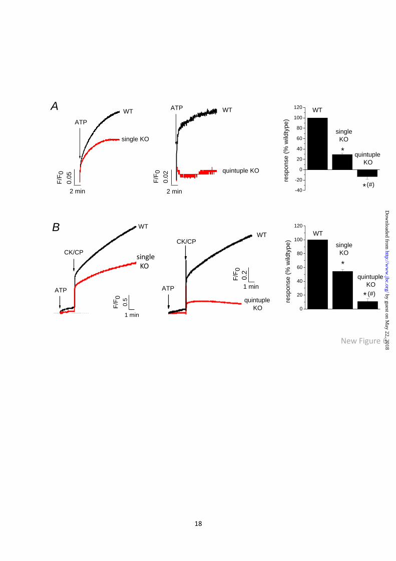
Fig. 6. P2X receptors mediate ATP evoked calcium mobilisation. Representative traces showing the
magnitude vacuolar calcium release evoked by ATP alone (A) or ATP plus creatine kinase-creatine
phosphate (CK-CP) (B) in AX4 wild-type (WT) vs P2XA receptor knockout (KO) cells and AX2 WT
vs P2XR null (quintuple KO) cells. Right, average peak responses. N=4-5; *P<0.01 KO vs WT
equivalent; #P<0.01 quintuple vs single KO.
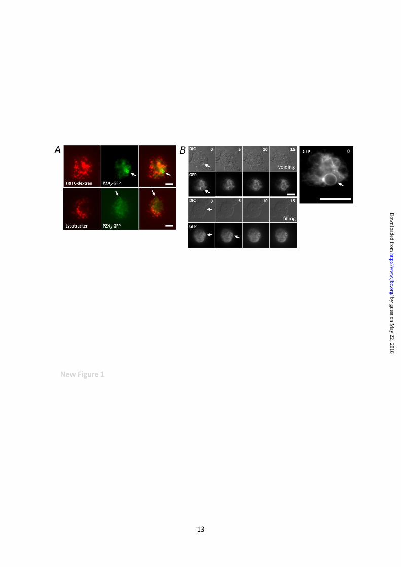
Fig. 7. Inhibitors of luminal ATP translocation or depletion of CV calcium impairs recovery from
osmotic swelling. (A and B) measurement of real-time changes in cell size by right-angled light
scattering. (A) cell swelling and recovery in response to hypotonic stress in control cells or cells with
depleted ER calcium (thapsigargin; 10µM, 30 mins) or depleted CV calcium (EGTA depletion). (B)
effect of luminal ATP translocation inhibitors on cell swelling and recovery. Control traces are
superimposed (grey) for comparison. (C) Images of control cells immediately after exposure to
distilled water (top right time 0) and following 60 mins exposure to water with and without (control)
inhibitors (bottom left). Average data on peak swelling given in (D). N=4; *P<0.05.
by guest on May 22, 2018
http://ww
w.jbc.org/
Dow
nloaded from
14
0 50 100 150 200 250 300
0.2
0.4
0.6
0.8
1.0
1.2
2
GF
P flu
ore
sce
nce
Time (s)
1
0 50 100 150 200 250 3000.0
0.2
0.4
0.6
0.8
1.0
1.2
2
GF
P flu
ore
sce
nce
Time (s)
1
1 2
calreticulin
1 2
P2XA
MGFSFDWDDIFQYSTVKIVRIRDRRLGILHLSFLVGIVAYIVVYSAIIKKG
YLFTEVPIGSVRTSLKGPNTFASNLTYCSNQQHNGSTYPFTPLECNYWDEQ
LALFPVGQDSTFTCTTRVRLSKQEANCNFTDPTCKFVDEPGSAKNIYIADI
ESFTILIDHTMYASSSGSQFNAVDLHGYILNQDGDEVQIDANGTSIGVSGK
PDIMTIGQLLSFGGVSLDQASPVDSNVSIRYDGVVLFVFITYSNTYTYSTS
DFKYVYSVQQIANTIYDVPETIILESIHSRLLYKRHGIRVIFIQTGTIGSF
HFQTLLLTLVSGLGLLAVATTVVDQLAIRLLPQRKSYSSLKFQVTESMSNP
MKKRITTDEGEDVLYTRIEGL 55
72
95
- +PNGase F
AX4
-GFP
Figure 2
NC
A B C
ED
by guest on May 22, 2018
http://ww
w.jbc.org/
Dow
nloaded from
15
Figure 3
0 5 10 15 20 25 30 35 400
5
10
15
20
25
% la
ten
t a
ctivity
Fraction
0 5 10 15 20 25 30 35 400
20
40
60
80
100
% G
FP
flu
ore
sce
nce
Fraction
25 30 350
600
1200
1800
ATP
Ab
sorb
ance 2
57n
m
Retention Time (min)
Vacuoles
0 5 10 15 20 25 30 350
2400
1800
1200
ATP
ADP
AMP
Ab
sorb
ance (
25
7n
m)
Retention Time (min)
Adenosine
600
0
400000
800000
1200000
1600000*
PelletL
um
ine
sce
nce
(A
U)
Sup.
P2XA
calreticulin
dajumin
A B
C D E
by guest on May 22, 2018
http://ww
w.jbc.org/
Dow
nloaded from
16
0 5 10 15 20
0.0
1.5
3.0
4.5
ATP + CK/CP****
**
****
AT
P t
ran
slo
ca
tio
n (
nm
ol/m
g p
rote
in)
Time (min)
ATP
A B
0
20
40
60
80
100
120
VO3-
4 (1
mM
)
Que
rcitin
Eva
ns b
lue
VO3-
4 (0
.1m
M)
Atra
ctylos
ide
CTP
UTP
GTP
ADP
****
****
***
AT
P t
ran
sp
ort
(%
co
ntr
ol)
**
ATP
alone
C D
0.5
1 min
F/F
0
CK/CP
ATP + ADP
+ GTP
+ UTP
control
ATP
0.0
5
2 min
F/F
0
vanadate
atractyloside
control
ATP0.0
5
2 min
F/F
0
E F
Figure 4
ADP
0.5
1 min
F/F
0
GTP
AMP
0.5
1 min
F/F
00.5
1 min
F/F
0
-imido-ATP
AMP
0.5
1 min
F/F
00
.5
1 min
F/F
0
-1.0
-0.5
0.0
0.5
1.0
1.5
Fraction
38
Fraction
21
Fraction
2
Re
lative
F/F
0
crude
by guest on May 22, 2018
http://ww
w.jbc.org/
Dow
nloaded from
17
+ 100M vanadate
ATP alone
ATP
0.2
5
1 min
F/F
0
Triton
1.0
1 min
F/F
0
A B
C
0.5
1 min
F/F
0
CK/CP
ATP
ADP
CK/CP
0
1
2
3
4
5steady-state rate
AT
P t
ran
slo
ca
tio
n
(nm
ol/m
g p
rote
in/m
in)
ADPATP0
1
2
3
4
ADPATP
initial rate
AT
P t
ran
slo
ca
tio
n
(nm
ol/m
g p
rote
in/m
in)
**
D
Figure 50 2 4 6 8 10
0
1
2
3
4
5
6
AT
P tra
nslo
ca
tio
n (
nm
ol/m
g p
rote
in)
Time (min)
E
by guest on May 22, 2018
http://ww
w.jbc.org/
Dow
nloaded from
18
New Figure 6
A
-40
-20
0
20
40
60
80
100
120
quintuple
KO
*
single
KO
resp
on
se
(%
wild
typ
e)
WT
*
(#)
B
CK/CP
ATP
KO
F/F
00.5
1 min
WT
2 min
ATP
single KO
WT
F/F
0
0.0
5
F/F
0
0.0
2 quintuple KO
WTATP
2 min
quintuple
KO
1 min
F/F
0WT
CK/CP
ATP
0.2
single KO
0
20
40
60
80
100
120
WT
resp
on
se
(%
wild
typ
e)
single
KO
*
quintuple
KO
* (#)
by guest on May 22, 2018
http://ww
w.jbc.org/
Dow
nloaded from
Venketesh Sivaramakrishnan and Samuel J. FountainA mechanism of intracellular P2X receptor activation
published online June 26, 2012J. Biol. Chem.
10.1074/jbc.M112.372565Access the most updated version of this article at doi:
Alerts:
When a correction for this article is posted•
When this article is cited•
to choose from all of JBC's e-mail alertsClick here
Supplemental material:
http://www.jbc.org/content/suppl/2012/06/26/M112.372565.DC1
by guest on May 22, 2018
http://ww
w.jbc.org/
Dow
nloaded from


![Molecular and functional properties of P2X receptors ... · receptors are localized in the membrane of the intracellular contractile vacuole [27, 30]. These findings demonstrate that](https://img.pdfslide.net/doc/110x75/61358e460ad5d206764773a3/molecular-and-functional-properties-of-p2x-receptors-receptors-are-localized.jpg)
![Mechanism Chest pain andthe hyperventilationsyndrome some … · [ ] =intracellular concentration, e.g. [Ca2+]It increased intracellular concentration of ionized calcium. LVDP=Left](https://img.pdfslide.net/doc/110x75/5e9803b4cc7cc5780210ca6f/mechanism-chest-pain-andthe-hyperventilationsyndrome-some-intracellular-concentration.jpg)

![Regulation of the intracellular Ca2+. Regulation of intracellular [H]:](https://img.pdfslide.net/doc/110x75/5a4d1b717f8b9ab0599b56a5/regulation-of-the-intracellular-ca2-regulation-of-intracellular-h.jpg)