Embed Size (px)
Citation preview
NOTES & NEW TECHNIQUES
ietersite is named after its discoverer, Sid Pieters, a well-known gem and mineral dealer
who first described it in 1962 from a locality inNamibia (Thomas, 2008). The term is now used gen-erally to describe brecciated varieties of tiger’s-eye.Tiger’s-eye sensu stricto occurs within Precambrianbanded iron formations as seams that run parallel tolayers of jasper. It is characterized by lustrous “gold-en” brown bands that exhibit a radiant chatoyancywhen polished due to the inclusion of crocidolitefibers within a microcrystalline silica host (Heaneyand Fisher, 2003). (Crocidolite is an asbestiform vari-ety of an amphibole called riebeckite.) AlthoughNamibian pietersite exhibits the same mineralogy astiger’s-eye, the chatoyant field is not observed as acontinuous band. Rather, pietersite contains angularfragments that are cemented as an irregular patch-
Kaifan Hu and Peter J. Heaney
Pietersite has been described as a brecciatedvariety of tiger’s-eye. This study examinedpieters ite specimens from Namibia and China(the main sources) using powder X-ray diffrac-tion, optical microscopy, environmental scan-ning electron microscopy, and conventionalgemological methods. On the basis of theresults, quantitative approaches were developedto distinguish pietersite samples from the twolocalities. It is also proposed that the petrogene-sis of this gem material is quite different from thatof South African tiger’s-eye.
A MICROSTRUCTURAL STUDY OF PIETERSITEFROM NAMIBIA AND CHINA
280 NOTES AND NEW TECHNIQUES GEMS & GEMOLOGY WINTER 2010
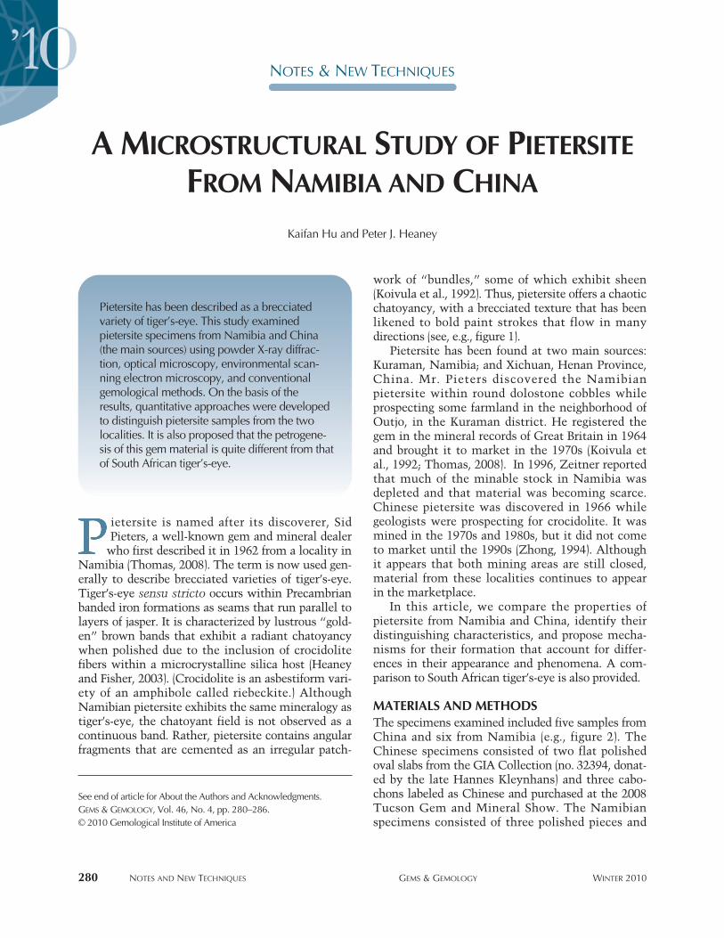
work of “bundles,” some of which exhibit sheen(Koivula et al., 1992). Thus, pietersite offers a chaoticchatoyancy, with a brecciated texture that has beenlikened to bold paint strokes that flow in manydirections (see, e.g., figure 1).
Pietersite has been found at two main sources:Kuraman, Namibia; and Xichuan, Henan Province,China. Mr. Pieters discovered the Namibianpieters ite within round dolostone cobbles whileprospecting some farmland in the neighborhood ofOutjo, in the Kuraman district. He registered thegem in the mineral records of Great Britain in 1964and brought it to market in the 1970s (Koivula etal., 1992; Thomas, 2008). In 1996, Zeitner reportedthat much of the minable stock in Namibia wasdepleted and that material was becoming scarce.Chinese pietersite was discovered in 1966 whilegeologists were prospecting for crocidolite. It wasmined in the 1970s and 1980s, but it did not cometo market until the 1990s (Zhong, 1994). Althoughit appears that both mining areas are still closed,material from these localities continues to appearin the marketplace.
In this article, we compare the properties ofpietersite from Namibia and China, identify theirdistinguishing characteristics, and propose mecha-nisms for their formation that account for differ-ences in their appearance and phenomena. A com-parison to South African tiger’s-eye is also provided.
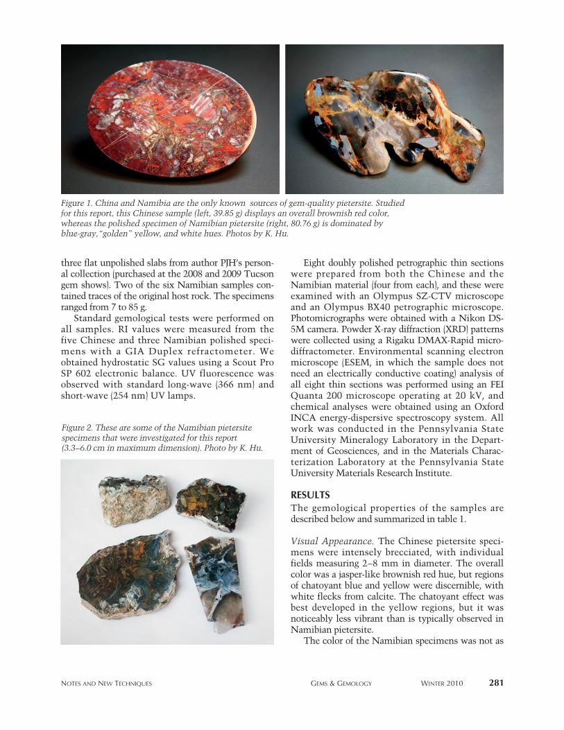
MATERIALS AND METHODSThe specimens examined included five samples fromChina and six from Namibia (e.g., figure 2). TheChinese specimens consisted of two flat polishedoval slabs from the GIA Collection (no. 32394, donat-ed by the late Hannes Kleynhans) and three cabo-chons labeled as Chinese and purchased at the 2008Tucson Gem and Mineral Show. The Namibianspecimens consisted of three polished pieces and
See end of article for About the Authors and Acknowledgments.GEMS & GEMOLOGY, Vol. 46, No. 4, pp. 280–286.© 2010 Gemological Institute of America
NOTES AND NEW TECHNIQUES GEMS & GEMOLOGY WINTER 2010 281
three flat unpolished slabs from author PJH’s person-al collection (purchased at the 2008 and 2009 Tucsongem shows). Two of the six Namibian samples con-tained traces of the original host rock. The specimensranged from 7 to 85 g.
Standard gemological tests were performed onall samples. RI values were measured from thefive Chinese and three Namibian polished speci-mens with a GIA Duplex refractometer. Weobtained hydrostatic SG values using a Scout ProSP 602 electronic balance. UV fluorescence wasobserved with standard long-wave (366 nm) andshort-wave (254 nm) UV lamps.
Eight doubly polished petrographic thin sectionswere prepared from both the Chinese and theNamibian material (four from each), and these wereexamined with an Olympus SZ-CTV microscopeand an Olympus BX40 petrographic microscope.Photomicrographs were obtained with a Nikon DS-5M camera. Powder X-ray diffraction (XRD) patternswere collected using a Rigaku DMAX-Rapid micro-diffractometer. Environmental scanning electronmicroscope (ESEM, in which the sample does notneed an electrically conductive coating) analysis ofall eight thin sections was performed using an FEIQuanta 200 microscope operating at 20 kV, andchemical analyses were obtained using an OxfordINCA energy-dispersive spectroscopy system. Allwork was conducted in the Pennsylvania StateUniversity Mineralogy Laboratory in the Depart -ment of Geosciences, and in the Materials Charac -terization Laboratory at the Pennsylvania StateUniversity Materials Research Institute.
RESULTSThe gemological properties of the samples aredescribed below and summarized in table 1.
Visual Appearance. The Chinese pietersite speci-mens were intensely brecciated, with individualfields measuring 2–8 mm in diameter. The overallcolor was a jasper-like brownish red hue, but regionsof chatoyant blue and yellow were discernible, withwhite flecks from calcite. The chatoyant effect wasbest developed in the yellow regions, but it wasnoticeably less vibrant than is typically observed inNamibian pietersite.
The color of the Namibian specimens was not as
Figure 2. These are some of the Namibian pietersitespecimens that were investigated for this report(3.3–6.0 cm in maximum dimension). Photo by K. Hu.
Figure 1. China and Namibia are the only known sources of gem-quality pietersite. Studiedfor this report, this Chinese sample (left, 39.85 g) displays an overall brownish red color,whereas the polished specimen of Namibian pietersite (right, 80.76 g) is dominated byblue-gray,“golden” yellow, and white hues. Photos by K. Hu.
282 NOTES AND NEW TECHNIQUES GEMS & GEMOLOGY WINTER 2010
varied as that of the Chinese material. Blue-gray and“golden” yellow fibrous regions predominated, withrare secondary brownish red fields, and the overallbodycolor of the Namibian specimens was blue-gray.The brecciated clasts ranged from 5 to 10 mm indiameter, but on average they were larger than thoseseen in the Chinese specimens. Chatoyancy was par-ticularly well developed in the blue fields. Three ofthe Namibian specimens had ~2-mm-thick veins ofcolorless translucent chalcedony. Chalcedony wasobservable in the Chinese specimens only with theaid of light microscopy.
Refractive Index. The RI values, around 1.54, wereconsistent with quartz for all samples. There was nodifference in RI values between the Chinese andNamibian specimens.
Specific Gravity. The SG values of the Chinese spec-imens ranged from 2.67 to 2.74. The SG values ofthe Namibian specimens were notably lower,2.50–2.58. The SG of quartz is 2.65.
UV Fluorescence. Most of the Namibian specimensluminesced a moderate-to-weak light green to long-wave UV radiation and a moderate-to-strong brightgreen to short-wave UV. This bright green lumines-cence is most likely explained by the greater chal-cedony content in those sectors. Portions of some ofthe Chinese specimens luminesced white to short-wave but were inert to long-wave UV; these areascorresponded to calcite.
Powder X-ray Diffraction. Our XRD patterns for theChinese and Namibian specimens were indistin-guishable, producing diffraction peaks only forquartz with minor calcite. No evidence of crocido-
lite was detected. This result is similar to our expe-rience with many tiger’s-eye specimens fromGriquatown, South Africa, for which crocidolitewas detected only by synchrotron X-ray radiation(Heaney and Fisher, 2003). We infer from theseresults that despite the intense chatoyancy ofpieters ite, the mass fraction of crocidolite is on theorder of a few weight percent or less.
Optical Microscopy. Examination of thin sections ofpietersite from both China and Namibia revealedthe presence of crocidolite embedded within micro-crystalline quartz. The crocidolite could be distin-guished on the basis of its moderately high relief,pleochroic grayish blue to greenish blue coloration,and optical extinction of 8–10° in cross-polarizedlight.
We also noted significant textural differences
NEED TO KNOW
• Pietersite, often described as a brecciated varietyof tiger’s-eye, is known from China and Namibia.
• Pietersite from the two localities has similar RI ranges, but the Namibian material has a lower SG.
• Crocidolite fibers are more densely intergrown (parallel, radial, and disordered textures) in Chinese samples. The fibers in Namibian speci-mens are generally oriented parallel to one another.
• Namibian pietersite formed under very differentgeologic conditions from those that producedSouth African tiger’s-eye.
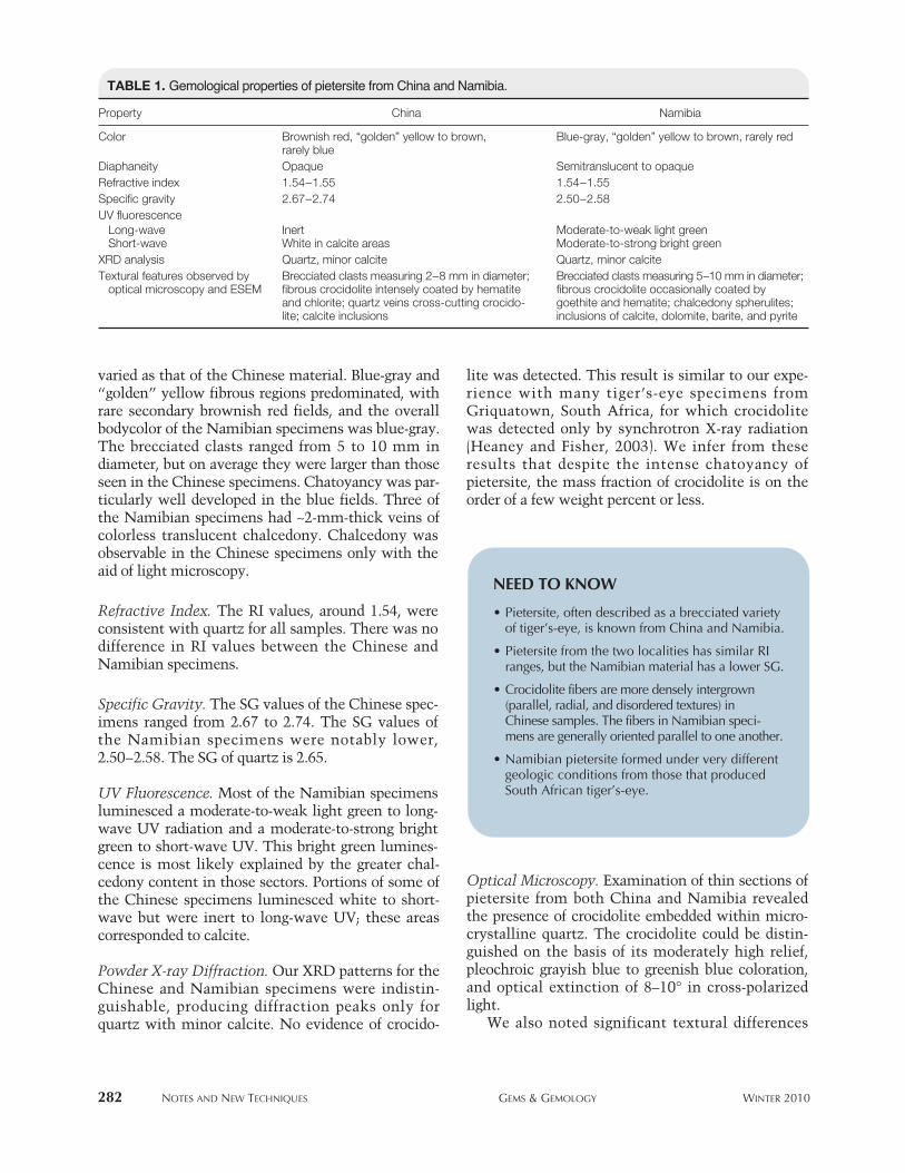
TABLE 1. Gemological properties of pietersite from China and Namibia.
Property China Namibia
Color Brownish red, “golden” yellow to brown, Blue-gray, “golden” yellow to brown, rarely redrarely blue
Diaphaneity Opaque Semitranslucent to opaqueRefractive index 1.54–1.55 1.54–1.55Specific gravity 2.67–2.74 2.50–2.58UV fluorescence
Long-wave Inert Moderate-to-weak light green Short-wave White in calcite areas Moderate-to-strong bright green
XRD analysis Quartz, minor calcite Quartz, minor calciteTextural features observed by Brecciated clasts measuring 2–8 mm in diameter; Brecciated clasts measuring 5–10 mm in diameter;
optical microscopy and ESEM fibrous crocidolite intensely coated by hematite fibrous crocidolite occasionally coated byand chlorite; quartz veins cross-cutting crocido- goethite and hematite; chalcedony spherulites;lite; calcite inclusions inclusions of calcite, dolomite, barite, and pyrite
NOTES AND NEW TECHNIQUES GEMS & GEMOLOGY WINTER 2010 283
between the Chinese and Namibian specimens. Inthe Chinese samples, the fibers were more denselyintergrown, and they showed a broader variety offabrics—parallel, radial, and disordered. They rangedfrom 20 µm to 2 mm long and rarely exceeded 2 µmwide. Both hematite and chlorite coated the fibers ofcrocidolite. The Chinese pietersite also differed fromthe Namibian samples in the presence of fibrouschlorite inclusions. The chlorite fibers exhibitedstrong pleochroism from deep green to yellowishbrown.
Unlike the Chinese material, the crocidolitefibers in the Namibian specimens were generally ori-ented parallel to one another. Fiber lengths wereshorter than in the Chinese material, typically10–50 µm, and they were less than 2 µm wide. The
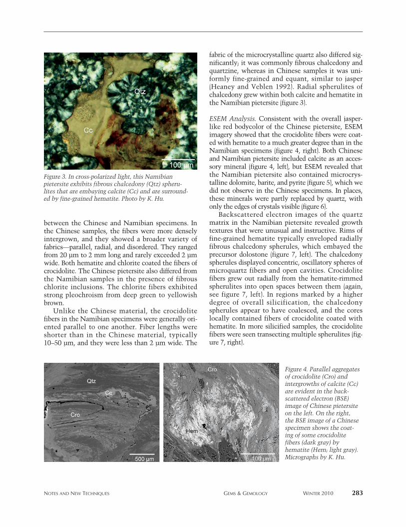
fabric of the microcrystalline quartz also differed sig-nificantly; it was commonly fibrous chalcedony andquartzine, whereas in Chinese samples it was uni-formly fine-grained and equant, similar to jasper(Heaney and Veblen 1992). Radial spherulites ofchalcedony grew within both calcite and hematite inthe Namibian pietersite (figure 3).
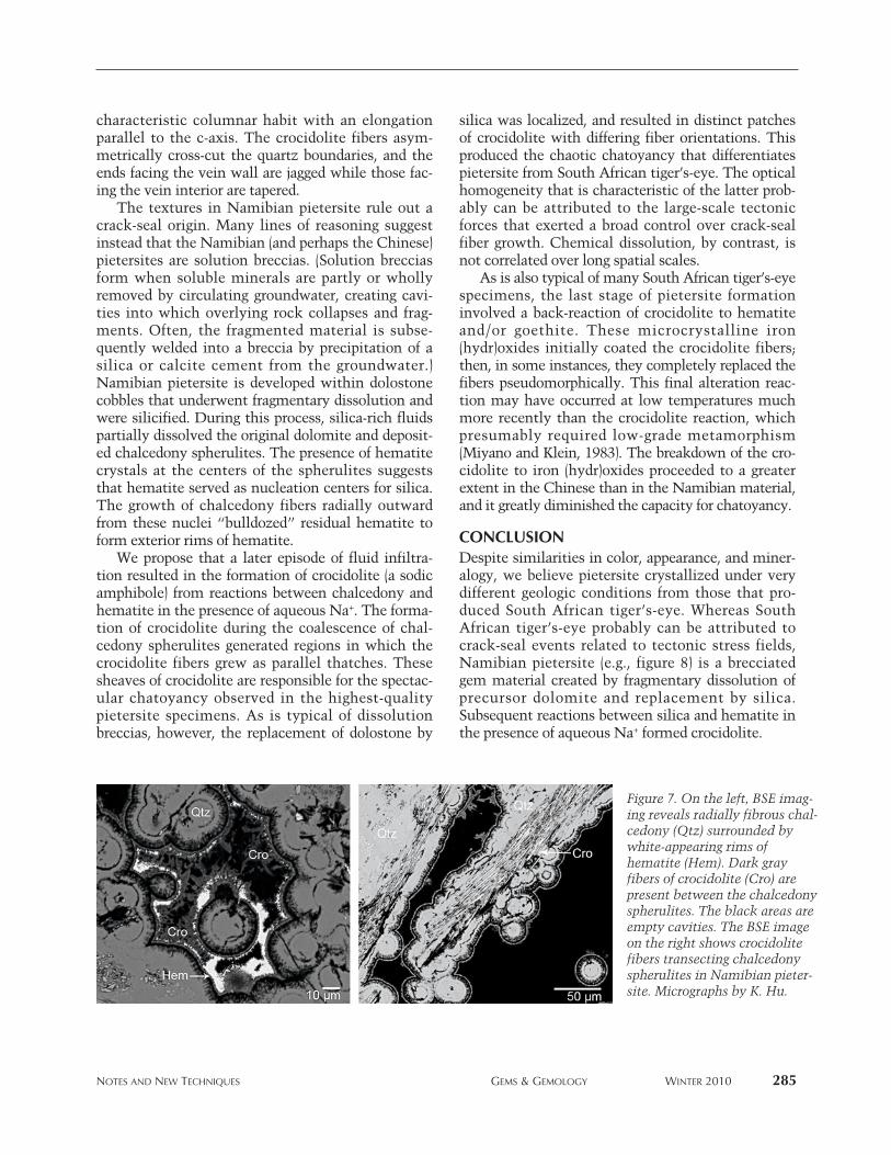
ESEM Analysis. Consistent with the overall jasper-like red bodycolor of the Chinese pietersite, ESEMimagery showed that the crocidolite fibers were coat-ed with hematite to a much greater degree than in theNamibian specimens (figure 4, right). Both Chineseand Namibian pietersite included calcite as an acces-sory mineral (figure 4, left), but ESEM revealed thatthe Namibian pietersite also contained microcrys-talline dolomite, barite, and pyrite (figure 5), which wedid not observe in the Chinese specimens. In places,these minerals were partly replaced by quartz, withonly the edges of crystals visible (figure 6).
Backscattered electron images of the quartzmatrix in the Namibian pietersite revealed growthtextures that were unusual and instructive. Rims offine-grained hematite typically enveloped radiallyfibrous chalcedony spherules, which embayed theprecursor dolostone (figure 7, left). The chalcedonyspherules displayed concentric, oscillatory spheres ofmicroquartz fibers and open cavities. Crocidolitefibers grew out radially from the hematite-rimmedspherulites into open spaces between them (again,see figure 7, left). In regions marked by a higherdegree of overall silicification, the chalcedonyspherules appear to have coalesced, and the coreslocally contained fibers of crocidolite coated withhematite. In more silicified samples, the crocidolitefibers were seen transecting multiple spherulites (fig-ure 7, right).
Figure 3. In cross-polarized light, this Namibianpietersite exhibits fibrous chalcedony (Qtz) spheru-lites that are embaying calcite (Cc) and are surround-ed by fine-grained hematite. Photo by K. Hu.
Figure 4. Parallel aggregatesof crocidolite (Cro) andintergrowths of calcite (Cc)are evident in the back -scattered electron (BSE)image of Chinese pietersiteon the left. On the right,the BSE image of a Chinesespecimen shows the coat-ing of some crocidolitefibers (dark gray) byhematite (Hem; light gray).Micrographs by K. Hu.

Consistent with a previous report (Leake et al.,1992), energy-dispersive spectroscopy revealed that thecrocidolite in both Chinese and Namibian materialcontains variable amounts of Mg in solid solution withFe and should be classified as magnesioriebeckite.
DISCUSSIONPietersite has been described as a “breccia aggregatemade up largely of hawk’s-eye and tiger’s-eye”(Schumann, 2009, p. 320) and as a “disoriented pseu-do-crocidolite mass with limonite” (Manutchehr-Danai, 2008, p. 368). Our analyses indicate thatpietersite specimens from Namibia and China doshare many hallmarks of tiger’s-eye. Miner alo gi -cally, both tiger’s-eye and pietersite contain asbesti-form fibers of crocidolite embedded within a fine-grained quartz host, and the included crocidolite isresponsible for the chatoyancy of the material.Chatoyancy is degraded where the crocidolite has
altered to iron (hydr)oxides. For example, much ofthe Chinese material that we examined containednonphenomenal areas in which a jasper-like dullnesssuperseded the original chatoyancy because of thisalteration reaction. Finally, like tiger’s-eye, thepietersite samples revealed no evidence for pseudo-morphism of quartz after crocidolite, despite popularassumptions to the contrary.
Nevertheless, our analyses suggest that the petro-genesis of pietersite is quite different from that ofthe tiger’s-eye found in Griquatown, South Africa.Heaney and Fisher (2003) proposed that SouthAfrican tiger’s-eye formed through a “crack-seal”process: The hydrofracture of banded-iron forma-tions generated flat seams parallel to the jasper bed-ding planes, and these cracks were sealed by quartzand crocidolite as an antitaxial infilling (i.e., growthfrom opposing crack walls toward the center of thevein). The quartz crystals in tiger’s-eye exhibit a
284 NOTES AND NEW TECHNIQUES GEMS & GEMOLOGY WINTER 2010
Figure 5. As revealed in BSE images (and identified by energy-dispersive spectroscopy),Namibian pietersite included barite (Brt; left), pyrite (Py; center), and dolomite (Dol; right).Micrographs by K. Hu.
Figure 6. These BSEimages of Namibianpietersite show thereplacement of bariteby quartz, leaving onlythe outer rims of thecrystals. Micrographsby K. Hu.
NOTES AND NEW TECHNIQUES GEMS & GEMOLOGY WINTER 2010 285
characteristic columnar habit with an elongationparallel to the c-axis. The crocidolite fibers asym-metrically cross-cut the quartz boundaries, and theends facing the vein wall are jagged while those fac-ing the vein interior are tapered.
The textures in Namibian pietersite rule out acrack-seal origin. Many lines of reasoning suggestinstead that the Namibian (and perhaps the Chinese)pietersites are solution breccias. (Solution brecciasform when soluble minerals are partly or whollyremoved by circulating groundwater, creating cavi-ties into which overlying rock collapses and frag-ments. Often, the fragmented material is subse-quently welded into a breccia by precipitation of asilica or calcite cement from the groundwater.)Namibian pietersite is developed within dolostonecobbles that underwent fragmentary dissolution andwere silicified. During this process, silica-rich fluidspartially dissolved the original dolomite and deposit-ed chalcedony spherulites. The presence of hematitecrystals at the centers of the spherulites suggeststhat hematite served as nucleation centers for silica.The growth of chalcedony fibers radially outwardfrom these nuclei “bulldozed” residual hematite toform exterior rims of hematite.
We propose that a later episode of fluid infiltra-tion resulted in the formation of crocidolite (a sodicamphibole) from reactions between chalcedony andhematite in the presence of aqueous Na+. The forma-tion of crocidolite during the coalescence of chal-cedony spherulites generated regions in which thecrocidolite fibers grew as parallel thatches. Thesesheaves of crocidolite are responsible for the spectac-ular chatoyancy observed in the highest-qualitypietersite specimens. As is typical of dissolutionbreccias, however, the replacement of dolostone by
silica was localized, and resulted in distinct patchesof crocidolite with differing fiber orientations. Thisproduced the chaotic chatoyancy that differentiatespietersite from South African tiger’s-eye. The opticalhomogeneity that is characteristic of the latter prob-ably can be attributed to the large-scale tectonicforces that exerted a broad control over crack-sealfiber growth. Chemical dissolution, by contrast, isnot correlated over long spatial scales.
As is also typical of many South African tiger’s-eyespecimens, the last stage of pietersite formationinvolved a back-reaction of crocidolite to hematiteand/or goethite. These microcrystalline iron(hydr)oxides initially coated the crocidolite fibers;then, in some instances, they completely replaced thefibers pseudomorphically. This final alteration reac-tion may have occurred at low temperatures muchmore recently than the crocidolite reaction, whichpresumably required low-grade metamorphism(Miyano and Klein, 1983). The breakdown of the cro-cidolite to iron (hydr)oxides proceeded to a greaterextent in the Chinese than in the Namibian material,and it greatly diminished the capacity for chatoyancy.
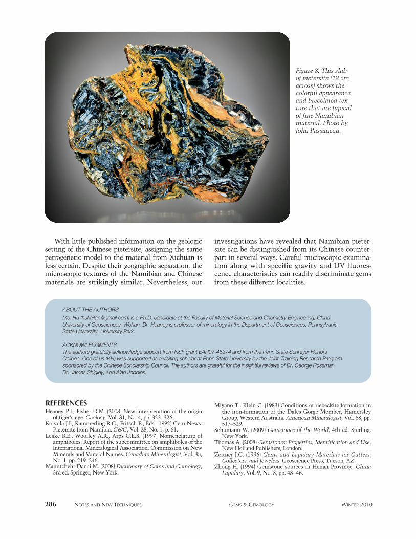
CONCLUSIONDespite similarities in color, appearance, and miner-alogy, we believe pietersite crystallized under verydifferent geologic conditions from those that pro-duced South African tiger’s-eye. Whereas SouthAfrican tiger’s-eye probably can be attributed tocrack-seal events related to tectonic stress fields,Namibian pietersite (e.g., figure 8) is a brecciatedgem material created by fragmentary dissolution ofprecursor dolomite and replacement by silica.Subsequent reactions between silica and hematite inthe presence of aqueous Na+ formed crocidolite.
Figure 7. On the left, BSE imag-ing reveals radially fibrous chal-cedony (Qtz) surrounded bywhite-appearing rims ofhematite (Hem). Dark grayfibers of crocidolite (Cro) arepresent between the chalcedonyspherulites. The black areas areempty cavities. The BSE imageon the right shows crocidolitefibers transecting chalcedonyspherulites in Namibian pieter-site. Micrographs by K. Hu.
286 NOTES AND NEW TECHNIQUES GEMS & GEMOLOGY WINTER 2010
With little published information on the geologicsetting of the Chinese pietersite, assigning the samepetrogenetic model to the material from Xichuan isless certain. Despite their geographic separation, themicroscopic textures of the Namibian and Chinesematerials are strikingly similar. Nevertheless, our
investigations have revealed that Namibian pieter-site can be distinguished from its Chinese counter-part in several ways. Careful microscopic examina-tion along with specific gravity and UV fluores-cence characteristics can readily discriminate gemsfrom these different localities.
REFERENCESHeaney P.J., Fisher D.M. (2003) New interpretation of the origin
of tiger’s-eye. Geology, Vol. 31, No. 4, pp. 323–326.Koivula J.I., Kammerling R.C., Fritsch E., Eds. (1992) Gem News:
Pietersite from Namibia. G&G, Vol. 28, No. 1, p. 61.Leake B.E., Woolley A.R., Arps C.E.S. (1997) Nomenclature of
amphiboles: Report of the subcommittee on amphiboles of theInternational Mineralogical Association, Commission on NewMinerals and Mineral Names. Canadian Mineralogist, Vol. 35,No. 1, pp. 219–246.
Manutchehr-Danai M. (2008) Dictionary of Gems and Gemo logy,3rd ed. Springer, New York.
Miyano T., Klein C. (1983) Conditions of riebeckite formation inthe iron-formation of the Dales Gorge Member, HamersleyGroup, Western Australia. American Mineralogist, Vol. 68, pp.517–529.
Schumann W. (2009) Gemstones of the World, 4th ed. Sterling,New York.
Thomas A. (2008) Gemstones: Properties, Identification and Use.New Holland Publishers, London.
Zeitner J.C. (1996) Gems and Lapidary Materials for Cutters,Collectors, and Jewelers. Geoscience Press, Tucson, AZ.
Zhong H. (1994) Gemstone sources in Henan Province. ChinaLapidary, Vol. 9, No. 3, pp. 43–46.
Figure 8. This slabof pietersite (12 cmacross) shows thecolorful appearanceand brecciated tex-ture that are typicalof fine Namibianmaterial. Photo byJohn Passaneau.
ABOUT THE AUTHORS
Ms. Hu ([email protected]) is a Ph.D. candidate at the Faculty of Material Science and Chemistry Engineering, ChinaUniversity of Geosciences, Wuhan. Dr. Heaney is professor of mineralogy in the Department of Geosciences, PennsylvaniaState University, University Park.
ACKNOWLEDGMENTSThe authors gratefully acknowledge support from NSF grant EAR07-45374 and from the Penn State Schreyer HonorsCollege. One of us (KH) was supported as a visiting scholar at Penn State University by the Joint-Training Research Programsponsored by the Chinese Scholarship Council. The authors are grateful for the insightful reviews of Dr. George Rossman, Dr. James Shigley, and Alan Jobbins.