Embed Size (px)
Citation preview

A molecular mechanism for osmolyte-inducedprotein stabilityTimothy O. Street*, D. Wayne Bolen†, and George D. Rose*‡
*T. C. Jenkins Department of Biophysics, The Johns Hopkins University, Jenkins Hall, 3400 North Charles Street, Baltimore, MD 21218; and †Department ofHuman Biological Chemistry, University of Texas Medical Branch, 301 University Boulevard, 5.154 Medical Research Building, Galveston, TX 77555-1052
Communicated by Carl Frieden, Washington University School of Medicine, St. Louis, MO, July 25, 2006 (received for review June 28, 2006)
Osmolytes are small organic compounds that affect protein stabil-ity and are ubiquitous in living systems. In the equilibrium proteinfolding reaction, unfolded (U) º native (N), protecting osmolytespush the equilibrium toward N, whereas denaturing osmolytespush the equilibrium toward U. As yet, there is no universalmolecular theory that can explain the mechanism by which os-molytes interact with the protein to affect protein stability. Here,we lay the groundwork for such a theory, starting with a keyobservation: the transfer free energy of protein backbone fromwater to a water/osmolyte solution, �gtr, is negatively correlatedwith an osmolyte’s fractional polar surface area. �gtr measures thedegree to which an osmolyte stabilizes a protein. Consequently, astraightforward interpretation of this correlation implies that theinteraction between the protein backbone and osmolyte polargroups is more favorable than the corresponding interaction withnonpolar groups. Such an interpretation immediately suggests theexistence of a universal mechanism involving osmolyte, backbone,and water. We test this idea by using it to construct a quantitativesolvation model in which backbone/solvent interaction energy is afunction of interactant polarity, and the number of energeticallyequivalent ways of realizing a given interaction is a function ofinteractant surface area. Using this model, calculated �gtr valuesshow a strong correlation with measured values (R � 0.99). Inaddition, the model correctly predicts that protecting/denaturingosmolytes will be preferentially excluded/accumulated around theprotein backbone. Taken together, these model-based results ra-tionalize the dominant interactions observed in experimental stud-ies of osmolyte-induced protein stabilization and denaturation.
organic osmolytes � osmolyte mechanism � protein folding
The equilibrium protein folding reaction, unfolded (U) ºnative (N), is not an ordinary chemical reaction because no
covalent bonds are made or broken in the interconversionbetween N and U. Instead, protein denaturation/renaturation isjust a reequilibration between the unfolded and folded popula-tions under changed solvent conditions. Accordingly, a thermo-dynamic description of protein folding can be framed in terms ofsolvent interactions with the unfolded and native states (1–3).
Osmolytes are small organic compounds that exert a dramaticinfluence on the protein folding reaction, again without makingor breaking covalent bonds. Protecting osmolytes push thefolding equilibrium toward N, whereas denaturing osmolytespush the equilibrium toward U. Both types of osmolytes are ofutmost significance. Protecting osmolytes are ubiquitous innature, where they play a vital role in stabilizing intracellularproteins against a wide variety of adverse environmental con-ditions (4–7). Alternatively, urea, a denaturing osmolyte foundnaturally in mammalian kidney, has been a key reagent through-out the long history of solvent denaturation studies (3, 8–10).
The solution thermodynamics of protein/osmolyte mixtureshas been well characterized in the literature (3, 11–19). In theemerging view (12, 17), protecting osmolytes raise the freeenergy of the unfolded state, favoring the folded population,whereas denaturing osmolytes lower the free energy of theunfolded state, favoring the unfolded population. Accordingly,
protecting/denaturing osmolytes interact unfavorably/favorablywith the unfolded state, resulting in preferential depletion/accumulation of osmolyte proximate to the protein surface. Suchosmolyte-induced behavior has been well characterized in ther-modynamic terms, but thermodynamics is a descriptive science,deliberately devoid of mechanism. As yet, there is no universaltheory that can account for the mechanism by which osmolytesinteract with the protein to affect stability.
What specific molecular interactions in a protein–osmolyte–water solution stabilize�destabilize the unfolded state of pro-teins? An important clue comes from recent work showing thatthe osmolyte effect operates predominantly on the proteinbackbone, a component common to all residues (11–13). Thisconclusion was reached by measuring transfer free energies, �gtr,of backbone models from water to 1 M osmolyte solutions.Although side chains do play a role, it is primarily the backbonetransfer free energy that determines the extent to which os-molytes either stabilize (i.e., �gtr � 0) or destabilize (i.e., �gtr �0) the protein relative to an equivalent aqueous solution.
Thus, the backbone �gtr value is the key metric for evaluatingthe relative denaturing/stabilizing strength of different os-molytes. The thermodynamic reference state for this metric isgiven by interactions of the peptide backbone unit with solventwater. When that backbone unit is transferred from water to anaqueous osmolyte solution, the very presence of a molecule thatexperiences backbone interactions which differ from corre-sponding interactions with water either raises (for a protectingosmolyte) or lowers (for a denaturing osmolyte) the �gtr valuerelative to this reference state. Furthermore, given the welldefined nature of the two solvent systems, the resultant �gtr willarise solely from differences between backbone/water and back-bone/water/osmolyte interactions. Any molecular interpretationof osmolyte interactions must be consistent with this experimen-tal reality.
We demonstrate that �gtr values for a wide variety of os-molytes are negatively correlated with their fractional polarsurface area (SA), f polar surface
osmolyte . The correlation suggests thatpolar and nonpolar osmolyte surfaces interact with the proteinbackbone at different energies, and that the extent of interactionis related to interactant SA. Specific instances in the literatureare in known agreement with this plausible idea. For example,the polar molecule urea that has long been known to interactfavorably with the amide backbone of proteins (20, 21). Also, ina related correlation, Record and colleagues (18) noted that �gtrfor glycine betaine is proportional to polar SA. To quantify ouridea, we develop a simple statistical mechanics backbone solva-tion model in which the interaction energy depends on interac-
Author contributions: T.O.S. and G.D.R. designed research; T.O.S. performed research;D.W.B. contributed key ideas; T.O.S. and G.D.R. analyzed data; and T.O.S., D.W.B., andG.D.R. wrote the paper.
The authors declare no conflict of interest.
Freely available online through the PNAS open access option.
Abbreviations: TMAO, trimethylamine N-oxide; SA, surface area.
‡To whom correspondence should be addressed. E-mail: [email protected].
© 2006 by The National Academy of Sciences of the USA
www.pnas.org�cgi�doi�10.1073�pnas.0606236103 PNAS � September 19, 2006 � vol. 103 � no. 38 � 13997–14002
BIO
PHYS
ICS
Dow
nloa
ded
by g
uest
on
June
3, 2
020
Dow
nloa
ded
by g
uest
on
June
3, 2
020
Dow
nloa
ded
by g
uest
on
June
3, 2
020
Dow
nloa
ded
by g
uest
on
June
3, 2
020

tant polarity, and the interaction degeneracy (i.e., the number ofenergetically equivalent ways of realizing the interaction) de-pends on the corresponding interactant SAs. Our goal is to learnwhether this minimal model, polar interactions in a statisticalmechanical framework, is sufficient to account for the diversityof experimental phenomena associated with protecting anddenaturing osmolytes.
ResultsCalculations described below were performed by using x-raystructures of eight stabilizing osmolytes [trimethylamine N-oxide(TMAO), betaine, sarcosine, proline, trehalose, sucrose, glyc-erol, and sorbitol], a destabilizing osmolyte (urea) and a relateddenaturant (guanidine). The �gtr values for these 10 compoundshave been measured by two independent methods in all casesexcept guanidine (11) (Table 1). Comparisons between osmolytestructures and the associated water to osmolyte �gtr values (Fig.1) indicate no evident correlation with either the total osmolyteSA or its polar SA. For example, TMAO and urea (Fig. 1 a andj) have similar total SAs but opposite effects on protein stability.Likewise, sucrose (Fig. 1c), an intermediate stabilizer, hasgreater polar SA than either TMAO, a strong stabilizer, or urea,a strong denaturant.
However, there is a clear correlation (R � 0.88) between �gtrand f polar surface
osmolyte for these osmolytes (Fig. 2). Specifically, asf polar surface
osmolyte increases, the osmolyte interaction with the proteinbackbone becomes increasingly favorable (i.e., their �gtr valuedecreases). This correlation suggests that the interaction be-tween the protein backbone and osmolyte polar groups is morefavorable than the corresponding interaction with nonpolar
groups. Furthermore, the correlation suggests that the proba-bility of interaction scales with interactant SA.
A Model for Solvent Interactions with the Protein Backbone. Giventhe chemical heterogeneity of these osmolytes, what accounts forthe observed correlation between f polar surface
osmolyte and �gtr? The mostdirect explanation would be the existence of a universal inter-action mechanism. Accordingly, we propose a quantitativemodel for solvent (water and osmolyte) interactions with back-bone polar groups (the amide nitrogen, bearing a partial positivecharge, and the carbonyl oxygen, bearing a partial negativecharge). Three types of protein/solvent interactions were definedfor these two backbone groups: favorable, unfavorable, andneutral, having energies of �1, 1, and 0 kcal/mol, respectively.Favorable interactions occur between polar groups with oppo-site charges, unfavorable interactions are between polar groupswith like charges, and neutral interactions involve nonpolargroups, as illustrated for TMAO in Fig. 3. The backbone amidenitrogen is assigned to have one solvent interaction site, whereasthe larger carbonyl oxygen, with its two lone pair electrons, isassigned to have two such sites. However, the overall results donot change appreciably if both backbone groups are treatedequivalently (discussed below). Solvent/solvent and backbone/backbone interactions are not included in the model.
A degeneracy term was included to quantify the number ofways of realizing a given backbone/solvent interaction in termsof the area of its available participating interactant surfaces (seeMethods). To implement this contribution, polar and nonpolarSAs were calculated for each osmolyte, with polar surfacefurther subdivided into contributions from groups with partialpositive (nitrogen) and negative (oxygen) charges (Table 1).
Table 1. Solvent accessible surface areas and �gtr values of osmolytes
Osmolyte* SA�, Å2† SA�, Å2† SAo, Å2† �SA, Å2‡ �gtr, cal�mol§
TMAO 0.0 43.2 168.4 211.6 89 � 2Betaine 3.6 82.7 166.7 253.0 65 � 3Sucrose 0.0 336.9 137.3 474.2 56 � 6Trehalose 0.0 340.6 145.2 485.8 54 � 8Sarcosine 24.5 43.3 141.6 209.4 50 � 2Sorbitol 0.0 233.6 97.8 331.4 43 � 7Proline 24.5 88.9 133.5 246.9 40 � 8Glycerol 0.0 142.7 84.1 226.8 22 � 8Urea 111.8 51.4 11.6 174.8 �41 � 2Guanidine 167.8 0.0 11.6 179.4 �59
*The 10 osmolytes listed in Fig. 1.†Osmolyte surface areas in Å2 with partial positive, negative, and neutral charge are indicated by SA�, SA�, andSAo, respectively.
‡Total surface area in Å2 � sum of SA�, SA�, and SAo.§�gtr is the free energy change that accompanies the transfer of a backbone unit from water to a 1 M osmolytesolution. Uncertainty in �gtr values is based on two independent measurement techniques (11). Value forguanidine was provided by S. Sarker (personal communication).
Fig. 1. Molecular structures of osmolytes. Protecting osmolytes are TMAO, betaine, sucrose, trehalose, sarcosine, sorbitol, proline, and glycerol (A–H), anddenaturants are urea and guanidine (J–K). Compounds are ordered by their measured �gtr values (see Table 1), shown in space-filling representations andcolor-coded by atom type: oxygen (red), nitrogen (blue), and carbon (green). Water polarity is represented by its surface electrostatic potential (I, Upper), usinga color saturation scale that runs from �0.07 (red) to 0.11 (blue) e/Å; white indicates neutral potential. The water surface is partitioned into discrete positive (red),negative (blue), and neutral (white) surfaces (I, Lower) using electrostatic potential cutoffs described in Methods.
13998 � www.pnas.org�cgi�doi�10.1073�pnas.0606236103 Street et al.
Dow
nloa
ded
by g
uest
on
June
3, 2
020
To treat water and osmolytes in a consistent manner, it wasalso necessary to subdivide a water molecule into polar andnonpolar regions. Although the decomposition of osmolytesurfaces into polar and nonpolar components is uncomplicated,
a similar decomposition of water requires a method to evaluateits surface charge distribution. To this end, an ab initio calcula-tion of the water electrostatic potential and electron density wasperformed, as described in Methods. When this potential ismapped onto a space-filling model (22), distinct regions ofpositive (blue), negative (red), and neutral (white) charge areapparent (Fig. 1i, top structure). After applying polar andnonpolar cutoff values (see Methods), the water surface is foundto have approximately equal regions of positive, negative, neutralcharge (37%, 33%, and 29%, respectively; Fig. 1i, lower struc-ture). Although the precise decomposition depends on thechosen electron density and the electrostatic potential threshold(data not shown), our overall results are quite insensitive to thisdecomposition, as discussed below. Consequently, the totalwater SA of 30 Å2 (the area of a water-sized sphere of radius1.5 Å) was subdivided into three equal-area regions of 10 Å2
each.
Comparisons Between Model-Based and Measured �gtr Values. Themodel was tested by using it to calculate �gtr values andcomparing them to experimentally measured values. For 1 Mosmolyte concentrations, the calculated and measured �gtr val-ues are in good agreement (Fig. 4A), with a correlation coeffi-cient of 0.99, a substantial improvement from the correspondingcorrelation with f polar surface
osmolyte (r � 0.88). Although the slope of thelinear regression line is less than unity (slope � 0.81), a slope of1 with a correlation coefficient of 0.98 can be obtained ifinteraction energies are set to �1.5, 1.5, and 0 kcal/mol (forfavorable, unfavorable, and neutral interactions, respectively).As a further test, �gtr values were calculated at osmolyteconcentrations beyond 1 M (Fig. 4B, solid lines); these valuescompare favorably with the corresponding experimentalvalues for sarcosine, urea, and guanidine (Fig. 4B, symbols),those osmolytes for which measurements beyond 1 M areavailable (13).
The success of the model in predicting �gtr values for adiversity of compounds across a range of concentrations isconsistent with our hypothesis that interactions between theprotein backbone and osmolytes are dominated by their SA andouter-group polarity. Moreover, this conclusion is insensitive tosubstantial changes in model parameters, as discussed below.
How Robust Are the Calculated �gtr Values? Our model has twoadjustable parameters: the polar and nonpolar SAs associatedwith water, and the interaction energy between solvent and theprotein backbone. To assess the sensitivity of calculated �gtrvalues to these parameters, both were varied extensively and �gtrvalues were recalculated.
Positive, negative, and neutral water SAs were simultaneouslyrandomized within a 5- to 15-Å2 interval, with solvent interactionenergies fixed at their original values. The resulting �gtr valuesremain in good agreement with the measured values (Fig. 6,which is published as supporting information on the PNAS website); 90% of the calculated �gtr values have correlationcoefficients with measured values that exceed 0.80.
In another test, interaction energies were assigned randomvalues in the range 0.5–5 kcal/mol (multiplied by �1 for favor-able interactions), with neutral interaction energy and the polarand nonpolar water SAs held fixed at their original values. Thecorrelation with measured �gtr values was recalculated for eachnew value (Fig. 7, which is published as supporting informationon the PNAS web site), and again, model-based and measured�gtr values were found to be in good agreement; all correlationcoefficients exceed 0.80.
As a final test, the correlation between model-based andmeasured �gtr values was recalculated under the alternativeassumption that the backbone carbonyl oxygen provides only onesolvent interaction site, not two. Water surface decomposition
Fig. 2. The polar fraction of osmolyte surface correlates with measured �gtr
values. Fractional polar SA, f polar surfaceosmolyte , is plotted against �gtr values from
Table 1 for the 10 osmolytes in Fig. 1. The linear regression line (solid line) hasa negative slope with a correlation coefficient of 0.88, indicating that back-bone/osmolyte interactions become increasingly favorable as osmolytes be-come increasingly polar.
Fig. 3. Illustrating TMAO/backbone interactions. Interactions between anosmolyte, such as TMAO (upper molecule), and the protein backbone (lowerstructure) can be favorable, neutral, or unfavorable. Favorable interactionsare between groups of opposite charge (A), neutral interactions involve atleast one nonpolar group (B), and unfavorable interactions are betweengroups of like charge (C). Atoms are color-coded as in Fig. 1. A large fractionof the TMAO surface is nonpolar, affording more opportunities (i.e., a higherdegeneracy) for this osmolyte to realize neutral interactions than eitherfavorable or unfavorable interactions.
Street et al. PNAS � September 19, 2006 � vol. 103 � no. 38 � 13999
BIO
PHYS
ICS
Dow
nloa
ded
by g
uest
on
June
3, 2
020

and interaction energies were held fixed at their original values.Still, model-based and measured �gtr values remain highlycorrelated (Fig. 8, which is published as supporting informationon the PNAS web site), although the correlation coefficient isreduced to 0.86.
Preferential Osmolyte Interactions with the Protein Backbone. Char-acteristically, stabilizing/destabilizing osmolytes are preferen-tially excluded/accumulated at the protein surface, respectively(14, 15, 23). To test this aspect of the model, the local concen-tration of osmolytes at backbone interaction sites was calculatedfor a 1 M osmolyte solution (see Methods). As shown in Fig. 5,stabilizing osmolytes are preferentially excluded from backbonepolar groups, whereas denaturing osmolytes are preferentiallyaccumulated there. In the model, the molecular basis of thesepreferential interactions is rooted in solvent/backbone interac-tions. Stabilizing osmolytes, such as TMAO, are preferentiallyexcluded from the backbone because water is more likely thanTMAO to interact favorably with backbone polar groups. Con-
versely, destabilizing osmolytes, such as urea, are preferentiallyaccumulated near the backbone because they have a strongerpropensity to interact with the backbone than water. However,it should be noted that the model does not take nonpolarbackbone regions into account, and interactions aroundthese regions can also contribute to the actual local osmolyteconcentration.
DiscussionThe model presented here establishes a connection between bulkthermodynamic quantities and the molecular interactions thatgive rise to these quantities. In particular, it was developed toexplain the experimentally determined backbone transfer freeenergies from water to osmolyte in terms of interactions betweenthe protein backbone and water or osmolyte molecules. Themodel was validated by using it to predict significant, experi-mentally observed behavior. In addition, the model predicts thatthe free energy change for folding/unfolding will be linearlydependent on osmolyte concentration or approximately so, andfor both protectants and denaturants, consistent with Pace’slinear extrapolation model (9, 10). Our model also correctlypredicts m values (d�gtr�d[osmolyte]) of opposite sign andapproximately equal magnitude for proteins that are eitherforced to fold by sarcosine or denatured by urea, therebyaccounting for the wide range of effects that natural osmolytescan exert on protein stability (12). Finally, �gtr values calculatedby using the model correlate extremely well with experimentalvalues (R � 0.99), illustrating that the relevant water–osmolyte–backbone energies are captured by the model.
Protein/osmolyte interactions are conspicuously weak, and insuch cases, classical binding models are notoriously deficient (24).In particular, urea/backbone interactions would have apparentbinding constants slightly greater than unity (Kbinding 1.2) (3),whereas protecting osmolyte/backbone interactions would haveapparent binding constants slightly less than unity (Kbinding 0.8).In this weak-binding regime, free energy effects are ostensiblyadditive because neither type of osmolyte occupies a significantfraction of the backbone surface, so there is essentially no compe-tition for backbone binding sites. Such additivity is observed inexperiments (25) and is consistent with the model.
Many previous studies have related SA calculations to thermo-dynamic quantities associated with protein folding (26–32), moti-vated by the early observation that the transfer of nonpolar groupsfrom organic to aqueous solvent is accompanied by an anomalouschange in heat capacity, �Cp (33). The observed correlationbetween �Cp and nonpolar surface is often interpreted to mean
Fig. 4. Comparison between calculated and measured �gtr values for osmolytes. (A) �gtr values, calculated from the model, are plotted against experimentallydetermined values from Table 1. Good agreement is apparent. The linear regression line (solid line) is given by �gtr
measured � 0.81 �gtrcalculated � 3.2, with correlation
coefficient 0.99. Data points corresponding to the 10 osmolytes in Fig. 1 are annotated in Inset. (B) Calculated �gtr values on higher osmolyte concentrations (�1M) are plotted against available experimental data for sarcosine (indigo triangles), urea (green squares), and guanidine (orange asterisks). Solid lines were drawnfrom model-based �gtr values, extended beyond 1 M osmolyte concentrations.
Fig. 5. Protecting/denaturing osmolytes are preferentially depleted/accumulated at the protein backbone. Concentration of osmolyte around thebackbone in a 1 M osmolyte solution plotted against measured �gtr valuesfrom Table 1. Data points corresponding to the 10 osmolytes in Fig. 1 areannotated in Inset. The local osmolyte concentration is given by the scaleddifference between Opref� and Obulk�, described in Methods. It is apparentthat the backbone concentration of protecting osmolytes (�gtr � 0) is com-paratively depleted ([osmolyte] � 1.0 M), whereas that of denaturing os-molytes (�gtr � 0) is comparatively enriched ([osmolyte] � 1.0 M).
14000 � www.pnas.org�cgi�doi�10.1073�pnas.0606236103 Street et al.
Dow
nloa
ded
by g
uest
on
June
3, 2
020

that water around nonpolar surfaces differs structurally and ther-modynamically from water in bulk solution (34). In contrast,the model proposed here focuses on solvent interactions with theprotein backbone, and SAs are only used here to estimate thebinding-competent fraction of interacting molecules.
Several other types of solvent interactions affecting proteinstability are neglected in our model, including crowding and ex-cluded volume (35–37), the structure of water in osmolyte solutions(38, 39), side chain/solvent interactions, and binding situations inwhich a large osmolyte molecule can occlude more than onebackbone unit. In addition, the model treats all nitrogens andoxygens as equally polar, an obvious simplification. However,despite such simplicity, the model captures key thermodynamicaspects of osmolyte behavior in a parameter-insensitive fashion.Therefore, it seems likely that the model’s anchoring suppositions,solvent/protein interactions that depend on polarity and SA, areprimarily responsible for the osmolyte effect in proteins.
MethodsAtom coordinates for TMAO, betaine, sucrose, trehalose,sarcosine, sorbitol, proline, glycerol, urea, and guanidine wereobtained from the HICUP database (40). Their solvent acces-sible polar and nonpolar SAs were calculated by using PyMOL(41) with a probe radius of 1.4 Å (Table 1). Guanidine differsfrom other osmolytes investigated here because it is intro-duced into solution as a salt, guanidinium hydrochloride. Tocorrect for the chloride ion associated with guanidine, a small,negatively charged surface of 30 Å2 was added to the guanidineSA, although this addition does not dramatically change itscalculated �gtr value (these values are �67 and �62 cal/molwith and without chloride ion addition, respectively).
The surface of a water molecule was defined at a thresholdelectron density of � � 0.005, giving a molecular volume of 11.5 Å3
(approximately the volume of a 1.5-Å sphere). The surface of waterwas decomposed into polar, nonpolar, and neutral regions bycalculating the electrostatic potential and mapping it onto thissurface. The thresholds used to delimit polar and nonpolar regionswere defined by the boundary where the potential decays to 1/e ofits minimum and maximum values, �0.023 and 0.037 e/Å, respec-tively. The neutral region was then defined as the complement tothese two regions. Both the electrostatic potential and the electrondensity were calculated ab initio by using CPMD (42).
The average energy of the protein backbone in variousosmolyte solutions was calculated by using a statistical me-chanics model in which the backbone has three interactionsites: one at the amide nitrogen and two at the carbonyloxygen; these sites are represented by the indices i, j, and k,respectively. At each interaction site, the solvent (either wateror osmolyte) can present a positively (�), neutral (o), ornegatively charged (�) surface. Accordingly, the indices i, j,and k can take the values �, o, or �, resulting in a total of33 � 27 possible microstates. Interactions between the back-bone and solvent are assigned energies of �1, 1, and 0 kcal/molcorresponding to interactions between opposite, like, or un-charged groups, respectively. The interaction energies for eachsite are assumed to be additive and independent. Thesemicrostates and their associated energies and degeneracies areenumerated in Table 2.
The degeneracy of a solvent interaction at a particularbackbone interaction site ref lects the number of energeticallyequivalent ways of making that interaction. In our model, thedegeneracy is given by the water or osmolyte SA that canparticipate in the interaction. Given that the three backboneinteraction sites (i, j, and k) are independent, the totaldegeneracy (�ijk) of a specific microstate, consisting of a �, o,or � solvent interaction at sites i, j, and k, will be the productof the degeneracies, as represented by their SAs, at individualinteraction sites
�ijk � SAi�SAj�SAk. [1]
These SAs will have a contribution from water (SAw,i, SAw,j, andSAw,k) and a contribution from osmolyte (SAo,i, SAo,j, and SAo,k)that depends on the osmolyte concentration. At Y molar:
SAi � 55.5�SAw,i � Y�SAo,i [2]
SAj � 55.5�SAw, j � Y�SAo, j [3]
SAk � 55.5�SAw,k � Y�SAo,k. [4]
In the model, �, o, and � water SAs are equal (10 Å2), and thecorresponding osmolyte SAs are given in Table 1. The SAcalculations treat the activity of water as a constant (i.e., thatmolarity of water in osmolyte solutions of varying concentrationis 55.5 M), a plausible approximation at the low cosolventconcentrations used here.
The probability of any given microstate is given by
pijk ��ijke�Eijk�kT
�i
�j
�k
�ijke�Eijk�kT, [5]
where k is Boltzmann’s constant and T is 298.15 K, the temper-ature at which �gtr values were measured experimentally. Theseprobabilities can be used to calculate the average energy of thesystem
Table 2. Solvent interactions with the protein backbone
Interaction site* Microstate†
N, i O1, j O2, k E, kcal�mol �
� � � �1 ����
� � o 0 ���o
� � � 1 ����
� o � 0 ��o�
� o o 1 ��oo
� o � 2 ��o�
� � � 1 ����
� � o 2 ���o
� � � 3 ����
o � � �2 �o��
o � o �1 �o�o
o � � 0 �o��
o o � �1 �oo�
o o o 0 �ooo
o o � 1 �oo�
o � � 0 �o��
o � o 1 �o�o
o � � 2 �o��
� � � �3 ����
� � o �2 ���o
� � � �1 ����
� o � �2 ��o�
� o o �1 ��oo
� o � 0 ��o�
� � � �1 ����
� � o 0 ���o
� � � 1 ����
The 27 microstates and their associated energies and degeneracies.*The single amide nitrogen and two carbonyl oxygen backbone interactionsites are indicated by N, O1, and O2, respectively. Solvent interactions withthese sites are given by the charge of the interacting solvent surface: positive(�), negative (�), and neutral (o), and i, j, and k indices are varied over therange of values for these interactions.
†E is the energy of a given microstate, and � is its degeneracy.
Street et al. PNAS � September 19, 2006 � vol. 103 � no. 38 � 14001
BIO
PHYS
ICS
Dow
nloa
ded
by g
uest
on
June
3, 2
020

E� � �i
�j
�k
Eijk�pijk, [6]
with �gtr values given by the difference between the averagesystem energy at 0 and 1 M osmolyte concentrations. Theaverage occupancy of osmolytes on the backbone interactionsites can also be calculated as
Opref� � �i
�j
�k
pijk� SAo,i
SAo,i � SAw,i
�SAo, j
SAo, j � SAw, j�
SAo,k
SAo,k � SAw,k� . [7]
This value can be compared with the expected osmolyte occu-pancy based solely on the bulk solution concentration (i.e., no
preferential interactions with the three backbone interactionsites)
Obulk� � 3 �SAo
SAo � SAw. [8]
The relative difference between Opref� and Obulk� yields a localosmolyte concentration when scaled to molarity.
All numerical calculations were performed in Python (www.python.org).
We thank Buzz Baldwin and two anonymous referees for suggestions.This work was supported by a Burroughs Welcome predoctoral fellow-ship (to T.O.S.), National Institutes of Health Grant GM-49760 (toD.W.B.), and by the Mathers Foundation (G.D.R.).
1. Wu H (1931) Chinese J Physiol V:321–344.2. Mirsky AE, Pauling L (1936) Proc Natl Acad Sci USA 22:439–447.3. Schellman JA (2002) Biophys Chem 96:91–101.4. Yancey PH, Clark ME, Hand SC, Bowlus RD, Somero GN (1982) Science
217:1214–1222.5. Record MT, Jr, Courtenay ES, Cayley DS, Guttman HJ (1998) Trends Biochem
Sci 23:143–148.6. Record MT, Jr, Courtenay ES, Cayley S, Guttman HJ (1998) Trends Biochem
Sci 23:190–194.7. Hochachka PW, Somero GN (2002) Biochemical Adaptation (Oxford Univ
Press, Oxford).8. Tanford C (1968) Adv Prot Chem 23:121–282.9. Greene RF, Jr, Pace CN (1974) J Biol Chem 249:5388–5393.
10. Santoro MM, Bolen DW (1988) Biochemistry 27:8063–8068.11. Auton M, Bolen DW (2004) Biochemistry 43:1329–1342.12. Auton M, Bolen DW (2005) Proc Natl Acad Sci USA 102:15065–15068.13. Liu Y, Bolen DW (1995) Biochemistry 34:12884–12891.14. Lee JC, Timasheff SN (1981) J Biol Chem 256:7193–7201.15. Timasheff SN (1992) Biochemistry 31:9857–9864.16. Makhatadze GI, Privalov PL (1992) J Mol Biol 226:491–505.17. Bolen DW, Baskakov IV (2001) J Mol Biol 310:955–963.18. Felitsky DJ, Cannon JG, Capp MW, Hong J, Van Wynsberghe AW, Anderson
CF, Record MT, Jr (2004) Biochemistry 43:14732–14743.19. Hong J, Capp MW, Anderson CF, Saecker RM, Felitsky DJ, Anderson MW,
Record MT, Jr (2004) Biochemistry 43:14744–14758.20. Nozaki Y, Tanford C (1963) J Biol Chem 238:4074–4080.21. Roseman M, Jencks WP (1974) J Am Chem Soc 97:631–640.
22. Pettersen EF, Goddard TD, Huang CC, Couch GS, Greenblatt DM, Meng EC,Ferrin TE (2004) J Comput Chem 25:1605–1612.
23. Lin TY, Timasheff SN (1994) Biochemistry 33:12695–12701.24. Schellman JA (1987) Biopolymers 26:549–559.25. Mello CC, Barrick D (2003) Protein Sci 12:1522–1529.26. Chothia C (1974) Nature 248:338–339.27. Eisenberg D, McLachlan AD (1986) Nature 319:199–203.28. Spolar RS, Ha JH, Record MT, Jr (1989) Proc Natl Acad Sci USA 86:8382–8385.29. Richards FM (1977) Annu Rev Biophys Bioeng 6:151–176.30. Lee B, Richards FM (1971) J Mol Biol 55:379–400.31. Hilser VJ, Gomez J, Freire E (1996) Proteins 26:123–133.32. Robertson AD, Murphy KP (1997) Chem Rev 97:1251–1268.33. Cohn EJ, Edsall JT (1943) Proteins, Amino Acids, and Peptides as Ions and
Dipolar Ions (Hafner, New York).34. Gallagher KR, Sharp KA (2003) J Am Chem Soc 125:9853–9860.35. Minton AP (1998) Methods Enzymol 295:127–149.36. Schellman JA (2003) Biophys J 85:108–125.37. Saunders AJ, Davis-Searles PR, Allen DL, Pielak GJ, Erie DA (2000)
Biopolymers 53:293–307.38. Batchelor JD, Olteanu A, Tripathy A, Pielak GJ (2004) J Am Chem Soc
126:1958–1961.39. Bennion BJ, Daggett V (2004) Proc Natl Acad Sci USA 101:6433–6438.40. Kleywegt GJ, Jones TA (1998) Acta Crystallogr D 54:1119–1131.41. DeLano WL (2002) The PYMOL Molecular Graphics System (DeLano Sci, San
Carlos, CA).42. Hutter J, Alavi A, Deutsch T, Bernasconi M, Goedecker S, Marx D, Tucker-
man M, Parrinello M (1997–2001) CPMD (Max-Planck-Institut fur Festkor-perforschung, Stuttgart), Vol 1997–2004.
14002 � www.pnas.org�cgi�doi�10.1073�pnas.0606236103 Street et al.
Dow
nloa
ded
by g
uest
on
June
3, 2
020

Corrections
NEUROSCIENCE. For the article ‘‘A molecular neuroethologicalapproach for identifying and characterizing a cascade of behav-iorally regulated genes,’’ by Kazuhiro Wada, Jason T. Howard,Patrick McConnell, Osceola Whitney, Thierry Lints, Miriam V.Rivas, Haruhito Horita, Michael A. Patterson, Stephanie A.White, Constance Scharff, Sebastian Haesler, Shengli Zhao,Hironobu Sakaguchi, Masatoshi Hagiwara, Toshiyuki Shiraki,Tomoko Hirozane-Kishikawa, Pate Skene, Yoshihide Hayash-izaki, Piero Carninci, and Erich D. Jarvis, which appeared inissue 41, October 10, 2006, of Proc Natl Acad Sci USA(103:15212–15217; first published October 3, 2006; 10.1073�pnas.0607098103), the authors note that Fig. 1 appeared incor-rectly due to a printer’s error. The corrected figure and its legendappear below.
BIOPHYSICS. For the article ‘‘A molecular mechanism for os-molyte-induced protein stability,’’ by Timothy O. Street, D.Wayne Bolen, and George D. Rose, which appeared in issue 38,September 19, 2006, of Proc Natl Acad Sci USA (103:13997–14002; first published September 12, 2006; 10.1073�pnas.0606236103), the authors note the following: ‘‘For Fig. 2 of ourarticle, we inadvertently published a plot of the contact surfacearea rather than the accessible surface area as intended. Also,the correlation coefficient given should be 0.81, not 0.88 as in theoriginal figure caption. All other aspects of the article remainunaffected by this correction. We regret the errors.’’ The cor-rected figure and legend appear below.
Fig. 1. Molecular functions and variant analysis. (A) Distribution of putativemolecular functions for 1,924 clusters and 2,449 subclusters of zebra finchbrain cDNAs that received gene ontology annotations (www.geneontology-.org), compared with 27,048 human genes. Genes can be represented in morethan one category because of multiple molecular functions, and thus catego-ries add up to �100%. Human values were obtained from ref. 24. (B) mRNAvariant analysis. Percentage represents the proportion of a specific varianttype relative to the total number of variants from 100 randomly selected cDNAclusters containing 256 subclusters and 668 clones. *, P � 0.01 from chancedistribution (horizontal line, t test across variant types in n � 10 bins of 10clusters each). Because not all clones have full sequence coverage, the abso-lute distribution may change when such sequences are present. Colors denotemRNA subdomains quantified. alt, Alternative.
www.pnas.org�cgi�doi�10.1073�pnas.0608997103
Fig. 2. The polar fraction of osmolyte surface correlates with measured �gtr
values. Fractional polar SA, f polar surfaceosmolyte , is plotted against �gtr values from
Table 1 for the 10 osmolytes in Fig. 1. The linear regression line (solid line) hasa negative slope with a correlation coefficient of 0.81, indicating that back-bone�osmolyte interactions become increasingly favorable as osmolytes be-come increasingly polar.
www.pnas.org�cgi�doi�10.1073�pnas.0608836103
17064–17065 � PNAS � November 7, 2006 � vol. 103 � no. 45 www.pnas.org
NEUROSCIENCE. For the article ‘‘Neurotoxic protein expressionreveals connections between the circadian clock and matingbehavior in Drosophila,’’ by Sebastian Kadener, Adriana Villella,Elzbieta Kula, Kristyna Palm, Elzbieta Pyza, Juan Botas, JeffreyC. Hall, and Michael Rosbash, which appeared in issue 36,September 5, 2006, of Proc Natl Acad Sci USA (103:13537–13542;first published August 28, 2006; 10.1073�pnas.0605962103), theauthors note that there were errors in the Acknowledgments.The corrected version appears below.
We thank Nancy Bonini (Howard Hughes Medical Institute, Universityof Pennsylvania, Philadelphia, PA) for the UAS-MJDtr lines; R. Allada,P. Emery, K. Abruzzi, K. Dower, D. Stoleru, and S. Lacadie for criticalreadings of the manuscript; and Heather Felton for administrativeassistance. S.K. is a recipient of a Human Frontier Science Programpostdoctoral fellowship. This work was supported in part by NationalInstitutes of Health Grants NS44232 (to M.R.), GM66778 (to J.C.H. andM.R.), and GM-21473 and NS33352 (to J.C.H.).
www.pnas.org�cgi�doi�10.1073�pnas.0608504103
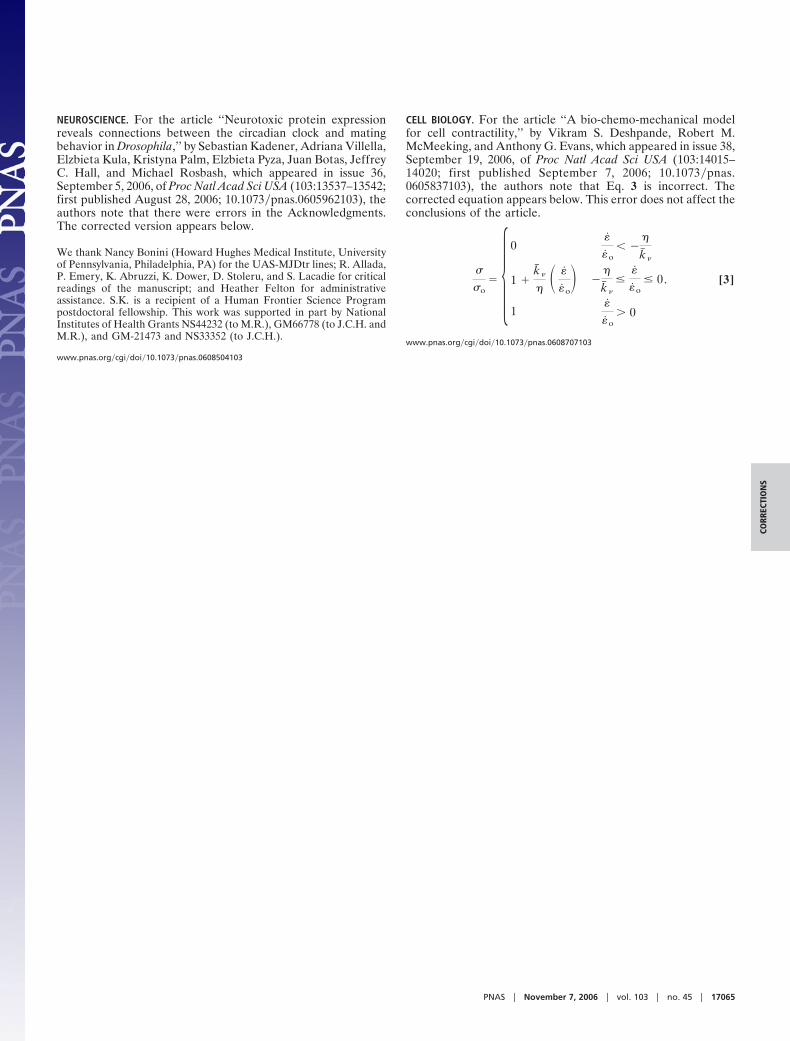
CELL BIOLOGY. For the article ‘‘A bio-chemo-mechanical modelfor cell contractility,’’ by Vikram S. Deshpande, Robert M.McMeeking, and Anthony G. Evans, which appeared in issue 38,September 19, 2006, of Proc Natl Acad Sci USA (103:14015–14020; first published September 7, 2006; 10.1073�pnas.0605837103), the authors note that Eq. 3 is incorrect. Thecorrected equation appears below. This error does not affect theconclusions of the article.
�
�o� �
0�
�o� �
�
k� �
1 �k� �
�� �
�o� �
�
k� �
��
�o� 0
1�
�o 0
. [3]
www.pnas.org�cgi�doi�10.1073�pnas.0608707103
PNAS � November 7, 2006 � vol. 103 � no. 45 � 17065
CORR
ECTI
ON
S