Embed Size (px)
Citation preview

Granquist et al. Acta Veterinaria Scandinavica 2010, 52:43http://www.actavetscand.com/content/52/1/43
Open AccessR E S E A R C H
ResearchA morphological and molecular study of Anaplasma phagocytophilum transmission events at the time of Ixodes ricinus tick biteErik G Granquist*1, Mona Aleksandersen2, Karin Bergström3, Stephen J Dumler4, Wenche O Torsteinbø1 and Snorre Stuen1
AbstractBackground: Anaplasma phagocytophilum is the causative agent of human granulocytic anaplasmosis (HGA) in humans and tick-borne fever (TBF) in ruminants. The bacterium invades and replicates in phagocytes, especially in polymorphonuclear granulocytes.
Methods: In the present study, skin biopsies and ticks (Ixodes ricinus) were collected from tick feeding lesions on 38 grazing lambs between two and three weeks after access to pastures. The histopathological changes associated with tick bites and A. phagocytophilum infection, were described. In addition the skin biopsies were examined by immunohistochemistry. Furthermore, samples from blood, skin biopsies and ticks were examined by serology, PCR amplification of msp2 (p44), genotyping of rrs (16S rRNA) variants, and compared with the results obtained from histological and immunohistochemical investigations.
Results: Tick bites were associated with chronic and hyperplastic inflammatory skin lesions in this study. A. phagocytophilum present in skin lesions were mainly associated with neutrophils and macrophages. Bacteria were occasionally observed in the Tunica media and Tunica adventitia of small vessels, but were rarely found in association with endothelial cells. PCR and genotyping of organisms present in blood, ticks and skin biopsies suggested a haematogenous and a local spread of organisms at the tick attachment sites.
Conclusions: The present study describes different aspects of A. phagocytophilum infection at the site of tick bite, and indicates that A. phagocytophilum rarely associates with endothelium during the early pathogenesis of infection.
IntroductionAnaplasma phagocytophilum is recognized as the caus-ative agent of Human Granulocytic Anaplasmosis (HGA)in humans and tick-borne fever (TBF) in ruminants [1-3].Although self-limiting in sheep, immune suppressionwith infection often results in secondary infections thatcomplicate the clinical picture [4]. TBF is of growing con-cern from the production and animal welfare perspec-tives in the sheep industry [5].
A. phagocytophilum is known to primarily infect andpropagate in polymorphonuclear leucocytes (PMN) [6-8].Its strict intracellular location provides a mechanism for
evading host defences, and also promotes chemotacticmechanisms (IL-8) that assist the attraction of neutro-phils to the tick bite site [9]. Degranulation of neutrophilsat the tick bite site increases the permeability of bloodvessels and increases the cellular infiltration of the area[10,11]. Because of the short-lived nature of circulatingneutrophils, the role of these cells in establishing andmaintaining infection has been questioned [10]. Earlierstudies have suggested that cells other than PMN areinvolved in the early pathogenesis, since ticks do notdirectly tap the blood vessels and thus cannot directlydeliver pathogens to circulating leukocytes [12-15].
Once inside the host cell however, a closed microenvi-ronment structurally designed to protect vital processeswithin the cell, gives shelter from extracellular humoraland cellular immune responses [16-20]. Earlier studies in
* Correspondence: [email protected] Department of Production Animal Clinical Sciences, Section of Small Ruminant Research, Norwegian School of Veterinary Science, Sandnes, N-4325 NorwayFull list of author information is available at the end of the article
© 2010 Granquist et al; licensee BioMed Central Ltd. This is an Open Access article distributed under the terms of the Creative CommonsAttribution License (http://creativecommons.org/licenses/by/2.0), which permits unrestricted use, distribution, and reproduction inany medium, provided the original work is properly cited.

Granquist et al. Acta Veterinaria Scandinavica 2010, 52:43http://www.actavetscand.com/content/52/1/43
Page 2 of 7
cell culture have shown that endothelial cells are capableof being infected with A. phagocytophilum and supportinfection in vitro [10,15,21].
The rationale of the present study was to examine thelocal skin inflammation, created during A. phagocytophi-lum infection, and if endothelial cells may act as in vivohost cells for A. phagocytophilum during natural infectionin lambs. Skin biopsies were collected from tick attach-ment sites and examined by histology, immunohis-tochemistry, PCR amplification of msp2 (p44) andgenotyping of A. phagocytophilum by PCR amplificationand sequencing of rrs (16S rRNA gene). Blood sampleswere also examined for the presence of bacteraemia byPCR amplification and rrs (16S rRNA gene) genotypingof A. phagocytophilum in addition to indirect fluores-cence antibody test (IFAT).
Materials and methodsAnimals and samplingSkin biopsies, EDTA blood and serum samples from 38lambs of the Norwegian White breed from two flockswere collected in May and June of the 2006 and 2007grazing seasons, in the Rogaland and Vest-Agder countyof Norway, respectively. The lambs were 4-6 weeks oldand the samples were collected between two and threeweeks after the lambs were put to pastures that were pre-viously known to be heavily infested with the sheep tick(Ixodes ricinus). The individual animals were selected forsampling based on the presence of at least two fresh tickbites. In addition, the rectal temperature was measured asan indicator of acute tick-borne fever [22]. If ticks werestill attached, they were collected and stored unfixed onindividual plastic tubes for later PCR amplification ofmsp2 (p44) to determine if they were infected by A.phagocytophilum. The wool in the tick bite area wassheared, and the skin surface was disinfected by 70% eth-anol, before a subcutaneous ring block of local anaesthe-sia was laid around the tick bite (0.5-1.0 ml 2%Carbocain™, AstraZeneca). A punch biopsy knife (8 mmin diameter) was used for collection of the skin biopsies[23]. Two biopsies from the tick bite sites and one controlbiopsy at least 20 cm from other ticks or tick bites werecollected from each lamb.
The biopsy wounds were closed by agraffe sutures. Theskin biopsies were cut in two halves with sterile scalpels.One half was stored on Zamboni's fixative before histo-logical processing and the other was kept on ice until fro-zen at -80°C for later DNA isolation. The experiment wasapproved by the National Animal Research Authority inNorway.
Real time PCR for identification of positive samples, targeting msp2 (p44)DNA was isolated from EDTA blood and skin biopsies,using a DNeasy® Blood and Tissue kit (Qiagen GmbH,
Hilden, Germany) according to protocols provided by theDNeasy® Blood and Tissue Handbook (2006). DNA fromticks was isolated using the DNeasy® Tissue kit (QIAGEN)for isolation from insects, according to protocols pro-vided by the DNeasy® Tissue kit Handbook (2004), withmodifications as follows; The volume of Proteinase K wasdoubled and the incubation time with Proteinase K wasextended to be 24 hours. The isolated DNA was dilutedaccording to spectrum readings and final template vol-ume was 5 μl containing 2.5 ng/μl total DNA. PCR posi-tive samples were detected by Real Time PCR using theLightcycler® 480 (LC480) (Roche Diagnostics Meylan,France) with Fast Start MASTERPLUS SYBR-green I Taqpolymerase mix and fluorescence detection. The specificprimers (Apmsp2f: 5'-ATG GAA GGT AGT GTT GGTTAT GGT ATT-3'and Apmsp2r: 5'-TTG GTC TTG AAGCGC TCG TA-3') were designed to amplify a 77 bp seg-ment at the conserved N-terminal coding region of msp2(p44)in the A. phagocytophilum genome [24]. Crossingpoints (CP) were determined by using the 2nd derivativemaximum method of the LightCycler® Software 1.5.0(Roche Diagnostics). The Cq (treshold cycle) was set tobe 40 since rrs (16S rRNA gene) sequences (see below)were obtained from two tissues having CP values of 39and 40, respectively. Further validation of msp2 (p44)amplicons was determined by melting point (Tm) analy-sis (range 82°C-83°C).
Semi nested conventional PCR and sequencing of the 16S rRNA geneDNA from blood and tissues were extracted according tothe protocols described in the above section. A semi-nested PCR was conducted for amplification of rrs (16SrRNA gene) on a PTC-200 instrument (MJ Research) aspreviously described [25]. Briefly, an initial PCR was per-formed using primers 16S-F5 (5'-AGTTTGATCATGGT-TCAGA-3') and ANA-R4B (5'-CGAACAACGCTTGC-3') for amplification of a 507 bp fragment of rrs (16SrRNA gene) in A. phagocytophilum, followed by a semi-nested reaction with primers 16S-F5 and ANA-R5 (5'-TCCTCTCAGACCAGCTATA-3') that produced a 282bp fragment. Positive amplification was verified by aga-rose gel electrophoresis and amplified PCR productswere sequenced directly, using Big Dye terminator cyclesequencing chemistry and capillary electrophoresis (ABI310; Applied Biosystems). A. phagocytophilum variantswere detected from visual inspection of the chromato-grams [25].
HaematologyDifferential blood cell counts were performed on EDTAblood samples using the Advia 120 Automated Hematol-ogy Analyzer (Bayer Corporation, Tarrytown, NY, USA)for evaluation of neutropenia (< 0.7 × 10⁹ cells/L).

Granquist et al. Acta Veterinaria Scandinavica 2010, 52:43http://www.actavetscand.com/content/52/1/43
Page 3 of 7
SerologyAn indirect immunofluorescence antibody assay (IFA)was used to determine the polyvalent antibody titres to A.phagocytophilum. Briefly, two-fold dilutions of sera wereadded to slides precoated with antigen obtained fromhorses (formerly Ehrlichia equi) (Protatek, St. Paul.Minn., USA). Bound antibodies were visualized by fluo-rescein-isothiocyanate (FITC)-conjugated rabbit-anti-sheep immunoglobulin (Cappel, Organon Teknika, WestChester, PA, USA). Sera were screened for antibodies atdilution 1:40. If positive, the sera were further diluted andretested. A titre of 1.6 (log10 reciprocal of 40) or more wasregarded as positive [26].
Histology and immunohistochemistrySkin samples fixed in Zamboni's fixative were routinelyprocessed and embedded in paraffin. Tissue sections of 3μm thickness were sectioned parallel to the tick bite andstained with haematoxylin and eosin for histologicalexamination.
For immunohistochemistry (IHC), 3 μm thick sectionswere collected on Menzel-Gläser SuperFrost Ultra Plus®
slides (Braunschweig, Germany) and dried over night at37°C. The sections were deparaffinised in xylene andrehydrated in graded alcohol solutions. The sections weretreated with 0.1M citrate buffer (pH 6.0) at 92°C for 20minutes in water bath or microwave oven for antigenretrieval and then cooled at room temperature for 30minutes. After washing in distilled water, slides wereplaced in phosphate buffered saline (PBS) for equilibra-tion. Endogenous peroxidase activity was inhibited byapplication of a methanol solution containing 1% H2O2for 10 minutes, followed by washing in PBS and incuba-tion for 20 minutes at room temperature with normalblocking serum (VECTASTAIN® Elite kit) (Vector Labo-ratories, Burlingham, CA, USA), diluted 1:50 in PBS con-taining 5% bovine serum albumin (BSA/PBS).
The sections were incubated with either a monoclonalanti A. phagocytophilum antibody or a polyclonal rabbitanti A. phagocytophilum antibody. The primary antibod-ies were diluted 1:400 in 1% BSA/PBS and incubation wasover night at 4°C. After washing in PBS, the sections wereincubated with the biotinylated universal antibody fromthe kit according to the protocol provided by the pro-ducer (VECTASTAIN). The sections were further incu-bated for 30 minutes with the VECTASTAIN® Elite ABCreagent after washing. Sections were exposed for the 3-amino-9-ethyl carbazole substrate (AEC) for 15 minutesand counterstained with non-alcoholic haematoxylin.Slides were washed three times in sterile water andmounted with poly vinyl alcohol (PVA).
ResultsExamination of the animalsTwenty-three of 38 lambs (60.5%) had rectal tempera-tures above 40°C and the highest recorded temperature
was 41.5°C. Thirteen lambs (34.2%) had neutropenia atthe time of sampling and nine lambs (23.7%) had feverand neutropenia. The number of engorged ticks on theanimals varied from one to more than 30 at the time ofsampling. Skin biopsies were mostly collected from theaxillary and inguinal regions as they were the most fre-quent tick attachment sites, registered. Tick bite sitesshowed typical mild erythema and local swelling.
PCR amplification of A. phagocytophilum msp2/p44 in blood, skin biopsies and ticksThirty-three lambs (86.8%) were positive for A. phagocy-tophilum by PCR analysis of peripheral blood. Thirty-seven (97.4%) had one or more skin biopsies that werepositive for A. phagocytophilum by PCR analysis. Seventyof 76 biopsies from tick attachment sites (92.1%) and 31of 38 control biopsies (81.5%) were positive by PCR for A.phagocytophilum infection. A total of 68 ticks were col-lected from the lambs. Fifty-eight (85.3%) were positivefor A. phagocytophilum by PCR. Two PCR positive ticks(2.9%) had a negative attachment site.
Sequencing of rrs (16S rRNA gene)Six different rrs (16S rRNA gene) isolates of A. phagocyto-philum were encountered during the study, that weresimilar to GenBank acc. no. U02521, M73220, AF336220,AY035312, AJ242784, and a novel variant GU459257. Allvariants except AY035312 were collected from the flockin Vest-Agder county. The variants M73220, AJ242784and AY035312 were collected from the flock in Rogalandcounty. A total of 38 partial rrs variant sequences wereobtained from the tick bite biopsies. Nineteen of 38sequences (50.0%) obtained from tick bite sites corre-sponded to the sequences obtained from the respectiveticks. The sequences obtained from control biopsies andthe blood samples were identical in all lambs where bothsequences were obtained (N = 10) (data not shown). Nodirect relations between variants, serum titre and inflam-matory changes were observed.
HistologyHistological examination of biopsies from infected skinareas showed inflammatory lesions in 35 of the 38 lambs(92.1%). The majority of lambs (60.5%) had focal histo-pathologic changes, characterized by thickened epider-mis, dermal fibroplasia and perivascular to diffuseinfiltration of mixed leucocytes (Fig. 1a). Twelve lambs(31.6%) had milder changes with perivascular inflamma-tory cell infiltration in affected areas, whereas histo-pathological changes were not observed in three of thelambs. Focal ulcerations of the epidermis were observedin skin biopsies from 10 (26.3%) animals (Fig. 1a). Theinflammatory exudate was composed of numerous neu-trophils and eosinophils in addition to mast cells, lym-phocytes and macrophages (Fig. 1b). The perivascularaggregates were mainly composed of mononuclearinflammatory cells. A substantial number of lambs, 14

Granquist et al. Acta Veterinaria Scandinavica 2010, 52:43http://www.actavetscand.com/content/52/1/43
Page 4 of 7
out of 38, showed cellulitis with subcutaneous infiltrationof neutrophils. Other lesions such as focal degenerationof dermal collagen (18.4%), vasculitis (10.5%), thrombosisof venules and lymphatics (15.8%) were observed (Fig 1c).
The different rrs (16S rRNA gene) variants of A. phago-cytophilum seemed to produce similar pathologicallesions. The control biopsies did not show inflammatorychanges.
Immunohistochemical examination, PCR and serologyVariable numbers of IHC positive organisms wereobserved in tick bite biopsies from 17/38 lambs (44.7%)and appeared as intracytoplasmic aggregates, known asmorulae. The observed organisms were associated withleucocytes in the inflammatory infiltrate in the biopsiesand were most often present in neutrophils or mac-rophages (Fig. 2b). In addition IHC positive organismswere occasionally observed in an extracellular location,either in the lumina of blood vessels or in the adventitialtunic. Bacteria were occasionally located in cells infiltrat-ing the vascular walls of venules or arterioles, usually inTunica media or Tunica adventitia and rarely in Tunicaintima (Fig. 2a). A. phagocytophilum organisms were alsopresent in intravascular inflammatory cells in lambsshowing vasculitis (Fig. 2a). IHC positive organisms weresometimes observed close to the vessel lumina (Fig. 2b).There was a large variation in the number of IHC positiveA. phagocytophilum organisms, observed in biopsiesbetween animals, and in different biopsies from the sameanimal (data not shown). Some biopsies had scatteredIHC positive labeling whereas intensively stained aggre-gates were observed in other lambs. Intensively stainedaggregates were mostly observed among inflammatorycell infiltrates of the dermis and subcutis. The controlbiopsies were IHC negative for A. phagocytophilum.
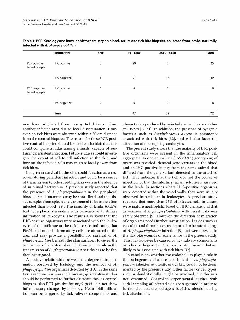
All lambs with IHC positive skin biopsies were alsopositive for msp2 (p44) by PCR on blood samples andwere seropositive for A. phagocytophilum infection. Theserological examination gave the best measures of timepost infection. Three lambs were seronegative (titre < 40)and had no visible organisms in the skin biopsies exam-ined by immunohistochemistry. Table 1 shows that 20(80%) of 25 IHC positive skin biopsies were collectedfrom animals with a serum titer ranging from 40 to 1280.Only 5 (20%) of the IHC positive biopsies were collectedfrom the group having titers ranging from 2560 to 5120(Table 1).
DiscussionLocal infection with A. phagocytophilum in tick attach-ment sites of lambs were characterized by hyperplasticskin changes and inflammatory infiltrates, similar to what
Figure 1 Skin lesions in lambs naturally infected with A. phagocy-tophilum. a) Skin biopsy with an attached tick (arrow) and ulceration of epidermis. Inflammatory cell infiltrates are present in dermis and subcutis underneath the tick bite. [Haematoxylin and eosin. Bar = 300 μm.]b) Photomicrograph of dermis. Thrombi (t) are present in lymphatics and venules, and a focal necrosis is observed in the wall of an artery (a). Infiltration of leukocytes, moderate oedema and proliferation of fi-brous tissue is found in dermis. [Haematoxylin and eosin. Bar = 100 μm.]c) Photomicrograph of dermis. A large thrombus is occluding the lu-men of a vein (arrows). Infiltration of neutrophils, macrophages and lymphocytes are present in dermis. [Haematoxylin and eosin. Bar = 50 μm.]

Granquist et al. Acta Veterinaria Scandinavica 2010, 52:43http://www.actavetscand.com/content/52/1/43
Page 5 of 7
is described for tick bite reactions even in the absence ofA. phagocytophilum infection. Immunohistochemistryshowed the presence of A. phagocytophilum in approxi-mately 45% of the lambs. These lambs were also positivefor A. phagocytophilum by PCR on blood and skin biop-sies. A. phagocytophilum organisms were mainly found ininflammatory cell infiltrates, particularly in PMNs andmacrophages of the dermis and subcutis. In the presentstudy, microorganisms were rarely observed in leucocytesin the blood stream of the skin biopsies, whereas Lepidiand coworkers reported that approximately 90% of theinfected neutrophils in deep tissues from sheep, humansand horses were seen within vessel lumens [9]. IHC posi-tive organisms were sometimes observed in the mid- andperipheral part of the vessel walls, but rarely in the inti-mal layer in the present study. The endothelium has ear-lier been suggested to function as a transition site for
transfer of A. phagocytophilum to neutrophils that areloosely bound and then released into the blood stream[15]. The present study however indicates that endothe-lium infection is a rare finding and it does not support therole of endothelium in the pathogenesis of A. phagocyto-philum infection in lambs, at least at the earliest phases oftick bite inoculation. This stands in contrast to studiesreported in mice, for which no morphological images areavailable [10]. The current study is however, limited inthat it is not an experimental study. The field conditionsdid neither allow control with attached ticks and infec-tious organisms, nor a longitudinal study of skin lesions,which is best estimated, based upon serum antibody titer,and at this point, endothelium could have played a role.
The very low number (7.9 %) of sero-negative animals(titre < 40), all which were IHC-negative, indicated thatmost lambs were sampled after seroconversion. Most ofthe IHC positive skin biopsies were collected from ani-mals with serum titres between 40 and 1280, which mayindicate that these had acute infections. However, pres-ence of maternal immunity cannot be neglected, since thehalf life of maternal antibodies has been estimated to be17.5 days [27]. Animals with titres between 2560 to 5120,were likely to have seroconverted. The IHC-positiveorganisms, observed in biopsies from this latter group,may therefore have been associated with an acute infec-tion.
PCR detection of msp2 (p44) in blood samples showedthat at least 86.8 % of the lambs, had A. phagocytophilumbacteraemia at the time of sampling. Five rrs (16S rRNAgene) variants were encountered. Organisms in biopsieswith sequences obtained from variants U02521, M73220,AF336220 and AJ242784 were detected by IHC. Differ-ences in local inflammatory responses to these variantshave never been described, but previous studies haveshown that different rrs (16S rRNA gene) variants of A.phagocytophilum can result in different immunologicalresponses and clinical reactions [28]. However, in thepresent study, no direct relationship between gene vari-ants, serum titre or inflammatory changes were observed.Similar histopathologic findings and inflammatory infil-trates with monocytes and neutrophils were associatedwith all rrs (16S rRNA gene) variants.
Nineteen of 38 sequences (50.0 %) obtained from tickbite sites corresponded to the sequences obtained fromthe respective ticks. All variants, except GU459257 (iso-lated from the skin and blood) have previously been iso-lated from the blood of infected sheep. The sequencesobtained from control biopsies and the blood sampleshowever were identical in all lambs, where bothsequences were obtained, indicating a haematogenousspread of organisms to the skin. In addition, two PCRpositive control biopsies were collected from animalshaving PCR negative blood, indicating that the organisms
Figure 2 Immunohistochemistry for Anaplasma phagocytophi-lum on skin tissue in lambs naturally infected with A. phagocyto-philum. a) Intracellular Anaplasma organisms (arrows) are present in the lumen of a small vessel. Vasculitis characterised by thickened wall and infiltration of numerous leukocytes is present in this vessel. A few bacteria are observed in the vessel wall, whereas more Anaplasma or-ganisms are found in leukocytes in dermis. [Msp2 (P44) immunostain, original magnification, ×1000]. b) Numerous Anaplasma organisms (ar-rows) are found in neutrophils and macrophages infiltrating the der-mis. [Msp2 (P44) immunostain, original magnification, ×1000]
Granquist et al. Acta Veterinaria Scandinavica 2010, 52:43http://www.actavetscand.com/content/52/1/43
Page 6 of 7
may have originated from nearby tick bites or fromanother infected area due to local dissemination. How-ever, no tick bites were observed within a 20 cm distancefrom the control biopsies. The reason for these PCR posi-tive control biopsies should be further elucidated as thiscould comprise a nidus among animals, capable of sus-taining persistent infection. Future studies should investi-gate the extent of cell-to-cell infection in the skin, andhow far the infected cells may migrate locally away fromtick bites.
Long-term survival in the skin could function as a res-ervoir during persistent infection and could be a sourceof transmission to other feeding ticks even in the absenceof sustained bacteremia. A previous study reported thatthe presence of A. phagocytophilum in the peripheralblood of small mammals may be short lived and that tis-sue samples from spleen and ear seemed to be more ofteninfected than blood [29]. The majority of lambs (60.5%)had hyperplastic dermatitis with perivascular to diffuseinfiltration of leukocytes. The results also show that theIHC-positive organisms were associated with the leuko-cytes of the infiltrate at the tick bite site, indicating thatPMNs and other inflammatory cells are attracted to thearea and may provide a possibility for survival of A.phagocytophilum beneath the skin surface. However, theoccurrence of persistent skin infections and its role in thetransmission of A. phagocytophilum to ticks has to be fur-ther investigated.
A positive relationship between the degree of inflam-mation observed by histology and the number of A.phagocytophilum organisms detected by IHC, in the sametissue sections was present. However, quantitative studiesshould be performed to further elucidate this, as controlbiopsies, also PCR positive for msp2 (p44), did not showinflammatory changes by histology. Neutrophil infiltra-tion can be triggered by tick salivary components and
chemotaxins produced by infected neutrophils and othercell types [30,31]. In addition, the presence of pyogenicbacteria such as Staphylococcus aureus is commonlyassociated with tick bites [32], and will also favor theattraction of neutrophil granulocytes.
The present study shows that the majority of IHC posi-tive organisms were present in the inflammatory cellaggregates. In one animal, rrs (16S rRNA) genotyping oforganisms revealed identical gene variants in the bloodand an IHC-positive biopsy from the same animal thatdiffered from the gene variant detected in the attachedtick. This indicates that the tick was not the source ofinfection, or that the infecting variant selectively survivedin the lamb. In sections where IHC-positive organismswere detected within the vessel walls, they were usuallyobserved intracellular in leukocytes. A previous studyreported that more than 95% of infected cells in tissueswere mature neutrophils, based on IHC analysis and thatassociation of A. phagocytophilum with vessel walls wasrarely observed [9]. However, the direction of migrationof organisms needs further investigation. Lesions such asvasculitis and thromboses are reported to be rare findingsof A. phagocytophilum infection [9], but were present inthe tick bite wounds of some lambs in the present study.This may however be caused by tick salivary componentsor other pathogens like S. aureus or streptococci that arelikely to be associated with tick bites [32].
In conclusion, whether the endothelium plays a role inthe pathogenesis of and establishment of A. phagocyto-philum infection at the site of tick bite could not be docu-mented by the present study. Other factors or cell types,such as dendritic cells, might be involved, but this wasnot examined. Controlled experimental studies withserial sampling of infected skin are suggested in order tofurther elucidate the pathogenesis of this infection duringtick attachment.
Table 1: PCR, Serology and immunohistochemistry on blood, serum and tick bite biopsies, collected from lambs, naturally infected with A. phagocytophilum
Serum titre ≤ 40 40 - 1280 2560 - 5120 Sum
PCR positiveblood sample
IHC positive 0 20 5 25
IHC negative 1 21 17 39
PCR negativeblood sample
IHC positive 0 0 0 0
IHC negative 2 6 0 8
Sum 3 47 22 72

Granquist et al. Acta Veterinaria Scandinavica 2010, 52:43http://www.actavetscand.com/content/52/1/43
Page 7 of 7
Competing interestsThe authors declare that they have no competing interests.
Authors' contributionsEGG performed the sampling, real time PCR, immunohistology, rrs sequenceanalysis and writing of the manuscript. MA participated in the design of thestudy, performed the histological examination, immunohistological interpreta-tion and created the figures. MA also revised the draft manuscript. KB per-formed the IFAT. WOT performed the sequencing of rrs (16S rRNA gene). JSDparticipated in the design of the study, provided reagents for IHC and helpedin revising the draft manuscript. SS designed the study and supervised thewriting of the draft manuscript. All authors read and approved the final manu-script.
AcknowledgementsWe thank Dr. Ulrike Munderloh for valuable help and contributions. We also thank Siri Hamre and Laila Aune for laboratory contributions.
Author Details1Department of Production Animal Clinical Sciences, Section of Small Ruminant Research, Norwegian School of Veterinary Science, Sandnes, N-4325 Norway, 2Department of Basic Sciences and Aquatic Medicine, Norwegian School of Veterinary Science, Oslo, N-0033 Norway, 3National Veterinary Institute, Uppsala 75189, Sweden and 4Department of Pathology, Division of Medical Microbiology, The Johns Hopkins Hospital, Baltimore, MD 21205 USA
References1. Dumler JS, Madigan JE, Pusterla N, Bakken JS: Ehrlichioses in humans:
epidemiology, clinical presentation, diagnosis, and treatment. Clin Infect Dis 2007, 45(Suppl 1):S45-51.
2. Bakken JS, Dumler JS, Chen SM, Eckman MR, Van Etta LL, Walker DH: Human granulocytic ehrlichiosis in the upper Midwest United States. A new species emerging? JAMA 1994, 272:212-218.
3. Foggie A: Studies on the infectious agent of tick-borne fever in sheep. J Pathol Bacteriol 1951, 63:1-15.
4. Woldehiwet Z: Immune evasion and immunosuppression by Anaplasma phagocytophilum, the causative agent of tick-borne fever of ruminants and human granulocytic anaplasmosis. Vet J 2008, 175:37-44.
5. Stuen S, Bergstrom K, Palmer E: Reduced weight gain due to subclinical Anaplasma phagocytophilum (formerly Ehrlichia phagocytophila) infection. Exp Appl Acarol 2002, 28:209-215.
6. Woldehiwet Z: Immune evasion and immunosuppression by Anaplasma phagocytophilum, the causative agent of tick-borne fever of ruminants and human granulocytic anaplasmosis. Vet J 2007, 175(1):37-44.
7. Chen SM, Dumler JS, Bakken JS, Walker DH: Identification of a granulocytotropic Ehrlichia species as the etiologic agent of human disease. J Clin Microbiol 1994, 32:589-595.
8. Dumler JS, Choi KS, Garcia-Garcia JC, Barat NS, Scorpio DG, Garyu JW, Grab DJ, Bakken JS: Human granulocytic anaplasmosis and Anaplasma phagocytophilum. Emerg Infect Dis 2005, 11:1828-1834.
9. Lepidi H, Bunnell JE, Martin ME, Madigan JE, Stuen S, Dumler JS: Comparative pathology, and immunohistology associated with clinical illness after Ehrlichia phagocytophila-group infections. Am J Trop Med Hyg 2000, 62:29-37.
10. Herron MJ, Ericson ME, Kurtti TJ, Munderloh UG: The interactions of Anaplasma phagocytophilum, endothelial cells, and human neutrophils. Ann N Y Acad Sci 2005, 1063:374-382.
11. Carlyon JA, Fikrig E: Invasion and survival strategies of Anaplasma phagocytophilum. Cell Microbiol 2003, 5:743-754.
12. Epperson DE, Pober JS: Antigen-presenting function of human endothelial cells. Direct activation of resting CD8 T cells. J Immunol 1994, 153:5402-5412.
13. Ferry B, Halttunen J, Leszczynski D, Schellekens H, vd Meide PH, Hayry P: Impact of class II major histocompatibility complex antigen expression on the immunogenic potential of isolated rat vascular endothelial cells. Transplantation 1987, 44:499-503.
14. Udo AK Hetzel, Zerai Woldehiwet: Anaplasma phagocytophilum: Sites of primary infection. In European Society of Veterinary Pathology 24th meeting Edinburgh, Scotland; 2006.
15. Munderloh UG, Lynch MJ, Herron MJ, Palmer AT, Kurtti TJ, Nelson RD, Goodman JL: Infection of endothelial cells with Anaplasma marginale and A. phagocytophilum. Vet Microbiol 2004, 101:53-64.
16. Niu H, Yamaguchi M, Rikihisa Y: Subversion of cellular autophagy by Anaplasma phagocytophilum. Cell Microbiol 2008, 10:593-605.
17. Niu H, Rikihisa Y, Yamaguchi M, Ohashi N: Differential expression of VirB9 and VirB6 during the life cycle of Anaplasma phagocytophilum in human leucocytes is associated with differential binding and avoidance of lysosome pathway. Cell Microbiol 2006, 8:523-534.
18. Rosen H: Bacterial responses to neutrophil phagocytosis. Curr Opin Hematol 2004, 11:1-6.
19. Carlyon JA, Fikrig E: Mechanisms of evasion of neutrophil killing by Anaplasma phagocytophilum. Curr Opin Hematol 2006, 13:28-33.
20. Rikihisa Y: Mechanisms to create a safe haven by members of the family Anaplasmataceae. Ann N Y Acad Sci 2003, 990:548-555.
21. Wamsley HL, Barbet AF: In situ detection of Anaplasma spp. by DNA target-primed rolling-circle amplification of a padlock probe and intracellular colocalization with immunofluorescently labeled host cell von Willebrand factor. J Clin Microbiol 2008, 46:2314-2319.
22. Woldehiwet Z, Scott GR: Immunological studies on tick-borne fever in sheep. J Comp Pathol 1982, 92:457-467.
23. Inman VD, Pariser RJ: Biopsy technique pearl: Obtaining an optimal split punch-biopsy specimen. J Am Acad Dermatol 2003, 48:273-274.
24. Courtney JW, Kostelnik LM, Zeidner NS, Massung RF: Multiplex real-time PCR for detection of Anaplasma phagocytophilum and Borrelia burgdorferi. J Clin Microbiol 2004, 42:3164-3168.
25. Stuen S, Dahl H, Bergstrom K, Moum T: Unidirectional suppression of Anaplasma phagocytophilum genotypes in infected lambs. Clin Diagn Lab Immunol 2005, 12:1448-1450.
26. Stuen S, Bergstrom K: Serological investigation of granulocytic Ehrlichia infection in sheep in Norway. Acta Vet Scand 2001, 42:331-338.
27. Stuen S, Hardeng F, Larsen HJ: Resistance to tick-borne fever in young lambs. Res Vet Sci 1992, 52:211-216.
28. Stuen S, Bergstrom K, Petrovec M, Van de Pol I, Schouls LM: Differences in clinical manifestations and hematological and serological responses after experimental infection with genetic variants of Anaplasma phagocytophilum in sheep. Clin Diagn Lab Immunol 2003, 10:692-695.
29. Liz JS, Anderes L, Sumner JW, Massung RF, Gern L, Rutti B, Brossard M: PCR detection of granulocytic ehrlichiae in Ixodes ricinus ticks and wild small mammals in western Switzerland. J Clin Microbiol 2000, 38:1002-1007.
30. Choi KS, Grab DJ, Dumler JS: Anaplasma phagocytophilum infection induces protracted neutrophil degranulation. Infect Immun 2004, 72:3680-3683.
31. Boppana DK, Wikel SK, Raj DG, Manohar MB, Lalitha J: Cellular infiltration at skin lesions and draining lymph nodes of sheep infested with adult Hyalomma anatolicum anatolicum ticks. Parasitology 2005, 131:657-667.
32. Foggie A: Studies on the relationship of tick-bite to tick pyaemia of lambs. Ann Trop Med Parasitol 1959, 53:27-34.
doi: 10.1186/1751-0147-52-43Cite this article as: Granquist et al., A morphological and molecular study of Anaplasma phagocytophilum transmission events at the time of Ixodes rici-nus tick bite Acta Veterinaria Scandinavica 2010, 52:43
Received: 26 February 2010 Accepted: 17 June 2010 Published: 17 June 2010This article is available from: http://www.actavetscand.com/content/52/1/43© 2010 Granquist et al; licensee BioMed Central Ltd. This is an Open Access article distributed under the terms of the Creative Commons Attribution License (http://creativecommons.org/licenses/by/2.0), which permits unrestricted use, distribution, and reproduction in any medium, provided the original work is properly cited.Acta Veterinaria Scandinavica 2010, 52:43