Embed Size (px)
Citation preview

METHODOLOGY ARTICLE Open Access
A mouse ear skin model to study thedynamics of innate immune responsesagainst Staphylococcus aureus biofilmsAizat Iman Abdul Hamid1, Laurence Nakusi1, Mickael Givskov2, Young-Tae Chang3, Claire Marquès1 andPascale Gueirard1*
Abstract
Background: Staphylococcus aureus is a human pathogen that is a common cause of nosocomial infections andinfections on indwelling medical devices, mainly due to its ability to shift between the planktonic and the biofilm/sessile lifestyle. Biofilm infections present a serious problem in human medicine as they often lead to bacterialpersistence and thus to chronic infections. The immune responses elicited by biofilms have been described asspecific and ineffective. In the few experiments performed in vivo, the importance of neutrophils and macrophagesas a first line of defence against biofilm infections was clearly established. However, the bilateral interactionsbetween biofilms and myeloid cells remain poorly studied and analysis of the dynamic processes at the cellularlevel in tissues inoculated with biofilm bacteria is still an unexplored field. It is urgent, therefore, to developbiologically sound experimental approaches in vivo designed to extract specific immune signatures from theplanktonic and biofilm forms of bacteria.
Results: We propose an in vivo transgenic mouse model, used in conjunction with intravital confocal microscopyto study the dynamics of host inflammatory responses to bacteria. Culture conditions were created to preparecalibrated inocula of fluorescent planktonic and biofilm forms of bacteria. A confocal imaging acquisition andanalysis protocol was then drawn up to study the recruitment of innate immune cells in the skin of LysM-EGFPtransgenic mice. Using the mouse ear pinna model, we showed that inflammatory responses to S. aureus can bequantified over time and that the dynamics of innate immune cells after injection of either the planktonic orbiofilm form can be characterized. First results showed that the ability of phagocytic cells to infiltrate the injectionsite and their motility is not the same in planktonic and biofilm forms of bacteria despite the cells beingconsiderably recruited in both cases.
Conclusion: We developed a mouse model of infection to compare the dynamics of the inflammatory responsesto planktonic and biofilm bacteria at the tissue and cellular levels. The mouse ear pinna model is a powerfulimaging system to analyse the mechanisms of biofilm tolerance to immune attacks.
Keywords: Staphylococcus aureus, Biofilm, Planktonic form, Inflammation, Mouse, Intravital imaging
© The Author(s). 2020 Open Access This article is distributed under the terms of the Creative Commons Attribution 4.0International License (http://creativecommons.org/licenses/by/4.0/), which permits unrestricted use, distribution, andreproduction in any medium, provided you give appropriate credit to the original author(s) and the source, provide a link tothe Creative Commons license, and indicate if changes were made. The Creative Commons Public Domain Dedication waiver(http://creativecommons.org/publicdomain/zero/1.0/) applies to the data made available in this article, unless otherwise stated.
* Correspondence: [email protected] Microorganismes : Génome et Environnement, UMR CNRS 6023,Université Clermont-Auvergne, Clermont Ferrand, FranceFull list of author information is available at the end of the article
Abdul Hamid et al. BMC Microbiology (2020) 20:22 https://doi.org/10.1186/s12866-019-1635-z

BackgroundStaphylococcus aureus (S. aureus) is a common com-mensal Gram-positive bacterium that colonizes the skinand mucous membranes of humans. It can also shift be-tween planktonic and biofilm lifestyles and colonize abi-otic surfaces such as indwelling medical devices andprosthetic implants [1]. Inside biofilms, bacteria are em-bedded in an extracellular matrix and are more tolerantto antibiotics and to host immune attacks [2]. Theresulting impact on human health is enormous sincebiofilm infections account for more than 80% of micro-bial infections in otherwise sterile tissue(s) and often be-come chronic [3].The immune responses elicited by biofilms have been
described as specific and ineffective thus promoting bac-terial persistence and the establishment of chronic infec-tions [4]. Different immune evasion mechanisms havebeen proposed to be involved, including phagocyte directkilling (macrophages, neutrophils), specific recruitmentof myeloid-derived suppressor cells (MDSCs) andmacrophage polarization towards an anti-inflammatoryphenotype [4, 5]. These results were mostly obtainedduring experiments performed in vitro in which biofilmswere exposed to monocytes or neutrophils, or both [6].In the few experiments performed in vivo with differentrodent models, several parameters vary, such as the pres-ence of a biomedical device, the tissue(s) that were inoc-ulated or implanted with a bacteria-free or loadeddevice, the bacteria delivery mode and the inoculumdose [7]. These studies illustrate the importance of bothneutrophils and monocytes/macrophages as a first lineof defence against biofilm infections. However, the bilat-eral interactions between biofilms and myeloid cells re-main poorly studied and analysis of the dynamicprocesses at the cellular level in tissues inoculated withbiofilms is still an unexplored field. The mouse ear pinnais currently one of the most frequently used tissues toperform intravital confocal live imaging. In particular, itallows the analysis of cellular behaviour in an inflamedtissue [8]. We previously developed a concept that waspotentially able to extract the biologically relevant fea-tures of the host and invasive bacteria after injectionof either the planktonic or biofilm form of bacteria inthe ear pinna [7]. In the present study, it was decidedto use the transgenic fluorescent reporter laboratorymice line LysM-EGFP. Owing to the relative thinnessof the ear pinna, the model enabled us to performlive imaging on recruited enhanced green fluorescentprotein (EGFP) fluorescent leukocytes, in particularneutrophils and monocytes/macrophages. When theLysM-EGFP mouse ear pinna dermis was loaded witheither planktonic or biofilm bacteria, the first resultsshowed that the inflammatory response to S. aureuscan be quantified in the skin.
Both bacterial forms induced a considerable inflammatoryresponse at the injection site. However, real-time analysisshowed different cellular dynamics with a limited access ofrecruited phagocytes to bacteria inside biofilms, resulting inless efficient phagocytosis. We also investigated the motilityof resident or recruited phagocytes and observed that cellsarrest at the injection site to interact with planktonic or bio-film bacteria. At early time points, biofilms slowed downphagocytes and modified their trajectory. Finally, the natureof the inoculum (planktonic or biofilm) influenced speedand straightness parameters differently, independently ofcell-bacteria interactions at the injection site.We therefore developed a mouse model of infection to
compare the inflammatory response to planktonic andbiofilm bacteria at the tissue and cellular levels. Our novelfindings show that the dynamics of the inflammatory re-sponses against the two bacterial forms are different.
ResultsPreparation and characterization of calibrated inocula ofStaphylococcus aureus biofilm and planktonic culturesA reproducible protocol of biofilm preparation was cre-ated to obtain a calibrated bacterial inoculum of 107
colony-forming units (CFUs) in 3.8 μL of biofilm suspen-sion (injection volume). As shown in Additional file 1:Figure S1A, titres of different aliquots of 24 h-old bio-films collected in the same well or in different wells forthree independent experiments were comparable (Add-itional file 2: Table S1). To compare host immune re-sponses to planktonic and biofilm forms of S. aureusLYO-S2 bacteria, calibrated inocula of planktonic bac-teria were also prepared. The titres of the inocula werecomparable for both bacterial forms and contained theexpected quantity of bacteria (Additional file 1: FigureS1B and Additional file 2: Table S1). However, the mor-phological characteristics of the two inocula were differ-ent, even after passing through the 34-gauge (34G)needle used for micro-injection into the mouse ear tis-sue. Scanning electron microscopy (SEM) ultrastructuralanalysis showed that planktonic bacteria were either dis-persed or organized in small clusters (Fig. 1a and Add-itional file 1: Figure S1C). In contrast, biofilms wereorganized in aggregates of 29.43 ± 7.06 μm across (Add-itional file 1: Figure S1D). When zoomed in, the extra-cellular matrix is clearly observed inside these aggregates(Fig. 1b, red arrows and Additional file 1: Figures S1E-H). However, the homogenization technique used toprepare biofilm inocula results in an inoculum contain-ing mainly biofilm aggregates but also detached bacteriaand planktonic bacteria. Future use of the term “biofilminoculum” or “biofilm” will be in reference to this typeof inoculum. Using the fluorescent probe CDy11, whichtargets amyloid fibrils, we observed that this biofilmmatrix component was detected more abundantly in our
Abdul Hamid et al. BMC Microbiology (2020) 20:22 Page 2 of 12

biofilm preparations than in the samples of planktonicbacteria (Fig. 1c-d) [9].
Micro-injection of calibrated inocula of planktonic orbiofilm forms of Staphylococcus aureus in the mouse earpinna induces a strong inflammatory responseLysM-EGFP transgenic mice were inoculated intradermallyinto the ear pinna with 107 CFUs of either planktonic orbiofilm mCherry-LYO-S2 fluorescent bacteria, or Trypti-case Soja (TS) culture medium, which was used as a con-trol. Inflammatory responses were followed at early (4–7 hpost-injection [hpi]) and late time points (after 24 hpi) bymeasuring the intensity of the EGFP signal for each group
(Fig. 2a-c). The image of the ear pinna enabled us to ana-lyse overall inflammation in the entire tissue (mosaic acqui-sition). To quantify this signal, we created the followingprotocol. A region of interest (ROI) was drawn on late timepoint images, where the EGFP signal was more easily de-tectable, and applied to early time point images. The ratioof the sum of EGFP fluorescence intensities to ROI areaswas calculated with this protocol and the inflammatory re-sponse was compared at early and late time points in thetwo groups of infected mice (Fig. 2d and Additional file 3:Table S2). We used the same protocol for the controlgroup and observed a non-specific recruitment of EGFP+innate immune cells due to the physical trauma from
Fig. 1 Characterization of calibrated inocula of Staphylococcus aureus biofilm and planktonic cultures. a and b SEM micrographs of S. aureus LYO-S2 planktonic (a) and 24 h biofilm (b) inocula after passing through the 34G needle used for micro-injections. Red arrows in panel B indicate thebiofilm extracellular matrix. Scale bar: 5 μm. c and d Fluorescence microscopy images of S. aureus biofilm (c) and planktonic (d) cultures stainedwith the green live cell fluorescent label SYTO9 and incubated with CDy11 red fluorescent probe. Scale bar: 50 μm
Abdul Hamid et al. BMC Microbiology (2020) 20:22 Page 3 of 12

Fig. 2 Micro-injection of calibrated inocula of Staphylococcus aureus in the mouse ear pinna. a–c Reconstituted confocal images of the mouse earpinna tissue showing the maximal projection intensities of the EGFP signal. LyM-EGFP transgenic mice were micro-injected with TS culture medium (a)or S. aureus mCherry-LYO-S2 in its planktonic (b) or biofilm (c) form at early (4–7 hpi) and late time points (after 24 hpi). The EGFP fluorescence (green)signal corresponds to phagocytic cells (neutrophils and macrophages). The yellow line indicates the ROI where the “Sum of EGFP fluorescenceintensities” was measured. Scale bar: 2 mm. One representative experiment is shown for each group of mice from four independent experiments. dRatio of the sum of EGFP fluorescence intensities to ROI area. Data are expressed as median and interquartile ranges for four mice per group
Abdul Hamid et al. BMC Microbiology (2020) 20:22 Page 4 of 12

injection and the introduction of TS culture medium(Additional file 4: Movie S1). At early time points, bothbacterial forms induced an inflammatory response, with a
statistically significant increase only in the group of miceinoculated with planktonic bacteria. Thus, planktonic bac-teria induced a greater response than biofilms after 4 hpi.
Fig. 3 Dynamics of recruited EGFP+ cells in the mouse ear pinna after inoculation of Staphylococcus aureus. a and b Live confocal imaging aftermicro-injection of S. aureus mCherry-LYO-S2 in its planktonic (a) or biofilm (b) form in the ear pinna of LysM-EGFP transgenic mice at early timepoints. Innate immune cell recruitment towards planktonic bacteria and biofilms was observed between 3.20 to 3.50 hpi and 4.20 to 4.40 hpi,respectively. A progressive recruitment of EGFP+ innate immune cells was observed at the injection site with cell-bacteria contact areas (filledwhite arrowheads). White empty circles show cell accumulation over time for the planktonic or biofilm inoculum at early time points. *:autofluorescent hair (also in magenta). Scale bar: 100 μm. (c and d) Live confocal imaging at late time points after micro-injection of planktonic(c) or biofilm (d) bacteria, at 24.20 hpi and 26.20 hpi, respectively. Empty white arrowhead indicates the presence of remaining planktonic formafter 24 h (low magenta signal) whereas biofilms were still easily detectable. Scale bar: 100 μm. a–d Images show average intensity projections ofgreen (innate immune cells) and magenta (bacteria) fluorescence. One representative experiment is shown for each group of mice from threeindependent experiments
Abdul Hamid et al. BMC Microbiology (2020) 20:22 Page 5 of 12

Between the early and late time points, the inflammatoryresponse was significantly greater for both bacterial forms.At late time points, the response was considerable in bothgroups of challenged mice compared to control mice, withno significant difference between mice inoculated withplanktonic or biofilm bacteria (Fig. 2d).
Dynamics of immune cell recruitment after the micro-injection of either planktonic or biofilm forms ofStaphylococcus aureus in the mouse ear pinna aredifferentLysM-EGFP transgenic mice were inoculated intrader-mally into the ear pinna with 107 CFUs of either plank-tonic or biofilm mCherry-LYO-S2 fluorescent bacteria,or with TS culture medium. We created a confocal ac-quisition protocol to analyse the dynamics of recruitedEGFP+ cells at the inoculation sites by real-time im-aging. A red autofluorescence signal is emitted by micehairs and could not be prevented by shaving the earpinna. Indeed, this operation would have induced a non-specific inflammatory response. In control mice, recruit-ment was low owing to injection trauma (Additional file4: Movie S1). In mice inoculated with bacteria, an influxof phagocytic cells was observed at early (3 to 6 hpi)(Fig. 3a-b, white circles) and late time points (after 24hpi) (Fig. 3c-d) for both bacterial forms. At early timepoints, immune cells were present over the entire sur-face of planktonic bacteria injection sites and multiplecontact points between cells and bacteria were observed.In addition, numerous immune cells infiltrated the injec-tion sites (Fig. 3a and Additional file 5: Figures S2A-B,white arrowheads; see also Additional file 6: Movie S2).In biofilms, the contact points were less numerous andwere mainly located at the periphery of the injection site.In contrast to planktonic inocula, a small number ofcells succeeded in infiltrating the biofilm (Fig. 3b andAdditional file 5: Figures S2C-D, white arrowheads; seealso Additional file 7: Movie S3). The fluorescent signalwas less detectable for planktonic bacteria after 24 h,suggesting that bacterial lysis after phagocytosis had oc-curred (Fig. 3c, white empty arrowhead). For biofilms,phagocytosis seemed to be less effective, since a fluores-cent signal was still clearly visible after 24 h (Fig. 3d).Overall, this real-time analysis using an intravital im-aging approach shows that the dynamics of the inflam-matory responses against planktonic and biofilm bacteriaare different.
Motility of recruited innate immune cells is different afterinjection of planktonic or biofilm forms of Staphylococcusaureus in the mouse ear pinnaUsing Imaris software, we created an analysis protocol totrack the motility properties (average speed andstraightness) of EGFP+ cells recruited at the injection zone
from the previously acquired time-lapse videos (Fig. 4).Using the “Spots” tool we attributed a sphere to a numberof immune cells observed in the acquisition field (Fig. 4a-b,white spheres). This enabled us to establish a trajectory(Fig. 4a-b, multicoloured lines) for each sphere correspond-ing to the path taken by each cell over time in the ear tis-sue. We then compared the average speed and straightnessof the trajectories of phagocytic cells in response to the twobacterial forms. In different zones of the injection site, cellsinteracted with bacteria (Fig. 4a) or not (Fig. 4b). We firstanalysed the motility of all cells in response to bacteria(planktonic or biofilm) or to TS culture medium withoutdistinction between cells that interacted with bacteria andcells that did not. At early time points, only biofilms in-duced a significant decrease in cell speed, compared to con-trol mice and mice inoculated with planktonic bacteria.Thus, biofilms slowed down recruited cells, an effect thatwas maintained 24 hpi. In contrast, planktonic bacteria sig-nificantly increased cell speed compared to the controlgroup (Fig. 4c and Additional file 8: Table S3). This differ-ential response induced by the two bacterial forms was alsoseen for cell trajectory straightness, which was significantlydecreased at early time points only by biofilms (less straighttrajectory of EGFP+ recruited cells) compared to the con-trol group. At late time points, we observed an opposite ef-fect, as both bacterial forms significantly increasedstraightness compared to the control group, with a morepronounced effect for planktonic bacteria (Fig. 4d and Add-itional file 9: Table S4). We further analysed the motility ofcells interacting with bacteria (bacteria contact) or not (nobacteria contact) in different zones of the cutaneous injec-tion site for the same time point (Fig. 4e-h). Cell motilitywas compared after inoculation of biofilm or planktonicbacteria. At early time points, the presence of the two formsof bacteria (bacteria contact) induced a significant decreasein both speed and straightness (Fig. 4e-f, Additional file 10:Table S5 and Additional file 11: Table S6). This indicatesthat cells arrest at the injection site to interact with inocu-lated bacteria. At early and late time points, cell speed wasreduced in biofilms compared to planktonic cells, inde-pendently of the presence of bacteria (Fig. 4e and g, Add-itional file 12: Table S7). Finally, at late time points,straightness was reduced for cells interacting with biofilms,compared to planktonic inocula (Fig. 4h and Add-itional file 13: Table S8). Taken together, these results dem-onstrate that the cell dynamics of the inflammatoryresponse are different after inoculation of biofilm or plank-tonic bacteria. The mouse ear pinna model evidences an in-flammatory response specific to biofilms that is probablyone mechanism of its tolerance to immune attacks.
DiscussionThe dynamics of the implementation of immune re-sponses during S. aureus infections in vivo is a key
Abdul Hamid et al. BMC Microbiology (2020) 20:22 Page 6 of 12

event. It is a determinant factor especially duringplanktonic-to-biofilm transition, as the bacterial persist-ence associated with the “chronicization” process of bio-film infection often depends on it. However, it is difficultto follow these events in mammals over time and this
hinders the clear understanding of the immune evasionmechanisms of S. aureus biofilms and therefore the de-sign of preventive strategies against biofilm infections.In the present study, we compared for the first time
the dynamics of early innate immune responses to
Fig. 4 Motility of recruited EGFP+ cells in the mouse ear pinna after micro-injection of Staphylococcus aureus. a and b Illustration of immune celltracking with Imaris software using the “Spots” tool to analyse the motility of recruited immune cells. The analysis was carried out in differentzones of the injection site where cells were either in contact with visible bacteria (a) or not (b). Each cell is represented by a white sphere and itstrajectory in the thickness of the tissue by a multicoloured line. Images shown were taken at 4.45 hpi (a) and 26 hpi (b). *: base of hair follicles.Scale bar: 50 μm. c–h Average speed and straightness of EGFP+ cells recruited to injection sites at early and late time points after inoculation ofTS culture medium (control), planktonic bacteria (planktonic form) or biofilms (biofilm form). Data are expressed as median and interquartileranges pooled from three different mice in three independent experiments for each group. Average speed (c) and straightness (d) of all cells (incontact with visible bacteria or not) in infected and control mice. Number of cells (N) analysed for each group at early and late time points,respectively: Control: N = 90 and 94 cells; Planktonic form: N = 315 and 433 cells; Biofilm form: N = 254 and 518 cells. Average speed (e and g) andstraightness (f and h) of cells either in contact (bacteria contact) or not (no bacteria contact) with planktonic or biofilm bacteria at early (e and f)and late (g and h) time points. Number of cells (N) analysed at early time points that were in contact or not in contact with bacteria, respectively:Planktonic form: N = 157 and 158 cells; Biofilm form: N = 142 and 112 cells. Number of cells (N) analysed at late time points that were in contactor not in contact with bacteria, respectively: Planktonic form: N = 298 and 135 cells; Biofilm form: N = 98 and 420 cells
Abdul Hamid et al. BMC Microbiology (2020) 20:22 Page 7 of 12

planktonic and biofilm S. aureus in the skin. The skin isa common target tissue for S. aureus infections and themouse ear pinna is frequently used as a cutaneous im-aging site. This accessible tissue can be rapidly and easilyprepared for imaging over long periods of time. Cali-brated inocula were injected intradermally in a verysmall volume with a 34G needle to limit inflammationresulting from injection trauma. The maturation state ofthe biofilm culture and the bacterial inocula were twomajor criteria in the finalization of the protocol. We pre-pared “young” biofilms (24 h-old) as previous studies re-ported that immature biofilms are more susceptible toneutrophil attack than mature biofilms [10]. We inocu-lated a high number of bacteria (107 CFUs) into the eartissue of LysM-EGFP transgenic mice, as in previouslypublished mouse models of S. aureus skin infectionssuch as the chronic diabetic wound model and the airpouch model [11, 12]. Once drawn up, our protocol en-abled us to compare qualitatively and quantitatively theinnate immune responses induced by a comparable doseof planktonic or biofilm bacteria. As described above,our biofilm inoculum contained bacterial aggregates andalso planktonic and detached bacteria. The phenotype ofthe latter is similar to that of biofilm bacteria. Indeed,previous studies have described differential gene expres-sion profiles for S. aureus in its planktonic or biofilmform [13, 14]. One major difference between the two in-ocula was the presence of the extracellular matrix in thebiofilm aggregates. Among the components of the LYO-S2 S. aureus biofilm matrix inoculated into mice, we de-tected amyloid fibrils. Small peptides called phenol-soluble modulins (PSMs) produce amyloids in S. aureusbiofilms and notably contribute to S. aureus biofilm sta-bility. They are also described as key virulence factorscapable of stimulating inflammatory responses or affect-ing leukocyte viability or functions [15, 16], and couldcontribute to the specific innate immune responses ob-served with biofilm bacteria.After inoculation, we used the mouse ear pinna model
to follow the inflammatory response to S. aureus overtime at the tissue level. Imaging analysis showed that bac-terial inocula induced an early inflammatory response atthe cutaneous injection site in LysM-EGFP transgenicmice, consisting of recruited EGFP+ phagocytes, in par-ticular neutrophils and monocytes/macrophages. Wequantified this response for the first time and showed thatit is significantly increased with the planktonic form after4 h, compared to that in control mice. The absence of sig-nificant differences between the control and biofilm con-ditions could have been due to early phagocyte killing, aspreviously reported [4, 5, 17]. After 24 h, the inflammatoryresponse was considerable and comparable for the twobacterial forms. In rodent models, neutrophils are usuallythe most rapidly recruited, and therefore most abundant,
cells in the proximity of biofilms [7]. In a catheter-relatedmodel, however, monocytes were the first cells to be re-cruited [18]. Although overall responses are comparable atlate time points, we postulate that the phenotype of re-cruited cells in our model differs according to whetherplanktonic or biofilm bacteria are injected with, for ex-ample, the specific recruitment of myeloid-derived sup-pressor cells (MDSCs) for the biofilm, as describedpreviously [19].The mouse ear pinna model then enabled us to follow
the inflammatory responses to planktonic or biofilm S.aureus over time at the cellular level. Analysis of the dy-namics of recruited EGFP+ cells at the inoculation sitesby real-time imaging showed that the dynamics differedbetween planktonic and biofilm bacteria. Our resultsshow that biofilm acts as a physical barrier [20]. Fewcells infiltrate the biofilm, with most recruited cells be-ing present at the periphery of the inoculum. After 24 h,when innate immune responses have been considered asset up, phagocytosis seemed to be limited. Indeed, thebacterial signal was still intense, compared to the lowsignal observed with planktonic bacteria at the sametime point. This impairment of phagocytosis observedwith biofilms is commonly known as “frustrated phago-cytosis” [20]. Complementary experiments are requiredto quantify the bacterial load in the ear tissue over time.The mouse ear pinna model further enabled us to ob-
tain reproducible quantitative measurements of thespeed and straightness of recruited innate immune cells.To obtain the most accurate representation of these mo-tility parameters, stringent algorithm settings were used.Then, manual corrections were applied to cell tracks.For example, only tracks lasting three or more frameswere considered during the time that cells were visiblein the observation field. Tracks that converged into onewere eliminated to avoid any uncertainty about theresulting cell trajectory. Likewise, cells near or exitingthe border of the image volume were carefullychecked to ensure that the same cell was not countedtwice with two different tracks. Analysis of innate im-mune cell migration showed that cells behaved differ-ently in presence of planktonic and biofilm bacteria.Study of the entire population of cells in the tissue(cells interacting or not with bacteria) showed thatbiofilms generally decreased cell speed andstraightness. In addition, when immune cells inter-acted with bacteria at the injection site, biofilms gen-erally decreased cell speed more significantly than didplanktonic bacteria. A possible correlation could bemade with previous observations describing immobi-lized neutrophils on Pseudomonas aeruginosa biofilmsin vitro after loss of their pseudopodia [21]. Interest-ingly, biofilms also induced a remote effect on cellspeed, as cells with no visible contact with bacteria
Abdul Hamid et al. BMC Microbiology (2020) 20:22 Page 8 of 12

moved more slowly when the inoculum was in thebiofilm form. This result suggests the potential diffu-sion of small molecules from the biofilm capable ofinfluencing the behaviour of proximal recruited cells[5]. We thus provide evidence that cell motility is af-fected differently by planktonic and biofilm bacteria.Notably, the latter has a greater effect on speed andstraightness. Further work is needed on the fine inter-actions between cells and bacteria in order to studyphagocytic cell arrest and subsequent phagocytosis (orlack of).
ConclusionsThe mouse ear skin model proposed here detects andmeasures the inflammatory responses induced by biofilmand planktonic bacterial challenge over time. It has greatpotential to elucidate the specific mechanisms used bybiofilms to circumvent host innate immune responsesand therefore to develop new preventive strategies spe-cifically targeting host immune responses during biofilminfections.
Material and methodsMice and ethical statementLysM-EGFP transgenic mice (6- to 8-week-old malesand females) were obtained from the bacteria-cell inter-actions unit, Pasteur Institute (Paris, France), and bredin the animal care facility at Université Clermont Au-vergne (Clermont-Ferrand, France). All experimentswere approved by the Ethics Committee on Animal Ex-perimentation of Auvergne C2E2A, Clermont-Ferrand,France (agreement number: 1725) and were carried outin accordance with the applicable guidelines andregulations.
mCherry-tagged strain constructionThe S. aureus LYO-S2 mCherry-tagged strain was con-structed after insertion of pAH9 plasmid [22] into theLYO-S2 clinical strain [23] by electroporation, as de-scribed previously [24]. The S. aureus LYO-S2 mCherry-tagged fluorescent strain, named S. aureus mCherry-LYO-S2, was selected onto Luria-Bertani (LB) agar con-taining erythromycin (10 μg/mL). The plasmid wasmaintained by growing the strain in TS culture mediumcontaining erythromycin (10 μg/mL). Fluorescence wasdetected in bacterial suspensions by fluorescencemicroscopy.
Bacterial growth conditionsS. aureus LYO-S2 or the mCherry-LYO-S2 fluorescentstrain were grown in TS culture medium at 37 °C withshaking and stored at − 80 °C in the same medium con-taining 15% glycerol. Planktonic bacteria were culturedat 37 °C in TS culture medium under aerobic conditions
and harvested after overnight growth (stationary phase).For biofilm preparations, overnight cultures were ad-justed to 2.107 CFUs/mL of TS culture medium andadded to 24-well cell culture plates (1 mL per well).Twenty-four-hour-old biofilms were obtained after incu-bation of plates at 37 °C without shaking.
Preparation of bacterial inoculaBefore injection, S. aureus mCherry-LYO-S2 planktonicinocula were prepared from the overnight growth, whichwas first homogenized. Bacterial concentration was thendeduced by measuring the OD600 and using the knownbacterial titre of the strain at 6.5.108 CFU/OD unit. Aspecific volume of the overnight growth containing 107
CFUs was then withdrawn and centrifuged at 3000 x gfor 5 min. The supernatant was eliminated and bacteriawere resuspended in TS culture medium to obtain afinal concentration of 107 CFUs per 3.8 μL of culturemedium. For S. aureus mCherry-LYO-S2 biofilms, inoc-ula were obtained by carefully eliminating 700 μL of thesupernatant from each well in the cell culture plate. Theremaining biofilm volume was then delicately homoge-nized and 3.8 μL, corresponding to 107 CFUs, was col-lected for further inoculation to mice. Serial dilutions ofboth planktonic and biofilm inocula were plated on LBagar plates for titration. Biofilm inocula were sonicatedthree times for 5 min each before dilution (Fisher Scien-tific, 80W, 37 kHz). CFUs were counted after 24 h at37 °C.
Inoculation of bacteria into miceMice were anesthetized by intraperitoneal injection of amixture of ketamine (50 mg/kg) and xylazine (5 mg/kg).A small volume (3.8 μL) of planktonic or biofilm inoculaor TS culture medium were injected into the dorsal eardermis of anesthetized mice with a 34G needle fitted toa NanoFil syringe (World Precision Instruments) [25]. Acharacteristic papule was observable at the injection site,evidence of an intradermal injection.
Scanning electron microscopy observation of bacterialpreparationsFor electron microscopy observations, biofilms andplanktonic inocula were prepared as described aboveand deposited on SEM Pore (Jeol filters) with a 34G nee-dle fitted to a NanoFil syringe. After absorption, bacteriawere fixed overnight at 4 °C with glutaraldehyde 1.6% in0.2M cacodylate buffer at pH 7.4, supplemented with ru-thenium red at 0.15%. They were then rinsed in thesame buffer. After post-fixation for 1 h with 1% osmiumtetroxide in cacodylate buffer at room temperature, sam-ples were washed for 20 min in distilled water and dehy-drated by graded ethanol from 25° to 100° (10 min each)to finish in hexamethyldisilazane (HMDS) evaporated
Abdul Hamid et al. BMC Microbiology (2020) 20:22 Page 9 of 12

overnight. After drying, samples were sputter-coatedwith gold-palladium (JFC-1300, JEOL, Japan). Morph-ology analyses were made with a scanning electronmicroscope JSM-6060LV (Jeol, Japan) at 5 kV in high-vacuum mode.
Detection of amyloid fibrils in biofilm preparationsPlanktonic suspensions and biofilms of S. aureus LYO-S2 were prepared as described previously. For planktonicbacteria, 5.6.108 CFU were withdrawn from the over-night culture. The suspension was then centrifuged asbefore and bacteria were resuspended in 200 μL of TSculture medium. For 24 h-old biofilm cultures, 700 μL ofsupernatant were carefully withdrawn from the cell cul-ture plates before homogenization of the remaining sus-pension. A 10 μM stock solution of the fluorescentprobe CDy11 [9] was prepared in dimethyl sulfoxide(DMSO). The solution was diluted in phosphate-buffered saline solution (PBS) to prepare a 100 μM solu-tion. Ten μL of the diluted probe was then added toeach bacterial preparation and incubated for 45 min inthe dark at room temperature. TS culture medium(800 μL) and 2 μL of the live cell fluorescent labelSYTO9 from the LIVE/DEAD BackLight Bacterial Via-bility Kit (Molecular probes) were then added to eachpreparation and left to incubate for 15 min in the darkat room temperature. Ten μL of planktonic and biofilmpreparation samples were deposited on glass slides forfurther observation by fluorescence microscopy. Imageacquisition was carried out on a ZEISS Cell ObserverSpinning Disk Confocal Microscope (Carl Zeiss Micros-copy, Germany), with two different lasers to observefluorescence emitted from SYTO9 and CDy11 (excita-tion at 488 and 590 nm, emission at 509 and 612, re-spectively, with exposure times set at 100 ms for bothchannels). Acquisition was performed with 20X (dry) ob-jectives. Each image corresponds to the Z-projectedaverage intensity signal for each channel.
In vivo confocal imaging: acquisitionTime-lapse video acquisitionThree to 6 hpi, mice were anesthetized by intraperito-neal injection of a mixture of ketamine (50 mg/kg) andxylazine (5 mg/kg). Infected ears were prepared as de-scribed previously [26] and imaged on a ZEISS Cell Ob-server Spinning Disk Confocal Microscope (Carl ZeissMicroscopy, Germany). Video acquisition was carriedout with two different lasers to observe EGFP andmCherry fluorescence (excitation at 488 and 590 nm,emission at 509 and 612, with exposure times set at 100and 300 ms, respectively). Acquisition was performedwith 10X (dry) and 20X (dry) objectives for periods of20 to 30 min. With the 10X objective, multiple fields ofobservation were required as the entire injection site was
imaged. Z-stacks and intervals between images were ad-justed according to the thickness of the ear tissue. Ac-quisition was repeated 24 hpi. Ear tissues of controlmice were inoculated with TS culture medium and im-aged at the same time points.
Mosaic acquisitionInfected ears were also imaged on a ZEISS LSM 800(Carl Zeiss Microscopy, Germany) confocal microscopewith a 10X objective (dry). Multiple fields of observationcovering the entirety of the tissue surface were imagedto get a reconstructed image of the ear. To set up acqui-sition parameters, multiple focal points distributedhomogenously over the acquisition zone were chosen.EGFP fluorescence signal was detected in six Z-stacksspanning 75 μm of tissue, with an exposure time of 9.5ms. The bright-field signal was also detected on a centralstack, with an exposure time of 10 ms. Acquisition wasrepeated after 24 h, with imaging sessions typically last-ing 30 to 45min. Ear tissues of control mice injectedwith TS culture medium were also imaged with thesame protocol.
In vivo confocal imaging: analysisTime-lapse video analysisVideos acquired with the 10X objective were firststitched together using ZEN software. Each image ex-tracted from time-lapse videos corresponds to the Z-projected average intensity signal for each channel at thecorresponding time point. Time- lapse videos at 20Xand 10X were then analysed with Imaris software usingthe “Spots” tool. For each cell, a track was generated bythe software and manually corrected according to spe-cific criteria: number of frames superior to three andelimination of converging tracks between two differentcells. Two different parameters (average speed andstraightness) of immune cell dynamics were then ex-tracted. For each time point, both parameters were ana-lysed in different zones of the cutaneous injection site,where cells were in contact or not with the bacterialinoculum.
Mosaic analysisImages acquired on the ZEISS LSM 800 confocal werestitched together using ZEN software to reconstitute anentire image of the ear tissue at early and late timepoints. A maximum intensity projection image was cre-ated from image Z-stacks. A ROI was then drawn manu-ally around the EGFP fluorescent zone of the 24 h imageto obtain the sum of EGFP fluorescence intensities ofeach pixel in the ROI. The shape of the ROI was con-served and applied to the early time point image. The ra-tio of the sum of intensities of EGFP fluorescence to thearea of the ROI was then calculated for both time points.
Abdul Hamid et al. BMC Microbiology (2020) 20:22 Page 10 of 12
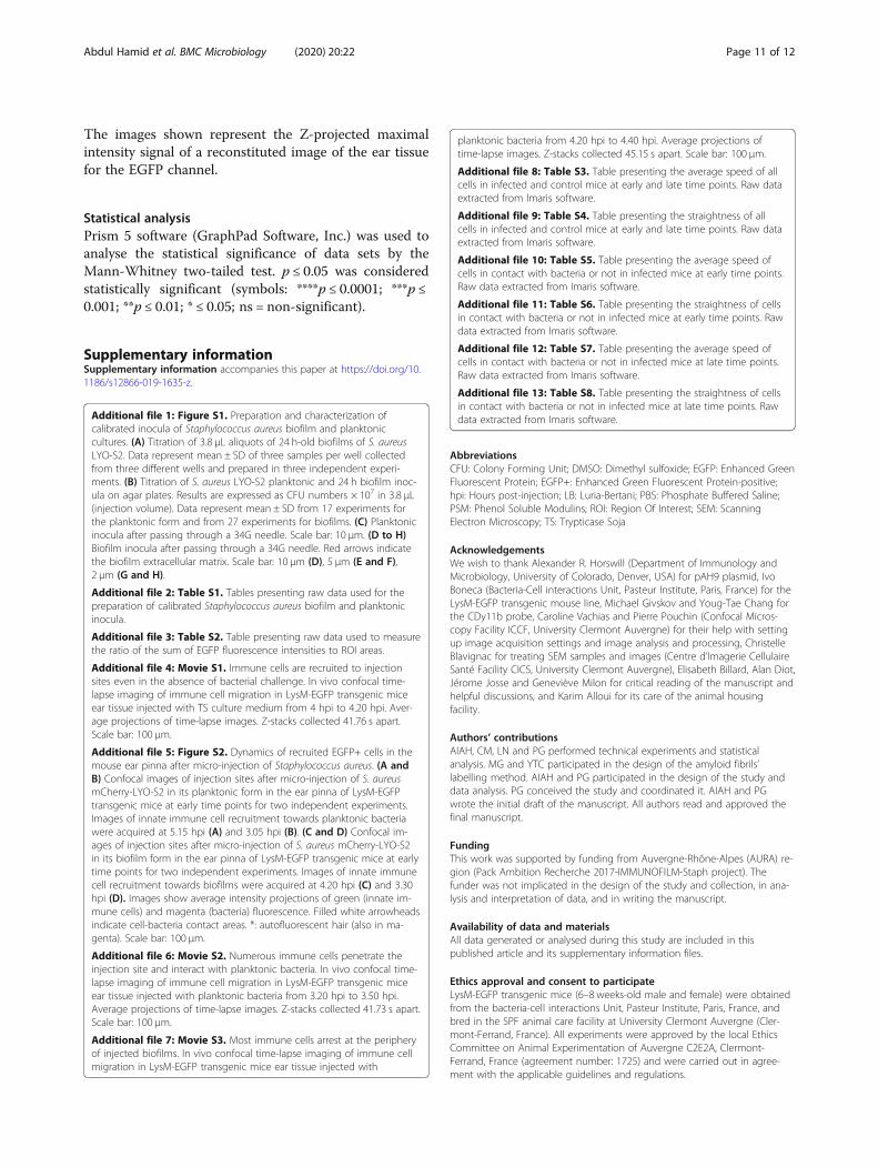
The images shown represent the Z-projected maximalintensity signal of a reconstituted image of the ear tissuefor the EGFP channel.
Statistical analysisPrism 5 software (GraphPad Software, Inc.) was used toanalyse the statistical significance of data sets by theMann-Whitney two-tailed test. p ≤ 0.05 was consideredstatistically significant (symbols: ****p ≤ 0.0001; ***p ≤0.001; **p ≤ 0.01; * ≤ 0.05; ns = non-significant).
Supplementary informationSupplementary information accompanies this paper at https://doi.org/10.1186/s12866-019-1635-z.
Additional file 1: Figure S1. Preparation and characterization ofcalibrated inocula of Staphylococcus aureus biofilm and planktoniccultures. (A) Titration of 3.8 μL aliquots of 24 h-old biofilms of S. aureusLYO-S2. Data represent mean ± SD of three samples per well collectedfrom three different wells and prepared in three independent experi-ments. (B) Titration of S. aureus LYO-S2 planktonic and 24 h biofilm inoc-ula on agar plates. Results are expressed as CFU numbers × 107 in 3.8 μL(injection volume). Data represent mean ± SD from 17 experiments forthe planktonic form and from 27 experiments for biofilms. (C) Planktonicinocula after passing through a 34G needle. Scale bar: 10 μm. (D to H)Biofilm inocula after passing through a 34G needle. Red arrows indicatethe biofilm extracellular matrix. Scale bar: 10 μm (D), 5 μm (E and F),2 μm (G and H).
Additional file 2: Table S1. Tables presenting raw data used for thepreparation of calibrated Staphylococcus aureus biofilm and planktonicinocula.
Additional file 3: Table S2. Table presenting raw data used to measurethe ratio of the sum of EGFP fluorescence intensities to ROI areas.
Additional file 4: Movie S1. Immune cells are recruited to injectionsites even in the absence of bacterial challenge. In vivo confocal time-lapse imaging of immune cell migration in LysM-EGFP transgenic miceear tissue injected with TS culture medium from 4 hpi to 4.20 hpi. Aver-age projections of time-lapse images. Z-stacks collected 41.76 s apart.Scale bar: 100 μm.
Additional file 5: Figure S2. Dynamics of recruited EGFP+ cells in themouse ear pinna after micro-injection of Staphylococcus aureus. (A andB) Confocal images of injection sites after micro-injection of S. aureusmCherry-LYO-S2 in its planktonic form in the ear pinna of LysM-EGFPtransgenic mice at early time points for two independent experiments.Images of innate immune cell recruitment towards planktonic bacteriawere acquired at 5.15 hpi (A) and 3.05 hpi (B). (C and D) Confocal im-ages of injection sites after micro-injection of S. aureus mCherry-LYO-S2in its biofilm form in the ear pinna of LysM-EGFP transgenic mice at earlytime points for two independent experiments. Images of innate immunecell recruitment towards biofilms were acquired at 4.20 hpi (C) and 3.30hpi (D). Images show average intensity projections of green (innate im-mune cells) and magenta (bacteria) fluorescence. Filled white arrowheadsindicate cell-bacteria contact areas. *: autofluorescent hair (also in ma-genta). Scale bar: 100 μm.
Additional file 6: Movie S2. Numerous immune cells penetrate theinjection site and interact with planktonic bacteria. In vivo confocal time-lapse imaging of immune cell migration in LysM-EGFP transgenic miceear tissue injected with planktonic bacteria from 3.20 hpi to 3.50 hpi.Average projections of time-lapse images. Z-stacks collected 41.73 s apart.Scale bar: 100 μm.
Additional file 7: Movie S3. Most immune cells arrest at the peripheryof injected biofilms. In vivo confocal time-lapse imaging of immune cellmigration in LysM-EGFP transgenic mice ear tissue injected with
planktonic bacteria from 4.20 hpi to 4.40 hpi. Average projections oftime-lapse images. Z-stacks collected 45.15 s apart. Scale bar: 100 μm.
Additional file 8: Table S3. Table presenting the average speed of allcells in infected and control mice at early and late time points. Raw dataextracted from Imaris software.
Additional file 9: Table S4. Table presenting the straightness of allcells in infected and control mice at early and late time points. Raw dataextracted from Imaris software.
Additional file 10: Table S5. Table presenting the average speed ofcells in contact with bacteria or not in infected mice at early time points.Raw data extracted from Imaris software.
Additional file 11: Table S6. Table presenting the straightness of cellsin contact with bacteria or not in infected mice at early time points. Rawdata extracted from Imaris software.
Additional file 12: Table S7. Table presenting the average speed ofcells in contact with bacteria or not in infected mice at late time points.Raw data extracted from Imaris software.
Additional file 13: Table S8. Table presenting the straightness of cellsin contact with bacteria or not in infected mice at late time points. Rawdata extracted from Imaris software.
AbbreviationsCFU: Colony Forming Unit; DMSO: Dimethyl sulfoxide; EGFP: Enhanced GreenFluorescent Protein; EGFP+: Enhanced Green Fluorescent Protein-positive;hpi: Hours post-injection; LB: Luria-Bertani; PBS: Phosphate Buffered Saline;PSM: Phenol Soluble Modulins; ROI: Region Of Interest; SEM: ScanningElectron Microscopy; TS: Trypticase Soja
AcknowledgementsWe wish to thank Alexander R. Horswill (Department of Immunology andMicrobiology, University of Colorado, Denver, USA) for pAH9 plasmid, IvoBoneca (Bacteria-Cell interactions Unit, Pasteur Institute, Paris, France) for theLysM-EGFP transgenic mouse line, Michael Givskov and Youg-Tae Chang forthe CDy11b probe, Caroline Vachias and Pierre Pouchin (Confocal Micros-copy Facility ICCF, University Clermont Auvergne) for their help with settingup image acquisition settings and image analysis and processing, ChristelleBlavignac for treating SEM samples and images (Centre d’Imagerie CellulaireSanté Facility CICS, University Clermont Auvergne), Elisabeth Billard, Alan Diot,Jérome Josse and Geneviève Milon for critical reading of the manuscript andhelpful discussions, and Karim Alloui for its care of the animal housingfacility.
Authors’ contributionsAIAH, CM, LN and PG performed technical experiments and statisticalanalysis. MG and YTC participated in the design of the amyloid fibrils’labelling method. AIAH and PG participated in the design of the study anddata analysis. PG conceived the study and coordinated it. AIAH and PGwrote the initial draft of the manuscript. All authors read and approved thefinal manuscript.
FundingThis work was supported by funding from Auvergne-Rhône-Alpes (AURA) re-gion (Pack Ambition Recherche 2017-IMMUNOFILM-Staph project). Thefunder was not implicated in the design of the study and collection, in ana-lysis and interpretation of data, and in writing the manuscript.
Availability of data and materialsAll data generated or analysed during this study are included in thispublished article and its supplementary information files.
Ethics approval and consent to participateLysM-EGFP transgenic mice (6–8 weeks-old male and female) were obtainedfrom the bacteria-cell interactions Unit, Pasteur Institute, Paris, France, andbred in the SPF animal care facility at University Clermont Auvergne (Cler-mont-Ferrand, France). All experiments were approved by the local EthicsCommittee on Animal Experimentation of Auvergne C2E2A, Clermont-Ferrand, France (agreement number: 1725) and were carried out in agree-ment with the applicable guidelines and regulations.
Abdul Hamid et al. BMC Microbiology (2020) 20:22 Page 11 of 12

Consent for publicationNot applicable for that section.
Competing interestsThe authors declare that they have no competing interests.
Author details1Laboratoire Microorganismes : Génome et Environnement, UMR CNRS 6023,Université Clermont-Auvergne, Clermont Ferrand, France. 2Costerton BiofilmCenter, department of Immunology and Microbiology, Faculty of HealthSciences, University of Copenhagen, Copenhagen, Denmark. 3Center forSelf-assembly and Complexity, IBS and Department of Chemistry, POSTECH,Pohang, Republic of Korea.
Received: 15 May 2019 Accepted: 7 November 2019
References1. Moormeier DE, Bayles KW. Staphylococcus aureus biofilm: a complex
developmental organism. Mol Microbiol. 2017;104(3):365–76.2. Ricciardi BF, Muthukrishnan G, Masters E, Ninomiya M, Lee CC, Schwarz EM.
Staphylococcus aureus evasion of host immunity in the setting of prostheticjoint infection: biofilm and beyond. Curr Rev Musculoskelet Med. 2018;11(3):389–400.
3. Jamal M, Ahmad W, Andleeb S, Jalil F, Imran M, Nawaz MA, et al. Bacterialbiofilm and associated infections. J Chin Med Assoc. 2018;81(1):7–11.
4. Yamada KJ, Kielian T. Biofilm-leukocyte cross-talk: impact on immunepolarization and Immunometabolism. J Innate Immun. 2018;22:1–9.
5. Gries CM, Kielian T. Staphylococcal biofilms and immune polarization duringprosthetic joint infection. J Am Acad Orthop Surg. 2017;25(Suppl 1):S20–4.
6. Watters C, Fleming D, Bishop D, Rumbaugh KP. Host responses to biofilm.Prog Mol Biol Transl Sci. 2016;142:193–239.
7. Forestier C, Billard E, Milon G, Gueirard P. Unveiling and characterizing earlybilateral interactions between biofilm and the mouse innate immunesystem. Front Microbiol. 2017;8:2309.
8. Peters NC, Egen JG, Secundino N, Debrabant A, Kimblin N, Kamhawi S, et al.In vivo imaging reveals an essential role for neutrophils in Leishmaniasistransmitted by sand flies. Science. 2008;321(5891):970–4.
9. Kim J-Y, Sahu S, Yau Y-H, Wang X, Shochat SG, Nielsen PH, et al. Detectionof pathogenic biofilms with bacterial amyloid targeting fluorescent probe,CDy11. J Am Chem Soc. 2016;138(1):402–7.
10. Günther F, Wabnitz GH, Stroh P, Prior B, Obst U, Samstag Y, et al. Hostdefence against Staphylococcus aureus biofilms infection: phagocytosis ofbiofilms by polymorphonuclear neutrophils (PMN). Mol Immunol. 2009;46(8–9):1805–13.
11. Guo Y, Ramos RI, Cho JS, Donegan NP, Cheung AL, Miller LS. In vivobioluminescence imaging to evaluate systemic and topical antibioticsagainst community-acquired methicillin-resistant Staphylococcus aureus-infected skin wounds in mice. Antimicrob Agents Chemother. 2013;57(2):855–63.
12. Torre A, Bacconi M, Sammicheli C, Galletti B, Laera D, Fontana MR, et al.Four-component Staphylococcus aureus vaccine 4C-staph enhances Fcγreceptor expression in neutrophils and monocytes and mitigates S. aureusinfection in neutropenic mice. Infect Immun. 2015;83(8):3157–63.
13. Resch A, Rosenstein R, Nerz C, Götz F. Differential gene expression profilingof Staphylococcus aureus cultivated under biofilm and planktonic conditions.Appl Environ Microbiol. 2005;71(5):2663–76.
14. Scherr TD, Roux CM, Hanke ML, Angle A, Dunman PM, Kielian T. GlobalTranscriptome analysis of Staphylococcus aureus biofilms in response toinnate immune cells. Infect Immun. 2013;81(12):4363–76.
15. Peschel A, Otto M. Phenol-soluble modulins and staphylococcal infection.Nat Rev Microbiol. 2013;11(10):667–73.
16. Zheng Y, Joo H-S, Nair V, Le KY, Otto M. Do amyloid structures formed byStaphylococcus aureus phenol-soluble modulins have a biological function?Int J Med Microbiol. 2018;308(6):675–82.
17. Hirschfeld J. Dynamic interactions of neutrophils and biofilms. J OralMicrobiol. 2014;6:26102.
18. Thurlow LR, Hanke ML, Fritz T, Angle A, Aldrich A, Williams SH, et al.Staphylococcus aureus biofilms prevent macrophage phagocytosis andattenuate inflammation in vivo. J Immunol. 2011;186(11):6585–96.
19. Heim CE, Vidlak D, Scherr TD, Kozel JA, Holzapfel M, Muirhead DE, et al.Myeloid-derived suppressor cells contribute to Staphylococcus aureusorthopedic biofilm infection. J Immunol. 2014;192(8):3778–92.
20. Costerton JW, Stewart PS, Greenberg EP. Bacterial biofilms: a common causeof persistent infections. Science. 1999;284(5418):1318–22.
21. Jesaitis AJ, Franklin MJ, Berglund D, Sasaki M, Lord CI, Bleazard JB, et al.Compromised host defense on Pseudomonas aeruginosa biofilms:characterization of neutrophil and biofilm interactions. J Immunol. 2003;171(8):4329–39.
22. Malone CL, Boles BR, Lauderdale KJ, Thoendel M, Kavanaugh JS, Horswill AR.Fluorescent reporters for Staphylococcus aureus. J Microbiol Methods. 2009;77(3):251–60.
23. Marquès C, Tasse J, Pracros A, Collin V, Franceschi C, Laurent F, et al. Effectsof antibiotics on biofilm and unattached cells of a clinical Staphylococcusaureus isolate from bone and joint infection. J Med Microbiol. 2015;64(9):1021–6.
24. Schenk S, Laddaga RA. Improved method for electroporation ofStaphylococcus aureus. FEMS Microbiol Lett. 1992;94(1–2):133–8.
25. Mac-Daniel L, Buckwalter MR, Gueirard P, Ménard R. Myeloid Cell Isolationfrom Mouse Skin and Draining Lymph Node Following IntradermalImmunization with Live Attenuated Plasmodium sporozoites. J Vis Exp. 2016;18(111):e53796.
26. Amino R, Thiberge S, Blazquez S, Baldacci P, Renaud O, Shorte S, et al.Imaging malaria sporozoites in the dermis of the mammalian host. NatProtoc. 2007;2(7):1705–12.
Publisher’s NoteSpringer Nature remains neutral with regard to jurisdictional claims inpublished maps and institutional affiliations.
Abdul Hamid et al. BMC Microbiology (2020) 20:22 Page 12 of 12