Embed Size (px)
Citation preview
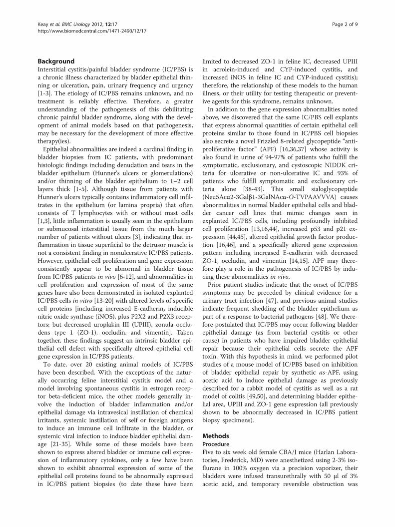
Keay et al. BMC Urology 2012, 12:17http://www.biomedcentral.com/1471-2490/12/17
RESEARCH ARTICLE Open Access
A mouse model for interstitial cystitis/painfulbladder syndrome based on APF inhibition ofbladder epithelial repair: a pilot studySusan Keay1,2*, Samantha Leitzell3, Ashley Ochrzcin3, George Clements3, Min Zhan4 and David Johnson2,3
Abstract
Background: Interstitial cystitis/painful bladder syndrome (IC/PBS) is a chronic bladder disorder with bladderepithelial thinning or ulceration, pain, urinary frequency and urgency. There is no reliably effective therapy forIC/PBS, and no generally accepted animal model for the disorder in which potential therapies can be tested. Bladderepithelial cells from IC/PBS patients make a small glycopeptide antiproliferative factor or "APF" that inhibitsproliferation, decreases tight junction protein expression, increases paracellular permeability, and induces changes ingene expression of bladder epithelial cells in vitro that mimic abnormalities in IC/PBS patient biopsy specimensin vivo. We therefore determined the ability of a synthetic APF derivative to inhibit bladder epithelial repair in mice.
Methods: The bladder epithelium of female CBA/J mice was stripped by transurethral infusion of 3% acetic acid, andmice were subsequently treated daily with one of three intravesical treatments [synthetic as-APF, inactiveunglycosylated control peptide, or phosphate buffered saline carrier (PBS)] for 1–21 days. Fixed bladder sectionswere either stained with haematoxylin and eosin for determination of epithelial area by image analysis, or incubatedwith anti-uroplakin III (UPIII) or anti-zonula occludens type 1 (ZO-1) antibodies for immunofluorescence microscopy.Epithelial measurement data were analyzed by a two-way analysis of variance (ANOVA); post hoc comparisons ofmultiple groups were carried out using the Tukey-Kramer method.
Results: Bladder epithelial repair was significantly attenuated in as-APF-treated mice as compared to control miceon days 3–21 (p< 0.05); the mean epithelial/total area over all measured days was also significantly lower inas-APF-treated mice vs. mice in either control group by post hoc analysis (p< 0.0001 for both comparisons). UPIIIand ZO-1 expression was also decreased in as-APF-treated mice as compared to mice in either control group by day7 (UPIII) or day 14 (ZO-1).
Conclusions: This model demonstrates in vivo effects of as-APF which abrogates bladder epithelial repair andexpression of UPIII and ZO-1 in CBA/J mice following transurethral acetic acid infusion. As bladder epithelial thinning,decreased UPIII expression, and decreased ZO-1 expression are histopathologic features of IC/PBS patient biopsies,this model may be useful for studying the pathophysiology of IC/PBS and the effect of potential therapies.
Keywords: Interstitial cystitis, Painful bladder syndrome, Mouse model
* Correspondence: [email protected] of Medicine, University of Maryland School of Medicine,Baltimore, MD, USA2Veterans Administration Maryland Health Care System, 10 North GreeneStreet, Room 3B-184, Baltimore, MD 21201, USAFull list of author information is available at the end of the article
© 2012 Keay et al.; licensee BioMed Central Ltd. This is an Open Access article distributed under the terms of the CreativeCommons Attribution License (http://creativecommons.org/licenses/by/2.0), which permits unrestricted use, distribution, andreproduction in any medium, provided the original work is properly cited.
Keay et al. BMC Urology 2012, 12:17 Page 2 of 9http://www.biomedcentral.com/1471-2490/12/17
BackgroundInterstitial cystitis/painful bladder syndrome (IC/PBS) isa chronic illness characterized by bladder epithelial thin-ning or ulceration, pain, urinary frequency and urgency[1-3]. The etiology of IC/PBS remains unknown, and notreatment is reliably effective. Therefore, a greaterunderstanding of the pathogenesis of this debilitatingchronic painful bladder syndrome, along with the devel-opment of animal models based on that pathogenesis,may be necessary for the development of more effectivetherapy(ies).Epithelial abnormalities are indeed a cardinal finding in
bladder biopsies from IC patients, with predominanthistologic findings including denudation and tears in thebladder epithelium (Hunner's ulcers or glomerulations)and/or thinning of the bladder epithelium to 1–2 celllayers thick [1-5]. Although tissue from patients withHunner's ulcers typically contains inflammatory cell infil-trates in the epithelium (or lamina propria) that oftenconsists of T lymphocytes with or without mast cells[1,3], little inflammation is usually seen in the epitheliumor submucosal interstitial tissue from the much largernumber of patients without ulcers [3], indicating that in-flammation in tissue superficial to the detrusor muscle isnot a consistent finding in nonulcerative IC/PBS patients.However, epithelial cell proliferation and gene expressionconsistently appear to be abnormal in bladder tissuefrom IC/PBS patients in vivo [6-12], and abnormalities incell proliferation and expression of most of the samegenes have also been demonstrated in isolated explantedIC/PBS cells in vitro [13-20] with altered levels of specificcell proteins [including increased E-cadherin, induciblenitric oxide synthase (iNOS), plus P2X2 and P2X3 recep-tors; but decreased uroplakin III (UPIII), zonula occlu-dens type 1 (ZO-1), occludin, and vimentin]. Takentogether, these findings suggest an intrinsic bladder epi-thelial cell defect with specifically altered epithelial cellgene expression in IC/PBS patients.To date, over 20 existing animal models of IC/PBS
have been described. With the exceptions of the natur-ally occurring feline interstitial cystitis model and amodel involving spontaneous cystitis in estrogen recep-tor beta-deficient mice, the other models generally in-volve the induction of bladder inflammation and/orepithelial damage via intravesical instillation of chemicalirritants, systemic instillation of self or foreign antigensto induce an immune cell infiltrate in the bladder, orsystemic viral infection to induce bladder epithelial dam-age [21-35]. While some of these models have beenshown to express altered bladder or immune cell expres-sion of inflammatory cytokines, only a few have beenshown to exhibit abnormal expression of some of theepithelial cell proteins found to be abnormally expressedin IC/PBS patient biopsies (to date these have been
limited to decreased ZO-1 in feline IC, decreased UPIIIin acrolein-induced and CYP-induced cystitis, andincreased iNOS in feline IC and CYP-induced cystitis);therefore, the relationship of these models to the humanillness, or their utility for testing therapeutic or prevent-ive agents for this syndrome, remains unknown.In addition to the gene expression abnormalities noted
above, we discovered that the same IC/PBS cell explantsthat express abnormal quantities of certain epithelial cellproteins similar to those found in IC/PBS cell biopsiesalso secrete a novel Frizzled 8-related glycopeptide “anti-proliferative factor” (APF) [16,36,37] whose activity isalso found in urine of 94-97% of patients who fulfill thesymptomatic, exclusionary, and cystoscopic NIDDK cri-teria for ulcerative or non-ulcerative IC and 93% ofpatients who fulfill symptomatic and exclusionary cri-teria alone [38-43]. This small sialoglycopeptide(Neu5Acα2-3Galβ1-3GalNAcα-O-TVPAAVVVA) causesabnormalities in normal bladder epithelial cells and blad-der cancer cell lines that mimic changes seen inexplanted IC/PBS cells, including profoundly inhibitedcell proliferation [13,16,44], increased p53 and p21 ex-pression [44,45], altered epithelial growth factor produc-tion [16,46], and a specifically altered gene expressionpattern including increased E-cadherin with decreasedZO-1, occludin, and vimentin [14,15]. APF may there-fore play a role in the pathogenesis of IC/PBS by indu-cing these abnormalities in vivo.Prior patient studies indicate that the onset of IC/PBS
symptoms may be preceded by clinical evidence for aurinary tract infection [47], and previous animal studiesindicate frequent shedding of the bladder epithelium aspart of a response to bacterial pathogens [48]. We there-fore postulated that IC/PBS may occur following bladderepithelial damage (as from bacterial cystitis or othercause) in patients who have impaired bladder epithelialrepair because their epithelial cells secrete the APFtoxin. With this hypothesis in mind, we performed pilotstudies of a mouse model of IC/PBS based on inhibitionof bladder epithelial repair by synthetic as-APF, usingacetic acid to induce epithelial damage as previouslydescribed for a rabbit model of cystitis as well as a ratmodel of colitis [49,50], and determining bladder epithe-lial area, UPIII and ZO-1 gene expression (all previouslyshown to be abnormally decreased in IC/PBS patientbiopsy specimens).
MethodsProcedureFive to six week old female CBA/J mice (Harlan Labora-tories, Frederick, MD) were anesthetized using 2-3% iso-flurane in 100% oxygen via a precision vaporizer, theirbladders were infused transurethrally with 50 μl of 3%acetic acid, and temporary reversible obstruction was
Keay et al. BMC Urology 2012, 12:17 Page 3 of 9http://www.biomedcentral.com/1471-2490/12/17
achieved by applying collodion U.S.P. (J.T. Baker, Phil-lipsburg, NJ) to the external urethral meatus. The collo-dion was removed 1 hour later with acetone, and themouse bladders were rinsed by transurethral infusionwith sterile phosphate buffered saline (PBS) (QualityBiologicals, Inc., Gaithersburg, MD) and further infusedwith one of three treatments (in 50 μl total volume):250 mM synthetic as-APF (PolyPeptide Laboratories,San Diego, CA), 250 mM inactive negative control pep-tide (PolyPeptide Laboratories), or PBS carrier alone.The bladders were then immediately obstructed for anadditional 3 hours with collodion, the collodion wasagain removed, and the animals were returned to theircages and allowed to void until the next day. Buprenor-phine (0.05-0.1 mg/kg) was administered subcutaneouslyto the mice every 8–12 hours for 48 hours to relieve anydiscomfort resulting from acetic acid treatment. Theintravesical treatment infusion (with as-APF, peptide, orPBS) was then repeated daily for 1–21 days until sacri-fice by CO2 inhalation. NIH guidelines for the care anduse of laboratory animals for experimental procedureswere followed throughout the project, and this work wasapproved by the Institutional Animal Care and UseCommittee of the University of Maryland, Baltimore.
Tissue fixation and stainingIn anesthetized mice, bladder tissue was fixed in situ byinfusing 50 μL of 10% buffered neutral formalin (EMD,Gibbstown, NJ) into the bladders with an infusion pump(Harvard Apparatus, Millis, MA) over 30 seconds. Blad-ders were then removed and fixed as a whole in 10% for-malin overnight, after which they were embedded inparaffin, cut into 6 μm sections, and either stained withhaematoxylin and eosin (H&E) or incubated with spe-cific antibodies for immunofluorescence microscopy.
A
PBS control
Peptide control
as-APF-treated
B
P
P
a
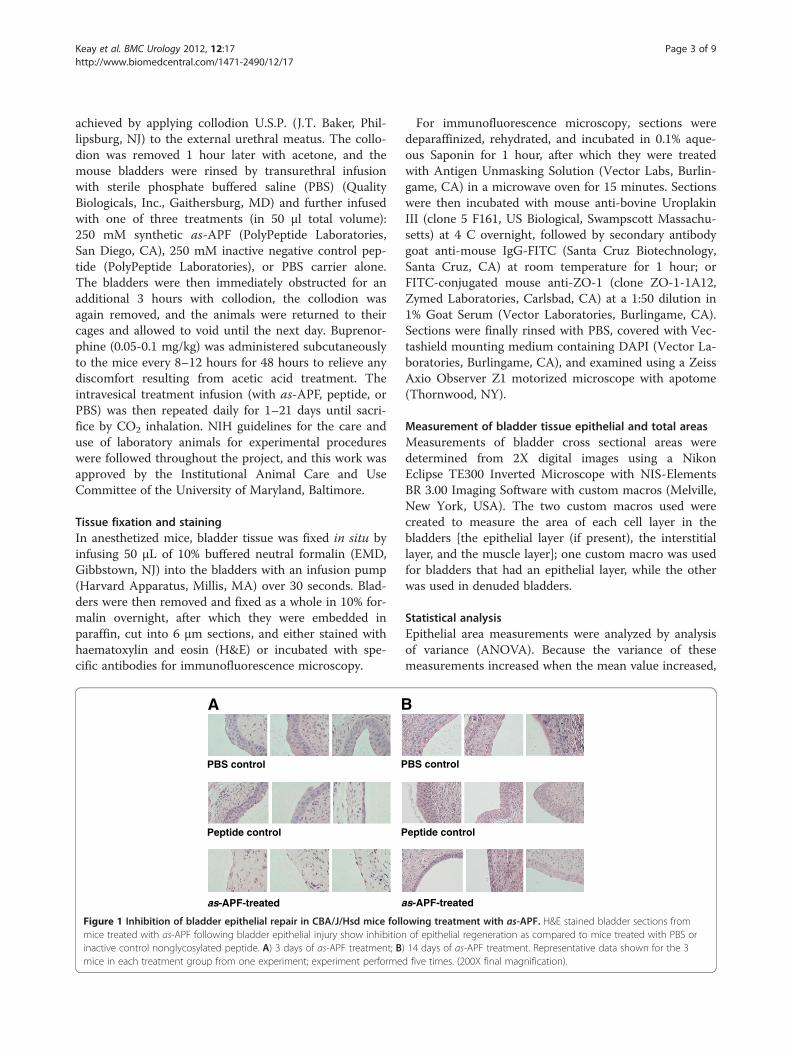
Figure 1 Inhibition of bladder epithelial repair in CBA/J/Hsd mice follmice treated with as-APF following bladder epithelial injury show inhibitioninactive control nonglycosylated peptide. A) 3 days of as-APF treatment; Bmice in each treatment group from one experiment; experiment performe
For immunofluorescence microscopy, sections weredeparaffinized, rehydrated, and incubated in 0.1% aque-ous Saponin for 1 hour, after which they were treatedwith Antigen Unmasking Solution (Vector Labs, Burlin-game, CA) in a microwave oven for 15 minutes. Sectionswere then incubated with mouse anti-bovine UroplakinIII (clone 5 F161, US Biological, Swampscott Massachu-setts) at 4 C overnight, followed by secondary antibodygoat anti-mouse IgG-FITC (Santa Cruz Biotechnology,Santa Cruz, CA) at room temperature for 1 hour; orFITC-conjugated mouse anti-ZO-1 (clone ZO-1-1A12,Zymed Laboratories, Carlsbad, CA) at a 1:50 dilution in1% Goat Serum (Vector Laboratories, Burlingame, CA).Sections were finally rinsed with PBS, covered with Vec-tashield mounting medium containing DAPI (Vector La-boratories, Burlingame, CA), and examined using a ZeissAxio Observer Z1 motorized microscope with apotome(Thornwood, NY).
Measurement of bladder tissue epithelial and total areasMeasurements of bladder cross sectional areas weredetermined from 2X digital images using a NikonEclipse TE300 Inverted Microscope with NIS-ElementsBR 3.00 Imaging Software with custom macros (Melville,New York, USA). The two custom macros used werecreated to measure the area of each cell layer in thebladders [the epithelial layer (if present), the interstitiallayer, and the muscle layer]; one custom macro was usedfor bladders that had an epithelial layer, while the otherwas used in denuded bladders.
Statistical analysisEpithelial area measurements were analyzed by analysisof variance (ANOVA). Because the variance of thesemeasurements increased when the mean value increased,
BS control
eptide control
s-APF-treated
owing treatment with as-APF. H&E stained bladder sections fromof epithelial regeneration as compared to mice treated with PBS or
) 14 days of as-APF treatment. Representative data shown for the 3d five times. (200X final magnification).
B
A
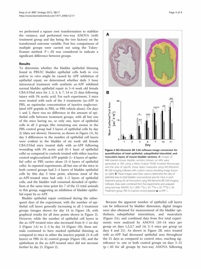
Figure 2 NIS-Elements BR 3.00 software image conversion forquantification of total epithelial, subepithelial interstitial, andmuscularis layers of mouse bladder sections. A) Images ofH&E-stained mouse bladder sections (shown on left) weregenerated at 20X using a Nikon Eclipse TE300 Inverted Microscopeand the area of specific tissue layers measured using NIS-ElementsBR 3.00 Imaging Software with custom macros (resulting image shownon right). B) These images were then used to determine the ratio ofepithelial area to total bladder cross-sectional area for mice in eachtreatment group for all time points using NIS-Elements BR 3.00 ImagingSoftware. Data were combined from five experiments and analyzedusing two-way ANOVA; *p< .0001; **p< .01; ***p< .02; ****p< .04.Treatment group: PBS □; inactive control peptide ■; as-APF ░:
Keay et al. BMC Urology 2012, 12:17 Page 4 of 9http://www.biomedcentral.com/1471-2490/12/17
we performed a square root transformation to stabilizethe variance, and performed two-way ANOVA (withtreatment group and day being the two factors) on thetransformed outcome variable. Post hoc comparisons ofmultiple groups were carried out using the Tukey-Kramer method. P< .05 was considered to indicate asignificant difference between groups.
ResultsTo determine whether the bladder epithelial thinningfound in PBS/IC bladder epithelial cells both in vivoand/or in vitro might be caused by APF inhibition ofepithelial repair, we determined whether daily 3 hourintravesical treatment with synthetic as-APF inhibitednormal bladder epithelial repair in 5–6 week old femaleCBA/J/Hsd mice for 1, 2, 3, 4, 7, 14 or 21 days followinginjury with 3% acetic acid. For each experiment, 3 micewere treated with each of the 3 treatments (as-APF inPBS, an equimolar concentration of inactive unglycosy-lated APF peptide in PBS, or PBS vehicle alone). On days1 and 2, there was no difference in the amount of epi-thelial cells between treatment groups, with all but oneof the mice having no, or only one, layer of epithelialcells in all 3 groups (the remaining one mouse in thePBS control group had 3 layers of epithelial cells by day2) (data not shown). However, as shown in Figure 1A, byday 3 differences in the number of epithelial cell layerswere evident in the bladder of six week old femaleCBA/J/Hsd mice treated daily with as-APF followingwounding with 3% acetic acid (0–1 layer of epithelialcells) as compared to controls treated with either inactivecontrol unglycosylated APF peptide (1–4 layers of epithe-lial cells) or PBS carrier alone (3–4 layers of epithelialcells). In repeated experiments, all but one of the mice inboth control groups had 2–4 layers of bladder epithelialcells by this day 3 time point, whereas most of theas-APF-treated mice had only 1–2 layers of epithelialcells, and the bladder wall remained denuded of epithe-lium at the same time point for 7 of the 15 total animalsin this group, suggesting an inhibition of bladder epithe-lial repair by as-APF.Bladder epithelial repair continued during the subse-
quent days of the experiment, with the number of epi-thelial cell layers generally increasing in all 3 treatmentgroups (images shown for day 14 in Figure 1B, withgraphical results for all time points shown in Figure 2).However, while the number of epithelial cell layers inthe as-APF-treated mice also increased from 0–1 on day3 (Figure 1A) to 2–3 by day 14 (Figure 1B), these ani-mals continued to have marked epithelial thinning ascompared to mice in either the inactive peptide (3–6 celllayers) or PBS (4–6) control groups (Figure 1B), and theepithelium in the as-APF-treated mice did not increasefurther by day 21 (Figure 2).
Because the apparent number of epithelial cell layerscan be influenced by bladder distension, digital imageswere also obtained for measurement of the bladder epi-thelium, subepithelial interstitium, and muscularis(Figure 2A), and combined data from five total experi-ments were analyzed by ANOVA (10–14 mice pergroup on days 1,2,3,7 and 14; 3–5 mice per group ondays 4 and 21). As shown in Figure 2B, mice treatedwith as-APF had decreased epithelial area throughoutthe 21 days as compared to control mice, reaching sig-nificance vs. one or both control groups on days 3–21(p< .05 for all groups by two-way ANOVA following
PBS PBS
Peptide Peptide
as-APF as-APF
A B
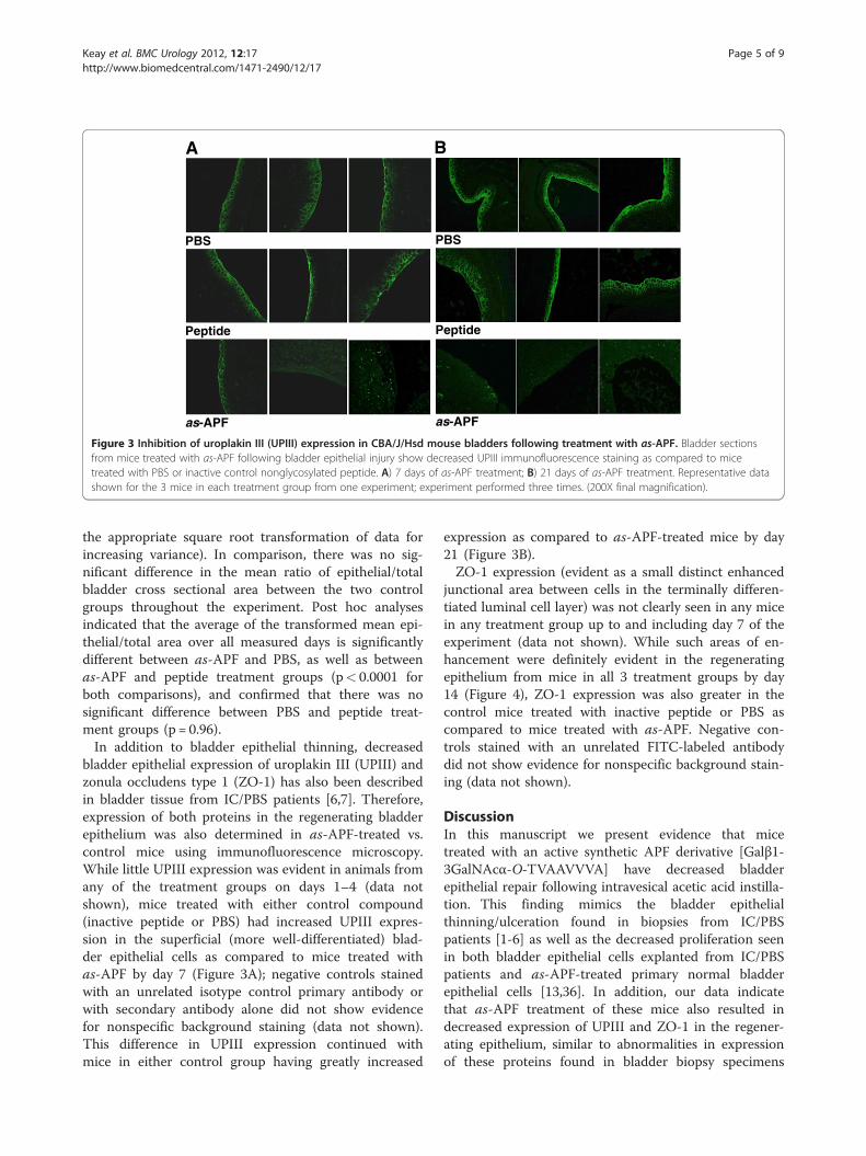
Figure 3 Inhibition of uroplakin III (UPIII) expression in CBA/J/Hsd mouse bladders following treatment with as-APF. Bladder sectionsfrom mice treated with as-APF following bladder epithelial injury show decreased UPIII immunofluorescence staining as compared to micetreated with PBS or inactive control nonglycosylated peptide. A) 7 days of as-APF treatment; B) 21 days of as-APF treatment. Representative datashown for the 3 mice in each treatment group from one experiment; experiment performed three times. (200X final magnification).
Keay et al. BMC Urology 2012, 12:17 Page 5 of 9http://www.biomedcentral.com/1471-2490/12/17
the appropriate square root transformation of data forincreasing variance). In comparison, there was no sig-nificant difference in the mean ratio of epithelial/totalbladder cross sectional area between the two controlgroups throughout the experiment. Post hoc analysesindicated that the average of the transformed mean epi-thelial/total area over all measured days is significantlydifferent between as-APF and PBS, as well as betweenas-APF and peptide treatment groups (p< 0.0001 forboth comparisons), and confirmed that there was nosignificant difference between PBS and peptide treat-ment groups (p = 0.96).In addition to bladder epithelial thinning, decreased
bladder epithelial expression of uroplakin III (UPIII) andzonula occludens type 1 (ZO-1) has also been describedin bladder tissue from IC/PBS patients [6,7]. Therefore,expression of both proteins in the regenerating bladderepithelium was also determined in as-APF-treated vs.control mice using immunofluorescence microscopy.While little UPIII expression was evident in animals fromany of the treatment groups on days 1–4 (data notshown), mice treated with either control compound(inactive peptide or PBS) had increased UPIII expres-sion in the superficial (more well-differentiated) blad-der epithelial cells as compared to mice treated withas-APF by day 7 (Figure 3A); negative controls stainedwith an unrelated isotype control primary antibody orwith secondary antibody alone did not show evidencefor nonspecific background staining (data not shown).This difference in UPIII expression continued withmice in either control group having greatly increased
expression as compared to as-APF-treated mice by day21 (Figure 3B).ZO-1 expression (evident as a small distinct enhanced
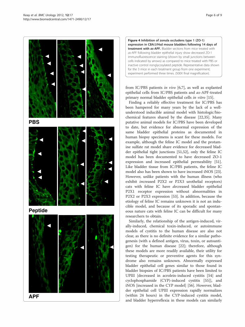
junctional area between cells in the terminally differen-tiated luminal cell layer) was not clearly seen in any micein any treatment group up to and including day 7 of theexperiment (data not shown). While such areas of en-hancement were definitely evident in the regeneratingepithelium from mice in all 3 treatment groups by day14 (Figure 4), ZO-1 expression was also greater in thecontrol mice treated with inactive peptide or PBS ascompared to mice treated with as-APF. Negative con-trols stained with an unrelated FITC-labeled antibodydid not show evidence for nonspecific background stain-ing (data not shown).
DiscussionIn this manuscript we present evidence that micetreated with an active synthetic APF derivative [Galβ1-3GalNAcα-O-TVAAVVVA] have decreased bladderepithelial repair following intravesical acetic acid instilla-tion. This finding mimics the bladder epithelialthinning/ulceration found in biopsies from IC/PBSpatients [1-6] as well as the decreased proliferation seenin both bladder epithelial cells explanted from IC/PBSpatients and as-APF-treated primary normal bladderepithelial cells [13,36]. In addition, our data indicatethat as-APF treatment of these mice also resulted indecreased expression of UPIII and ZO-1 in the regener-ating epithelium, similar to abnormalities in expressionof these proteins found in bladder biopsy specimens
PBS
Peptide
APF
Figure 4 Inhibition of zonula occludens type 1 (ZO-1)expression in CBA/J/Hsd mouse bladders following 14 days oftreatment with as-APF. Bladder sections from mice treated withas-APF following bladder epithelial injury show decreased ZO-1immunofluorescence staining (shown by small junctions betweencells indicated by arrows) as compared to mice treated with PBS orinactive control nonglycosylated peptide. Representative data shownfor the 3 mice in each treatment group from one experiment;experiment performed three times. (500X final magnification).
Keay et al. BMC Urology 2012, 12:17 Page 6 of 9http://www.biomedcentral.com/1471-2490/12/17
from IC/PBS patients in vivo [6,7], as well as explantedepithelial cells from IC/PBS patients and as-APF-treatedprimary normal bladder epithelial cells in vitro [15].Finding a reliably effective treatment for IC/PBS has
been hampered for many years by the lack of a well-understood inducible animal model with histologic/bio-chemical features shared by the disease [22,35]. Manyputative animal models for IC/PBS have been developedto date, but evidence for abnormal expression of thesame bladder epithelial proteins as documented inhuman biopsy specimens is scant for these models. Forexample, although the feline IC model and the protam-ine sulfate rat model share evidence for decreased blad-der epithelial tight junctions [51,52], only the feline ICmodel has been documented to have decreased ZO-1expression and increased epithelial permeability [51].Like bladder tissue from IC/PBS patients, the feline ICmodel also has been shown to have increased iNOS [23].However, unlike patients with the human illness (whoexhibit increased P2X2 or P2X3 urothelial receptors)cats with feline IC have decreased bladder epithelialP2X1 receptor expression without abnormalities inP2X2 or P2X3 expression [53]. In addition, because theetiology of feline IC remains unknown it is not an indu-cible model, and because of its sporadic and spontan-eous nature cats with feline IC can be difficult for manyresearchers to obtain.Similarly, the relationship of the antigen-induced, vir-
ally-induced, chemical toxin-induced, or autoimmunemodels of cystitis to the human disease are also notclear, as there is no definite evidence for a similar patho-genesis (with a defined antigen, virus, toxin, or autoanti-gen) for the human disease [22]; therefore, althoughthese models are more readily available, their utility fortesting therapeutic or preventive agents for this syn-drome also remains unknown. Abnormally expressedbladder epithelial cell genes similar to those found inbladder biopsies of IC/PBS patients have been limited toUPIII [decreased in acrolein-induced cystitis [54] andcyclophosphamide (CYP)-induced cystitis [55]], andiNOS [increased in the CYP model] [56]. However, blad-der epithelial cell UPIII expression rapidly normalizes(within 24 hours) in the CYP-induced cystitis model,and bladder hyperreflexia in these models can similarly
Keay et al. BMC Urology 2012, 12:17 Page 7 of 9http://www.biomedcentral.com/1471-2490/12/17
resolve within days [57] indicating the transient natureof some of these changes in these models. Therefore,additional studies are also required to compare the dur-ability of abnormal bladder epithelial gene expression inall of these models for us to understand the relative util-ity of each model for studies of IC/PBS pathogenesis andtreatment.The model described in this preliminary report has
certain advantages over the other animal models thathave been described to date. It is an inducible rodentmodel, making it potentially more readily available thanthe spontaneous feline model. This mouse model alsohas changes in expression of two proteins similar tochanges found consistently in patients with IC/PBS –UPIII and ZO-1. Unlike some protein expression abnor-malities found in the other models to date, thesechanges appear to be durable (sustainable) for up to3 weeks. And whereas most of the other models areinduced by factors that are almost certainly not causativein the pathogenesis of IC/PBS, this model is based on apotential etiology of IC/PBS (APF) that was first discov-ered in urine from patients with this illness, subse-quently shown to be made by bladder epithelial cells inthese patients and to induce abnormal expression of atleast six epithelial proteins known to be abnormallyexpressed in bladder tissue from IC/PBS patients[14-16,36]. Because APF inhibits bladder epithelial cellreplication but does not by itself induce cell deathin vitro [13,16] we previously hypothesized that APFmay inhibit bladder epithelial repair following injurycaused by other inciting factors such as a urinary tractinfection [16,58]. However, the use of acetic acid to in-duce such bladder epithelial injury prior to APF instilla-tion in the current model is also by itself not related tothe pathogenesis of IC; future refinements to the modelmay therefore include induction of epithelial sheddingby bacteria known to cause cystitis [48] prior to APFinstillation.Disadvantages of this model, however, include its re-
quirement for daily intravesical instillations of active syn-thetic APF (or its control peptide), making it relativelylabor-intensive and expensive for a rodent model. Inaddition, some of the variability seen in this model regard-ing the effects of APF on epithelial repair or gene expres-sion may result from limited (only 3 hours daily) exposureto the instilled APF. However, based on the preliminaryresults from this model, development of a continuousAPF release model (via an implanted pump), or a trans-genic mouse model that would express active APF eitherconstitutively or inducibly, appear to be warranted.
ConclusionsBladder instillation of as-APF inhibits bladder epithelialrepair and expression of UPIII and ZO-1 in CBA/J mice
following transurethral acetic acid infusion. Becausebladder epithelial thinning, decreased UPIII expression,and decreased ZO-1 expression are consistently found inIC/PBS patient bladder epithelial biopsies, this modelmay be useful for studying the pathophysiology of thisillness, and the effect of potential therapies.
AbbreviationsAPF: Antiproliferative factor; as-APF: Asialylated APF; FITC: Fluoresceinisothiocyanate; H&E: Haematoxylin and eosin; IC: Interstitial cystitis;IC/PBS: Interstitial cystitis/painful bladder syndrome; PBS: Phosphate bufferedsaline; UPIII: Uroplakin III; ZO-1: Zonula occludens type 1.
Competing interestsSusan Keay is named as an inventor on patents for the composition and useof APF (patents owned by the University of Maryland, Baltimore and theDepartment of Veterans Affairs).
Authors’ contributionsSL performed the animal experiments, as well as tissue specimen fixationand immunofluorescence staining, plus light and immunofluorescencemicroscopy. AO and GC performed measurements of bladder cross sectionalareas by light microscopy. MZ performed the statistical analysis. SK and DJsupervised the performance of the experiments, data analysis andpreparation of the manuscript. All authors read and approved the finalmanuscript.
AcknowledgementsThe authors thank Grazyna Zaidel for assistance with preparation of tissuesections and staining, and Eunice Katz for assistance with manuscriptpreparation. This work was supported in part by grants from Acologix, Inc.and Trillium Therapeutics, Inc. Facility usage and some salary support wasdonated by the Department of Veterans Affairs.
Author details1Department of Medicine, University of Maryland School of Medicine,Baltimore, MD, USA. 2Veterans Administration Maryland Health Care System,10 North Greene Street, Room 3B-184, Baltimore, MD 21201, USA. 3BaltimoreResearch and Education Foundation, Baltimore, MD, USA. 4Department ofEpidemiology and Public Health, University of Maryland School of Medicine,Baltimore, MD, USA.
Received: 7 March 2012 Accepted: 4 May 2012Published: 8 June 2012
References1. Johansson SL, Fall M: Clinical features and spectrum of light microscopic
changes in interstitial cystitis. J Urol 1990, 143:1118–1124.2. Skoluda D, Wegner K, Lemmel EM: Critical notes: respective immune
pathogenesis of interstitial cystitis. (article in German). Urologe A 1974,13:15–23.
3. Tomaszewski JE, Landis JR, Russack V, Williams TM, Wang LP, Hardy C,Brensinger C, Matthews YL, Abele ST, Kusek JW, Nyberg LM, InterstitialCystitis Database Study: Biopsy features are associated with primarysymptoms in interstitial cystitis: results from the Interstitial cystitisdatabase study. Urology 2001, 57:67–81.
4. Smith BH, Dehner LP: Chronic ulcerating interstitial cystitis (Hunner’sulcer). Arch Pathol 1972, 93:76–81.
5. Ratliff TL, Klutke CG, McDougall EM: The etiology of interstitial cystitis.Urology Clinics of North America 1994, 21:21–30.
6. Slobodov G, Feloney M, Gran C, Kyker KD, Hurst RE, Culkin DJ: Abnormalexpression of molecular markers for bladder impermeability anddifferentiation in the urothelium of patients with interstitial cystitis.J Urol 2004, 171:1554–1558.
7. Sanchez Freire V, Burkhard FC, Kessler TM, Kuhn A, Draeger A,Monastyrskaya K: MicroRNAs may mediate the down-regulation ofneurokinin-1 receptor in chronic bladder pain syndrome. Am J Pathol2010, 176:288–303.
Keay et al. BMC Urology 2012, 12:17 Page 8 of 9http://www.biomedcentral.com/1471-2490/12/17
8. Liebert M, Wedemeyer G, Stein JA, Washington R Jr, Faerber G, Flint A,Grossman HB: Evidence for urothelial cell activation in interstitial cystitis.J Urol 1993, 149:470–475.
9. Hauser PJ, Dozmorov MG, Bane BL, Slobodov G, Culkin DJ, Hurst RE:Abnormal expression of differentiation related proteins andproteoglycan core proteins in the urothelium of patients with interstitialcystitis. J Urol 2008, 179:764–769.
10. Laguna P, Smedts F, Nordling J, Horn T, Bouchelouche K, Hopman A, de laRosette J: Keratin expression profiling of transitional epithelium in thepainful bladder syndrome/interstitial cystitis. Am J Clin Pathol 2006,125:105–110.
11. Tempest HV, Dixon AK, Turner WH, Elneil S, Sellers LA, Ferguson DR: P2Xand P2X receptor expression in human bladder urothelium and changesin interstitial cystitis. Br J Urol Int 2004, 93:1344–1348.
12. Koskela LR, Thiel T, Ehren I, De Verdier PJ, Wiklund NP: Localization andexpression of inducible nitric oxide synthase in biopsies from patientswith interstitial cystitis. J Urol 2008, 180:737–741.
13. Keay S, Zhang C-O, Shoenfelt JL, Chai TC: Decreased in vitro proliferationof bladder epithelial cells from patients with interstitial cystitis. Urology2003, 61:1278–1284.
14. Keay S, Seillier-Moiseiwitsch F, Zhang C-O, Chai TC, Zhang J: Changes inhuman bladder cell gene expression associated with interstitial cystitisor antiproliferative factor treatment. Physiol Genomics 2003, 14:107–115.
15. Zhang CO, Wang JY, Koch KR, Keay S: Regulation of tight junction proteinsand bladder epithelial paracellular permeability by an antiproliferativefactor from patients with interstitial cystitis. J Urol 2005, 174:2382–2387.
16. Keay S, Kleinberg M, Zhang C-O, Hise MK, Warren JW: Bladder epithelialcells from patients with interstitial cystitis produce an inhibitor ofheparin-binding epidermal growth factor-like growth factor production.J Urol 2000, 164:2112–2118.
17. Hurst RE, Roy JB, Min KW, Veltri RW, Marley G, Patton K, Shackelford DL,Stein P, Parsons CL: A deficit of chondroitin sulfate proteoglycans on thebladder uroepithelium in interstitial cystitis. Urology 1996, 48:817–821.
18. Hurst RE, Moldwin RM, Mulholland SG: Bladder defense molecules,urothelial differentiation, urinary biomarkers, and interstitial cystitis.Urology 2007, 69(Suppl 4A):17–23.
19. Southgate J, Varley CL, Garthwaite MA, Hinley J, Marsh F, Stahlschmidt J,Trejdosiewicz LK, Eardley I: Differentiation potential of urothelium frompatients with benign bladder dysfunction. BJU Int 2007, 99:1506–1516.
20. Sun Y, Chai TC: Up-regulation of P2X3 receptor during stretch of bladderurothelial cells from patients with interstitial cystitis. J Urol 2004,17:448–452.
21. Bon K, Lichtensteiger CA, Wilson SG, Mogil JS: Characterization ofcyclophosphamide cystitis, a model of visceral and referred pain, in themouse: species and strain differences. J Urol 2003, 170:1008–1012.
22. Bjorling DE, Wang Z-Y, Bushman W: Models of inflammation of the lowerurinary tract. Neurourol Urodyn 2011, 30:673–682.
23. Birder LA, Wolf-Johnston A, Buffington CA, Roppolo JR, de Groat WC, KanaiAJ: Altered inducible nitric oxide synthase expression and nitric oxideproduction in the bladder of cats with feline interstitial cystitis. J Urol2005, 173:625–629.
24. Chen MC, Mudge CS, Klumpp DJ: Urothelial lesion formation is mediatedby TNFR1 during neurogenic cystitis. Am J Physiol Renal Physiol 2006,291:F741–F749.
25. Chuang Y-C, Chancellor MB, Seki S, Yoshimura N, Tyagi P, Huang L, LavelleJP, De Groat WC, Fraser MO: Intravesical protamine sulfate and potassiumchloride as a model for bladder hyperactivity. Urology 2003, 61:664–670.
26. Fraser MO, Chuang Y-C, Lavella JP, Yoshimura N, de Groat WC, ChancellorMB: A reliable, nondestructive animal model for interstitial cystitis:intravesical low-dose protamine sulfate combined with physiologicalconcentrations of potassium chloride. Urology 2001, 57(6 Suppl 1):112.
27. Guerios SD, Wang Z-Y, Bjorling DE: Nerve growth factor mediatesperipheral mechanical hypersensitivity that accompanies experimentalcystitis in mice. Neurosci Lett 2006, 392:193–197.
28. Hauser PJ, Buethe DA, Califano J, Sofinowski TM, Culkin DJ, Hurst RE:Restoring barrier function to acid damaged bladder by intravesicalchondroitin sulfate. J Urol 2009, 182:2477–2482.
29. Kirimoto T, Nakano K, Irimura K, Hayashi Y, Matsuura N, Kiniwa M, Oka T,Yoshimura N: Beneficial effects of suplatast tosilate (IPD-1151 T) in a ratcystitis model induced by intravesical hydrochloric acid. BJU Int 2007,100:935–939.
30. Lavelle J, Meyers S, Ramage R, Bastacky S, Doty D, Apodaca G, Zeidel ML:Bladder permeability barrier: recovery from selective injury of surfaceepithelial cells. Am J Physiol Renal Physiol 2002, 283:F242–F253.
31. Lin Y-H, Liu G, Kavran M, Altuntas CZ, Gasbarro G, Tuohy VK, Daneshgari F:Lower urinary tract phenotype of experimental autoimmune cystitis inmouse: a potential animal model for interstitial cystitis. BJU Int 2008,102:1724–1730.
32. Liu W, DeYoung BR, Chen X, Evanoff DP, Luo Y: RDP58 inhibits T cell-mediated bladder inflammation in an autoimmune cystitis model.J Autoimmun 2008, 30:257–265.
33. Randich A, Mebane H, Ness TJ: Ice water testing reveals hypersensitivity inadult rats that experienced neonatal bladder inflammation: implicationsfor painful bladder syndrome/interstitial cystitis. J Urol 2009, 182:337–342.
34. Soler R, Bruschini H, Freire MP, Alves MT, Srougi M, Ortiz V: Urine isnecessary to provoke bladder inflammation in protamine sulfateinduced urothelial injury. J Urol 2008, 180:1527–1531.
35. Westropp JL, Buffington CA: In vivo models of interstitial cystitis. J Urol2002, 167(2 Pt 1):694–702.
36. Keay SK, Szekely Z, Conrads TP, Veenstra TD, Barchi JJ Jr, Zhang C-O, KochKR, Michejda CJ: An antiproliferative factor from interstitial cystitispatients is a frizzled 8 protein-related sialoglycopeptide. Proc Natl AcadSci, USA 2004, 101:11803–11808.
37. Chai TC, Keay S: New theories in interstitial cystitis. Nat Clin Pract Urol2004, 1:85–89.
38. Keay S, Zhang C-O, Shoenfelt J, Erickson DE, Whitmore K, Warren JW, MarvelR, Chai T: Sensitivity and specificity of antiproliferative factor, heparin-binding epidermal growth factor-like growth factor, and epidermalgrowth factor as urine markers for interstitial cystitis. Urology 2001,57(6 Suppl 1):9–14.
39. Keay S, Reeder J, Koch K, Zhang C-O, Grkovic D, Peters K, Zhang Y, Kusek JW,Nyberg LM, Payne CK, Propert K, the Interstitial Cystitis Clinical Trials Group(ICCTG): Prospective evaluation of candidate urine and cell markers inpatients with interstitial cystitis enrolled in a randomized clinical trial ofBacillus Calmette Guerin (BCG). World J Urol 2007, 25:499–504.
40. Keay S, Zhang C-O, Hise MK, Hebel JR, Jacobs SC, Gordon D, Whitmore K,Bodison S, Gordon N, Warren JW: A diagnostic in vitro assay for interstitialcystitis. Urology 1998, 52(6):974–978.
41. Keay S, Warren JW, Zhang C-O, Tu LM, Gordon DA, Whitmore KE:Antiproliferative activity is present in bladder but not renal pelvic urinefrom interstitial cystitis patients. J Urol 1999, 162:1487–1489.
42. Zhang C-O, Li Z-L, Shoenfelt JL, Kong C-Z, Chai TC, Erickson DE, Peters KM,Rovner ES, Keay S: Comparison of APF activity and epithelial growthfactor levels in urine from Chinese, African American and caucasianAmerican patients with interstitial cystitis. Urology 2003, 61:897–901.
43. Keay S, Zhang C-O, Chai T, Warren J, Koch K, Grkovic D, Colville H,Alexander R: Antiproliferative factor, heparin-binding epidermal growthfactor-like growth factor, and epidermal growth factor in men withinterstitial cystitis vs. chronic pelvic pain syndrome. Urology 2004,63:22–26.
44. Shahjee HM, Koch KR, Guo L, Zhang C-O, Keay S: Antiproliferative factordecreases Akt phosphorylation and alters gene expression via CKAP4 inT24 bladder carcinoma cells. J Exp Clin Cancer Res 2010, 29:160–170.
45. Kim J, Keay SK, Dimitrakov JD, Freeman MR: p53 mediates interstitialcystitis antiproliferative factor (APF)-induced growth inhibition of humanurothelial cells. FEBS Lett 2007, 581:3795–3799.
46. Koch KR, Zhang C-O, Kaczmarek P, Barchi J Jr, Guo L, Shahjee HM, Keay S:The effect of a novel frizzled 8-related antiproliferative factor on in vitrocarcinoma and melanoma cell proliferation and invasion. Invest NewDrugs 2011. [Epub ahead of print – doi 10.1007/S10637-011-9746-x].
47. Warren JW, Brown V, Jacobs S, Horne L, Langenberg P, Greenberg P:Urinary tract infection and inflammation at onset of interstitial cystitis/painful bladder syndrome. Urology 2008, 71:1085–1090.
48. Davis CP: Bladder cytopathic effects associated with Gram-negativebacterial genera. Current Microbiol 1981, 6:37–42. 49.
49. Takahashi K, Takeuchi J, Takahashi T, Miyauchi S, Horie K, Uchiyama Y:Effects of sodium hyaluronate on epithelial healing of the vesicalmucosa and vesical fibrosis in rabbits with acetic acid induced cystitis.J Urol 2001, 166:710–713.
50. Daneshmand A, Rahimian R, Mohammadi H, Ejtemaee-Mehr S, Tavangar SM,Kelishomi RB, Dehpour AR: Protective effects of lithium on acetic acid-induced colitis in the rat. Dig Dis Sci 2009, 54:1901–7.
Keay et al. BMC Urology 2012, 12:17 Page 9 of 9http://www.biomedcentral.com/1471-2490/12/17
51. Lavelle JP, Meyers SA, Ruiz WG, Buffington CA, Zeidel ML, Apodaca G:Urothelial pathophysiological changes in feline interstitial cystitis:a human model. Am JPhysiol Renal Physiol 2000, 278:F540–F553.
52. Cetinel S, Ercan F, Sirvanci S, Sehirli O, Ersoy Y, San T, Sener G: Theameliorating effect of melatonin on protamine sulfate induced bladderinjury and its relationship to interstitial cystitis. J Urol 2003, 169:1564–8.
53. Birder LA, Ruan HZ, Chopra B, Xiang Z, Barrick S, Buffington CA, Roppolo JR,Ford AP, de Groat WC, Burnstock G: Alterations in P2X and P2Y purinergicreceptor expression in urinary bladder from normal cats and cats withinterstitial cystitis. Am J Physiol Renal Physiol 2004, 287:F1084–91.
54. Bjorling DE, Elkahwaji JE, Bushman W, Janda LM, Boldon K, Hopkins WJ,Wang Z-Y: Acute acrolein-induced cystitis in mice. BJU Int 2007,99:1523–9.
55. Choi SH, Byun Y, Lee G: Expressions of uroplakins in the mouse urinarybladder with cyclophosphamide-induced cystitis. J Korean Med Sci 2009,24:684–9.
56. Cho KH, Hyun JH, Chang YS, Na YG, Shin JH, Song KH: Expression of nitricoxide synthase and aquaporin-3 in cyclophosphamide treated ratbladder. Int Neurourol J 2010, 14:149–56.
57. Birder LA, Hanna-Mitchell AT, Mayer E, Buffington CA: Cystitis, co-morbiddisorders and associated epithelial dysfunction. Neurourol Urodyn 2011,30:668–72.
58. Keay S, Warren JW: A hypothesis for the etiology of interstitial cystitisbased upon inhibition of bladder epithelial repair. Med Hypotheses 1998,51:79–83.
doi:10.1186/1471-2490-12-17Cite this article as: Keay et al.: A mouse model for interstitial cystitis/painful bladder syndrome based on APF inhibition of bladder epithelialrepair: a pilot study. BMC Urology 2012 12:17.
Submit your next manuscript to BioMed Centraland take full advantage of:
• Convenient online submission
• Thorough peer review
• No space constraints or color figure charges
• Immediate publication on acceptance
• Inclusion in PubMed, CAS, Scopus and Google Scholar
• Research which is freely available for redistribution
Submit your manuscript at www.biomedcentral.com/submit


![GLOBAL INTERSTITIAL CYSTITIS, BLADDER PAIN ......Newsletter “Registration Started” Visit GLOBAL INTERSTITIAL CYSTITIS, BLADDER PAIN SOCIETY VOLUME 2, ISSUE 1 [FEBRUARY 2020] GIBS](https://img.pdfslide.net/doc/110x75/5f0b648e7e708231d4304bc6/global-interstitial-cystitis-bladder-pain-newsletter-aoeregistration-starteda.jpg)


![Interstitial cystitis[1]](https://img.pdfslide.net/doc/110x75/55a728d31a28ab885e8b4702/interstitial-cystitis1.jpg)