Embed Size (px)
Citation preview

A mouse model of conditional lipodystrophySuyeon Kim*, Li-Wen Huang*, Kathy J. Snow*, Vitaly Ablamunits†, Muneer G. Hasham*, Timothy H. Young*,Angelique C. Paulk*, Joel E. Richardson*, Jason P. Affourtit*, Tali Shalom-Barak*, Carol J. Bult*, and Yaacov Barak*‡
*The Jackson Laboratory, 600 Main Street, Bar Harbor, ME 04609; and †St. Luke’s–Roosevelt Hospital Center and Columbia University,404 West 114th Street, New York, NY 10025
Communicated by Ronald M. Evans, Salk Institute for Biological Studies, San Diego, CA, August 20, 2007 (received for review April 26, 2007)
Lipodystrophies are syndromes of adipose tissue degenerationassociated with severe defects in lipid and glucose homeostasis.We report here the generation and analysis of Ppargldi, a targetedallele that confers conditional dominant lipodystrophy in mice. ThePpargldi allele was generated by insertion of the Tet activator (tTA)and a tTA-regulated Flag-Pparg1 transgene into the Pparg gene.Unexpectedly, tTA elicits mild lipodystrophy, insulin resistance,and dyslipidemia, and the Flag-PPAR�1 transgene surprisinglyexacerbates these traits. Doxycycline can both completely preventand reverse these phenotypes, providing a mouse model of induc-ible lipodystrophy. Embryonic fibroblasts from either Ppargldi/� orthe phenotypically similar aP2-nSrebp1c (Sr) transgenic mice un-dergo robust adipogenesis, suggesting that neither strain developslipodystrophy because of defective adipocyte differentiation. Inaddition, Ppargldi/� adipose tissue shares extensive gene expres-sion aberrations with that of Sr mice, authenticating the pheno-type at the molecular level and revealing a common expressionsignature of lipodystrophic fat. Thus, the Ppargldi/� mouse providesa conditional animal model for studying lipodystrophy and itsassociated physiology and gene expression.
doxycycline � adipocyte � insulin resistance
Generalized and partial lipodystrophies are rare syndromes ofheritable or acquired adipose tissue degeneration and mis-
distribution, often accompanied by severe defects in lipid andglucose homeostasis (reviewed in refs. 1 and 2). Genetic causeshave been identified for several familial forms of the disease inhumans and include mutations in the AGPAT2, BSCL2 (Seipin),LMNA (Lamin A), ZMPSTE24 (a lamin-processing protease),PPARG, or AKT2 genes. Acquired lipodystrophies are eitheridiopathic or arise because of immunological disorders, such asautoimmunity or complement activation. An increasingly prev-alent type of acquired lipodystrophy occurs in HIV-infectedpatients subject to prolonged protease inhibitor therapy (3). Thislipodystrophic syndrome, whose molecular etiology is unclear, istypified by loss of s.c., peripheral, facial, and gluteal fat andaccumulation of excess fat around the neck and trunk, along withinsulin resistance, type II diabetes, and dyslipidemia.
Several mouse models have been developed and characterizedto date for the study of lipodystrophy and its consequences. Micewith deliberate subversion of adipocyte differentiation or via-bility include: aP2-DTA mice, in which lipoatrophy was engi-neered by adipocyte-specific expression of diphtheria toxin (4);A-ZIP/F mice, which were rendered lipoatrophic by artificialinterference with the adipogenic C/EBP transcription factors(5); and FAT-ATTAC mice, which undergo transient lipoatrophyafter temporal induction of adipocyte apoptosis (6). Thesemodels are useful for exploring the outcomes of lipodystrophyand general treatment modalities. Models in which lipodystrophyarose expectedly or serendipitously because of mutation ormisexpression of endogenous genes include the following: aP2-nSrebp1c mice (Sr), which express a truncated, constitutivelyactive Srebp-1c transgene in adipocytes, and whose lipodystro-phic phenotype is neither expected nor mechanistically under-stood (7); fld (‘‘fatty liver dystrophy’’) mice, which suffer fromsevere lipoatrophy because of spontaneous mutations in the
Lpin1 gene (8); and several knockout mouse strains with varyingdegrees of lipodystrophy, including mice deficient for Lmna (9),Zmpste24 (10), ribosomal S6 kinase (Rsk2) (11), Irs1/Irs3 double-knockout mice (12), and adipocyte-specific Pparg-null mice(13–15). These mouse models are useful for studying both theetiology and consequences of lipodystrophy.
Here, we report a mouse model of lipodystrophy, which is theserendipitous outcome of a genetically engineered allele ofperoxisome proliferator-activated receptor � (PPAR�), termedPpargldi. The Ppargldi allele confers dominantly heritable lipo-dystrophy, which shares extensive anatomic, metabolic, and geneexpression properties with Sr transgenic mice. The pathology iscaused by unexpected combined effects of a knocked-in Tet-activator protein, tTA (16), and a Tet-regulated Flag-Pparg1transgene and is completely inhibited by doxycycline, making thePpargldi/� mouse a unique model of conditional lipodystrophy.We further show that neither Ppargldi/� nor Sr fibroblasts exhibitmajor adipogenic defects, suggesting that both arise subsequentto normal adipocyte differentiation. The Ppargldi/� mouse offersan ideal inducible platform for studying both systemic andadipose tissue-specific attributes of lipodystrophy.
ResultsThe Ppargldi Allele. The Ppargldi allele was originally designed forsubstituting the endogenous Pparg gene with recombinant, in-ducible PPAR�. It was generated by homologous disruption ofthe second coding exon of Pparg with a cassette encoding thetetracycline-regulated transactivator (tTA; ref. 16), Pgk-Neo, andTet operator-controlled Flag-Pparg1 cDNA (Fig. 1 A and B). Thepredicted fusion transcript between the 5� portion of the endog-enous Pparg gene and the knocked-in tTA (AF1-IRES-tTA) wasabundantly expressed in adipose tissue of Ppargldi/� mice, as wasthe tTA protein (Fig. 1 C–E). An �1.5-kb transcript, corre-sponding to Tet operator-driven Flag-Pparg1 cDNA, was mod-estly expressed in Ppargldi/� adipose tissue (Fig. 1C). However,expression of the Flag-PPAR�1 protein in adipose tissue wasbelow the detection threshold (Fig. 1F). Both tTA and Flag-Pparg1 were poorly expressed in other Pparg-expressing tissuesof Ppargldi/� mice, such as placenta and liver (data not shown).Interestingly, endogenous PPAR�2, but not the PPAR�1 iso-form, was drastically reduced in the heterozygous fat pads,despite integrity of the companion WT Pparg allele (Fig. 1F).
Author contributions: S.K., L.-W.H., K.J.S., M.G.H., T.H.Y., A.C.P., and Y.B. performedresearch; S.K., L.-W.H., K.J.S., J.P.A., C.J.B., and Y.B. analyzed data; V.A., J.E.R., and T.S.-B.contributed new reagents/analytic tools; Y.B. designed research; and Y.B. wrote the paper.
The authors declare no conflict of interest.
Abbreviations: BAT, brown adipose tissue; dox, doxycycline; EFP, epididymal fat pad;PPAR�; peroxisome proliferator-activated receptor �; Sr, aP2-nSrebp1c; tTA, tetracycline-regulated transactivator; WAT, white adipose tissue.
Data deposition: Microarray data have been deposited in the Gene Expression Omnibus(GEO) database, www.ncbi.nlm.nih.gov/geo (accession no. GSE9132).
‡To whom correspondence should be addressed. E-mail: [email protected].
This article contains supporting information online at www.pnas.org/cgi/content/full/0707797104/DC1.
© 2007 by The National Academy of Sciences of the USA
www.pnas.org�cgi�doi�10.1073�pnas.0707797104 PNAS � October 16, 2007 � vol. 104 � no. 42 � 16627–16632
GEN
ETIC
S

Ppargldi/� Mice Exhibit Severe, Doxycycline-Treatable Lipodystrophy.Unexpectedly, heterozygous Ppargldi/� mice derived from threeindependent ES cell clones exhibited overt, dominantly inheritedlipodystrophy. Anatomical and histological aspects of this phe-
notype included: (i) ‘‘buffalo humps’’ (Fig. 2A), comprisingswollen interscapular fat pads due to hypertrophy and unilocularlipid deposition in mutant brown adipocytes (compare Fig. 2Cwith Fig. 2B); (ii) complete depletion of s.c. adipocytes (compareFig. 2E with Fig. 2D); (iii) severely reduced size of gonadal whiteadipose tissue (WAT) (Fig. 2G vs. Fig. 2F); (iv) irregular shapesand sizes of residual adipocytes, ranging from overtly hypertro-phic (compare Fig. 2I with Fig. 2H) to minuscule cells (Fig. 2 J);and (v) fibrotic WAT stroma filled with fragmented lipid drop-lets and leukocytes (Fig. 2K). Importantly, all of these defectswere completely prevented by continuous administration ofdoxycycline (dox) in the drinking water from midgestationonward (Fig. 2 L–O). Likewise, a 6-wk dox treatment of pubertalPpargldi/� mice fully reversed all of their defects, with theexception of WAT, whose histology was entirely correctedwithout regaining normal size (data not shown).
Ppargldi/� mice displayed typical metabolic sequelae of lipo-dystrophy. These included the following: (i) early onset increasesin the levels of circulating triglycerides, cholesterol, and free fattyacids (Fig. 3 A–C); (ii) hyperglycemia in prepubertal Ppargldi/�
males (P � 0.05), which normalized later in life, with sporadic,transient hyperglycemia in individual mutant mice (Fig. 3D); (iii)hyperinsulinemia (Fig. 3E), pancreatic islet hyperplasia (com-pare Fig. 3G with Fig. 3F), and severe glucose intolerance (Fig.
Fig. 1. The Ppargldi allele. (A) The Ppargldi targeting cassette contains, from5� to 3�, the following: an internal ribosome entry site (IRES), the Tet activatorgene, tTA (15), Pgk-neo, a synthetic polyA module, and a Tet operator(TetO)-driven Flag-Pparg1 cDNA. The cassette was targeted into the secondcoding exon of the mouse Pparg gene via homologous recombination, asshown. (B) Southern blots of BamHI-digested genomic DNA were hybridizedto 5� and 3� probes external to the genomic arms of the targeting construct(see A). Correctly targeted Ppargldi/� ES cell clones (T) exhibit the predictedrestriction fragment lengths. (C) A Northern blot of two WT (�/�) and twoPpargldi/� epididymal WAT pads was hybridized to the 5� portion of PpargcDNA. Ppargldi/� WAT expresses both WT Pparg and an abundant fusiontranscript of the 5� part of Pparg, IRES, and tTA (AF1-IRES-tTA), as well asmodest levels of an �1.5-kb transcript, corresponding to the transgenicFlag-Pparg1; expression and identity of the latter transcript were confirmedby RNase protection analysis (data not shown). (D) A tTA probe corroboratesthe identity of the AF1-IRES-tTA transcript in Ppargldi/� WAT and BAT. (E)Anti-VP16 antibodies detect ample tTA in Ppargldi/� WAT and BAT proteinextracts (tTA is a Tet repressor–VP16 fusion protein). BAT exhibits an addi-tional, nonspecific reactive band (NS). (F) PPAR� isoforms in WT and Ppargldi/�
WAT. A SDS/7.5% PAGE Western blot was reacted with a custom-madeanti-PPAR� antibody. Identities of PPAR�1, Flag-PPAR�1, and PPAR�2 proteinswere determined based on migration patterns of in vitro-translated counter-parts (IVT) (PPAR�2 data not shown). Flag-PPAR�1 protein could not bedetected in Ppargldi/� WAT extracts, whereas PPAR�2 was suppressed.
Fig. 2. The histological Ppargldi/� phenotype. (A) A characteristic buffalo humpin a 5 day-old Ppargldi/� pup (arrow). (B and C) BAT of WT (�/�) and Ppargldi/�
mice. Ppargldi/� BAT exhibits markedly enlarged lipid droplets with unilocular vs.multilocular deposition (C vs. B). (D and E) WT and Ppargldi/� skin. Subdermal fatis eliminated in Ppargldi/� mice. (F and G) Epididymal fat pads (EFP) of WT andPpargldi/� mice. White adipose mass is substantially decimated in Ppargldi/� mice.(H–K) Histology of WT and Ppargldi/� WAT. (I) A domain of marked adipocytehypertrophy in the Ppargldi/� tissue. (J ) Another domain in the same EFP withminuscule cells, fragmented lipid droplets, and marked leukocyte infiltration. (K)A higher power micrograph of J Inset, with clear leukocytes and fragmented lipiddroplets. (L–O) Complete phenotypic reversal in Ppargldi/� mice treated with doxfrom gestation onwards. Shown are BAT (L), skin (M), EFP (N), and WAT (O);compare with untreated mutants in C, E, G, I–J, respectively. All tissue specimens,except in A, are from 10-wk-old male cohorts. (Scale bars: B, C, H–J, L, and, O,50 �m; D, E, and M, 250 �m; K, 10 �m.)
16628 � www.pnas.org�cgi�doi�10.1073�pnas.0707797104 Kim et al.

3H); and (iv) a 60–150% increase in liver size (Fig. 3I) accom-panied by hepatocyte hypertrophy and vacuolization, but not theprototypic lipid droplet accumulation observed in classical ste-atosis (Fig. 3 J and K). Like the histological defects, all metabolicanomalies of Ppargldi/� mice were completely reversed by doxtreatment (Fig. 3 L and M and data not shown). Thus, theTet-regulated components of the Ppargldi allele, and not struc-tural interference with endogenous Pparg, are responsible for theunexpected lipodystrophic phenotype in its entirety.
Despite dyslipidemia and severe insulin resistance throughoutlife, Ppargldi/� mice developed neither bona fide steatosis nortype II diabetes. Mice as old as 12–18 months exhibited hyper-plastic yet intact and insulin-laden pancreatic islets (Fig. 3N),
with hyperinsulinemia and normoglycemia (data not shown).These observations suggest that steatosis and diabetes are notautomatic complications of chronic hyperlipidemic or hyperin-sulinemic burdens, respectively, and the development of eachrequires additional physiological factors.
Pathogenic Determinants of the Ppargldi Allele. To dissect the causesof the Ppargldi phenotype, we generated and analyzed a targeted
Fig. 4. tTA causes mild lipodystrophy with modest metabolic anomalies. (A)The PpargtTA allele contains an exact replica of the IRES, tTA, Pgk-neo, andsynthetic polyA modules of the Ppargldi allele, targeted to the same locationwithin the second coding exon of the Pparg gene. (B) Southern blots ofBamHI-digested genomic DNA hybridized to 5� and 3� external probes. Cor-rectly targeted PpargtTA/� ES cell clones (T) exhibit the digestion patternspredicted in A. (C) A Northern blot of two WT (�/�) and two PpargtTA/� EFPswas hybridized the 5� Pparg cDNA probe, as described in Fig. 1C. PpargtTA/�
WAT expresses the same AF1-IRES-tTA fusion transcript seen in Ppargldi/� fat.(D–K) BAT (D and E), skin (F and G), EFPs (H and I), and WAT (J and K) from10-wk-old WT (D, F, H, and J) and PpargtTA/� (E, G, I, and K) males. BAT exhibitsapproximately the same phenotype of Ppargldi/� mice (see Fig. 2C), whereassubdermal fat and WAT exhibit a markedly attenuated phenotype (comparewith Fig. 2 D, I, and J. (Scale bars: D, E, J, and K, 50 �m; F and G, 250 �m.) (L andM) Fasting triglyceride (L) and insulin (M) levels in 5.5-wk-old WT, PpargtTA/�,and Ppargldi/� males (n � 5–6 mice). All cohorts were born within the sameweek and assayed simultaneously. Notice the intermediate hypertriglyceride-mic and hyperinsulinemic phenotypes of PpargtTA/� compared with Ppargldi/�
mice. * and **, levels of both analytes are significantly different in each straincompared with the other two (P � 0.05).
Fig. 3. The metabolic Ppargldi/� phenotype. (A–E) Levels of plasma triglyc-erides (A), free fatty acids (B), cholesterol (C), glucose (D), and insulin (E) inmatched, 6-h-fasted WT (white bars; n � 6–10) and Ppargldi/� cohorts (blackbars; n � 6–10). Values are presented for 4-wk-old females and a longitudinalanalysis of males aged 4–12 wk. Asterisks designate statistically significantdifferences (P � 0.05) between Ppargldi/� and WT mice. (F and G) Pancreaticislet hyperplasia in Ppargldi/� mice. (H) Severe glucose intolerance in Ppargldi/�
(n � 5) as compared with WT mice (n � 5). (I) Hepatomegaly in Ppargldi/� mice.(J and K) Hepatic histology of WT and Ppargldi/� mice. Ppargldi/� hepatocytesare hypertrophic, and contain large unstained vacuoles, but do not display theprototypic lipid droplet accumulation of steatotic livers. (L and M) Dox treat-ment circumvents hypertriglyceridemia and hyperinsulinemia in Ppargldi/�
mice. Values were measured in untreated 5.5-wk-old WT and Ppargldi/� co-horts (�) compared with matched cohorts exposed to dox from midgestationonwards (�). (N) Aldehyde Fuchsin-stained pancreas from a 16-month-oldPpargldi/� male demonstrates rich insulin content in �-cells of aged mutants.Tissue specimens were collected from 10-wk-old male cohorts, except in N.(Scale bars: F, G, and N, 100 �m; I, 1 cm; J and K, 50 �m.)
Kim et al. PNAS � October 16, 2007 � vol. 104 � no. 42 � 16629
GEN
ETIC
S
allelic variant, PpargtTA, which expressed the exact IRES-tTAknockin configuration as in Ppargldi but lacked the TetO-drivenFlagPparg1 module (Fig. 4 A–C). Surprisingly, mice derived fromtwo independent PpargtTA/� ES clones manifested histologicaland metabolic characteristics of lipodystrophy, albeit substan-tially more modest than Ppargldi/� mice. Specifically, (i) brownadipocytes were pale, unilocular, and hypertrophic (Fig. 4 D andE), yet not to the overt extent of buffalo humps; (ii) s.c. fat wasreduced, but not eliminated (Fig. 4 F and G); (iii) the gonadalfat pads were smaller than those of WT mice (Fig. 4 H and I),although not as severely as in Ppargldi/� mice, and exhibitedneither adipocyte hypertrophy nor fibrosis and almost no leu-kocyte infiltration (Fig. 4 J and K); (iv) triglyceride and insulinlevels were elevated, albeit to a significantly lesser extent than inPpargldi/� mice (Fig. 4 L and M); and (v) hepatomegaly wasmodest (data not shown). Dox treatment completely suppressedthese pathologies (data not shown), suggesting that basal lipo-dystrophy results from promiscuous transcriptional activity oftTA, rather than generalized transcription cofactor titration byits VP16 moiety (17). Most critically, the markedly attenuatedPpargtTA/� phenotype indicated that the Flag-Pparg1 transgene isresponsible for the dramatically exacerbated pathology ofPpargldi/� mice. Resequencing of the Ppargldi targeting constructconfirmed that apart from the engineered Flag epitope in its Nterminus, this cDNA was 100% identical to WT Pparg1, and,thus, its pathogenic activity is not due to accidental mutation.
Ppargldi/� and Sr Fibroblasts Undergo Normal Adipogenesis. Congen-ital lipodystrophy is commonly ascribed to defective adipocytedifferentiation (1, 2, 5, 7, 8). Adipogenic disorders clearlyunderlie lipodystrophy in both A-ZIP/F-1 transgenic mice (5)and Lpin1-deficient Fld mouse (8). However, such an etiology is
harder to reconcile with the normal elaboration of Ppargldi/�
brown adipose tissue (BAT), as hypertrophic and dysfunctionalas it eventually becomes, or with the presence of WAT in thesemice, as residual as it may be. Similar considerations apply to theetiologically distinct yet phenotypically similar Sr mice (7).Indeed, in vitro adipogenesis of both Ppargldi/� and Sr embryonicfibroblasts was consistently normal, as judged by both lipidaccumulation and expression of adipogenic markers in at leastfive independent preparations of each (Fig. 5). These observa-tions suggest that lipodystrophy in these two mouse models maynot be the result of perturbed adipocyte differentiation per se.
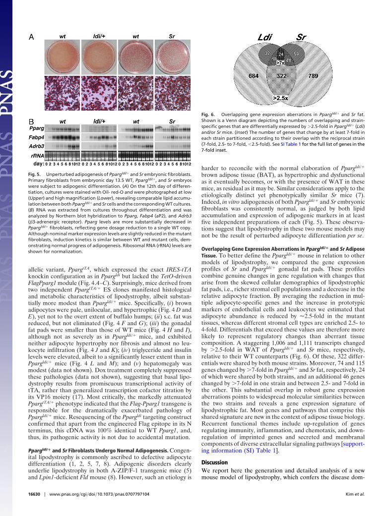
Overlapping Gene Expression Aberrations in Ppargldi/� and Sr AdiposeTissue. To better define the Ppargldi/� mouse in relation to othermodels of lipodystrophy, we compared the gene expressionprofiles of Sr and Ppargldi/� gonadal fat pads. These profilescombine genuine changes in gene regulation with changes thatarise from the skewed cellular demographics of lipodystrophicfat pads, i.e., richer stromal cell populations and a decrease in therelative adipocyte fraction. By averaging the reduction in mul-tiple adipocyte-specific genes and the increase in prototypicmarkers of endothelial cells and leukocytes we estimated thatadipocyte abundance is reduced by �2.5-fold in the mutanttissues, whereas different stromal cell types are enriched 2.5- to4-fold. Differentials that exceed these values are therefore morelikely to represent regulatory changes than aberrant tissuecomposition. A staggering 1,006 and 1,111 transcripts changedby �2.5-fold in WAT of Ppargldi/� and Sr mice, respectively,relative to their WT counterparts (Fig. 6). Of these, 322 differ-entials were shared by both mouse strains. Moreover, 74 and 115genes changed by �7-fold in Ppargldi/� and Sr fat, respectively, 24of which were shared by both strains, and an additional 46 geneschanged by �7-fold in one strain and between 2.5- and 7-fold inthe other. This substantial overlap in robust gene expressionaberrations points to widespread molecular similarities betweenthe two strains and reveals a gene expression signature oflipodystrophic fat. Most genes and pathways that comprise thisshared signature are new in the context of adipose tissue biology.Recurrent functional themes include up-regulation of genesregulating immunity, inflammation, and chemotaxis, and down-regulation of imprinted genes and secreted and membranalcomponents of diverse extracellular signaling pathways [support-ing information (SI) Table 1].
DiscussionWe report here the generation and detailed analysis of a newmouse model of lipodystrophy, which confers the disease dom-
Fig. 5. Unperturbed adipogenesis of Ppargldi/� and Sr embryonic fibroblasts.Primary fibroblasts from embryonic day 13.5 WT, Ppargldi/�, and Sr embryoswere subject to adipogenic differentiation. (A) On the 12th day of differen-tiation, cultures were stained with Oil- red-O and were photographed at low(Upper) and high magnification (Lower), revealing comparable lipid accumu-lation between both Ppargldi/� and Sr cells and the corresponding WT cultures.(B) RNA was extracted from cultures throughout differentiation and wasanalyzed by Northern blot hybridization to Pparg, Fabp4 (aP2), and Adrb3(�3-adrenergic receptor). Pparg levels are more substantially decreased inPpargldi/� fibroblasts, reflecting gene dosage reduction to a single WT copy.Although nominal marker expression levels are slightly reduced in the mutantfibroblasts, induction kinetics is similar between WT and mutant cells, dem-onstrating normal progress of adipogenesis. Ribosomal RNA (rRNA) levels areshown for normalization.
Fig. 6. Overlapping gene expression aberrations in Ppargldi/� and Sr fat.Shown is a Venn diagram depicting the numbers of overlapping and strain-specific genes that are differentially expressed by �2.5-fold in Ppargldi/� (Ldi)and/or Sr mice. (Inset) The number of genes that change by at least 7-fold ineach strain partitioned according to their overlap with the reciprocal strain(7-fold, 2.5- to 7-fold, �2.5-fold). See SI Table 1 for the full list of genes in the7-fold inset.
16630 � www.pnas.org�cgi�doi�10.1073�pnas.0707797104 Kim et al.
inantly through cooperative unanticipated effects of the syn-thetic transcription factor tTA and a minutely expressed Flag-PPAR�1 transgene. Although most mouse models oflipodystrophy exhibit indiscriminate degeneration of all adiposetissue depots, the Ppargldi/� phenotype is strikingly similar to thatof aP2-nSREBP1c (Sr) mice in the unique combination ofhypoplastic WAT and hypertrophic, isoplastic BAT. This traitand its metabolic comorbidities resemble the lipodystrophicsyndrome of HIV patients, in which some fat depots degeneratewhereas others expand (3). This analogy raises the hypothesisthat all three cases of lipodystrophy arise from insults thatconverge on similar cellular processes and renders the Ppargldi/�
mouse and its milder PpargtTA/� counterpart attractive animalmodels for severe and mild grades of this serious complication.The ability to fully suppress lipodystrophy by doxycycline in bothstrains makes them unique models of conditional lipodystrophy,further expanding their utility.
Ppargldi/� mice teach us several valuable lessons, both ingeneral and with regard to PPAR� and lipodystrophy. Thecautionary tale of tTA-induced lipodystrophy is the first re-ported deleterious activity of the system in vitro or in vivothroughout its rather extensive history (18). The completesuppression of the PpargtTA/� phenotype by doxycycline suggeststhat its molecular mechanism of action likely entails promiscu-ous transcriptional induction of endogenous genes rather thangeneralized transcription factor titration by the VP16 moiety(17). This finding is surprising in light of the bacterial origin andcomplex response element of the Tet repressor but is stronglysupported by follow-up studies that identify at least one cellulargene, Gpr56, as a robust, dox-repressible cellular target of tTA(S.K., L.-W.H., and Y.B., unpublished work).
The strong pathogenic impact of Flag-PPAR�1 provides thesecond surprise. The flag-tagged receptor recapitulates the DNA-binding, transactivation potential and adipogenic activity of nativePPAR�1 (ref. 19 and data not shown) and was expected to benefitadipocyte functions and systemic metabolism. However, not onlydoes it markedly exacerbate lipodystrophy in Ppargldi/� overPpargtTA/� mice, it does so with minute expression levels relative tothe companion WT PPAR�1 allele. Possible explanations to thisconfounding effect include the following: (i) The N-terminal block-ade might generate a defective PPAR� variant with strong domi-nant-negative effect on the product of the companion WT PPAR�allele. This idea is based on the lipodystrophy of patients withdominant negative PPARG mutations but is harder to reconcilewith the difference between N-terminal blockade here and muta-tions in the ligand-binding domain of PPAR� in patients (20–22).(ii) The N-terminal modification may underlie a gain-of-function,which either exaggerates normal deleterious functions of PPAR� orelicits altogether new off-target pathogenic interactions. (iii) Thetransgene may have non-protein-mediated functions at the level ofeither its RNA product or allele structure. This possibility gainssome support from the dramatic decline in PPAR�2 protein levelsin Ppargldi/� adipose tissue (Fig. 1F). However, endogenousPPAR�1 levels are intact and PPAR�2 is not repressed in fibro-blast-derived Ppargldi/� adipocytes in vitro (data not shown), sug-gesting that its down-regulation is likely not a direct effect ofFlag-PPAR�1.
The common contention that heritable lipodystrophy resultsfrom defective adipocyte differentiation has been proven inseveral instances (5, 8). However, the adipogenic potential ofboth Ppargldi/� and Sr fibroblasts is unperturbed. Although wecannot rule out an adipogenic defect that is manifested only invivo, the most plausible interpretation is that lipodystrophy inthese two mouse models is independent of and subsequent toadipogenesis. This conclusion should not be surprising in thebroader sense, as congenital and acquired degenerative diseasesof other tissues, such as muscle, heart, brain, and kidney,frequently arise in properly differentiated tissues. Moreover, the
phenotypically analogous HIV-associated lipodystrophy clearlyevolves in mature adipose tissue.
Last, the gene expression profiles of Ppargldi/� and Sr fat padsrecapitulate their high phenotypic similarity and imply a putativemolecular signature of lipodystrophic fat. The vast majority ofgenes involved in the regulation of adipogenesis and classicallipid metabolism are glaringly unchanged in both strains, withthe obvious exception of up-regulated cholesterol-repressedgenes in Sr mice. This observation is consistent with the con-clusion that neither phenotype arises from defective adipogen-esis and suggests that aberrant lipid metabolism is also not atfault. Instead, several genes associated with immunity andinflammation, most conspicuously Mhc2q10 and Ccl8 (see SITable 1), are up-regulated in the two strains, hinting at potentialinvolvement of immune and inflammatory processes in bothphenotypes. A broader array of inflammation-related genes isup-regulated in Ppargldi/� mice, suggesting that these processesare not general attributes of lipodystrophy, but rather strain-specific events that may contribute to discrete aspects of thephenotype. Most of these genes are not changed in PpargtTA/�
mice (data not shown), suggesting that inflammation and im-munity are involved primarily in severe, but not basal lipodys-trophy. The substantial leukocytic infiltrate in adipose tissue ofPpargldi/� and Sr, but not PpargtTA/� mice is consistent with thisinterpretation. Multiple imprinted genes and genes encoding cellsurface receptors and secreted factors from diverse signalingpathways are highly down-regulated in adipose tissue of bothstrains, two phenomena whose biological significance remains tobe determined. On the whole, most of the acutely affected genesare previously unrecognized in the context of adipocyte biology,hindering current understanding of these phenotypes, but offer-ing fresh research directions. Importantly, a preliminary screenrevealed significant similarities to gene expression anomalies ofadipose tissue from HIV patients (F. Villarroya, personal com-munication). This observation buttresses the functional analogybetween HIV-associated lipodystrophy and the two mousestrains and validates their utility as models for studying thispoorly understood syndrome.
Experimental ProceduresPpargldi/� and PpargtTA/� Mice. Targeting vectors were constructedby standard recombinant DNA technology and introduced intothe Pparg locus by homologous recombination as described inref. 23. Correct integration was validated by Southern blots byusing probes external to the homology arms of the vectors, asshown. Germ-line chimeras were established from three distinctPpargldi/� and two distinct PpargtTA/� ES clones. Mice carryingeither allele were subsequently propagated on the closely related129S1/SvImJ (129S) background. All histological, metabolic, andgene expression analyses were performed on F1 hybrid progenyof 129S-Ppargldi/� or -PpargtTA/� sires and WT C57BL/6J (B6)dams. This configuration ensured genetic homogeneity of thecohorts and was required because of poor fecundity of Ppargldi/�
mice after introgression onto B6. Where indicated, doxycyclinewas administered continuously in drinking water at 0.5 mg/ml.All mouse studies were approved by The Jackson Laboratory’sAnimal Care and Use Committee.
Nucleic Acid and Protein Detection. Northern and Western blotanalyses were performed using standard methods (24). tTA wasvisualized with an anti-VP16 antibody (Clontech, MountainView, CA). Custom PPAR� antiserum was raised in rabbits(QCB, Hopkinton, MA) against a bacterially expressed, 6X-His-tagged polypeptide comprising the 98 N-terminal amino acidsand was affinity-purified using the same antigen.
Histology and Metabolic Phenotyping. All tissues were fixed inBouin’s fixative, except liver, which was fixed in formalin. Fixed
Kim et al. PNAS � October 16, 2007 � vol. 104 � no. 42 � 16631
GEN
ETIC
S
tissues were embedded in paraffin, sectioned, and stained withhematoxylin and eosin. Pancreas sections were also stained withaldehyde fuchsin to detect insulin granules. Plasma analytes weredetermined in retroorbital blood collected from animals subjectto 5–6 h of fasting. Glucose, triglycerides, free fatty acids, andcholesterol were measured in a Beckman (Fullerton, CA) Syn-chron CX5 Delta Clinical Chemistry Analyzer. Insulin wasmeasured using a rat insulin RIA kit (LINCO, St. Charles, MO).For glucose tolerance tests, retroorbital blood was collectedfrom 6 h-fasted animals, followed by i.p. injection of 2 g glucoseper kilogram of body weight, collection of retroorbital bloodsamples 30, 60, 90, and 120 min thereafter, and determination ofglucose concentrations in the Synchron Analyzer.
Adipogenesis of Embryonic Fibroblasts. To prepare fibroblasts car-casses of embryonic day 13.5 embryos were individually minced by4–5 passages through an 18-guage syringe needle. Cells were platedon 6-cm dishes, brought to complete confluence, and 2 days laterwere treated for 72 h with insulin (10 �g/ml), 5 �M dexamethazone,0.2 mM isobutyl-methyl-xanthine, and 1 �M rosiglitazone. Cultureswere fed with fresh insulin-containing medium at 72-h intervalsthereafter. Lipid content was assessed by Oil-red-O staining at theend of differentiation. In other experiments, RNA was extractedfrom cultures at different stages of differentiation and analyzed.
Affymetrix Microarrays and Statistical Analysis. RNA from epidid-ymal fat pads of three 10-wk-old mutant males (Ppargldi/� or Sr) andthree litter-matched WT controls for each strain was treated withDNase I and probes were generated according to manufacturer’sinstructions (Affymetrix, Santa Clara, CA). Each sample hybridizedto one MOE430 version 2.0 GeneChip array in a Fluidics Station450 instrument (Affymetrix). Arrays were imaged with a GeneChip3000 Laser Confocal Slide Scanner, were quantified using Gene-Chip Operating Software version 1.2 (Affymetrix), and expressionvalues were summarized using the robust multichip average (RMA)function. P values were assigned using ANOVA methodology (25).False discovery rates (FDR) were assigned by computing q valuesfrom test statistics (26).
We thank Dr. Ed Leiter for discussions and encouragement; Dr.Francesc Villarroya for sharing unpublished microarray data; Drs. SueAckerman, Jurgen Naggert, and Ron Evans for helpful comments on themanuscript; The Jackson Laboratory’s diagnostic laboratory, microin-jection, histology, and gene expression services for expert technicalassistance; and Pat Cherry for administrative assistance. Support for thiswork was received through pilot funding from National Institute ofDiabetes and Digestive and Kidney Diseases Grant 5 P30 DK32520 (toY.B.), Grant CA34196 (to The Jackson Laboratory), an American HeartAssociation Postdoctoral Fellowship (to S.K.), and private gifts from theFraternal Order of Eagles.
1. Garg A (2004) N Engl J Med 350:1220–1234.2. Capeau J, Magre J, Lascols O, Caron M, Bereziat V, Vigouroux C, Bastard JP
(2005) Biochem Soc Trans 33:1073–1077.3. Carr A, Samaras K, Burton S, Law M, Freund J, Chisholm DJ, Cooper DA
(1998) AIDS 12:F51–F58.4. Ross SR, Graves RA, Spiegelman BM (1993) Genes Dev 7:1318–1324.5. Moitra J, Mason MM, Olive M, Krylov D, Gavrilova O, Marcus-Samuels B,
Feigenbaum L, Lee E, Aoyama T, Eckhaus M, et al. (1998) Genes Dev12:3168–3181.
6. Pajvani UB, Trujillo ME, Combs TP, Iyengar P, Jelicks L, Roth KA, Kitsis RN,Scherer PE (2005) Nat Med 11:797–803.
7. Shimomura I, Hammer RE, Richardson JA, Ikemoto S, Bashmakov Y,Goldstein JL, Brown MS (1998) Genes Dev 12:3182–3194.
8. Peterfy M, Phan J, Xu P, Reue K (2001) Nat Genet 27:121–124.9. Cutler DA, Sullivan T, Marcus-Samuels B, Stewart CL, Reitman ML (2002)
Biochem Biophys Res Commun 291:522–527.10. Pendas AM, Zhou Z, Cadinanos J, Freije JM, Wang J, Hultenby K, Astudillo
A, Wernerson A, Rodriguez F, Tryggvason K, et al. (2002) Nat Genet 31:94–99.11. El-Haschimi K, Dufresne SD, Hirshman MF, Flier JS, Goodyear LJ, Bjorbaek
C (2003) Diabetes 52:1340–1346.12. Laustsen PG, Michael MD, Crute BE, Cohen SE, Ueki K, Kulkarni RN, Keller
SR, Lienhard GE, Kahn CR (2002) Genes Dev 16:3213–3222.13. He W, Barak Y, Hevener A, Olson P, Liao D, Le J, Nelson M, Ong E, Olefsky
JM, Evans RM (2003) Proc Natl Acad Sci USA 100:15712–15717.
14. Jones JR, Barrick C, Kim KA, Lindner J, Blondeau B, Fujimoto Y, Shiota M,Kesterson RA, Kahn BB, Magnuson MA (2005) Proc Natl Acad Sci USA102:6207–6212.
15. Imai T, Takakuwa R, Marchand S, Dentz E, Bornert JM, Messaddeq N,Wendling O, Mark M, Desvergne B, Wahli W, et al. (2004) Proc Natl Acad SciUSA 101:4543–4547.
16. Gossen M, Bujard H (1992) Proc Natl Acad Sci USA 89:5547–5551.17. Tasset D, Tora L, Fromental C, Scheer E, Chambon P (1990) Cell 62:1177–
1187.18. Zhu Z, Zheng T, Lee CG, Homer RJ, Elias JA (2002) Semin Cell Dev Biol
13:121–128.19. Hummasti S, Tontonoz P (2006) Mol Endocrinol 20:1261–1275.20. Barroso I, Gurnell M, Crowley VE, Agostini M, Schwabe JW, Soos MA,
Maslen GL, Williams TD, Lewis H, Schafer AJ, et al. (1999) Nature 402:880–883.
21. Agarwal AK, Garg A (2002) J Clin Endocrinol Metab 87:408–411.22. Hegele RA, Cao H, Frankowski C, Mathews ST, Leff T (2002) Diabetes
51:3586–3590.23. Barak Y, Nelson MC, Ong ES, Jones YZ, Ruiz-Lozano P, Chien KR, Koder
A, Evans RM (1999) Mol Cell 4:585–595.24. Shalom-Barak T, Nicholas JM, Wang Y, Zhang X, Ong ES, Young TH,
Gendler SJ, Evans RM, Barak Y (2004) Mol Cell Biol 24:10661–10669.25. Churchill GA (2004) BioTechniques 37:173–177.26. Storey JD, Tibshirani R (2003) Proc Natl Acad Sci USA 100:9440–9445.
16632 � www.pnas.org�cgi�doi�10.1073�pnas.0707797104 Kim et al.