Embed Size (px)
Citation preview

A
JS
a
ARAA
KGST
1
bluilAwra
(snhpbat
(L((T
h0
BioSystems 151 (2017) 21–26
Contents lists available at ScienceDirect
BioSystems
jo ur nal home p age: www.elsev ier .com/ locate /b iosystems
new integrated symmetrical table for genetic codes
ian-Jun Shuchool of Mechanical & Aerospace Engineering, Nanyang Technological University, 50 Nanyang Avenue, 639798, Singapore
r t i c l e i n f o
rticle history:eceived 16 November 2016ccepted 17 November 2016vailable online 23 November 2016
a b s t r a c t
Degeneracy is a salient feature of genetic codes, because there are more codons than amino acids. Theconventional table for genetic codes suffers from an inability of illustrating a symmetrical nature amonggenetic base codes. In fact, because the conventional wisdom avoids the question, there is little agreementas to whether the symmetrical nature actually even exists. A better understanding of symmetry and an
eywords:enetic codesymmetryable
appreciation for its essential role in the genetic code formation can improve our understanding of nature’scoding processes. Thus, it is worth formulating a new integrated symmetrical table for genetic codes,which is presented in this paper. It could be very useful to understand the Nobel laureate Crick’s wobblehypothesis — how one transfer ribonucleic acid can recognize two or more synonymous codons, whichis an unsolved fundamental question in biological science.
© 2016 Elsevier Ireland Ltd. All rights reserved.
. Introduction
The discovery of double-helix molecular structure of deoxyri-onucleic acid (DNA) by Watson and Crick (1953) is one of
andmarks in the history of science. It represents the birth of molec-lar biology. On the cellular level, the living organisms are classified
nto prokaryotes and eukaryotes. The prokaryotes are unicellularife forms while the eukaryotes include human, animal and fungus.ll prokaryotic and eukaryotic cells share a common process byhich information encoded by a gene is used to produce the cor-
esponding protein. This process is called protein biosynthesis andccomplished in two steps: transcription and translation.
During transcription, DNA is transcribed into ribonucleic acidRNA). DNA carries the genetic information, while RNA is used toynthesize proteins. DNA consists of a strand of bases, namely Ade-ine (A), Thymine (T), Guanine (G) and Cytosine (C), whereas RNAas A, G, C and Uracil (U) instead of T. Then, translation occurs whereroteins (molecules composed of a long chain of amino acids) areuilt upon the codons in RNA. Each codon, which is a set of threedjoined nucleotides (triplet), specifies one amino acid or termina-ion signal (Crick et al., 1961).
There are 20 amino acids, namely Histidine (His/H), ArginineArg/R), Lysine (Lys/K), Phenylalanine (Phe/F), Alanine (Ala/A),eucine (Leu/L), Methionine (Met/M), Isoleucine (Ile/I), Tryptophan
Trp/W), Proline (Pro/P), Valine (Val/V), Cysteine (Cys/C), GlycineGly/G), Glutamine (Gln/Q), Asparagine (Asn/N), Serine (Ser/S),yrosine (Tyr/Y), Threonine (Thr/T), Aspartic acid (Asp/D) and Glu-E-mail address: [email protected]
ttp://dx.doi.org/10.1016/j.biosystems.2016.11.004303-2647/© 2016 Elsevier Ireland Ltd. All rights reserved.
tamic acid (Glu/E). For the formation of proteins in living organismcells, it is found that each amino acid can be specified by eithera minimum of one codon or up to a maximum of six possiblecodons. In other words, different codons specify the different num-ber of amino acids. A table for genetic codes is a representationof translation for illustrating the different amino acids with theirrespectively specifying codons, that is, a set of rules by which infor-mation encoded in genetic material (RNA sequences) is translatedinto proteins (amino acid sequences) by living cells. There are a totalof 64 possible codons, but there are only 20 amino acids specifiedby them. Therefore, degeneracy is a salient feature of genetic codes.Genetic information is stored in DNA in the form of sequences ofnucleotides which is made clearly in the double-helix model, butit does not provide any clue on how one transfer ribonucleic acid(tRNA) can recognize two or more synonymous codons. Therefore,deciphering the genetic codes becomes a problem. Up to now, it isstill unable to find out the reason or explanation for these kinds ofcharacteristics and relationships between codons and amino acids.Therefore, it has always been an interesting area for us to exploreand obtain any explanation further.
The table for genetic codes allows us to identify a codon andthe individual amino acid assigned to the codon by nature. Theseassignment tables may come in a variety of forms, but they all sufferfrom an inability of illustrating a symmetrical nature among geneticbase codes. In fact, because the conventional wisdom avoids thequestion, there is little agreement as to whether the symmetrical
nature actually even exists. A better understanding of symmetryand an appreciation for its essential role in the genetic code forma-tion can improve our understanding of nature’s coding processes.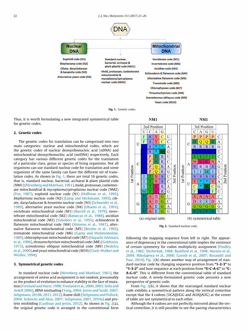
22 J.-J. Shu / BioSystems 151 (2017) 21–26
enetic codes.
Tf
2
mtmcooolt(a(ba1vtmflnt1e1eW
3
aatAS2tt
Fig. 1. G
hus, it is worth formulating a new integrated symmetrical tableor genetic codes.
. Genetic codes
The genetic codes for translation can be categorized into twoain categories: nuclear and mitochondrial codes, which are
he genetic codes of nuclear deoxyribonucleic acid (nDNA) anditochondrial deoxyribonucleic acid (mtDNA) respectively. Each
ategory has various different genetic codes for the translationf a particular class, genus or species of living organisms. Not allrganisms can use standard nuclear code for translation and somerganisms of the same family can have the different set of trans-ation codes. As shown in Fig. 1, there are total 16 genetic codes,hat is, standard nuclear, bacterial, archaeal & plant plastid codeNM1) (Nirenberg and Matthaei, 1961), mold, protozoan, coelenter-te mitochondrial & mycoplasma/spiroplasma nuclear code (NM2)Fox, 1987), euplotid nuclear code (N1) (Hoffman et al., 1995),lepharisma nuclear code (N2) (Liang and Heckmann, 1993), cili-te, dasycladacean & hexamita nuclear code (N3) (Schneider et al.,989), alternative yeast nuclear code (N4) (Ohama et al., 1993),ertebrate mitochondrial code (M1) (Barrell et al., 1979), inver-ebrate mitochondrial code (M2) (Batuecas et al., 1988), ascidian
itochondrial code (M3) (Yokobori et al., 1993), echinoderm &atworm mitochondrial code (M4) (Himeno et al., 1987), alter-ative flatworm mitochondrial code (M5) (Bessho et al., 1992),rematode mitochondrial code (M6) (Garey and Wolstenholme,989), chlorophycean mitochondrial code (M7) (Hayashi-Ishimarut al., 1996), thraustochytrium mitochondrial code (M8) (Goldstein,973), scenedesmus obliquus mitochondrial code (M9) (Nedelcut al., 2000) and yeast mitochondrial code (M10) (Clark-Walker andeiller, 1994).
. Symmetrical genetic codes
In standard nuclear code (Nirenberg and Matthaei, 1961), therrangement of amino acid assignment is not random, presumablys the product of evolution to enhance stability in the face of muta-ion (Freeland and Hurst, 1998; Freeland et al., 2000, 2003; Sella andrdell 2006), tRNA misloading (Yang, 2004; Jestin and Soulé, 2007;
eligmann, 2010b, 2011, 2012), frame shift (Seligmann and Pollock,004; Itzkovitz and Alon, 2007; Seligmann, 2007, 2010a) and pro-ein misfolding (Guilloux and Jestin, 2012). As shown in Fig. 2(a),he original genetic code is arranged in the conventional formFig. 2. Standard nuclear code.
following the mapping sequence from left to right. The appear-ance of degeneracy in the conventional table implies the existenceof certain symmetry for codon multiplicity assignment (Findleyet al., 1982; Shcherbak, 1988; Bashford et al., 1998; Hornos et al.,2004; Nikolajewa et al., 2006; Gavish et al., 2007; Rosandic andPaar, 2014). Fig. 2(b) shows another way of arrangement of stan-dard nuclear code by changing sequence position from “1-2-3” to“1-3-2” and base sequence at each position from “U-C-A-G” to “C-A-G-U”. This is different from the conventional table of standardnuclear code. A newly-formulated genetic code presents a newperspective of genetic code.
From Fig. 2(b), it shows that the rearranged standard nuclearcode exhibits a symmetrical pattern along the vertical centerlineexcept that the 4 codons (UGA||UGG and AUA||AUG) at the center
of table are not symmetrical to each other.Although the 4 codons are not perfectly mirrored about the ver-tical centerline, it is still possible to see the pairing characteristics

J.-J. Shu / BioSystems 151 (2017) 21–26 23
Fig. 3. Rearranged codes.

24 J.-J. Shu / BioSystems 151 (2017) 21–26
d tabl
bTt
mFcmis
Fig. 4. An integrate
etween “C|U”-pyrimidine and “A|G”-purine at the third position.he third (or wobble) base plays a significant role in encoding theype of amino acid.
The rearrangement of other genetic codes for both nDNA andtDNA also exhibits such a symmetrical pattern. As shown in
ig. 3, the majority of codon pairs are symmetric about the verticalenterline. However there are still some codon pairs that are asym-
etric. The four rearranged genetic codes, namely, vertebrate (M1),nvertebrate (M2), ascidian (M3) and yeast (M10), exhibit a perfectymmetrical pattern along the vertical centerline and all belong to
e for genetic codes.
the mitochondrial genetic family. It is worth pointing out, that theonly perfect symmetry of vertebrate mitochondrial code (M1) wasnoted (Lehmann, 2000; Gonzalez et al., 2013).
4. An integrated table for genetic codes
In view of the symmetrical and asymmetrical characteristicsof all 16 rearranged genetic codes, there is a question that manymay raise whether a perfect symmetrical genetic code is the ori-gin or ultimate product of evolutionary progress. The total 16 sets

ems 15
ogvmgrgintsoiut
5
iTncuwwn&cnaaS2
atbpLe
tecafba
R
A
B
B
B
B
C
C
J.-J. Shu / BioSyst
f genetic codes that are used in different biological species mayive us the clue of how the evolutionary process happened. Todayarious species are quite different in terms of appearance, but theyay have the same ancestor. Therefore, a new integrated table for
enetic codes is needed to discover any possible regularity occur-ing in all genetic codes. As shown in Fig. 4, the integrated table forenetic codes has a standardized template in the center which isdentical for all genetic codes and surrounded with the unique sig-ature of each rearranged genetic code. The proposed integratedable for genetic codes aims to provide the user with a comprehen-ive and simple genetic translation interface, which is comprisedf the entire different genetic translation codes. Simply by replac-ng the blank center of standardized template with the surroundingnique signature, the user can then obtain desired genetic transla-ion table for each organism.
. Concluding remarks
As shown in Fig. 4, the STOP (*) codon of standard nuclear codes mutated to other amino acids in almost every non-standard code.he only two that do not contain STOP (*) codon mutation are alter-ative yeast nuclear code (N4) and thraustochytrium mitochondrialode (M8). This seems to imply that, the STOP (*) codon is the mostnstable codon in genetic codes, or could be seen as an empty shell,hich could easily be replaced by the nearby amino acids. It isorth mentioning to this end that the five genetic codes, euplotiduclear code (N1), alternative yeast nuclear code (N4), echinoderm
flatworm mitochondrial code (M4), alternative flatworm mito-hondrial code (M5) and trematode mitochondrial code (M6), doot follow the intuition based on standard nuclear code (Nirenbergnd Matthaei, 1961), which asymmetry is restricted to the ‘punctu-tion’ codons, START (Met/M) and STOP (*) codons (Rumer, 1966;hcherbak, 1989; Kozyrev and Khrennikov, 2010; Rosandic et al.,013; Seligmann, 2015).
There is the existence of symmetrical and asymmetrical char-cteristics in genetic codes. The presence of symmetry enableshe ease/efficiency of live formation and the role of symmetry-reaking (asymmetry) is to enable evolution/adaptation to takelace (Elitzur, 1997; Seligmann, 2000; Antoneli and Forger, 2011;enstra, 2014). In short, lives are formed due to symmetry butvolved due to asymmetry.
The newly-formulated integrated symmetrical table provideshe new perspective of possible codon-amino acid relationship andxplains the hidden meaning/logic behind the degenerative geneticodes, and can be used to build programmable biomolecular medi-ted processors (Shu et al., 2011, 2015, 2016; Wong et al., 2015)or efficient genome editing (Ishino et al., 1987; Jinek et al., 2012)y taking both symmetrical and asymmetrical characteristics intoccount.
eferences
ntoneli, F., Forger, M., 2011. Symmetry breaking in the genetic code: finitegroups. Math. Comput. Modell. 53 (7-8), 1469–1488.
arrell, B.G., Bankier, A.T., Drouin, J., 1979. A different genetic code in humanmitochondria. Nature 282 (5735), 189–194.
ashford, J.D., Tsohantjis, I., Jarvis, P.D., 1998. A supersymmetric model for theevolution of the genetic code. Proc. Natl. Acad. Sci. U. S. A. 95 (3), 987–992.
atuecas, B., Garesse, R., Calleja, M., Valverde, J.R., Marco, R., 1988. Genomeorganization of artemia mitochondrial DNA. Nucleic Acids Res. 16 (14A),6515–6529.
essho, Y., Ohama, T., Osawa, S., 1992. Planarian mitochondria II. The uniquegenetic code as deduced from cytochrome c oxidase subunit I gene sequences.J. Mol. Evol. 34 (4), 331–335.
lark-Walker, G.D., Weiller, G.F., 1994. The structure of the small mitochondrialDNA of Kluyveromyces thermotolerans is likely to reflect the ancestral geneorder in fungi. J. Mol. Evol. 38 (6), 593–601.
rick, F.H.C., Barnett, L., Brenner, S., Watts-Tobin, R.J., 1961. General nature of thegenetic code for proteins. Nature 192 (4809), 1227–1232.
1 (2017) 21–26 25
Elitzur, A.C., 1997. Constancy, uniformity and symmetry of living systems: thecomputational functions of morphological invariance. Biosystems 43 (1),41–53.
Findley, G.L., Findley, A.M., McGlynn, S.P., 1982. Symmetry characteristics of thegenetic code. Proc. Natl. Acad. Sci. U. S. A.-Phys. Sci. 79 (22), 7061–7065.
Fox, T.D., 1987. Natural variation in the genetic code. Annu. Rev. Genet. 21, 67–91.Freeland, S.J., Hurst, L.D., 1998. The genetic code is one in a million. J. Mol. Evol. 47
(3), 238–248.Freeland, S.J., Knight, R.D., Landweber, L.F., Hurst, L.D., 2000. Early fixation of an
optimal genetic code. Mol. Biol. Evol. 17 (4), 511–518.Freeland, S.J., Wu, T., Keulmann, N., 2003. The case for an error minimizing
standard genetic code. Origins Life Evol. Biospheres 33 (4), 457–477.Garey, J.R., Wolstenholme, D.R., 1989. Platyhelminth mitochondrial DNA: Evidence
for early evolutionary origin of a tRNAserAGN that contains a dihydrouridinearm replacement loop, and of serine-specifying AGA and AGG codons. J. Mol.Evol. 28 (5), 374–387.
Gavish, M., Peled, A., Chor, B., 2007. Genetic code symmetry and efficient design ofGC-constrained coding sequences. Bioinformatics 23 (2), E57–E63.
Goldstein, S., 1973. Zoosporic marine fungi (Thraustochytriaceae anddermocystidiaceae). Annu. Rev. Microbiol. 27, 13–26.
Gonzalez, D.L., Giannerini, S., Rosa, R., 2013. On the origin of the mitochondrialgenetic code: towards a unified mathematical framework for the managementof genetic information. Nat. Precedings, 1–20.
Guilloux, A., Jestin, J.-L., 2012. The genetic code and its optimization for kineticenergy conservation in polypeptide chains. Biosystems 109 (2), 141–144.
Hayashi-Ishimaru, Y., Ohama, T., Kawatsu, Y., Nakamura, K., Osawa, S., 1996. UAGis a sense codon in several chlorophycean mitochondria. Curr. Genet. 30 (1),29–33.
Himeno, H., Masaki, H., Kawai, T., Ohta, T., Kumagai, I., Miura, K., Watanabe, K.,1987. Unusual genetic codes and a novel gene structure for tRNASerAGY instarfish mitochondrial DNA. Gene 56 (2–3), 219–230.
Hoffman, D.C., Anderson, R.C., DuBois, M.L., Prescott, D.M., 1995. Macronucleargene-sized molecules of hypotrichs. Nucleic Acids Res. 23 (8), 1279–1283.
Hornos, J.E.M., Braggion, L., Magini, M., Forger, M., 2004. Symmetry preservation inthe evolution of the genetic code. IUBMB Life 56 (3), 125–130.
Ishino, Y., Shinagawa, H., Makino, K., Amemura, M., Nakata, A., 1987. Nucleotidesequence of the iap gene, responsible for alkaline phosphatase isozymeconversion in Escherichia-coli, and identification of the gene product. J.Bacteriol. 169 (12), 5429–5433.
Itzkovitz, S., Alon, U., 2007. The genetic code is nearly optimal for allowingadditional information within protein-coding sequences. Genome Res. 17 (4),405–412.
Jestin, J.-L., Soulé, C., 2007. Symmetries by base substitutions in the genetic codepredict 2’ or 3’ aminoacylation of tRNAs. J. Theor. Biol. 247 (2), 391–394.
Jinek, M., Chylinski, K., Fonfara, I., Hauer, M., Doudna, J.A., Charpentier, E., 2012. Aprogrammable dual-RNA-guided DNA endonuclease in adaptive bacterialimmunity. Science 337 (6096), 816–821.
Kozyrev, S.V., Khrennikov, A.Y., 2010. 2-Adic numbers in genetics and Rumer’ssymmetry. Doklady Math. 81 (1), 128–130.
Lehmann, J., 2000. Physico-chemical constraints connected with the codingproperties of the genetic system. J. Theor. Biol. 202 (2), 129–144.
Lenstra, R., 2014. Evolution of the genetic code through progressive symmetrybreaking. J. Theor. Biol. 347, 95–108.
Liang, A., Heckmann, K., 1993. Blepharisma uses UAA as a termination codon.Naturwissenschaften 80 (5), 225–226.
Nedelcu, A.M., Lee, R.W., Lemieux, C., Gray, M.W., Burger, G., 2000. The completemitochondrial DNA sequence of Scenedesmus obliquus reflects an intermediatestage in the evolution of the green algal mitochondrial genome. Genome Res.10 (6), 819–831.
Nikolajewa, S., Friedel, M., Beyer, A., Wilhelm, T., 2006. The new classificationscheme of the genetic code, its early evolution, and tRNA usage. J. Bioinform.Comput. Biol. 4 (2), 609–620.
Nirenberg, M.W., Matthaei, J.H., 1961. The dependence of cell-free proteinsynthesis in E. coli upon naturally occurring or synthetic polyribonucleotides.Proc. Natl. Acad. Sci. U. S. A. 47 (10), 1588–1602.
Ohama, T., Suzuki, T., Mori, M., Osawa, S., Ueda, T., Watanabe, K., Nakase, T., 1993.Non-universal decoding of the leucine codon CUG in several Candida species.Nucleic Acids Res. 21 (17), 4039–4045.
Rosandic, M., Paar, V., 2014. Codon sextets with leading role of serine create idealsymmetry classification scheme of the genetic code. Gene 543 (1), 45–52.
Rosandic, M., Paar, V., Gluncic, M., 2013. Fundamental role of start/stop regulatorsin whole DNA and new trinucleotide classification. Gene 531 (2), 184–190.
Rumer, Y.B., 1966. Systematization of codons in the genetic code. Dokl. Akad. NaukSSSR 167 (6), 1393–1394 (in Russian).
Schneider, S.U., Leible, M.B., Yang, X.-P., 1989. Strong homology between the smallsubunit of ribulose-1,5-bisphosphate carboxylase/oxygenase of two species ofAcetabularia and the occurrence of unusual codon usage. Mol. Gen. Genet. 218(3), 445–452.
Seligmann, H., Pollock, D.D., 2004. The ambush hypothesis: hidden stop Ccodonsprevent off-frame gene reading. DNA Cell Biol. 23 (10), 701–705.
Seligmann, H., 2000. Evolution and ecology of developmental processes and of the
resulting morphology: directional asymmetry in hindlimbs of Agamidae andLacertidae (Reptilia: lacertilia). Biol. J. Linn. Soc. 69 (4), 461–481.Seligmann, H., 2007. Cost minimization of ribosomal frameshifts. J. Theor. Biol. 249(1), 162–167.

2 ems 15
S
S
S
S
S
S
S
S
Yang, C.M., 2004. On the 28-gon symmetry inherent in the genetic codeintertwined with aminoacyl-tRNA synthetases—the Lucas series. Bull. Math.Biol. 66 (5), 1241–1257.
Yokobori, S.-I., Ueda, T., Watanabe, K., 1993. Codons AGA and AGG are read as
6 J.-J. Shu / BioSyst
eligmann, H., 2010a. The ambush hypothesis at the whole-organism level: offframe, ‘hidden’ stops in vertebrate mitochondrial genes increasedevelopmental stability. Comput. Biol. Chem. 34 (2), 80–85.
eligmann, H., 2010b. Do anticodons of misacylated tRNAs preferentially mismatchcodons coding for the misloaded amino acid? BMC Mol. Biol. 11 (41), 1–5.
eligmann, H., 2011. Error compensation of tRNA misacylation bycodon-anticodon mismatch prevents translational amino acid misinsertion.Comput. Biol. Chem. 35 (2), 81–95.
eligmann, H., 2012. Coding constraints modulate chemically spontaneousmutational replication gradients in mitochondrial genomes. Curr. Genomics 13(1), 37–54.
eligmann, H., 2015. Phylogeny of genetic codes and punctuation codes withingenetic codes. Biosystems 129, 36–43.
ella, G., Ardell, D.H., 2006. The coevolution of genes and genetic codes: crick’s
frozen accident revisited. J. Mol. Evol. 63 (3), 297–313.hcherbak, V.I., 1988. The co-operative symmetry of the genetic-code. J. Theor.Biol. 132 (1), 121–124.
hcherbak, V.I., 1989. Rumer’s rule and transformation in the context of theco-operative symmetry of the genetic-code. J. Theor. Biol. 139 (2), 271–276.
1 (2017) 21–26
Shu, J.-J., Wang, Q.-W., Yong, K.-Y., 2011. DNA-based computing of strategicassignment problems. Phys. Rev. Lett. 106 (18), 188702.
Shu, J.-J., Wang, Q.-W., Yong, K.-Y., Shao, F., Lee, K.J., 2015. ProgrammableDNA-mediated multitasking processor. J. Phys. Chem. B 119 (17), 5639–5644.
Shu, J.-J., Wang, Q.-W., Yong, K.-Y., 2016. Programmable DNA-mediated decisionmaker. Int. J. Bio-Inspired Comput. 8 (6), 1–5.
Watson, J.D., Crick, F.H.C., 1953. Molecular structure of nucleic acids − A structurefor deoxyribose nucleic acid. Nature 171 (4356), 737–738.
Wong, J.R., Lee, K.J., Shu, J.-J., Shao, F., 2015. Magnetic fields facilitateDNA-mediated charge transport. Biochemistry 54 (21), 3392–3399.
glycine in ascidian mitochondria. J. Mol. Evol. 36 (1), 1–8.