Embed Size (px)
Citation preview
A New Method for Rapidly Generating Inhibitors of Glyoxalase I inside TumorCells Using S-(N-Aryl-N-hydroxycarbamoyl)ethylsulfoxides
Diana S. Hamilton,† Malcolm J. Kavarana,† Ellyn M. Sharkey,† Julie L. Eiseman,‡ and Donald J. Creighton*,†
Department of Chemistry and Biochemistry, University of Maryland, Baltimore County, Baltimore, Maryland 21250, andDivision of Developmental Therapeutics, Greenebaum Cancer Center, and Department of Pathology, University of Maryland,Baltimore, Maryland 21201
Received December 18, 1998
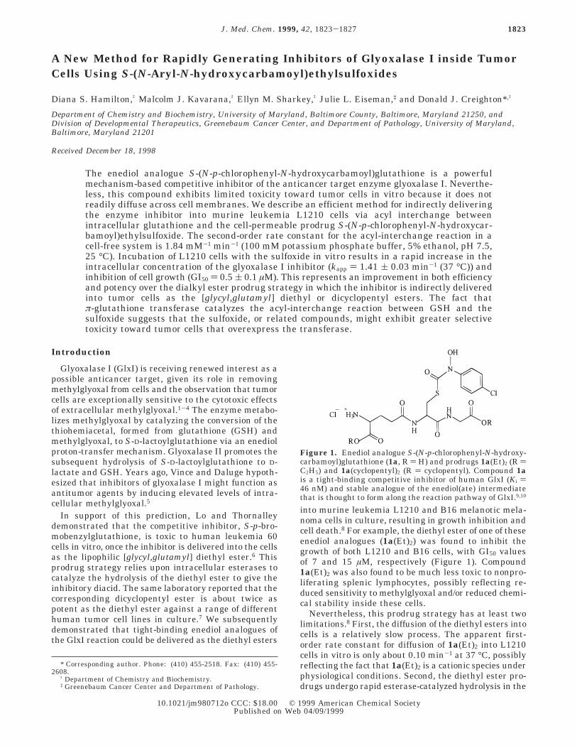
The enediol analogue S-(N-p-chlorophenyl-N-hydroxycarbamoyl)glutathione is a powerfulmechanism-based competitive inhibitor of the anticancer target enzyme glyoxalase I. Neverthe-less, this compound exhibits limited toxicity toward tumor cells in vitro because it does notreadily diffuse across cell membranes. We describe an efficient method for indirectly deliveringthe enzyme inhibitor into murine leukemia L1210 cells via acyl interchange betweenintracellular glutathione and the cell-permeable prodrug S-(N-p-chlorophenyl-N-hydroxycar-bamoyl)ethylsulfoxide. The second-order rate constant for the acyl-interchange reaction in acell-free system is 1.84 mM-1 min-1 (100 mM potassium phosphate buffer, 5% ethanol, pH 7.5,25 °C). Incubation of L1210 cells with the sulfoxide in vitro results in a rapid increase in theintracellular concentration of the glyoxalase I inhibitor (kapp ) 1.41 ( 0.03 min-1 (37 °C)) andinhibition of cell growth (GI50 ) 0.5 ( 0.1 µM). This represents an improvement in both efficiencyand potency over the dialkyl ester prodrug strategy in which the inhibitor is indirectly deliveredinto tumor cells as the [glycyl,glutamyl] diethyl or dicyclopentyl esters. The fact thatπ-glutathione transferase catalyzes the acyl-interchange reaction between GSH and thesulfoxide suggests that the sulfoxide, or related compounds, might exhibit greater selectivetoxicity toward tumor cells that overexpress the transferase.
Introduction
Glyoxalase I (GlxI) is receiving renewed interest as apossible anticancer target, given its role in removingmethylglyoxal from cells and the observation that tumorcells are exceptionally sensitive to the cytotoxic effectsof extracellular methylglyoxal.1-4 The enzyme metabo-lizes methylglyoxal by catalyzing the conversion of thethiohemiacetal, formed from glutathione (GSH) andmethylglyoxal, to S-D-lactoylglutathione via an enediolproton-transfer mechanism. Glyoxalase II promotes thesubsequent hydrolysis of S-D-lactoylglutathione to D-lactate and GSH. Years ago, Vince and Daluge hypoth-esized that inhibitors of glyoxalase I might function asantitumor agents by inducing elevated levels of intra-cellular methylglyoxal.5
In support of this prediction, Lo and Thornalleydemonstrated that the competitive inhibitor, S-p-bro-mobenzylglutathione, is toxic to human leukemia 60cells in vitro, once the inhibitor is delivered into the cellsas the lipophilic [glycyl,glutamyl] diethyl ester.6 Thisprodrug strategy relies upon intracellular esterases tocatalyze the hydrolysis of the diethyl ester to give theinhibitory diacid. The same laboratory reported that thecorresponding dicyclopentyl ester is about twice aspotent as the diethyl ester against a range of differenthuman tumor cell lines in culture.7 We subsequentlydemonstrated that tight-binding enediol analogues ofthe GlxI reaction could be delivered as the diethyl esters
into murine leukemia L1210 and B16 melanotic mela-noma cells in culture, resulting in growth inhibition andcell death.8 For example, the diethyl ester of one of theseenediol analogues (1a(Et)2) was found to inhibit thegrowth of both L1210 and B16 cells, with GI50 valuesof 7 and 15 µM, respectively (Figure 1). Compound1a(Et)2 was also found to be much less toxic to nonpro-liferating splenic lymphocytes, possibly reflecting re-duced sensitivity to methylglyoxal and/or reduced chemi-cal stability inside these cells.
Nevertheless, this prodrug strategy has at least twolimitations.8 First, the diffusion of the diethyl esters intocells is a relatively slow process. The apparent first-order rate constant for diffusion of 1a(Et)2 into L1210cells in vitro is only about 0.10 min-1 at 37 °C, possiblyreflecting the fact that 1a(Et)2 is a cationic species underphysiological conditions. Second, the diethyl ester pro-drugs undergo rapid esterase-catalyzed hydrolysis in the
* Corresponding author. Phone: (410) 455-2518. Fax: (410) 455-2608.
† Department of Chemistry and Biochemistry.‡ Greenebaum Cancer Center and Department of Pathology.
Figure 1. Enediol analogue S-(N-p-chlorophenyl-N-hydroxy-carbamoyl)glutathione (1a, R ) H) and prodrugs 1a(Et)2 (R )C2H5) and 1a(cyclopentyl)2 (R ) cyclopentyl). Compound 1ais a tight-binding competitive inhibitor of human GlxI (Ki )46 nM) and stable analogue of the enediol(ate) intermediatethat is thought to form along the reaction pathway of GlxI.9,10
1823J. Med. Chem. 1999, 42, 1823-1827
10.1021/jm980712o CCC: $18.00 © 1999 American Chemical SocietyPublished on Web 04/09/1999
plasma of most inbred strains of laboratory mice,complicating efficacy studies using murine models.
Here we show that enediol analogue 1a can be rapidlygenerated inside L1210 cells via an acyl-interchangereaction between intracellular GSH and the cell-perme-able prodrug S-(N-p-chlorophenyl-N-hydroxycarbam-oyl)ethylsulfoxide (2a), Scheme 1. This prodrug strategyhas important advantages over the dialkyl ester prodrugstrategy.
ChemistryThe dicyclopentyl ester of enediol analogue 1a was
obtained by acid-catalyzed esterification of 1a in cyclo-pentanol/HCl. The synthetic route to the acylatingreagents used with GSH to produce different enediolanalogues is outlined in Scheme 2. Oxidation of thioesters3a-c to the corresponding sulfoxides 2a-c followedpublished methods.11,12 N-(p-Chlorophenyl)hydroxyl-amine was prepared by reduction of p-chloronitroben-zene with hydrazine hydrate in the presence of 5% Rhon carbon.13
Results and DiscussionChemical Kinetics. The studies described in this
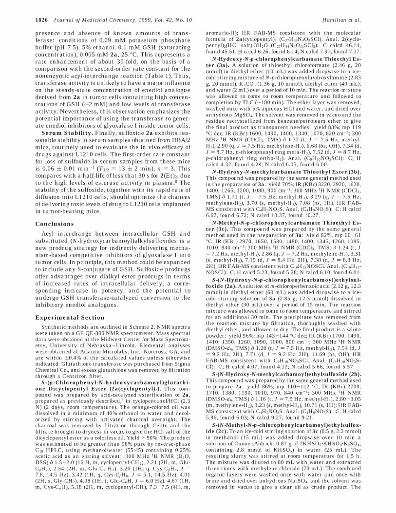
report were originally inspired by the observation thatintracellular sulfoxidation of herbicidal N,N-di-n-pro-pylcarbamate thioalkyl esters promotes their catabolismby facilitating acyl interchange with GSH.11 We rea-soned that sulfoxides of N-aryl/alkyl-N-hydroxycarbam-ate thioethyl esters might also undergo rapid acylinterchange with GSH, providing an efficient methodfor generating enediol analogue inhibitors of glyoxalaseI inside tumor cells. Indeed, sulfoxides 2a,b are at least106-fold more reactive than the thioester 3a towardGSH, on the basis of a comparison of the second-orderrate constants for the acyl-interchange reactions, Table1. The N-OH group inductively activates the sulfoxidesfor acyl interchange, as the rate constant for 2a exceedsthat of the corresponding N-CH3 derivative 2c by 13-fold. Ultraviolet and NMR spectroscopy confirmed the
products of the interchange reactions to be the corre-sponding enediol analogues.
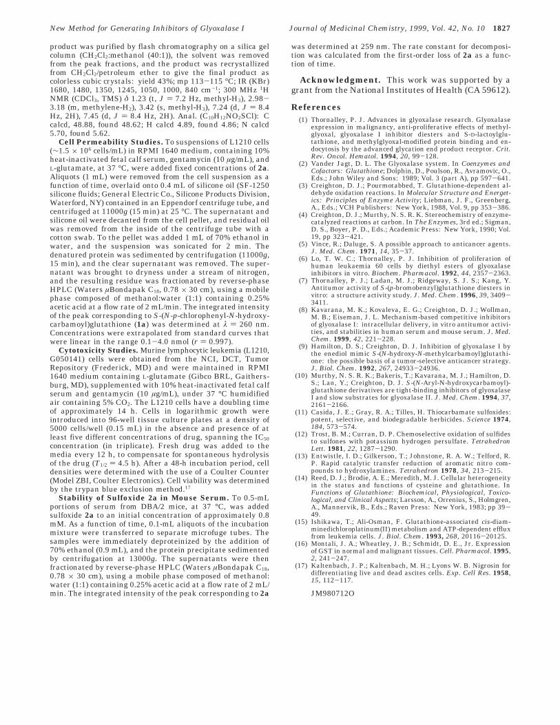
Cell Permeability Studies. Thus, enediol analogueshould rapidly form inside tumor cells incubated in thepresence of sulfoxide, provided that the sulfoxide candiffuse across the cell membrane and intracellular GSHis free to react with the sulfoxide. Indeed, incubation ofL1210 cells in the presence of 0.19 mM 2a results inthe rapid appearance of intracellular enediol analogue1a, Figure 2. Compound 1a was isolated from cellextracts by reverse-phase C18 column chromatographyand quantitated by interpolation from standard curves.The maximum concentration of 1a (1.6 mM) observedafter about 20 min is close to the reported concentrationof GSH in L1210 cells (1.9 mM),14 indicating that mostof the intracellular GSH is free to react with 2a.Moreover, the maximum intracellular concentration ofenediol analogue exceeds the extracellular concentrationof the sulfoxide by 8-fold, indicating that the rateconstant for efflux of the enediol analogue is much lessthan that for influx of the sulfoxide. Similarly, incuba-tion of the cells with 0.01 mM sulfoxide for 30 minresults in a 13-fold higher concentration of intracellular1a, Figure 2. Thus, the enediol analogue can be con-centrated inside tumor cells under conditions where the
Scheme 1. Acyl-Interchange Reaction betweenGlutathione (GSH) andS-(N-p-Chlorophenyl-N-hydroxycarbamoyl)ethylsulfoxide(2a) To Give Enediol Analogue 1a
Scheme 2. Synthetic Route to the Acylating ReagentsShown in Table 1a
a Reagents and conditions: (i) K2CO3/Et2O; (ii) for 3a,b, m-chloroperbenzoic acid was used; for 3c, Oxone (2KHSO5‚KHSO4‚K2HSO4) was used.
Table 1. Second-Order Rate Constants (k2) forAcyl-Interchange Reactions between Glutathione and SelectedCarbamoyl Estersa
CH3CH2XC(O)N(Y)Z + GSH f GSC(O)N(Y)Z + CH3CH2XH
substituentcarbamoylester X Y Z
k2(mM-1 min-1)
3a S OH C6H4Cl ∼3.5 × 10⁻⁷ b
2a S(O) OH C6H4Cl 1.84 ( 0.07c
2b S(O) OH CH3 0.59 ( 0.02c
2c S(O) CH3 C6H4Cl 0.14 ( 0.01c
a Conditions: potassium phosphate buffer (0.1 M), pH 7.5, 5%ethanol, 25 °C. b Calculated from the initial rate of appearance ofproduct (∼0.1% reaction) in the presence of a 15-fold excess ofglutathione, monitored by reverse-phase high-performance liquidchromatography. c Calculated from the first-order rate of loss ofreactants under pseudo-first-order conditions (g10-fold excess ofglutathione), monitored spectrophotometrically.
Figure 2. Time-dependent change in the intracellular con-centration of S-(N-p-chlorophenyl-N-hydroxycarbamoyl)glu-tathione (1a) in L1210 cells incubated in the presence of 0.19mM S-(N-p-chlorophenyl-N-hydroxycarbamoyl)ethylsulfoxide(2a) (0) and in the presence of 0.01 mM 2a (4). Error barsrepresent standard deviations for quadruplicate determina-tions. Conditions: RPMI 1640 medium containing 10% heat-inactivated fetal calf serum, 37 °C.
1824 Journal of Medicinal Chemistry, 1999, Vol. 42, No. 10 Hamilton et al.
extracellular concentration of sulfoxide is low. Theexplanation for the decrease in concentration of enediolanalogue after 20 min is not known but could involveseveral factors including enzymatic degradation and/orexport of the enediol analogue from the cells by theGSH-conjugate export pump that is present in L1210cells.15
The sulfoxide prodrug strategy is more efficient thanthe dialkyl ester prodrug strategy at delivering enediolanalogue 1a into L1210 cells. The apparent first-orderrate constant for formation of 1a in L1210 cells incu-bated with sulfoxide is 14-fold larger than that with thediethyl ester prodrug 1a(Et)2 and 2-fold larger than thatwith the dicyclopentyl ester 1a(cyclopentyl)2, Table 2.These rate constants were derived from the slopes ofplots of the initial rates of appearance of 1a inside thecells versus extracellular [prodrug], e.g., Figure 3.
Cytotoxicity Studies. Sulfoxide 2a is both cytostaticand cytotoxic to L1210 cells in tissue culture, asreflected in the GI50 and LC50 values obtained byinterpolation from the dose-response curve of Figure4. We believe that this most likely reflects rapid reactionof intracellular sulfoxide with GSH to give enediolanalogue, which induces elevated levels of methylgly-oxal in the L1210 cells by inhibiting glyoxalase I. Thetoxicity of the sulfoxide is unlikely to be due to depletionof intracellular GSH, because the total concentration ofGSH in L1210 cells is large in comparison to the valueof the growth inhibition parameter (GI50). As previously
noted, the minimum concentration of GSH in the L1210cells used in this study is 1.6 mM. Therefore, theconcentration of GSH will be reduced by less than 1%in the presence of a sulfoxide concentration correspond-ing to the GI50 (0.5 µM), assuming that the maximumachievable concentration of enediol analogue inside thecells will be about 10-fold greater than that of theextracellular sulfoxide. In principle, the toxicity of thesulfoxide might arise from acylation of critical sulfhy-dryl-containing proteins inside the cell. However, thispossibility seems less likely, given that the concentra-tion of free GSH inside the cells is orders of magnitudehigher than that of protein-associated sulfhydryl groups.Therefore, the sulfoxide is much more likely to reactwith GSH than with intracellular proteins. Importantly,the GI50 value for the sulfoxide is significantly lowerthan those of the diethyl ester 1a(Et)2 and dicyclopentylester 1a(cyclopentyl)2, Table 2. This can be explainedby the greater rate at which the sulfoxide enters thecells in comparison to the dialkyl esters.
Substrate Properties with GSH Transferase.N,N-Di-n-propyl ethylsulfoxide has been reported to bea substrate for the GSH transferase in plant cells.11 Thisprompted us to examine the kinetic properties of sul-foxide 2a with the π isozyme of GSH transferase fromhuman placenta. Multidrug-resistant tumors of thecolon, lung, and breast commonly overexpress the πisozyme of GSH transferase.16 Therefore, resistanttumors might be exceptionally sensitive to sulfoxidessuch as 2a, provided that the sulfoxides are catalyticallyconverted to the inhibitory enediol analogue.
Sulfoxide 2a proved to be a poor substrate for thetransferase from human placenta with an estimated kcat/Km value of 54 mM-1 min-1. This value was derivedfrom the first-order rates of loss of 2a (∆OD260) in the
Table 2. Apparent First-Order Rate Constants for Delivery ofEnediol Analogue 1a into L1210 Cells via Different Prodrugsand Comparative Toxicities of the Prodrugs to L1210 Cellsa
prodrugrate constant
(min-1)bGI50(µM)c
TGI(µM)c
LC50(µM)c
2a 1.41 ( 0.03 0.5 ( 0.1 3.9 ( 0.4 13 ( 81a(cyclopentyl)2 0.59 ( 0.01 3.4 ( 0.1 5.3 ( 0.1 8.3 ( 1.01a(Et)2 0.10 ( 0.01d 7 ( 3d 27 ( 10d 54 ( 5d
a The mean and standard deviations for the cell toxicityparameters for each prodrug were calculated from three differentexperiments carried out on different days. Conditions: RPMI 1640/10% fetal calf serum, 37 °C. b Derived from the slopes of plots ofinitial rates of appearance of the inhibitory diacid 2 inside L1210cells versus the extracellular concentration of prodrug. c Definedin the legend to Figure 4. d From ref 8.
Figure 3. Initial rates of appearance of intracellular S-(N-p-chlorophenyl-N-hydroxycarbamoyl)glutathione (1a) in L1210cells as a function of the extracellular concentration of sulfox-ide 2a. Error bars represent standard deviations for triplicatedeterminations. Conditions: RPMI 1640 medium containing10% heat-inactivated fetal calf serum, 37 °C.
Figure 4. Growth inhibition of L1210 cells by S-(N-p-chlorophenyl-N-hydroxycarbamoyl)ethylsulfoxide (2a) (RPMI1640/10% fetal calf serum, 37 °C, 48 h). Percentage growth(PG) was calculated from the following relationships: When(Ftest - Ftzero) g 0, PG ) 100 × (Ftest - Ftzero)/(Fctrl - Ftzero), whereFtzero ) cell density prior to exposure to drug, Ftest ) cell densityafter 48 h exposure to drug, Fctrl ) cell density after 48 h withno exposure to drug. When (Ftest - Ftzero) < 0, PG ) 100 × (Ftest
- Ftzero)/Ftzero. Different symbols correspond to experimentscarried out on different days. Error bars represent standarddeviations for triplicate determinations. The toxicity param-eters were obtained by interpolation from the dose-responsecurve and are defined as follows: GI50, concentration producing50% growth inhibition in comparison to no-drug controls; TGI,concentration producing 100% growth inhibition; LC50, con-centration producing 50% cell killing.
New Method for Generating Inhibitors of Glyoxalase I Journal of Medicinal Chemistry, 1999, Vol. 42, No. 10 1825
presence and absence of known amounts of trans-ferase: conditions of 0.09 mM potassium phosphatebuffer (pH 7.5), 5% ethanol, 0.1 mM GSH (saturatingconcentration), 0.005 mM 2a, 25 °C. This represents arate enhancement of about 30-fold, on the basis of acomparison with the second-order rate constant for thenonenzymic acyl-interchange reaction (Table 1). Thus,transferase activity is unlikely to have a major influenceon the steady-state concentration of enediol analoguederived from 2a in tumor cells containing high concen-trations of GSH (∼2 mM) and low levels of transferaseactivity. Nevertheless, this observation emphasizes thepotential importance of using the transferase to gener-ate enediol inhibitors of glyoxalase I inside tumor cells.
Serum Stability. Finally, sulfoxide 2a exhibits rea-sonable stability in serum samples obtained from DBA/2mice, routinely used to evaluate the in vivo efficacy ofdrugs against L1210 cells. The first-order rate constantfor loss of sulfoxide in serum samples from these miceis 0.06 ( 0.01 min-1 (T1/2 ) 13 ( 2 min), n ) 3. Thiscompares with a half-life of less that 30 s for 2(Et)2, dueto the high levels of esterase activity in plasma.8 Thestability of the sulfoxide, together with its rapid rate ofdiffusion into L1210 cells, should optimize the chancesof delivering toxic levels of drug to L1210 cells implantedin tumor-bearing mice.
Conclusions
Acyl interchange between intracellular GSH andsubstituted (N-hydroxycarbamoyl)alkylsulfoxides is anew prodrug strategy for indirectly delivering mecha-nism-based competitive inhibitors of glyoxalase I intotumor cells. In principle, this method could be expandedto include any S-conjugate of GSH. Sulfoxide prodrugsoffer advantages over dialkyl ester prodrugs in termsof increased rates of intracellular delivery, a corre-sponding increase in potency, and the potential toundergo GSH transferase-catalyzed conversion to theinhibitory enediol analogues.
Experimental SectionSynthetic methods are outlined in Scheme 2. NMR spectra
were taken on a GE QE-300 NMR spectrometer. Mass spectraldata were obtained at the Midwest Center for Mass Spectrom-etry, University of Nebraska-Lincoln. Elemental analyseswere obtained at Atlantic Microlabs, Inc., Norcross, GA, andare within (0.4% of the calculated values unless otherwiseindicated. Glutathione transferase was purchased from SigmaChemical Co., and excess glutathione was removed by filtrationthrough a Centricon filter.
S-(p-Chlorophenyl-N-hydroxycarbamoyl)glutathi-one Dicyclopentyl Ester (2a(cyclopentyl)2). This com-pound was prepared by acid-catalyzed esterification of 2a,prepared as previously described,8 in cyclopentanol/HCl (2.3N) (2 days, room temperature). The orange-colored oil wasdissolved in a minimum of 40% ethanol in water and decol-orized by stirring with activated charcoal overnight. Thecharcoal was removed by filtration through Celite and thefiltrate brought to dryness in vacuo to give the HCl salt of thedicyclopentyl ester as a colorless oil. Yield > 90%. The productwas estimated to be greater than 98% pure by reverse-phaseC18 HPLC, using methanol/water (55:45) containing 0.25%acetic acid as an eluting solvent: 300 MHz 1H NMR (D2O,DSS) δ 1.5-2.0 (16 H, m, cyclopentyl-CH2), 2.21 (2H, m, Glu-CâH2), 2.54 (2H, m, Glu-Cγ H2), 3.20 (1H, q, Cys-CâHa, J )7.8, 14.5 Hz), 3.42 (1H, q, Cys-CâHb, J ) 5.1, 14.5 Hz), 4.01(2H, s, Gly-CH2), 4.08 (1H, t, Glu-CRH, J ) 6.0 Hz), 4.67 (1H,m, Cys-CRH), 5.18 (2H, m, cyclopentyl-CH), 7.3-7.5 (4H, m,
aromatic-H); HR FAB-MS consistent with the molecularformula of 2a(cyclopentyl)2 (C27H38N4O8SCl). Anal. 2(cyclo-pentyl)2(HCl salt)‚3H2O (C27H44N4O11SCl2): C calcd 46.14,found 45.51; H calcd 6.26, found 6.14; N calcd 7.97, found 7.17.
N-Hydroxy-N-p-chlorophenylcarbamate Thioethyl Es-ter (3a). A solution of thioethyl chloroformate (2.46 g, 20mmol) in diethyl ether (10 mL) was added dropwise to a ice-cold stirring mixture of N-p-chlorophenylhydroxylamine (2.83g, 20 mmol), K2CO3 (1.36 g, 10 mmol), diethyl ether (40 mL),and water (2 mL) over a period of 10 min. The reaction mixturewas allowed to come to room temperature and followed tocompletion by TLC (∼180 min). The ether layer was removed,washed once with 5% aqueous HCl and water, and dried overanhydrous MgSO4. The solvent was removed in vacuo and theresidue recrystallized from benzene/petroleum ether to givethe final product as transparent needles: yield 83%; mp 119°C dec; IR (KBr) 1600, 1490, 1400, 1340, 1070, 820 cm-1; 300MHz 1H NMR (CDCl3, TMS) δ 1.32 (t, J ) 7.5 Hz, methyl-H3), 2.90 (q, J ) 7.5 Hz, methylene-H2), 6.68 (bs, OH), 7.34 (d,J ) 8.7 Hz, p-chlorophenyl ring meta-H2), 7.52 (d, J ) 8.7 Hz,p-chlorophenyl ring ortho-H2). Anal. (C9H10NO2SCl): C; Hcalcd 4.32, found 4.29; N calcd 6.05, found 6.00.
N-Hydroxy-N-methylcarbamate Thioethyl Ester (3b).This compound was prepared by the same general method usedin the preparation of 3a: yield 70%; IR (KBr) 3220, 2920, 1620,1400, 1265, 1200, 1080, 900 cm-1; 300 MHz 1H NMR (CDCl3,TMS) δ 1.71 (t, J ) 7.5 Hz, methyl-H3), 3.29 (q, J ) 7.5 Hz,methylene-H2), 3.70 (s, methyl-H3), 7.08 (bs, 1H), HR FAB-MS consistent with C4H9NO2S. Anal. (C4H9NO2S): C; H calcd6.67, found 6.72; N calcd 10.37, found 10.27.
N-Methyl-N-p-chlorophenylcarbamate Thioethyl Es-ter (3c). This compound was prepared by the same generalmethod used in the preparation of 3a: yield 82%, mp 60-61°C; IR (KBr) 2970, 1650, 1580, 1480, 1400, 1345, 1260, 1085,1010, 840 cm-1; 300 MHz 1H NMR (CDCl3, TMS) δ 1.24 (t, J) 7.2 Hz, methyl-H3), 2.86 (q, J ) 7.2 Hz, methylene-H2), 3.31(s, methyl-H3), 7.19 (d, J ) 8.4 Hz, 2H), 7.38 (d, J ) 8.8 Hz,2H); HR FAB-MS consistent with C10H12NOSCl. Anal. (C10H12-NOSCl): C; H calcd 5.23, found 5.28; N calcd 6.10, found 6.01.
S-(N-Hydroxy-N-p-chlorophenylcarbamoyl)ethylsul-foxide (2a). A solution of m-chloroperbenzoic acid (2.12 g, 12.3mmol) in diethyl ether (60 mL) was added dropwise to a ice-cold stirring solution of 3a (2.85 g, 12.3 mmol) dissolved indiethyl ether (30 mL) over a period of 15 min. The reactionmixture was allowed to come to room temperature and stirredfor an additional 30 min. The precipitate was removed fromthe reaction mixture by filtration, thoroughly washed withdiethyl ether, and allowed to dry. The final product is a whitepowder: yield 96%; mp 143-144 °C dec; IR (KBr) 1700, 1490,1410, 1350, 1260, 1090, 1000, 800 cm-1; 300 MHz 1H NMR(DMSO-d6, TMS) δ 1.20 (t, J ) 7.5 Hz, methyl-H3), 7.54 (d, J) 9.2 Hz, 2H), 7.71 (d, J ) 9.2 Hz, 2H), 11.69 (bs, OH); HRFAB-MS consistent with C9H10NO3SCl. Anal. (C9H10NO3S-Cl): C; H calcd 4.07, found 4.12; N calcd 5.66, found 5.57.
S-(N-Hydroxy-N-methylcarbamoyl)ethylsulfoxide (2b).This compound was prepared by the same general method usedto prepare 2a: yield 86%; mp 110-112 °C; IR (KBr) 2700,1710, 1380, 1190, 1010, 970, 840 cm-1; 300 MHz 1H NMR(DMSO-d6, TMS) δ 1.16 (t, J ) 7.5 Hz, methyl-H3), 2.80-3.05(m, methylene-H2), 3.23 (s, methyl-H3), 10.71 (s, 1H); HR FAB-MS consistent with C4H9NO3S. Anal. (C4H9NO3S): C; H calcd5.96, found 6.03; N calcd 9.27, found 9.21.
S-(N-Methyl-N-p-chlorophenylcarbamoyl)ethylsulfox-ide (2c). To an ice-cold stirring solution of 3c (0.5 g, 2.2 mmol)in methanol (15 mL) was added dropwise over 10 min asolution of Oxone (Aldrich; 0.87 g of 2KHSO5‚KHSO4‚K2SO4,containing 2.8 mmol of KHSO5) in water (25 mL). Theresulting slurry was stirred at room temperature for 1.5 h.The mixture was diluted to 80 mL with water and extractedthree times with methylene chloride (70 mL). The combinedorganic layers were washed once with water and once withbrine and dried over anhydrous Na2SO4, and the solvent wasremoved in vacuo to give a clear oil as crude product. The
1826 Journal of Medicinal Chemistry, 1999, Vol. 42, No. 10 Hamilton et al.
product was purified by flash chromatography on a silica gelcolumn (CH2Cl2:methanol (40:1)), the solvent was removedfrom the peak fractions, and the product was recrystallizedfrom CH2Cl2/petroleum ether to give the final product ascolorless cubic crystals: yield 43%; mp 113-115 °C; IR (KBr)1680, 1480, 1350, 1245, 1050, 1000, 840 cm-1; 300 MHz 1HNMR (CDCl3, TMS) δ 1.23 (t, J ) 7.2 Hz, methyl-H3), 2.98-3.18 (m, methylene-H2), 3.42 (s, methyl-H3), 7.24 (d, J ) 8.4Hz, 2H), 7.45 (d, J ) 8.4 Hz, 2H). Anal. (C10H12NO2SCl): Ccalcd, 48.88, found 48.62; H calcd 4.89, found 4.86; N calcd5.70, found 5.62.
Cell Permeability Studies. To suspensions of L1210 cells(∼1.5 × 10⁶ cells/mL) in RPMI 1640 medium, containing 10%heat-inactivated fetal calf serum, gentamycin (10 µg/mL), andL-glutamate, at 37 °C, were added fixed concentrations of 2a.Aliquots (1 mL) were removed from the cell suspension as afunction of time, overlaid onto 0.4 mL of silicone oil (SF-1250silicone fluids; General Electric Co., Silicone Products Division,Waterford, NY) contained in an Eppendorf centrifuge tube, andcentrifuged at 11000g (15 min) at 25 °C. The supernatant andsilicone oil were decanted from the cell pellet, and residual oilwas removed from the inside of the centrifuge tube with acotton swab. To the pellet was added 1 mL of 70% ethanol inwater, and the suspension was sonicated for 2 min. Thedenatured protein was sedimented by centrifugation (11000g,15 min), and the clear supernatant was removed. The super-natant was brought to dryness under a stream of nitrogen,and the resulting residue was fractionated by reverse-phaseHPLC (Waters µBondapak C18, 0.78 × 30 cm), using a mobilephase composed of methanol:water (1:1) containing 0.25%acetic acid at a flow rate of 2 mL/min. The integrated intensityof the peak corresponding to S-(N-p-chlorophenyl-N-hydroxy-carbamoyl)glutathione (1a) was determined at λ ) 260 nm.Concentrations were extrapolated from standard curves thatwere linear in the range 0.1-4.0 nmol (r ) 0.997).
Cytotoxicity Studies. Murine lymphocytic leukemia (L1210,G050141) cells were obtained from the NCI, DCT, TumorRepository (Frederick, MD) and were maintained in RPMI1640 medium containing L-glutamate (Gibco BRL, Gaithers-burg, MD), supplemented with 10% heat-inactivated fetal calfserum and gentamycin (10 µg/mL), under 37 °C humidifiedair containing 5% CO2. The L1210 cells have a doubling timeof approximately 14 h. Cells in logarithmic growth wereintroduced into 96-well tissue culture plates at a density of5000 cells/well (0.15 mL) in the absence and presence of atleast five different concentrations of drug, spanning the IC50
concentration (in triplicate). Fresh drug was added to themedia every 12 h, to compensate for spontaneous hydrolysisof the drug (T1/2 ) 4.5 h). After a 48-h incubation period, celldensities were determined with the use of a Coulter Counter(Model ZBI, Coulter Electronics). Cell viability was determinedby the trypan blue exclusion method.17
Stability of Sulfoxide 2a in Mouse Serum. To 0.5-mLportions of serum from DBA/2 mice, at 37 °C, was addedsulfoxide 2a to an initial concentration of approximately 0.8mM. As a function of time, 0.1-mL aliquots of the incubationmixture were transferred to separate microfuge tubes. Thesamples were immediately deproteinized by the addition of70% ethanol (0.9 mL), and the protein precipitate sedimentedby centrifugation at 13000g. The supernatants were thenfractionated by reverse-phase HPLC (Waters µBondapak C18,0.78 × 30 cm), using a mobile phase composed of methanol:water (1:1) containing 0.25% acetic acid at a flow rate of 2 mL/min. The integrated intensity of the peak corresponding to 2a
was determined at 259 nm. The rate constant for decomposi-tion was calculated from the first-order loss of 2a as a func-tion of time.
Acknowledgment. This work was supported by agrant from the National Institutes of Health (CA 59612).
References(1) Thornalley, P. J. Advances in glyoxalase research. Glyoxalase
expression in malignancy, anti-proliferative effects of methyl-glyoxal, glyoxalase I inhibitor diesters and S-D-lactoylglu-tathione, and methylglyoxal-modified protein binding and en-docytosis by the advanced glycation end product receptor. Crit.Rev. Oncol. Hematol. 1994, 20, 99-128.
(2) Vander Jagt, D. L. The Glyoxalase system. In Coenzymes andCofactors: Glutathione; Dolphin, D., Poulson, R., Avramovic, O.,Eds.; John Wiley and Sons: 1989; Vol. 3 (part A), pp 597-641.
(3) Creighton, D. J.; Pourmotabbed, T. Glutathione-dependent al-dehyde oxidation reactions. In Molecular Structure and Energet-ics: Principles of Enzyme Activity; Liebman, J. F., Greenberg,A., Eds.; VCH Publishers: New York, 1988, Vol. 9, pp 353-386.
(4) Creighton, D. J.; Murthy, N. S. R. K. Stereochemistry of enzyme-catalyzed reactions at carbon. In The Enzymes, 3rd ed.; Sigman,D. S., Boyer, P. D., Eds.; Academic Press: New York, 1990; Vol.19, pp 323-421.
(5) Vince, R.; Daluge, S. A possible approach to anticancer agents.J. Med. Chem. 1971, 14, 35-37.
(6) Lo, T. W. C.; Thornalley, P. J. Inhibition of proliferation ofhuman leukaemia 60 cells by diethyl esters of glyoxalaseinhibitors in vitro. Biochem. Pharmacol. 1992, 44, 2357-2363.
(7) Thornalley, P. J.; Ladan, M. J.; Ridgeway, S. J. S.; Kang, Y.Antitumor activity of S-(p-bromobenzyl)glutathione diesters invitro: a structure activity study. J. Med. Chem. 1996, 39, 3409-3411.
(8) Kavarana, M. K.; Kovaleva, E. G.; Creighton, D. J.; Wollman,M. B.; Eiseman, J. L. Mechanism-based competitive inhibitorsof glyoxalase I: intracellular delivery, in vitro antitumor activi-ties, and stabilities in human serum and mouse serum. J. Med.Chem. 1999, 42, 221-228.
(9) Hamilton, D. S.; Creighton, D. J. Inhibition of glyoxalase I bythe enediol mimic S-(N-hydroxy-N-methylcarbamoyl)glutathi-one: the possible basis of a tumor-selective anticancer strategy.J. Biol. Chem. 1992, 267, 24933-24936.
(10) Murthy, N. S. R. K.; Bakeris, T.; Kavarana, M. J.; Hamilton, D.S.; Lan, Y.; Creighton, D. J. S-(N-Aryl-N-hydroxycarbamoyl)-glutathione derivatives are tight-binding inhibitors of glyoxalaseI and slow substrates for glyoxalase II. J. Med. Chem. 1994, 37,2161-2166.
(11) Casida, J. E.; Gray, R. A.; Tilles, H. Thiocarbamate sulfoxides:potent, selective, and biodegradable herbicides. Science 1974,184, 573-574.
(12) Trost, B. M.; Curran, D. P. Chemoselective oxidation of sulfidesto sulfones with potassium hydrogen persulfate. TetrahedronLett. 1981, 22, 1287-1290.
(13) Entwistle, I. D.; Gilkerson, T.; Johnstone, R. A. W.; Telford, R.P. Rapid catalytic transfer reduction of aromatic nitro com-pounds to hydroxylamines. Tetrahedron 1978, 34, 213-215.
(14) Reed, D. J.; Brodie, A. E.; Meredith, M. J. Cellular heterogeneityin the status and functions of cysteine and glutathione. InFunctions of Glutathione: Biochemical, Physiological, Toxico-logical, and Clinical Aspects; Larsson, A., Orrenius, S., Holmgren,A., Mannervik, B., Eds.; Raven Press: New York, 1983; pp 39-49.
(15) Ishikawa, T.; Ali-Osman, F. Glutathione-associated cis-diam-minedichloroplatinum(II) metabolism and ATP-dependent effluxfrom leukemia cells. J. Biol. Chem. 1993, 268, 20116-20125.
(16) Montali, J. A.; Wheatley, J. B.; Schmidt, D. E., Jr. Expressionof GST in normal and malignant tissues. Cell. Pharmacol. 1995,2, 241-247.
(17) Kaltenbach, J. P.; Kaltenbach, M. H.; Lyons W. B. Nigrosin fordifferentiating live and dead ascites cells. Exp. Cell Res. 1958,15, 112-117.
JM980712O
New Method for Generating Inhibitors of Glyoxalase I Journal of Medicinal Chemistry, 1999, Vol. 42, No. 10 1827


![4β-[4’-(1-(Aryl)ureido)benzamide]podophyllotoxins as DNA ... · 1 4 -[4 -(1-(Aryl)ureido)benzamide]podophyllotoxins as DNA Topoisomerase I and IIα Inhibitors and Apoptosis Inducing](https://img.pdfslide.net/doc/110x75/5e7ee9e4bc140f3b9414d72f/4-4a-1-arylureidobenzamidepodophyllotoxins-as-dna-1-4-4-1-arylureidobenzamidepodophyllotoxins.jpg)


![5-Dibromo-3-hexylthiophene via Suzuki Cross Coupling ... · anti-depressant, BASE1 inhibitors, anti-HIV PR inhibitors and anti-breast cancer activities [4,5]. In ... Entry Aryl Boronic](https://img.pdfslide.net/doc/110x75/5fb3c95e19b64f0bb5481afb/5-dibromo-3-hexylthiophene-via-suzuki-cross-coupling-anti-depressant-base1.jpg)