Embed Size (px)
Citation preview
RESEARCH ARTICLE Open Access
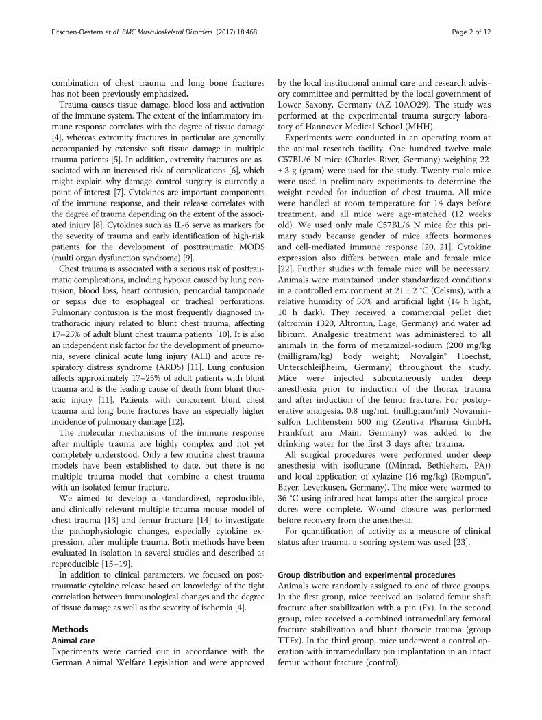
A new multiple trauma model of themouseStefanie Fitschen-Oestern1*, Sebastian Lippross1, Tim Klueter1, Matthias Weuster1, Deike Varoga1,Mersedeh Tohidnezhad2, Thomas Pufe2, Stefan Rose-John3, Hagen Andruszkow4, Frank Hildebrand4,Nadine Steubesand1, Andreas Seekamp1 and Claudia Neunaber4
Abstract
Background: Blunt trauma is the most frequent mechanism of injury in multiple trauma, commonly resultingfrom road traffic collisions or falls. Two of the most frequent injuries in patients with multiple trauma are chesttrauma and extremity fracture. Several trauma mouse models combine chest trauma and head injury, but notrauma mouse model to date includes the combination of long bone fractures and chest trauma. Outcome isessentially determined by the combination of these injuries. In this study, we attempted to establish areproducible novel multiple trauma model in mice that combines blunt trauma, major injuries and simplepracticability.
Methods: Ninety-six male C57BL/6 N mice (n = 8/group) were subjected to trauma for isolated femur fractureand a combination of femur fracture and chest injury. Serum samples of mice were obtained by heart punctureat defined time points of 0 h (hour), 6 h, 12 h, 24 h, 3 d (days), and 7 d.
Results: A tendency toward reduced weight and temperature was observed at 24 h after chest trauma andfemur fracture. Blood analyses revealed a decrease in hemoglobin during the first 24 h after trauma. Someanimals were killed by heart puncture immediately after chest contusion; these animals showed the most severelung contusion and hemorrhage. The extent of structural lung injury varied in different mice but was evident inall animals. Representative H&E-stained (Haematoxylin and Eosin-stained) paraffin lung sections of mice withmultiple trauma revealed hemorrhage and an inflammatory immune response. Plasma samples of mice withchest trauma and femur fracture showed an up-regulation of IL-1β (Interleukin-1β), IL-6, IL-10, IL-12p70 and TNF-α(Tumor necrosis factor- α) compared with the control group. Mice with femur fracture and chest trauma showeda significant up-regulation of IL-6 compared to group with isolated femur fracture.
Conclusions: The multiple trauma mouse model comprising chest trauma and femur fracture enables manyanalogies to clinical cases of multiple trauma in humans and demonstrates associated characteristic clinical andpathophysiological changes. This model is easy to perform, is economical and can be used for further researchexamining specific immunological questions.
BackgroundMultiple trauma accounts for a significant proportion ofdeaths worldwide [1]. The most frequent injuries intrauma patients are chest trauma, extremity fracturesand head injuries [2].Blunt chest trauma can result in significant morbidity
in injured patients, and both chest wall and intrathoracic
visceral injuries can lead to life-threatening complica-tions if not anticipated and treated [3]. Extremityfractures such as a femur fracture must be stabilized.The time point of operative treatment is still contro-
versially discussed, although most of the literaturerecommends early surgical stabilization of thesefractures. Respiratory deterioration can be exacerbatedby the presence of unstable long bone fractures.Several trauma mouse models focus on blunt chest
trauma and head injury, but to our knowledge the
* Correspondence: [email protected] of Trauma Surgery, University Medical Center ofSchleswig-Holstein, Arnold-Heller Straße 7, 24105, Campus Kiel, Kiel, GermanyFull list of author information is available at the end of the article
© The Author(s). 2017 Open Access This article is distributed under the terms of the Creative Commons Attribution 4.0International License (http://creativecommons.org/licenses/by/4.0/), which permits unrestricted use, distribution, andreproduction in any medium, provided you give appropriate credit to the original author(s) and the source, provide a link tothe Creative Commons license, and indicate if changes were made. The Creative Commons Public Domain Dedication waiver(http://creativecommons.org/publicdomain/zero/1.0/) applies to the data made available in this article, unless otherwise stated.
Fitschen-Oestern et al. BMC Musculoskeletal Disorders (2017) 18:468 DOI 10.1186/s12891-017-1813-9
combination of chest trauma and long bone fractureshas not been previously emphasized.Trauma causes tissue damage, blood loss and activation
of the immune system. The extent of the inflammatory im-mune response correlates with the degree of tissue damage[4], whereas extremity fractures in particular are generallyaccompanied by extensive soft tissue damage in multipletrauma patients [5]. In addition, extremity fractures are as-sociated with an increased risk of complications [6], whichmight explain why damage control surgery is currently apoint of interest [7]. Cytokines are important componentsof the immune response, and their release correlates withthe degree of trauma depending on the extent of the associ-ated injury [8]. Cytokines such as IL-6 serve as markers forthe severity of trauma and early identification of high-riskpatients for the development of posttraumatic MODS(multi organ dysfunction syndrome) [9].Chest trauma is associated with a serious risk of posttrau-
matic complications, including hypoxia caused by lung con-tusion, blood loss, heart contusion, pericardial tamponadeor sepsis due to esophageal or tracheal perforations.Pulmonary contusion is the most frequently diagnosed in-trathoracic injury related to blunt chest trauma, affecting17–25% of adult blunt chest trauma patients [10]. It is alsoan independent risk factor for the development of pneumo-nia, severe clinical acute lung injury (ALI) and acute re-spiratory distress syndrome (ARDS) [11]. Lung contusionaffects approximately 17–25% of adult patients with blunttrauma and is the leading cause of death from blunt thor-acic injury [11]. Patients with concurrent blunt chesttrauma and long bone fractures have an especially higherincidence of pulmonary damage [12].The molecular mechanisms of the immune response
after multiple trauma are highly complex and not yetcompletely understood. Only a few murine chest traumamodels have been established to date, but there is nomultiple trauma model that combine a chest traumawith an isolated femur fracture.We aimed to develop a standardized, reproducible,
and clinically relevant multiple trauma mouse model ofchest trauma [13] and femur fracture [14] to investigatethe pathophysiologic changes, especially cytokine ex-pression, after multiple trauma. Both methods have beenevaluated in isolation in several studies and described asreproducible [15–19].In addition to clinical parameters, we focused on post-
traumatic cytokine release based on knowledge of the tightcorrelation between immunological changes and the degreeof tissue damage as well as the severity of ischemia [4].
MethodsAnimal careExperiments were carried out in accordance with theGerman Animal Welfare Legislation and were approved
by the local institutional animal care and research advis-ory committee and permitted by the local government ofLower Saxony, Germany (AZ 10AO29). The study wasperformed at the experimental trauma surgery labora-tory of Hannover Medical School (MHH).Experiments were conducted in an operating room at
the animal research facility. One hundred twelve maleC57BL/6 N mice (Charles River, Germany) weighing 22± 3 g (gram) were used for the study. Twenty male micewere used in preliminary experiments to determine theweight needed for induction of chest trauma. All micewere handled at room temperature for 14 days beforetreatment, and all mice were age-matched (12 weeksold). We used only male C57BL/6 N mice for this pri-mary study because gender of mice affects hormonesand cell-mediated immune response [20, 21]. Cytokineexpression also differs between male and female mice[22]. Further studies with female mice will be necessary.Animals were maintained under standardized conditionsin a controlled environment at 21 ± 2 °C (Celsius), with arelative humidity of 50% and artificial light (14 h light,10 h dark). They received a commercial pellet diet(altromin 1320, Altromin, Lage, Germany) and water adlibitum. Analgesic treatment was administered to allanimals in the form of metamizol-sodium (200 mg/kg(milligram/kg) body weight; Novalgin® Hoechst,Unterschleiβheim, Germany) throughout the study.Mice were injected subcutaneously under deepanesthesia prior to induction of the thorax traumaand after induction of the femur fracture. For postop-erative analgesia, 0.8 mg/mL (milligram/ml) Novamin-sulfon Lichtenstein 500 mg (Zentiva Pharma GmbH,Frankfurt am Main, Germany) was added to thedrinking water for the first 3 days after trauma.All surgical procedures were performed under deep
anesthesia with isoflurane ((Minrad, Bethlehem, PA))and local application of xylazine (16 mg/kg) (Rompun®,Bayer, Leverkusen, Germany). The mice were warmed to36 °C using infrared heat lamps after the surgical proce-dures were complete. Wound closure was performedbefore recovery from the anesthesia.For quantification of activity as a measure of clinical
status after trauma, a scoring system was used [23].
Group distribution and experimental proceduresAnimals were randomly assigned to one of three groups.In the first group, mice received an isolated femur shaftfracture after stabilization with a pin (Fx). In the secondgroup, mice received a combined intramedullary femoralfracture stabilization and blunt thoracic trauma (groupTTFx). In the third group, mice underwent a control op-eration with intramedullary pin implantation in an intactfemur without fracture (control).
Fitschen-Oestern et al. BMC Musculoskeletal Disorders (2017) 18:468 Page 2 of 12
Group Fx and TTFx were divided into six subgroups(n = 8) depending on the time point of sacrifice: 0 h, 6 h,12 h, 24 h, 3 d and 7 d. The control group was sacrificedat 0 h (n = 8).One hundred twelve multiple trauma, femur fracture
and control mice were tested (48 multiple trauma and48 isolated femur fracture). The control group consistedof 16 mice (16,6% (percent)) that underwent an oper-ation (femur stabilization) in the absence of fracture orchest trauma (Fig. 1c). Experiments were undertaken bythree different surgeons. There were no significant dif-ferences with regard to moribund animals (surgeon 1: 6moribund mice, surgeon 2: 4 moribund mice, surgeon 3:6 moribund mice).
Blunt thoracic traumaAfter induction of the femur fracture, a blunt thoracictrauma was induced by a modified version of a previ-ously described model for rats of bilateral lung contu-sion in the TTFx group [13, 24]. The method has beenpreviously described as reproducible.Blunt thoracic trauma was induced in anesthetized
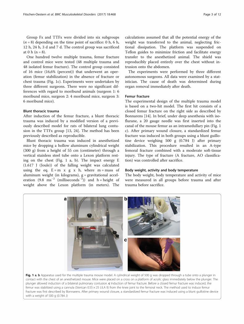
mice by dropping a hollow aluminum cylindrical weight(300 g) from a height of 55 cm (centimeter) through avertical stainless steel tube onto a Lexon platform rest-ing on the chest (Fig. 1 a, b). The impact energy E(1.617 J (Joule)) of the falling weight was calculatedusing the eq. E =m x g x h, where m =mass ofaluminum weight (in kilograms), g = gravitational accel-eration (9.8 ms−2 (milliseconds−2)) and h = height ofweight above the Lexon platform (in meters). The
calculations assumed that all the potential energy of theweight was transferred to the animal, neglecting fric-tional dissipation. The platform was suspended onTeflon guides to minimize friction and facilitate energytransfer to the anesthetized animal. The shield wasreproducibly placed entirely over the chest without in-trusion onto the abdomen.The experiments were performed by three different
autonomous surgeons. All data were examined by a stat-istician. The cause of death was determined duringorgan removal immediately after death.
Femur fractureThe experimental design of the multiple trauma modelis based on a two-hit model. The first hit consists of aclosed femur fracture on the right side as described byBonnarens [14]. In brief, under deep anesthesia with iso-flurane, a 20 gauge needle was first inserted into thecanal of the mouse femur as an intramedullary pin (Fig. 1c). After primary wound closure, a standardized femurfracture was induced in both groups using a blunt guillo-tine device weighing 500 g (0.784 J) after primarystabilization. This procedure resulted in an A-typefemoral fracture combined with a moderate soft-tissueinjury. The type of fracture (A fracture, AO classifica-tion) was controlled after sacrifice.
Body weight, activity and body temperatureThe body weight, body temperature and activity of micewere measured in all groups before trauma and aftertrauma before sacrifice.
Fig. 1 a, b Apparatus used for the multiple trauma mouse model. A cylindrical weight of 500 g was dropped through a tube onto a plunger incontact with the chest of an anesthetized mouse. Mice were placed on a cross on a platform of acrylic glass immediately below the plunger. Theplunger allowed induction of a bilateral pulmonary contusion. c Induction of femur fracture. Before a closed femur fracture was induced, thefemur was stabilized using a cannula (Sterican 0.55 × 25 LILA lI) from the knee joint to the femoral neck. The method used to induce femurfracture was first described by Bonnarens. After primary wound closure, a standardized femur fracture was induced using a blunt guillotine devicewith a weight of 500 g (0.784 J)
Fitschen-Oestern et al. BMC Musculoskeletal Disorders (2017) 18:468 Page 3 of 12
Assessment of blood parametersSamples for the control group were collected using theretrobulbar technique during the preliminary test. Post-traumatic control was performed by heart puncture. Byusing 2-mL (milliliter) syringes (Pico50, RadiometerMedical, Brønshøj, Denmark) containing 80 IUelectrolyte-balanced heparin, blood samples (0.7 ml) forblood gas analysis and assessment of marker enzyme ac-tivities were collected from heart. The animals weresacrificed by heart puncture under deep anesthesia. Thehemoglobin concentration, hematocrit and metabolicparameters (lactate, glucose), and osmolality wereassessed using a blood gas analyzer (ABL 715, Radiom-eter, Copenhagen, Denmark).
SpecimensAnimals were sacrificed immediately after trauma, after6 h, 12 h, 24 h, 3 d and 7 d to obtain samples for histo-logic examination. Tissue samples from lung werecollected and stored at −20 °C until processed.
HistologyTissue samples were embedded in paraffin. Sections (5 μm(micrometer)) were obtained from the central portion ofthe lung with a sliding microtome (HM 430; MicromInternational), placed on Superfrost Plus microscope slides(Thermo Scientific) and incubated overnight at 60 °C. Thesections were routinely stained with hematoxylin and eosin(H&E). Safranin O staining was carried out for 6 min usinga 0.1% aqueous solution at pH 3.0.
Micro computed tomographyChest trauma of multiple trauma mice was assessed bymicro computed tomography (μCT).The CT scan was performed at the Molecular
Imaging North Competence Center (Am BotanischenGarten 14, 24,118 Kiel). Micro computed tomographyin Kiel has been applied previously to mice in severalstudies [25, 26]. Two radiologists planed every scan.The total scan time was approximately 14 min. Scan-
ning of mice lungs has been described previously [27].The lungs of mice, which were killed immediately aftertrauma, were scanned using a Novotec MicroScope(Novotec Medical GmbH, Pforzheim) at an isotropicnominal spatial resolution (voxel size) of 15–20 μm.Samples were transported on ice before the scannedlungs were positioned on a special platform toprevent artifacts. Image analysis was performed usingImageJ software.
Harvesting procedureAnimals were sacrificed under deep anesthesia with isoflur-ane at 0 h, 6 h, 12 h, 24 h and 3 d after trauma induction.Heparinized blood was obtained via cardiac puncture.
Blood was centrifuged at 2500×g for 5 min (minutes) atroom temperature (Eppendorf 3200, Hamburg, Germany).After centrifugation, the plasma was transferred into a freshtube, snap-frozen and stored at −80 °C.
Protein analysis of cytokinesTo analyze concentrations of different cytokines, bloodsamples obtained by heart puncture of the mice werecentrifuged for five minutes. The supernatant wasremoved and stored at 20 °C until processing. The con-centrations of IL-1β, IL-6, IL-10, IL-12p70 and TNFα inplasma samples were analyzed using a Luminex assay ac-cording to standard protocols with LiquiChip200(Qiagen). A Milliplex cytokine multiplex immunoassaykit (MPXHCYTO-60 K-01; Millipore) was used forprotein detection.
StatisticsStatistical analysis was performed using a standard soft-ware application (SPSS Inc., Chicago, IL, USA). Differ-ences between the sham group and the other groupswere evaluated using the Wilcoxon signed-rank test. Forcompromise of mice with an isolated fracture and micewith chest trauma and a femur fracture we used furtherthe Mann-Whitney U test. Probability values less than0.05 were considered statistically significant. The Dataare shown as box-and-whisker plot with median andinterquartile range.
ResultsSurvivalRegarding the reproducibility of the model in the group ofmultiple trauma mice, 16 mice (33%) died after chest traumabecause of hemorrhage and 32 mice (66%) survived.
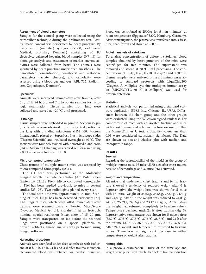
Weight and temperatureAll mice that underwent chest trauma and femur frac-ture showed a tendency of reduced weight after 6 h.Representative the weight loss was shown for 5 micewith an initial weight of 25,62 g, 25,58 g, 26,54 g, 24,40 gand 24,83 g. After 6 h the weight was reduced to 24,06 g,24,95 g, 25,39 g, 24,16 g and 23,17 g (Fig. 2). After 3 daysthe weight had returned completely to baseline values.Temperature declined until 24 h after trauma (Fig. 3).Representative temperature was shown for 5 mice before(38,7 °C, 37,6 °C, 37,8 °C, 37,2 °C, 38,7 °C) and 24 h afterthe trauma (37,2 °C, 36,8 °C, 37,6 °C, 37 °C, 37,5 °C).After 24 h weight and temperature returned to baselinevalues. There was no significant decrease in eithertemperature or weight after trauma.
HemoglobinIn a previous examination 5 mice of the same age andweight were punctured retrobulbar before trauma induction
Fitschen-Oestern et al. BMC Musculoskeletal Disorders (2017) 18:468 Page 4 of 12
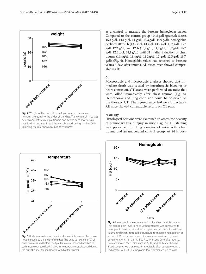
as a control to measure the baseline hemoglobin values.Compared to the control group (14,8 g/dl (gram/deciliter),15,3 g/dl, 14,4 g/dl, 14 g/dl, 15,3 g/dl, 14,9 g/dl), hemoglobindeclined after 6 h (13,7 g/dl, 13 g/dl, 13,5 g/dl, 11,7 g/dl, 13,7g/dl, 12,2 g/dl) and 12 h (13,7 g/dl, 11,7 g/dl, 13,2 g/dl, 14,7g/dl, 12,5 g/dl, 14,1 g/dl) until 24 h after induction of chesttrauma (14,4 g/dl, 13,4 g/dl, 13,2 g/dl, 12 g/dl, 12,3 g/dl, 13,7g/dl) (Fig. 4). Hemoglobin values had returned to baselinevalues 3 days after trauma. All tested mice showed compar-able results.
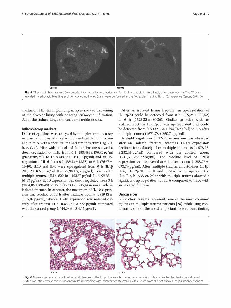
CtMacroscopic and microscopic analyses showed that im-mediate death was caused by intrathoracic bleeding orheart contusion. CT scans were performed on mice thatwere killed immediately after chest trauma (Fig. 5).Hemothorax and lung contusion could be observed onthe thoracic CT. The injured mice had no rib fractures.All mice showed comparable results on CT scan.
HistologyHistological sections were examined to assess the severityof pulmonary tissue injury in mice (Fig. 6). HE stainingwas performed for lung samples of mice with chesttrauma and an unoperated control group. At 24 h post-
Fig. 2 Weight of the mice after multiple trauma. The mousenumbers are equal to the order of the data. The weight of mice wasdetermined before multiple trauma and before each mouse wassacrificed. A decrease in weight was observed during the first 24 hfollowing trauma (shown for 6 h after trauma)
Fig. 3 Body temperature of the mice after multiple trauma. The mousemice are equal to the order of the data. The body temperature (°C) ofmice was measured before multiple trauma was induced and beforeeach mouse was sacrificed. A drop in temperature was observed duringthe first 24 h after trauma (shown for 6 h after trauma)
Fig. 4 Hemoglobin measurements in mice after multiple trauma.The hemoglobin level in mice without trauma was compared tohemoglobin level in mice after multiple trauma. Five mice withouttrauma underwent retrobulbar puncture to measure hemoglobin asa control. Mice that underwent trauma were sacrificed by heartpuncture at 6 h, 12 h, 24 h, 3 d, 7 d, 14 d, and 28 d after trauma.Data are shown for 5 mice each at 6, 12, and 24 h after trauma.Blood samples were analyzed immediately after puncture using aRadiometer ABL 700. Hemoglobin levels decreased up to 24 h
Fitschen-Oestern et al. BMC Musculoskeletal Disorders (2017) 18:468 Page 5 of 12
contusion, HE staining of lung samples showed thickeningof the alveolar lining with ongoing leukocytic infiltration.All of the stained lungs showed comparable results.
Inflammatory markersDifferent cytokines were analyzed by multiplex immunoassayin plasma samples of mice with an isolated femur fractureand in mice with a chest trauma and femur fracture (Fig. 7 a,b, c, d, e). Mice with an isolated femur fracture showed adown-regulation of IL1β from 0 h (808,84 ± 190,93 pg/ml(picogram/ml)) to 12 h (492,81 ± 190,93 pg/ml) and an up-regulation of IL-6 from 0 h (39,32 ± 18,58) to 6 h (76,67 ±16,40). IL1β and IL-6 were up-regulated from 0 h (IL1β209,12 ± 166,51 pg/ml; IL-6 22,98 ± 9,59 pg/ml) to 6 h aftermultiple trauma (IL1β 829,40 ± 163,87 pg/ml; IL-6 99,88 ±65,18 pg/ml). IL-10 expression was down-regulated from 0 h(2464,06 ± 894,49) to 12 h (1773,15 ± 742,4) in mice with anisolated fracture. In contrast, the maximum of IL-10 expres-sion was reached at 12 h after multiple trauma (2519,12 ±1782,87 pg/ml), whereas IL-10 expression was reduced dir-ectly after trauma (0 h 1085,22 ± 702,85 pg/ml) comparedwith the control group (1644,08 ± 1001,46 pg/ml).
After an isolated femur fracture, an up-regulation ofIL-12p70 could be detected from 0 h (679,24 ± 578,52)to 6 h (1523,32 ± 480,26). Similar to mice with anisolated fracture, IL-12p70 was up-regulated and couldbe detected from 0 h (321,64 ± 294,74 pg/ml) to 6 h aftermultiple trauma (1671,78 ± 350,74 pg/ml).A slight regulation of TNFα expression was observed
after an isolated fracture, whereas TNFα expressiondeclined immediately after multiple trauma (0 h 578,93± 232,48 pg/ml) compared with the control group(1241,5 ± 266,22 pg/ml). The baseline level of TNFαexpression was recovered at 6 h after trauma (1288,76 ±693,74 pg/ml). After multiple trauma all cytokines (IL1β,IL-6, IL-12p70, IL-10 and TNFα) were up-regulated(Fig. 7 a, b, c, d, e). Mice with multiple trauma showed asignificant up-regulation for IL-6 compared to mice withan isolated fracture.
DiscussionBlunt chest trauma represents one of the most commoninjuries in multiple trauma patients [28], while lung con-tusion is one of the most important factors contributing
Fig. 5 CT scan of chest trauma. Computerized tomography was performed for 5 mice that died immediately after chest trauma. The CT scansrevealed intrathoracic bleeding and hemopneumothorax. Scans were performed in the Molecular Imaging North Competence Center, CAU Kiel
Fig. 6 Microscopic evaluation of histological changes in the lung of mice after pulmonary contusion. Mice subjected to chest injury showedextensive intra-alveolar and intrabronchial hemorrhaging with consecutive atelectasis, while sham mice did not show such pulmonary changes
Fitschen-Oestern et al. BMC Musculoskeletal Disorders (2017) 18:468 Page 6 of 12

a
b
c
d
e
Fig. 7 a, b, c, d, e Cytokine analysis assessed by the Luminex assay. Serum samples of mice with an isolated femur fracture (Fx) and mice withchest trauma and femur fracture (TTFx) were analysed by multiplex immunoassay. All Data are shown as box-and-whisker plot with median andinterquartile range. Concentration of different cytokines (IL-1β, IL-6, IL-10, IL-12p70 and TNFα) was measured in different groups of mice 0 h, 6 h,12 h, 3 d and 7 d (n = 8) after trauma. Figures show regulation of IL-1α, IL-6, IL-10, IL-12p70 and TNFα after an isolated femur fracture from 0 h to7 d after trauma and cytokine regulation after induction of chest trauma and femur fracture from 0 h to 7 d after trauma. Cytokine expressionwas compared respectively to Sham group
Fitschen-Oestern et al. BMC Musculoskeletal Disorders (2017) 18:468 Page 7 of 12
to the increased morbidity and mortality of multipletrauma patients [29]. Femoral fractures represent one ofthe most prevalent associated injuries in multiple traumapatients with blunt thoracic trauma [30]. The presenceof long bone fractures causing respiratory deteriorationand respiratory dysfunction may preclude orthopedicsurgical intervention for several days.In our study, we investigated a reproducible new mul-
tiple trauma mouse model using the combination ofthese two major injuries. The main questions of thepresent study may be summarized as follows:Why were mice chosen as our experimental animal?Why did we choose the combination of chest trauma
and femur fracture?What are the influences of chest trauma and femur
fractures?How is the immune response altered in terms of cyto-
kine expression?Mice are currently the experimental tool of choice for
the majority of immunologists, and the study of theirimmune responses has offered tremendous insight intothe functions of the human immune system [31].Humans and mice share approximately 80% of theirgenes [32], and unlike large animal models, mice aretechnically easier to implement, have lower acquisitionand housing costs and superior ethical acceptance andare available as knockout animals.Traumatic brain injury, thoracic trauma, hemorrhagic
shock and long bone fracture are the focus of mostmural trauma models. All these models have advantagesand disadvantages.Some trauma models focus on an isolated organ or tis-
sue injury, and some models focus on the combinationof several severe injuries. To concentrate on a particularinjury might be an advantage in some ways, but it doesnot replicate multiple trauma in humans [33].Several trauma studies focus on traumatic brain injur-
ies [34–36]. The knowledge about outcome rates afterconcomitant traumatic brain injuries may help prioritizethe research in this regard. However traumatic brain in-jury models have the limitation of not reflecting exactlythe clinical setting and posttraumatic intensive monitor-ing in humans [37].Hemorrhagic shock ist the leading cause of morbidity
and mortality in trauma patients [38]. In mouse modelshemorrhagic shock can be induced volume controlled,pressure controlled or uncontrolled [39]. While volume-controlled hemorrhagic shock shows compensatoryphysiological mechanisms and is easy and less invasiveto perform, it provides the disadvantage of an uncertainseverity of hemorrhage [40]. Pressure-controlledhemorrhagic shock models are standardized and repro-ducible models that allow the analysis of severe shockstates and the monitoring of physiological parameters;
however, they show suppression of compensatorymechanisms. Uncontrolled hemorrhagic shock modelsrepresent the clinical situation but are less standardized[40]. The manipulation of a single variable such as vol-ume, blood pressure and time may cause unpredictable,irreproducible results so that hemorrhagic shock modelsare difficult to compare [40].Chest trauma in small animals can be induced by, for
instance, a blast wave generator [41] or weight-inducedbilateral lung contusion [13], which we used in ourmodel. Blast injury is an important cause of trauma inmilitary conflicts or terrorism, whereas weight-inducedtrauma imitates the trauma that occurs in traffic acci-dents [42].A blast generator created laser induced stress waves
and the intensity of the shock wave is flexible by varyingthe laser energy [42]. The trauma model of bilateral lungcontusion induced by a focused external blunt chesttrauma (Fig. 1a, b) has the advantages of being specificin terms of lung contusion (Figs 5 and 6) and uses amethod of impact induction, which is reproducible andhighly relevant to the chest trauma that occurs in motorvehicle accidents [13].Despite the high incidence of chest trauma and femur
fracture, there are no trauma mouse models combiningthese two injuries to date, and the immunologicalterations following pulmonary contusion remain insuf-ficiently elucidated.Apart from thoracic injuries, long bone fractures are
particularly critical and represent one of the most preva-lent associated injuries in multiple trauma patients withblunt thoracic injuries [30, 43]. Tibia fracture modelshave the advantage of easier intramedullary access com-pared to the femur [44]. The diameter of the femur isrelatively consistent and large compared with that of thetibia, which facilitates the use of larger implants and thebone is more thickly covered by muscle [44].In an open femur fracture model the bone is exposed
and fractured via osteotomy or by weakening the bonewith several drill holes [45]. Open fracture models arestabilized by extramedullary fixation techniques like alocking plate or an external fixator [39]. Induction of anopen fracture and extramedullary stabilization generatesconsiderable soft tissue injury. External fixation has thedisadvantage of high implant weight and the large vari-ation in implant stiffness [46]. An external plate fixationmay damage the periosteum and perfusion and nutrition[46]. In contrast to most of the intramedullarystabilization techniques the external stabilization pro-vides rotation stability after fracture [47].In a closed femur fracture model the fracture usually
followed by placement of intramedullary screws, pins orlocking nails [14, 46, 47]. Closed fracture models havethe advantage of reduced risk of wound infection
Fitschen-Oestern et al. BMC Musculoskeletal Disorders (2017) 18:468 Page 8 of 12
compared to an open osteotomy [14]. Several intrame-dullary stabilization systems are not stable against longi-tudinal and rotational deformations [44]. A locking nailoffers higher stability compared to pin fixation but is nota rigid fixation technique and the operation time is lon-ger which might be a relevant disadvantage in a multipletrauma model [44]. Rotation stability can be achieved byflattening the tip and the distal end of a needle or a pin[46]. In our trauma mouse model, femur fracture wasstabilized by minimal invasive surgery before the fracturewas induced. Prior to fracturing the femur a 20 gaugeneedle was inserted in the medullary cavity of the femurto maintain axial alignment during the fracture andavoid large displacements. We chose this method de-scribed by Bonnarens [14] because it offers accurate re-duction, a reduced operation time, less costs and lessblood loss than external fixation [48]. While stabilizationis performed immediately before the induction of frac-ture, the second hit inflammatory reaction can also beprohibited similar to damage control. Nevertheless, ahigher incidence of complications and higher mortalityafter fracture stabilization is always observed in the pres-ence of severe thoracic injuries [49].Although early fracture stabilization can minimize sev-
eral pulmonary complications such as fat embolism [50],damage control surgery and the timing of optimal treat-ment in multiple trauma patients are still points of interest[51]. Damage control during femur fracture stabilizationhas been shown to be beneficial for preventing the secondhit inflammatory reaction and is associated with decreasedblood loss and decreased mortality and morbidity intrauma patients [52]. There is evidence that early fracturefixation reduces the incidence of fracture-related compli-cations and improves fracture outcome.The mortality rates of multiple trauma patients range
from 7% to 45%, depending on the injury severity [53].In multiple trauma patients, 20–25% of deaths are at-tributed to chest injury [54]. In our study, 16 mice (33%)died within the first 30 min after chest trauma, and 64mice (66%) survived. The mortality rate in our traumamodel was high compared to other studies [41, 42, 55]but most of the thoracic trauma models focus on an iso-lated thoracic injury [41, 42, 55]. Examining isolatedorgan injury may be of benefit; however, this does notaccurately replicate human trauma, which often involvesmultiple organ systems [33].Additionally, in some models only one side or a special
part of the chest is affected [42, 55] whereas our modelis a bilateral contusion model [13]. In preliminary testwe determined a high impact energy to generate a severechest trauma. The thoracic trauma was first describedfor rats (body weight 250–300 g) with an impact energyof 2,45 J [13]. The falling weight in our study had an im-pact energy of 1617 J, and the mice had a body weight
of 22 ± 3 g. The impact energy we choose might be highin relation to the small mural chest.The animals that did not survive died immediately
after the chest trauma due to intrathoracic hemorrhage,which was confirmed by CT scan and removal of the or-gans. Interestingly, no rib fractures were found in ourstudy or mentioned in previous evaluations [56, 57]. Themurine chest exhibits high flexibility because of the cos-tal dorso-ventral joints, which are not present in the hu-man thorax [58, 59].In humans, the metabolic response to severe injury
results in hypothermia and weight loss. Similar toclinical conditions, the mice displayed a decrease inbody temperature, weight loss and blood loss aftermultiple trauma during the first 24 h after trauma(Fig. 2, Fig. 3, Fig. 4).Accidental hypothermia is a serious problem in
multiple trauma patients because of the negativepathophysiological effects [60]. Early rewarming ap-pears to be essential for the treatment of hypothermictrauma patients. In our study, the mice were placedunder an incubator lamp for the first 12 h after in-duction of trauma, but body temperature did not re-cover until 24 h post-trauma potentially because onlyexternal warming was applied without the donation ofwarm infusions or a blood supply, which is normallyadministered to trauma patients.The posttraumatic inflammatory reaction in humans
and mice is essentially regulated by cytokine expression[61, 62]. The magnitude of cytokine expression is regu-lated by the trauma severity in humans [63]. TNFα, IL-6,IL-1β and IL-10 correlate with the systemic inflamma-tory response and injury severity [64, 65], and thereforewe focused on these mediators in mice. Multiple traumapatients with severe damage or limited lung function ex-hibit significantly higher cytokine patterns in the earlypost-injury phase, with elevations of TNFα, IL-6, IL-10and IL-1β compared with other trauma patients [66].We detected an increase in TNFα, IL-6 and IL-1β in theplasma samples from multiple trauma mice.We focused on IL-12, which is produced at high
levels by monocytes and macrophages. Blunt chesttrauma induces mediator-dependent monocyte migra-tion to the lung [67], and high expression of IL-12can be detected in the monocytes of trauma patients[68]. An increase of IL-12p70 could also be detectedin multiple trauma mice.In comparison to humans, a correlation between
cytokine expression and the severity of trauma couldalso be detected in our trauma model. Mice with a singlefracture generally showed reduced cytokine expressioncompared with multiple trauma mice.TNFα, IL-6 and IL-1β are rapidly acting cytokines in
humans, and peak levels can be detected within 24 h
Fitschen-Oestern et al. BMC Musculoskeletal Disorders (2017) 18:468 Page 9 of 12
after trauma [69]. Similar results were obtained for miceafter trauma, with an increase observed at 24 h. Our re-sults using the murine trauma model were also consist-ent with the results of an isolated blast wave traumamodel or burn injury model [41]. In summary, miceseems to show comparable cytokine expression patternsto human trauma patients.
ConclusionWe have established a new multiple trauma mousemodel that better recapitulates the immunologicalresponse of severely injured patients. Despite clear dif-ferences between humans and animals, animal studiesare necessary to gain further insight into the physio-logical mechanisms underlying multiple trauma.This trauma model will be extremely helpful to answer
outstanding questions concerning whether cytokineblockade, which is available in the clinic, is helpful forthe treatment of trauma patients. Such studies can befurther complemented by the evaluation of geneticallymodified mice that lack particular cytokines, in terms ofthe course of multiple traumatic situations. These stud-ies will eventually lead to better therapeutic approachesto this life-threatening condition. Specifically, it is evenmore important to develop new animal models with themost frequently encountered injuries for further medicalimprovement, necessitating further studies.
Abbreviations%: percent; °C: celsius; ALI: acute lung injury; ARDS: acute respiratory distresssyndrome; Cm: centimeter; D: day; dl: deciliter; E: impact energy; Fxgroup: fracture group; g: gram; g: gravitational acceleration; h: height ofweight above the Lexon platform (in meters); h: hour; H&E-stained: Haematoxylin and Eosin-stained; IL: Interleukin; J: Joule; Kg: kilogram;m: mass of aluminum weight; mg: milligram; min: minutes; ml: milliliter;MODS: multi organ dysfunction syndrome; ms: millisecond; pg: picogram;TNF-α: Tumor necrosis factor- α; TTFx group: thorax trauma fracture;μCT: micro computed tomography; μm: micrometer
AcknowledgementsNot applicable.
FundingAll experiments were financed by research funds of the department. Thefunds are not bound to any data collection, data analysis or result. Furtheran AO funds (Arbeitsgemeinschaft Osteosynthese, F379014) was used tofinance part of elisa experiments. Before starting with the experiments anapplication with the experimental set-up ws sent to the AO. AO funding hadno influence on data analysis, process, results or writing the manuscript.
Availability of data and materialsThe datasets used and analysed during the current study are available fromthe corresponding author on reasonable request. All data generated oranalysed during this study are included in this published article. Themanuscript, including related data, figures and tables have not beenpreviously published and are not under consideration elsewhere.
Authors’ contributionsWe declare, that all authors have made substantial contributions to theconception and design of the study, acquisition of data, analysis andinterpretation of data. All authors drafted the article critically for importantintellectual content and made substantial contributions to final approval ofthe version to be submitted. SFO and CN operated all animals and retrieved
samples from all animals. HA helped to control all animals in the first 24 h.Elisa experiments were performed by SFO with the help of MT, TP, NS andSRJ. Data analysis was supported by AS, SL, FH, MW, TK and DV. All authorswere involved in reading and approving the final manuscript.
Ethics approval and consent to participateExperiments were carried out in accordance with the German AnimalWelfare Legislation, and were approved by the local institutional animal careand research advisory committee (Kiel and Hannover, Germany) andpermitted by the local government of Lower Saxony, Germany (referencenumber: AZ 10AO29).
Consent for publicationThe manuscript does not contain any individual person’s data.Not applicable
Competing interestsThe authors declare that they have no competing interests.
Publisher’s NoteSpringer Nature remains neutral with regard to jurisdictional claims inpublished maps and institutional affiliations.
Author details1Department of Trauma Surgery, University Medical Center ofSchleswig-Holstein, Arnold-Heller Straße 7, 24105, Campus Kiel, Kiel,Germany. 2Department of Anatomy and Cell Biology, RWTH AachenUniversity, Wendlingweg 2, D-52074 Aachen, Germany. 3Department ofBiochemistry, Medical Faculty, Olshausenstr. 40, 24098 Kiel, Germany.4Department of Trauma Surgery, RWTH Aachen University, Pauwelsstraße 30,52074 Aachen, Germany.
Received: 11 June 2017 Accepted: 6 November 2017
References1. Lenz A, Franklin GA, Cheadle WG. Systemic inflammation after trauma.
Injury, Int. J. Care Injured. 2007;38:1336–45.2. Bardenheuer M, Obertacke U, Waydhas C, Nast-Kolb D. Epidemiology of the
severely injured patient. A prospective assessment of preclinical and clinicalmanagement. AG Polytrauma of DGU. Unfallchirurg. 2000;103:355–63.
3. Wanek S, Mayberry JC. Blunt thoracic trauma: flail chest, pulmonarycontusion, and blast injury. Crit Care Clin. 2004;20:71–81.
4. Hildebrand F, Pape HC, Krettek C. The importance of cytokines in theposttraumatic inflammatory reaction. Unfallchirurg. 2005;108(10):793–4. 796–803
5. Kauvar D, Sarfati MR, Kraiss LW. National trauma databank analysis ofmortality and limb loss in isolated lower extremity vascular trauma. J VascSurg. 2011;53(6)
6. Kobbe P, Lichte P, Pape HC. Complex extremity fractures following highenergy injuries: the limited value of existing classifications and a proposalfor a treatment-guide. Injury. 2009;40(4):69–74.
7. Nahm NJ, Vallier HA. Timing of definitive treatment of femoral shaftfractures in patients with multiple injuries: a systematic review ofrandomized and nonrandomized trials. J Trauma Acute Care Surg. 2012;73(5):1046–63.
8. Seekamp A, Jochum M, Ziegler M, van Griensven M, Martin M, Regel G.Cytokines and adhesion molecules in elective and accidental trauma-relatedischemia/reperfusion. J Trauma. 1998;44(5):874–82.
9. Guisasola MC, Ortiz A, Chana F, Alonso B, Vaquero J. Early inflammatoryresponse in polytraumatized patients: cytokines and heat shock proteins. Apilot study. Orthop Traumatol Surg Res. 2015;101(5):607–11.
10. Crohn SM. Pulmonary contusion: review of the clinical entity. J Trauma.1997;42:973–9.
11. Miller PR, Croce MA, Bee TK, Qaisi WG, Smith CP, Collins GL, Fabian TC.ARDS after pulmonary contusion: accurate measurement of contusionvolume identifies high-risk patients. J Trauma. 2001;51:223–8.
12. Pelias ME, Townsend MC, Flancbaum L. Long bone fractures predispose topulmonary dysfunction in blunt chest trauma despite early operativefixation. Surgery. 1992;111(5):576–9.
Fitschen-Oestern et al. BMC Musculoskeletal Disorders (2017) 18:468 Page 10 of 12
13. Raghavendran K, Davidson BA, Helinski JD, Marschke CJ, Manderscheid P,Woytash JA, Notter RH, Knight PR. A rat model for isolated bilateral lungcontusion from blunt chest trauma. Anesth Analg. 2005;101(5):1482–9.
14. Bonnarens F, Einhorn TA. Production of a standard closed fracture inlaboratory animal bone. J Orthop Res. 1984;2:97–101.
15. Suresh MV, Ramakrishnan SK, Thomas B, Machado-Aranda D, Bi Y, Talarico N,Anderson E, Yatrik SM, Raghavendran K. Activation of hypoxia-induciblefactor-1α in type 2 alveolar epithelial cell is a major driver of acuteinflammation following lung contusion. Crit Care Med. 2014;42(10):642–53.
16. Dolgachev VA, Yu B, Sun L, Shanley TP, Raghavendran K, Hemmila MR.Interleukin 10 overexpression alters survival in the setting of gram-negativepneumonia following lung contusion. Shock. 2014;41(4):301–10.
17. Dolgachev VA, Yu B, Reinke JM, Raghavendran K, Hemmila MR. Hostsusceptibility to gram-negative pneumonia after lung contusion. J TraumaAcute Care Surg. 2012;72(3):614–22.
18. Drissi H, Paglia DN. Surgical procedures and experimental outcomes ofclosed fractures in rodent models. Femur fracture. Methods Mol Biol.2015;1226:193–211.
19. Aurégan JC, Coyle RM, Danoff JR, Burky RE, Akelina Y, Rosenwasser MP. Therat model of femur fracture for bone and mineral research: An improveddescription of expected comminution, quantity of soft callus and incidenceof complications. Bone Joint Res. 2013;2(8):149–54.
20. Nadkarni S, McArthur S. Oestrogen and immunomodulation: newmechanisms that impact on peripheral and central immunity. Curr OpinPharmacol. 2013;13:576–81.
21. Hernández-Bello R, Nava-Castro K, Muñiz-Hernández S, Nava-Luna P, Trejo-Sánchez I, Tiempos-Guzmán N, Mendoza-Rodríguez Y, Morales-Montor J.Beyond the reproductive effect of sex steroids: their role during immunityto helminth parasite infections. Mini Rev Med Chem. 2012;12:1071–80.
22. Crockett ET, Spielman W, Dowlatshahi S, He J. Sex differences ininflammatory cytokine production in hepatic ischemia-reperfusion. JInflamm (Lond). 2006;3:16.
23. Neunaber C, Oestern S, Andruszkow H, Zeckey C, Mommsen P, Kutter D,Stöfen M, Krettek C, Hildebrand F. Cytokine productive capacity of alveolarmacrophages and Kupffer cells after femoral fracture and blunt chesttrauma in a murine trauma model. Immunol Lett. 2013;152(2):159–66.
24. Stockmann U, Roscher R. Pulmonary contusion after blunt chest trauma: anexperimental model. Thoraxchir Vask Chir. 1977;25:211–3.
25. Tower RJ, Campbell GM, Müller M, Will O, Glüer CC, Tiwari S. Binding kinetics ofa fluorescently labeled bisphosphonate as a tool for dynamic monitoring ofbone mineral deposition in vivo. J Bone Miner Res. 2014;29(9):1993–2003.
26. Tiwari S, Schem C, Lorenzen AC, Kayser O, Wiese C, Graeff C, Peña J,Marshall RP, Heller M, Kalthoff H, Jonat W, Glüer CC. Application of ex vivomicro-computed tomography for assessment of in vivo fluorescence andplain radiographic imaging for monitoring bone metastases and osteolyticlesions. J Bone Miner Metab. 2012;30(3):373–80.
27. Jackson IL, Vujaskovic Z, Down JD. Revisiting strain-related differences inradiation sensitivity of the mouse lung: recognizing and avoiding theconfounding effects of pleural effusions. Radiat Res. 2010;173(1):10–20.
28. Hildebrand F, Giannoudis PV, Mv G, Zelle B, Ulmer B, Krettek C, Bellamy MC, PapeHC. Management of polytraumatized patients with associated blunt chesttrauma: a comparison of two European countries. Injury. 2005;36(2):293–302.
29. Ganie FA, Lone H, Lone GN, Wani ML, Singh S, Dar AM, Wani N, Wani SN,Nazeer N. Lung contusion: a Clinico-pathological entity with unpredictableclinical course. Bull Emerg Trauma. 2013;1(1):7–16.
30. Hildebrand F, Giannoudis P, van Griensven M, Chawda M, Probst C, HarmsO. Secondary effects of femoral instrumentation on pulmonary physiologyin a standardised sheep model: what is the effect of lung contusion andreaming. Injury 2005; 36: 544–555.
31. Mestas J, Hughes C. Of mice and not men: differences between mouse andhuman immunology. J Immunol. 2004;172:2731–8.
32. Tsukamoto T, Pape HC. Animal models for trauma research: what are theoptions? Shock. 2009;31(1):3110.
33. Stortz JA, Raymond SL, Mira JC, Moldawer LL, Mohr AM, Efron PA. Murine modelsof sepsis and trauma: can we bridge the gap? ILAR J. 2017;58(1):90–105.
34. Carbonell WS, Maris DO, McCall T, Grady MS. Adaptation of the fluidpercussion injury model to the mouse. J Neurotrauma. 1998;15(3):217–29.
35. Smith DH, Soares HD, Pierce JS, Perlman KG, Saatman KE, Meaney DF, DixonCE, McIntosh TKA. Model of parasagittal controlled cortical impact in themouse: cognitive and histopathologic effects. J Neurotrauma.1995;12(2):169–78.
36. Flierl MA, Stahel PF, Beauchamp KM, Morgan SJ, Smith WR, Shohami E.Mouse closed head injury model induced by a weight-drop device. NatProtoc. 2009;4(9):1328–37.
37. Marklund N. Rodent models of traumatic brain injury: methods andchallenges. Methods Mol Biol. 2016;1462:29–46.
38. Nunez TC, Cotton BA. Transfusion therapy in hemorrhagic shock. Curr OpinCrit Care. 2009;15(6):536–41.
39. Frink M, Andruszkow H, Zeckey C, Krettek C, Hildebrand F. ExperimentalTrauma Models: An Update. J Biomed Biotechnol. 2011;2011:797383.
40. Pfeifer R, Lichte P, Schreiber H, Sellei RM, Dienstknecht T, Sadeghi C, PapeHC, Kobbe P. Models of hemorrhagic shock: differences in the physiologicaland inflammatory response. Cytokine. 2013;61(2):585–90.
41. Knöferl MW, Liener UC, Seitz DH, Perl M, Brückner UB, Kinzl L, Gebhard F.Cardiopulmonary, histological, and inflammatory alterations after lung contusionin a novel mouse model of blunt chest trauma. Shock. 2003;19:519–25.
42. Satoh Y, Sato S, Saitoh D, Tokuno S, Hatano B, Shimokawaji T, Kobayashi H,Takishima K. Pulmonary blast injury in mice: a novel model for studyingblast injury in the laboratory using laser-induced stress waves. Lasers SurgMed. 2010;42(4):313–8.
43. Rixen D, Sauerland S, Oestern H-J, Bouillon B. Management strategies in thefirst operative phase after long-bone injury of the lower extremity inmultiple-injured patients. A systematic literature review. Unfallchirurg.2005;108(10):829–42.
44. Qi B, Yu J, Zhao Y, Zhu D, Yu T. Review article mouse fracture models: aprimer. Int J Clin Exp Med. 2016;9(7):12418–29.
45. Klein M, Stieger A, Stenger D, Scheuer C, Holstein JH, Pohlemann T, MengerMD, Histing T. Comparison of healing process in open osteotomy modeland open fracture model: Delayed healing of osteotomies afterintramedullary screw fixation. J Orthop Res. 2015;33(7):971-8.
46. Holstein JH, Menger MD, Culemann U, Meier C, Pohlemann T. Developmentof a locking femur nail for mice. J Biomech. 2007;40(1):215–9.
47. Manigrasso MB, O’Connor JP. Characterization of a closed femur fracturemodel in mice. J Orthop Trauma. 2004;18(10):687–95.
48. Cheung KM, Kaluarachi K, Andrew G, Lu W, Chan D, Cheah KS. An externallyfixed femoral fracture model for mice. J Orthop Res. 2003;21(4):685–90.
49. Bone LB, Giannoudis P. Femoral shaft fracture fixation and chest injury afterPolytrauma. J Bone Joint Surg Am. 2011;93:311–7.
50. Riska EB, von Bonsdorff H, Hakkinen S, Jaroma H, Kiviluoto O, Paavilainen T.Prevention of fat embolism by early internal fixation of fractures in patientswith multiple injuries. Injury 1976, 8: 110–116.
51. Schreiber VM, Tarkin IS, Hildebrand F, Darwiche S, Pfeifer R, Chelly J,Giannoudis P, Pape HC. The timing of definitive fixation for major fracturesin polytrauma-a matched-pair comparison between a US and Europeanlevel I centres: analysis of current fracture management practice inpolytrauma. Injury. 2011;42(7):650–4.
52. Roberts CS, Pape HC, Jones AL, Malkani AL, Rodriguez JL, Giannoudis PV.Damage control orthopaedics: evolving concepts in the treatment of patientswho have sustained orthopaedic trauma. Instr Course Lect. 2005;54:447–62.
53. Chawda MN, Hildebrand F, Pape HC, Giannoudis PV. Predicting outcomeafter multiple trauma: which scoring system? Injury. 2004;35(4):347–58.
54. Clark GC, Schecter WP, Trunkey DD. Variables affecting outcome in blunt chesttrauma: flail chest vs. pulmonary contusion. J Trauma. 1988;28(3):298–304.
55. Hoth JJ, Hudson WP, Brownlee NA, Yoza BK, Hiltbold EM, Meredith JW, CEMC. Toll-like receptor 2 participates in the response to lung injury in amurine model of pulmonary contusion. Shock. 2007;28(4):447–52.
56. Raghavendran K, Davidson B, Woytash JA, Helinski JD, Marschke CJ,Manderscheid P, Notter R, Knight P. The evolution of isolated bilateral lungcontusion from blunt chest trauma in rats: cellular and cytokine responses.Shock. 2005;24(2):132–8.
57. Perl M, Gebhard F, Braumüller S, Tauchmann B, Brückner U, Kinzl L, KnöferlM. The pulmonary and hepatic immune microenvironment and itscontribution to the early systemic inflammation following blunt chesttrauma. Crit Care Med. 2006;34(4)
58. Genton L, Pichard C. Protein catabolism and requirements in severe illness.Int J Vitam Nutr Res. 2011;81(2–3):143–52.
59. Omerbegović M, Durić A, Muratović N, Mulalić L, Hamzanija E. Metabolicresponse to trauma and stress. Med Arh. 2003, 57;4(1):57–60.
60. Mommsen P, Zeckey C, Frink M, Krettek C, Hildebrand F. Accidentalhypothermia in multiple trauma patients. Zentralbl Chir. 2012;137(3):264-9.
61. Gentile LF, Nacionales DC, Cuenca AG, Armbruster M, Ungaro RF,Abouhamze AS, Lopez C, Baker HV, Moore FA, Ang DN, Efron PA.
Fitschen-Oestern et al. BMC Musculoskeletal Disorders (2017) 18:468 Page 11 of 12
Identification and description of a novel murine model for polytrauma andshock. Crit Care Med. 2013;41(4):1075–85.
62. Bone RC. Towards a theory regarding the pathogenesis of the systemicinflammatory response syndrome: what we do and do not know aboutcytokine regulation. Crit Care Med. 1996;24(1):163–72.
63. Pfeifer R, Darwiche S, Kohut L, Billiar T, Pape HC. Cumulative effects of boneand soft tissue injury on systemic inflammation: a pilot study. Clin OrthopRelat Res. 2013;471(9):2815–21.
64. Akkose S, Ozgurer A, Bulut M, Koksal O, Ozdemir F, Ozguc H. Relationshipsbetween markers of inflammation, severity of injury, and clinical outcomesin hemorrhagic shock. Adv Ther. 2007;24:955–62.
65. Jaffer U, Wade RG, Gourlay T. Cytokines in the systemic inflammatoryresponse syndrome: a review. HSR Proc Intensive Care Cardiovasc Anesth.2010;2(3):161–75.
66. Roumen RM, Hendriks T, van der Ven-Jongekrijg J, Nieuwenhuijzen GA,Sauerwein RW, van der Meer JW, Goris RJ. Cytokine patterns in patientsafter major vascular surgery, hemorrhagic shock, and severe blunt trauma.Relation with subsequent adult respiratory distress syndrome and multipleorgan failure. Ann Surg. 1993;218(6):769–76.
67. Seitz DH, Niesler U, Palmer A, Sulger M, Braumüller ST, Perl M, Gebhard F,Knöferl MW. Blunt chest trauma induces mediator-dependent monocytemigration to the lung. Crit Care Med. 2010;38(9):1852–9.
68. Köller M, Dávid A, Hahn MP, Muhr G. Liberation of interleukin 12 (IL12) aftertrauma and polytrauma. Langenbecks Arch Chir Suppl Kongressbd.1998;115(1):453–6.
69. Li J, Li NP, YF G, Yang X, XB L, Cong JN, Ling Y, Tang JA, Yuan XY, Wang H.Dynamic activity of NF-kappaB in multiple trauma patients and protectiveeffects of ulinastain. Chin J Traumatol. 2011;14(6):354–8.
• We accept pre-submission inquiries
• Our selector tool helps you to find the most relevant journal
• We provide round the clock customer support
• Convenient online submission
• Thorough peer review
• Inclusion in PubMed and all major indexing services
• Maximum visibility for your research
Submit your manuscript atwww.biomedcentral.com/submit
Submit your next manuscript to BioMed Central and we will help you at every step:
Fitschen-Oestern et al. BMC Musculoskeletal Disorders (2017) 18:468 Page 12 of 12