Embed Size (px)
Citation preview
PROCEEDINGS Open Access
A new scheme to discover functionalassociations and regulatory networks of E3ubiquitin ligasesKai-Yao Huang1, Julia Tzu-Ya Weng1,2, Tzong-Yi Lee1,2* and Shun-Long Weng3,4,5*
From The Fourteenth Asia Pacific Bioinformatics Conference (APBC 2016)San Francisco, CA, USA. 11 - 13 January 2016
Abstract
Background: Protein ubiquitination catalyzed by E3 ubiquitin ligases play important modulatory roles in variousbiological processes. With the emergence of high-throughput mass spectrometry technology, the proteomicsresearch community embraced the development of numerous experimental methods for the determination ofubiquitination sites. The result is an accumulation of ubiquitinome data, coupled with a lack of available resourcesfor investigating the regulatory networks among E3 ligases and ubiquitinated proteins. In this study, by integratingexisting ubiquitinome data, experimentally validated E3 ligases and established protein-protein interactions, wehave devised a strategy to construct a comprehensive map of protein ubiquitination networks.
Results: In total, 41,392 experimentally verified ubiquitination sites from 12,786 ubiquitinated proteins of humanshave been obtained for this study. Additional 494 E3 ligases along with 1220 functional annotations and 28588protein domains were manually curated. To characterize the regulatory networks among E3 ligases and ubiquitinatedproteins, a well-established network viewer was utilized for the exploration of ubiquitination networks from 40892protein-protein interactions. The effectiveness of the proposed approach was demonstrated in a case study examiningE3 ligases involved in the ubiquitination of tumor suppressor p53. In addition to Mdm2, a known regulator of p53, theinvestigation also revealed other potential E3 ligases that may participate in the ubiquitination of p53.
Conclusion: Aside from the ability to facilitate comprehensive investigations of protein ubiquitination networks, byintegrating information regarding protein-protein interactions and substrate specificities, the proposed method coulddiscover potential E3 ligases for ubiquitinated proteins. Our strategy presents an efficient means for the preliminaryscreen of ubiquitination networks and overcomes the challenge as a result of limited knowledge about E3ligase-regulated ubiquitination.
Keywords: Ubiquitination, Ubiquitin, E3 ubiquitin ligase, Protein-protein interaction, Ubiquitination network
* Correspondence: [email protected]; [email protected] of Computer Science and Engineering, Yuan Ze University,Taoyuan 320, Taiwan3Department of Obstetrics and Gynecology, Hsinchu Mackay MemorialHospital, Hsin-Chu 300, TaiwanFull list of author information is available at the end of the article
© 2016 Huang et al. Open Access This article is distributed under the terms of the Creative Commons Attribution 4.0International License (http://creativecommons.org/licenses/by/4.0/), which permits unrestricted use, distribution, andreproduction in any medium, provided you give appropriate credit to the original author(s) and the source, provide a link tothe Creative Commons license, and indicate if changes were made. The Creative Commons Public Domain Dedication waiver(http://creativecommons.org/publicdomain/zero/1.0/) applies to the data made available in this article, unless otherwise stated.
Huang et al. BMC Systems Biology 2016, 10(Suppl 1):3DOI 10.1186/s12918-015-0244-1
IntroductionProtein ubiquitination involves a series of enzymaticreactions such as E1 activation, E2 conjugation, and E3ligation, resulting in the conjugation of single or multipleubiquitin proteins at a target lysine residue [1]. Numeroussubstrate proteins with ubiquitination sites have beencharacterized to date, owing to the emergence ofhigh-throughput mass spectrometry-based proteomicsapproaches [2–4]. Identified to play key roles in tran-scriptional regulation, signal transduction, develop-ment, apoptosis, endocytosis, cell proliferation andcancers, ubiquitination of the lysine residue has beenregarded as an essential mediator of various biologicalprocesses [5–7]. Among the enzymes that catalyzeprotein ubiquitination, E3 ligases are particularly im-portant for the recognition of substrate sites to facili-tate ubiquitin-mediated protein degradation [8]. Therelationships between E3 ligase and substrates arecomplex. Multiple substrates could be targeted by a singleE3 ligase; alternatively, multiple E3 ligases could catalyzethe ubiquitination of a single substrate [9]. Thesesubstrate-enzyme correlations could be used to constructE3-specific regulatory networks and map to the associatedcellular pathways, making possible the characterization ofcomplex cellular processes and functional analysis ofE3-sbustrate relationships [9]. This approach has allowedthe discovery of the role that anaphase-promoting complex(APC)/cyclosome plays in modulating key targets of the cellcycle, such as cyclins and their related E3 ligases [10–12].To date, a significant amount of research efforts have
been invested towards the characterization of E3 struc-tures and examination of the mechanisms underlyingE3-mediated regulatory networks, as well as E3-relateddiseases [13–21]. Based on their catalytic mechanisms inthe ubiquitination process, E3 ligases can be classifiedinto three major types: the HECT (homologous to E6-AP C-terminus), the RING (really interesting new gene),and U-box domain types [22]. The HECT-type is re-sponsible for catalyzing the attachment of ubiquitin tosubstrate proteins. In contrast, the RING-type and U-box-type, similar in both structure and function, facili-tate the interaction between an E2 enzyme and the tar-get proteins. Regardless of the types, the significance ofE3-mediated ubiquitination is obvious from their associ-ation with diseases [23]. Several studies have suggestedthat the inhibition of E3 ligases may cause growthsuppression or cell death, as evidenced by the over-expression of Mdm2/Hdm2, IAPs, and SCF in varioushuman cancers [24]. Therefore, regulation of E3 ligaseactivities and functions may be a promising approachfor cancer treatments.Many databases and tools have been developed to aid
in the study of E3 ligases. For example, E3Miner [25] of-fers a text mining approach to identify ubiquitin-protein
ligases, whereas E3Net [9] allows users to search througha collection of 1671 E3-substrate relationships among 493E3s and 1277 substrates in 42 organisms. In contrast, byanalyzing protein sequence similarities, domains, and dis-tributions across different species, Sakiyama et al. [26] con-structed a useful database for the exploration of proteinsinvolved in the ubiquitin signaling cascade. Unfortunately,the present accumulation of large-scale ubiquitinome datademands for the development of tools that investigate theregulatory networks of E3 ligases and their substrates.Here, we present a new strategy that utilizes an interactivenetwork viewer to assist with the discovery of novel pro-tein ubiquitination networks. Furthermore, to effectivelyinvestigate the relationships between E3 ligases and theirsubstrates, metabolic pathways and protein-protein inter-actions (PPIs) were integrated to construct comprehensiveprotein ubiquitination networks. The ability of theproposed method to identify E3 ligase-mediated ubi-quitination networks and their biological significancewas demonstrated by case studies. The results indicatedthat, despite the current limited knowledge about regula-tory relationship between E3 ligases and ubiquitinatedproteins, our approach could uncover potential E3 ligase-substrate relationships based on based on protein-proteininteraction information and substrate site specificities.
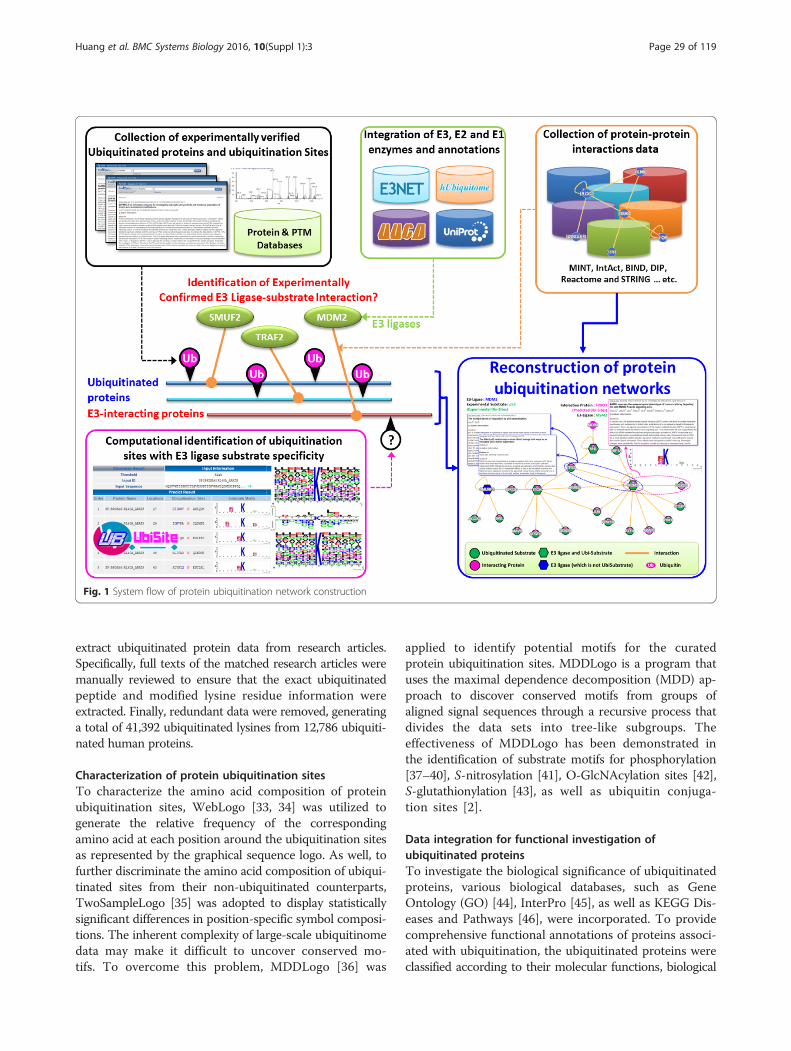
Materials and methodConstruction of the protein ubiquitination networks in-volved collection of E3 ligase and ubiquitinated proteindata, integration of ubiquitinated proteins’ functionaldata, computational identification of ubiquitination sitesbased on substrate motifs, as well as network construc-tion using protein-protein interactions and metabolicpathways (Fig. 1). A network viewer was employed toprovide a visualization of the ubiquitination regulatorynetwork, with implemented functional information, for agroup of proteins of interest. The detailed workflow isdescribed as follows.
Data collection of E3 ubiquitin ligases and ubiquitinatedproteinsExperimentally validated E1 activating, E2 conjugating,and E3 ligating enzyme data were obtained from varioussources. From UUCD-Version 1.0 [27], seven distinct E1activating enzymes were collected. From E3Net, UUCD[27], hUbiquitome [28], and UniProtKB [29], 494 non-redundant E3 ubiquitin ligases and their biologicalfunctions were extracted. In addition, a total of 46non-redundant E2 conjugating enzymes were collectedfrom UUCD [27], hUbiquitome [28] and UniProtKB[29]. Experimentally verified ubiquitination sites fromdbPTM [30–32] were also included. Next, search keywords,such as “ubiquitinated”, “ubiquitination”, “ubiquitylated”, or“ubiquitylation”, were entered on the PubMed database to
Huang et al. BMC Systems Biology 2016, 10(Suppl 1):3 Page 28 of 119
extract ubiquitinated protein data from research articles.Specifically, full texts of the matched research articles weremanually reviewed to ensure that the exact ubiquitinatedpeptide and modified lysine residue information wereextracted. Finally, redundant data were removed, generatinga total of 41,392 ubiquitinated lysines from 12,786 ubiquiti-nated human proteins.
Characterization of protein ubiquitination sitesTo characterize the amino acid composition of proteinubiquitination sites, WebLogo [33, 34] was utilized togenerate the relative frequency of the correspondingamino acid at each position around the ubiquitination sitesas represented by the graphical sequence logo. As well, tofurther discriminate the amino acid composition of ubiqui-tinated sites from their non-ubiquitinated counterparts,TwoSampleLogo [35] was adopted to display statisticallysignificant differences in position-specific symbol composi-tions. The inherent complexity of large-scale ubiquitinomedata may make it difficult to uncover conserved mo-tifs. To overcome this problem, MDDLogo [36] was
applied to identify potential motifs for the curatedprotein ubiquitination sites. MDDLogo is a program thatuses the maximal dependence decomposition (MDD) ap-proach to discover conserved motifs from groups ofaligned signal sequences through a recursive process thatdivides the data sets into tree-like subgroups. Theeffectiveness of MDDLogo has been demonstrated inthe identification of substrate motifs for phosphorylation[37–40], S-nitrosylation [41], O-GlcNAcylation sites [42],S-glutathionylation [43], as well as ubiquitin conjuga-tion sites [2].
Data integration for functional investigation ofubiquitinated proteinsTo investigate the biological significance of ubiquitinatedproteins, various biological databases, such as GeneOntology (GO) [44], InterPro [45], as well as KEGG Dis-eases and Pathways [46], were incorporated. To providecomprehensive functional annotations of proteins associ-ated with ubiquitination, the ubiquitinated proteins wereclassified according to their molecular functions, biological
Fig. 1 System flow of protein ubiquitination network construction
Huang et al. BMC Systems Biology 2016, 10(Suppl 1):3 Page 29 of 119
processes, and cellular components. Since ubiquitination isknown to regulate the cellular localization, interactions,and degradation of proteins [47–49], the biological roles ofubiquitination sites within a specific protein domain couldbe inferred from the functional annotation of the domain.For this purpose, essential protein family, domain, andfunctional site information was obtained from InterPro[45], a database which integrates data from various sourcessuch as the PROSITE [50], PRINTS [51], Pfam [52], andProDom [53].
Network construction using protein-protein interactionsand metabolic pathwaysSubstantial evidence supports the role that protein ubi-quitination plays in the regulation of cellular processes.Thus, by integrating experimentally validated mammalianE3 ubiquitin ligases and their functional information, wehoped to provide a foundation for navigating ubiquitina-tion regulatory networks in mammals. To facilitate theexploration of regulatory relationships between E3 ligasesand their ubiquitinated substrates, associated metabolicpathways and protein-protein interactions (PPIs) wereincluded for the comprehensive construction of proteinubiquitination networks. The human metabolic pathwayswere extracted from KEGG [54]. Experimentally veri-fied PPIs were obtained from over ten PPI databases(Additional file 1: Table S1). Potential PPIs predictedbased on co-regulation, co-occurrence in the literature,co-expression, and genomic context were curated fromthe STRING database [55]; each interaction included aconfidence score calculated by the STRING built-infunction.Next, a graph theory [56, 57] approach has been
adopted to illustrate the relationships between E3 ligasesand substrates. Specifically, we use a directed and cyclicgraph G = ( V , E ) to symbolize a protein ubiquitinationnetwork, where x , y ∈ V and ( x , y ) ∈ E. The E3 ligasesand substrate proteins were represented by x and y,respectively, and protein ubiquitination was denotedby (x, y) ∈ E to indicate the recognition of a specificsubstrate y by E3 ligase x (Additional file 2: Figure S1).Due to limited knowledge about ubiquitinated substratesthat are recognized by E3 ligases, (x, y) could also repre-sent a type of protein-protein interaction between E3ligase x and ubiquitinated protein y. We used V to refer toall human proteins and E, to all experimentally confirmedPPIs. Cytoscape [58], a publicly available network viewer,was employed for the visualization of regulatory networksamong E3 ligases and ubiquitinated substrates.
Results and discussionData statistics in this investigationData used for building the protein ubiquitination net-works in this study were experimentally validated and
supported with 39,814 research articles. Over 500 re-search articles were manually reviewed via a text miningmethod. In total, 41,392 ubiquitination sites from 12,786ubiquitinated proteins in humans were extracted from406 literatures. After removing redundant data amongheterogeneous online resources, 494 experimentally veri-fied human E3 ubiquitin ligases remained in the result-ing data. PPIs between E3 ligases and ubiquitinatedproteins were retrieved to deduce potential regulatoryrelationships between E3 ligases and ubiquitinated sub-strates to compensate for the limited information aboutE3 ligase targets. As shown in Table 1, 9,271 physicalPPIs between 426 E3 ligases and 2,649 ubiquitinatedproteins were curated. In particular, by incorporatingthe substrate motifs identified by the MDDLogoubiquitination site prediction method [36], potentialsubstrates of E3 ligases could be inferred from the27,227 PPIs between E3 ligases and other proteins.Moreover, 377,117 PPIs that appeared to involve ubiquiti-nated proteins could be integrated for the investiga-tion of their functional associations in the context ofubiquitination.
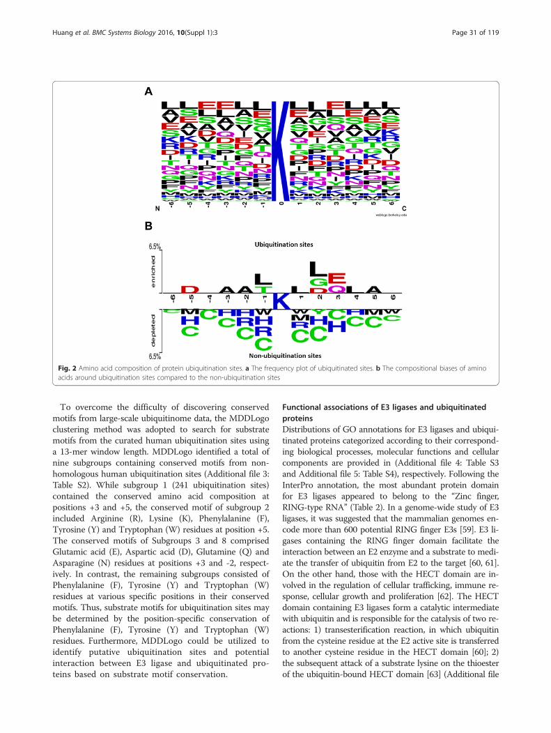
Substrate specificities of human ubiquitination sitesThe entropy plots generated by the sequence logo wasused to graphically visualize the amino acid sequencesflanking the substrate sites (at position 0). This allowsfor the easy observation of amino acid conservationsurrounding the ubiquitination sites. Figure 2a showsLeu (L), Glu (E), and Ala (A) to be the most conservedamino acid residues as indicated by the position-specific amino aicd composition around the ubiquiti-nated lysines. Furthermore, using TwoSampleLogo, thedifferences in position-specific amino acid compositionbetween ubiquitinated and non-ubiquitinated sites wererevealed (Fig. 2b). The residues surrounding the ubiqui-tination sites were significantly enriched with Ala (A),Asp (D), Glu (E), Leu (L), Gly (G) and Thr (T), and de-pleted in Cys (C), His (H), Arg (R), Trp (W) and Met(M) (p < 0.005).
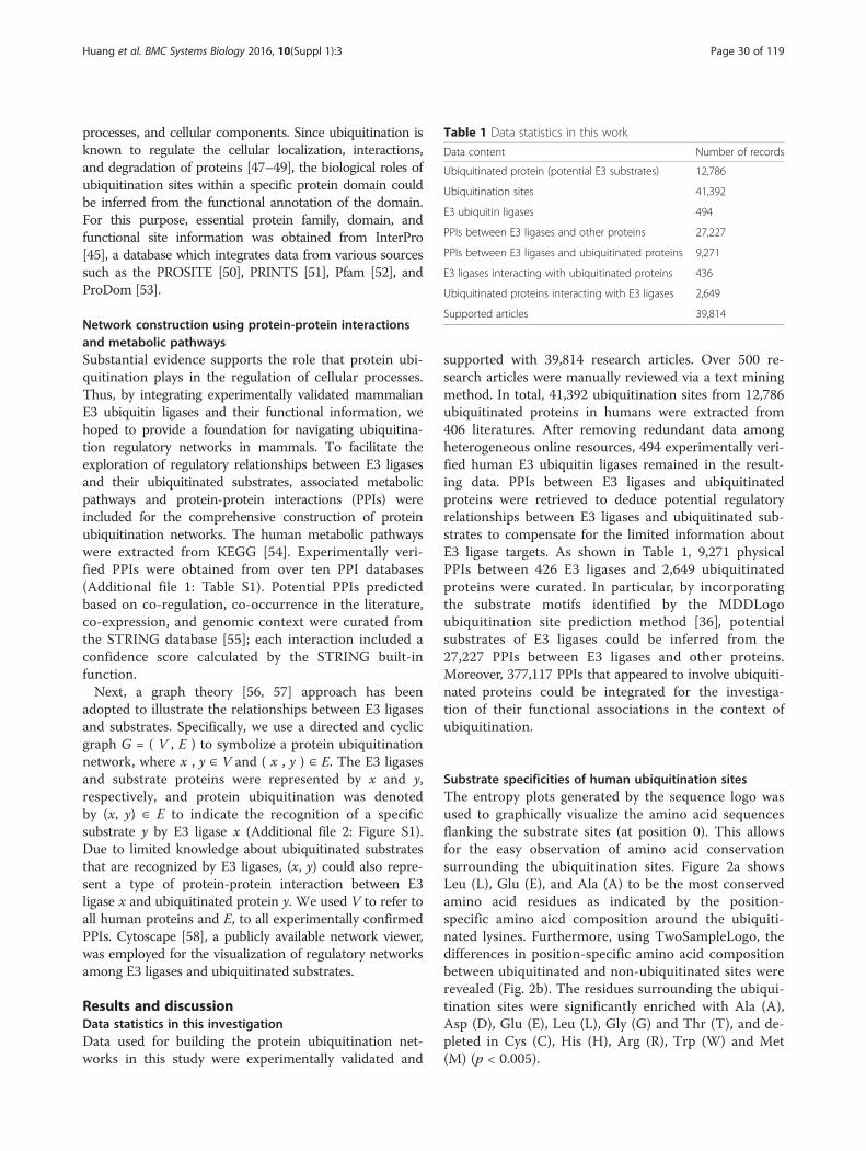
Table 1 Data statistics in this work
Data content Number of records
Ubiquitinated protein (potential E3 substrates) 12,786
Ubiquitination sites 41,392
E3 ubiquitin ligases 494
PPIs between E3 ligases and other proteins 27,227
PPIs between E3 ligases and ubiquitinated proteins 9,271
E3 ligases interacting with ubiquitinated proteins 436
Ubiquitinated proteins interacting with E3 ligases 2,649
Supported articles 39,814
Huang et al. BMC Systems Biology 2016, 10(Suppl 1):3 Page 30 of 119
To overcome the difficulty of discovering conservedmotifs from large-scale ubiquitinome data, the MDDLogoclustering method was adopted to search for substratemotifs from the curated human ubiquitination sites usinga 13-mer window length. MDDLogo identified a total ofnine subgroups containing conserved motifs from non-homologous human ubiquitination sites (Additional file 3:Table S2). While subgroup 1 (241 ubiquitination sites)contained the conserved amino acid composition atpositions +3 and +5, the conserved motif of subgroup 2included Arginine (R), Lysine (K), Phenylalanine (F),Tyrosine (Y) and Tryptophan (W) residues at position +5.The conserved motifs of Subgroups 3 and 8 comprisedGlutamic acid (E), Aspartic acid (D), Glutamine (Q) andAsparagine (N) residues at positions +3 and -2, respect-ively. In contrast, the remaining subgroups consisted ofPhenylalanine (F), Tyrosine (Y) and Tryptophan (W)residues at various specific positions in their conservedmotifs. Thus, substrate motifs for ubiquitination sites maybe determined by the position-specific conservation ofPhenylalanine (F), Tyrosine (Y) and Tryptophan (W)residues. Furthermore, MDDLogo could be utilized toidentify putative ubiquitination sites and potentialinteraction between E3 ligase and ubiquitinated pro-teins based on substrate motif conservation.
Functional associations of E3 ligases and ubiquitinatedproteinsDistributions of GO annotations for E3 ligases and ubiqui-tinated proteins categorized according to their correspond-ing biological processes, molecular functions and cellularcomponents are provided in (Additional file 4: Table S3and Additional file 5: Table S4), respectively. Following theInterPro annotation, the most abundant protein domainfor E3 ligases appeared to belong to the “Zinc finger,RING-type RNA” (Table 2). In a genome-wide study of E3ligases, it was suggested that the mammalian genomes en-code more than 600 potential RING finger E3s [59]. E3 li-gases containing the RING finger domain facilitate theinteraction between an E2 enzyme and a substrate to medi-ate the transfer of ubiquitin from E2 to the target [60, 61].On the other hand, those with the HECT domain are in-volved in the regulation of cellular trafficking, immune re-sponse, cellular growth and proliferation [62]. The HECTdomain containing E3 ligases form a catalytic intermediatewith ubiquitin and is responsible for the catalysis of two re-actions: 1) transesterification reaction, in which ubiquitinfrom the cysteine residue at the E2 active site is transferredto another cysteine residue in the HECT domain [60]; 2)the subsequent attack of a substrate lysine on the thioesterof the ubiquitin-bound HECT domain [63] (Additional file
Fig. 2 Amino acid composition of protein ubiquitination sites. a The frequency plot of ubiquitinated sites. b The compositional biases of aminoacids around ubiquitination sites compared to the non-ubiquitination sites
Huang et al. BMC Systems Biology 2016, 10(Suppl 1):3 Page 31 of 119
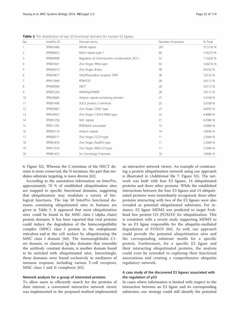
6: Figure S2). Whereas the C-terminus of the HECT do-main is more conserved, the N-terminus, the part that me-diates substrate targeting, is more diverse [62].According to the annotation information on InterPro,
approximately 70 % of established ubiquitination sitesare mapped to specific functional domains, suggestingthat ubiquitination may modulate a variety of bio-logical functions. The top 50 InterPro functional do-mains containing ubiquitinated sites in humans aregiven in Table 3. It appeared that most ubiquitinationsites could be found in the MHC class I (alpha chain)protein domains. It has been reported that viral proteinscould induce the degradation of the histocompatibilitycomplex (MHC) class I protein in the endoplasmicreticulum and at the cell surface by ubiquitinating theMHC class I domain [64]. The immunoglobulin C1-set domain, or classical Ig-like domains that resemblethe antibody constant domain, is another domain foundto be enriched with ubiquitinated sites. Interestingly,these domains were found exclusively in mediators ofimmune response, including various T-cell receptors,MHC class I and II complexes [65].
Network analysis for a group of interested proteinsTo allow users to efficiently search for the proteins oftheir interest, a convenient interactive network viewerwas implemented in the proposed method implemented
an interactive network viewer. An example of construct-ing a protein ubiquitination network using our approachis illustrated in (Additional file 7: Figure S3). The net-work was built with four E3 ligases, 14 ubiquitinatedproteins and three other proteins. While the establishedinteractions between the four E3 ligases and 14 ubiquiti-nated proteins were immediately recognized, three otherproteins interacting with two of the E3 ligases were alsorevealed as potential ubiquitinated substrates. For in-stance, E3 ligase MDM2 was predicted to target Fork-head box protein O3 (FOXO3) for ubiquitination. Thisis consistent with a recent study supporting MDM2 tobe an E3 ligase responsible for the ubiquitin-mediateddegradation of FOXO3 [66]. As well, our approachcould provide the potential ubiquitination sites andthe corresponding substrate motifs for a specificprotein. Furthermore, for a specific E3 ligase andtheir interacting ubiquitinated proteins, the analysiscould even be extended to exploring their functionalassociations and creating a comprehensive ubiquitinregulatory network.
A case study of the discovered E3 ligases associated withthe regulation of p53In cases where information is limited with respect to theinteraction between an E3 ligase and its correspondingsubstrates, our strategy could still identify the potential
Table 2 The distribution of top 20 functional domains for human E3 ligases
No. InterPro ID Domain terms Number of proteins % Total
1 IPR001680 WD40 repeat 287 57.5150 %
2 IPR006652 Kelch repeat type 1 68 13.6273 %
3 IPR000408 Regulator of chromosome condensation, RCC1 55 11.0220 %
4 IPR001841 Zinc finger, RING-type 54 10.8216 %
5 IPR000315 Zinc finger, B-box 48 9.6192 %
6 IPR003877 SPla/RYanodine receptor SPRY 38 7.6152 %
7 IPR013069 BTB/POZ 28 5.6112 %
8 IPR000569 HECT 28 5.6112 %
9 IPR001202 WW/Rsp5/WWP 28 5.6112 %
10 IPR020683 Ankyrin repeat-containing domain 27 5.4108 %
11 IPR001496 SOCS protein, C-terminal 25 5.0100 %
12 IPR002867 Zinc finger, C6HC-type 23 4.6092 %
13 IPR018957 Zinc finger, C3HC4 RING-type 22 4.4088 %
14 IPR001258 NHL repeat 21 4.2084 %
15 IPR011705 BTB/Kelch-associated 15 3.0060 %
16 IPR002110 Ankyrin repeat 14 2.8056 %
17 IPR000571 Zinc finger, CCCH-type 11 2.2044 %
18 IPR001876 Zinc finger, RanBP2-type 11 2.2044 %
19 IPR011016 Zinc finger, RING-CH-type 11 2.2044 %
20 IPR001452 Src homology-3 domain 10 2.0040 %
Huang et al. BMC Systems Biology 2016, 10(Suppl 1):3 Page 32 of 119
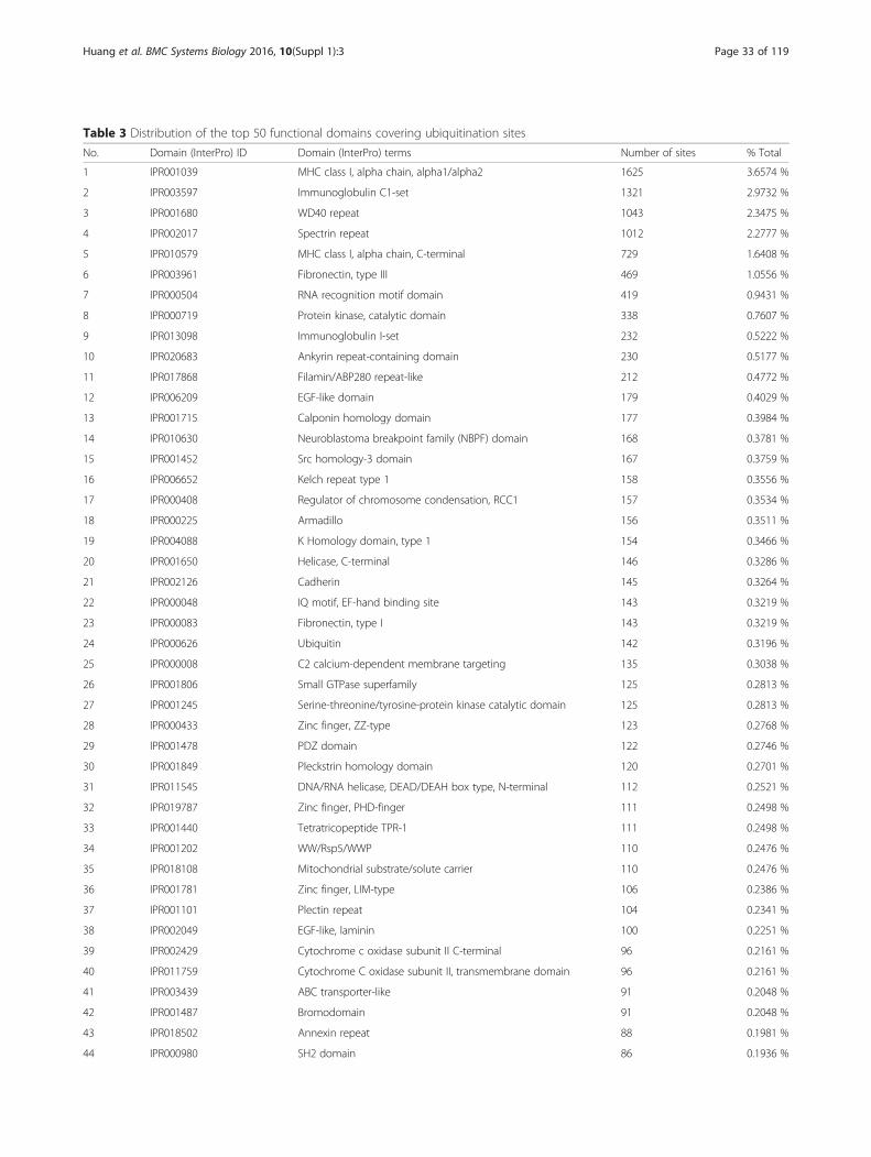
Table 3 Distribution of the top 50 functional domains covering ubiquitination sites
No. Domain (InterPro) ID Domain (InterPro) terms Number of sites % Total
1 IPR001039 MHC class I, alpha chain, alpha1/alpha2 1625 3.6574 %
2 IPR003597 Immunoglobulin C1-set 1321 2.9732 %
3 IPR001680 WD40 repeat 1043 2.3475 %
4 IPR002017 Spectrin repeat 1012 2.2777 %
5 IPR010579 MHC class I, alpha chain, C-terminal 729 1.6408 %
6 IPR003961 Fibronectin, type III 469 1.0556 %
7 IPR000504 RNA recognition motif domain 419 0.9431 %
8 IPR000719 Protein kinase, catalytic domain 338 0.7607 %
9 IPR013098 Immunoglobulin I-set 232 0.5222 %
10 IPR020683 Ankyrin repeat-containing domain 230 0.5177 %
11 IPR017868 Filamin/ABP280 repeat-like 212 0.4772 %
12 IPR006209 EGF-like domain 179 0.4029 %
13 IPR001715 Calponin homology domain 177 0.3984 %
14 IPR010630 Neuroblastoma breakpoint family (NBPF) domain 168 0.3781 %
15 IPR001452 Src homology-3 domain 167 0.3759 %
16 IPR006652 Kelch repeat type 1 158 0.3556 %
17 IPR000408 Regulator of chromosome condensation, RCC1 157 0.3534 %
18 IPR000225 Armadillo 156 0.3511 %
19 IPR004088 K Homology domain, type 1 154 0.3466 %
20 IPR001650 Helicase, C-terminal 146 0.3286 %
21 IPR002126 Cadherin 145 0.3264 %
22 IPR000048 IQ motif, EF-hand binding site 143 0.3219 %
23 IPR000083 Fibronectin, type I 143 0.3219 %
24 IPR000626 Ubiquitin 142 0.3196 %
25 IPR000008 C2 calcium-dependent membrane targeting 135 0.3038 %
26 IPR001806 Small GTPase superfamily 125 0.2813 %
27 IPR001245 Serine-threonine/tyrosine-protein kinase catalytic domain 125 0.2813 %
28 IPR000433 Zinc finger, ZZ-type 123 0.2768 %
29 IPR001478 PDZ domain 122 0.2746 %
30 IPR001849 Pleckstrin homology domain 120 0.2701 %
31 IPR011545 DNA/RNA helicase, DEAD/DEAH box type, N-terminal 112 0.2521 %
32 IPR019787 Zinc finger, PHD-finger 111 0.2498 %
33 IPR001440 Tetratricopeptide TPR-1 111 0.2498 %
34 IPR001202 WW/Rsp5/WWP 110 0.2476 %
35 IPR018108 Mitochondrial substrate/solute carrier 110 0.2476 %
36 IPR001781 Zinc finger, LIM-type 106 0.2386 %
37 IPR001101 Plectin repeat 104 0.2341 %
38 IPR002049 EGF-like, laminin 100 0.2251 %
39 IPR002429 Cytochrome c oxidase subunit II C-terminal 96 0.2161 %
40 IPR011759 Cytochrome C oxidase subunit II, transmembrane domain 96 0.2161 %
41 IPR003439 ABC transporter-like 91 0.2048 %
42 IPR001487 Bromodomain 91 0.2048 %
43 IPR018502 Annexin repeat 88 0.1981 %
44 IPR000980 SH2 domain 86 0.1936 %
Huang et al. BMC Systems Biology 2016, 10(Suppl 1):3 Page 33 of 119
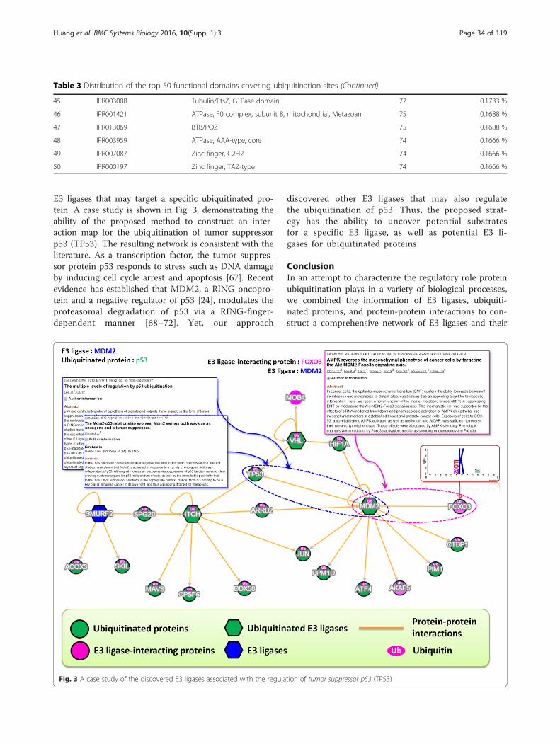
E3 ligases that may target a specific ubiquitinated pro-tein. A case study is shown in Fig. 3, demonstrating theability of the proposed method to construct an inter-action map for the ubiquitination of tumor suppressorp53 (TP53). The resulting network is consistent with theliterature. As a transcription factor, the tumor suppres-sor protein p53 responds to stress such as DNA damageby inducing cell cycle arrest and apoptosis [67]. Recentevidence has established that MDM2, a RING oncopro-tein and a negative regulator of p53 [24], modulates theproteasomal degradation of p53 via a RING-finger-dependent manner [68–72]. Yet, our approach
discovered other E3 ligases that may also regulatethe ubiquitination of p53. Thus, the proposed strat-egy has the ability to uncover potential substratesfor a specific E3 ligase, as well as potential E3 li-gases for ubiquitinated proteins.
ConclusionIn an attempt to characterize the regulatory role proteinubiquitination plays in a variety of biological processes,we combined the information of E3 ligases, ubiquiti-nated proteins, and protein-protein interactions to con-struct a comprehensive network of E3 ligases and their
Table 3 Distribution of the top 50 functional domains covering ubiquitination sites (Continued)
45 IPR003008 Tubulin/FtsZ, GTPase domain 77 0.1733 %
46 IPR001421 ATPase, F0 complex, subunit 8, mitochondrial, Metazoan 75 0.1688 %
47 IPR013069 BTB/POZ 75 0.1688 %
48 IPR003959 ATPase, AAA-type, core 74 0.1666 %
49 IPR007087 Zinc finger, C2H2 74 0.1666 %
50 IPR000197 Zinc finger, TAZ-type 74 0.1666 %
Fig. 3 A case study of the discovered E3 ligases associated with the regulation of tumor suppressor p53 (TP53)
Huang et al. BMC Systems Biology 2016, 10(Suppl 1):3 Page 34 of 119
ubiquitinated substrates. Designed to serve as not only ameaningful platform for investigating E3-substrate regula-tory networks but also a new strategy to uncover potentialE3 ligases for ubiquitinated substrates, the proposed ap-proach allows for the efficient characterization of proteinubiquitination networks from large-scale ubiquitinomedata. With access to more updated data, the proposedscheme can be further refined for the study of E1 activat-ing enzymes, E2 conjugating enzymes, and E3 ubiquitinligases. Also, recent publications regarding the structuralenvironment of experimentally validated ubiquitinationsites based on protein tertiary structures [73–76] could beincorporated to infer the functional interactions betweenthe enzymes and substrates. Finally, confirmed functionalannotations of ubiquitination sites could be extractedfrom the literature via a more advanced informationretrieval system to collect more adequate informationrequired for further functional analyses.
Additional files
Additional file 1: Table S1. Data statistics of protein-protein interactionsobtained from public resources. (PDF 8 kb)
Additional file 2: Figure S1. A conceptual diagram of exploitingCytoscape software to construct protein ubiquitination networks basedon graph theory. (PDF 86 kb)
Additional file 3: Table S2. The entropy plot of sequence logos forMDDLogo-identified motifs obtained from non-homologous ubiquitinationsites in humans. (PDF 86 kb)
Additional file 4: Table S3. Distribution of the top 15 GO annotationsfor human E3 ligases. (PDF 31 kb)
Additional file 5: Table S4. Distribution of the top 20 GO annotationsfor human ubiquitinated proteins. (PDF 52 kb)
Additional file 6: Figure S2. The HECT-type structure of E3 ligases.(PDF 133 kb)
Additional file 7: Figure S3. An example to construct proteinubiquitination network for 21 proteins (containing four E3 ligases and 14ubiquitinated proteins). (PDF 345 kb)
Competing interestsThe authors have declared that no competing interests exist.
Authors’ contributionsTYL and SLW conceived and supervised the project. KYH was responsible forthe project design and computational analyses. KYH and JTYW drafted themanuscript with revisions by TYL and SLW. All authors read and approvedthe final manuscript.
AcknowledgementsThe authors sincerely appreciate the Ministry of Science and Technologyof Taiwan for financially supporting this research under Contract Numberof MOST 103-2221-E-155-020-MY3, MOST 103-2633-E-155-002 andMOST104-2221-E-155-036-MY2.
DeclarationsPublication charge for this work was funded by MOST grant under contractnumber of MOST 103-2221-E-155-020-MY3 and MOST 104-2221-E-155-036-MY2to TYL.
Author details1Department of Computer Science and Engineering, Yuan Ze University,Taoyuan 320, Taiwan. 2Innovation Center for Big Data and Digital
Convergence, Yuan Ze University, Taoyuan 320, Taiwan. 3Department ofObstetrics and Gynecology, Hsinchu Mackay Memorial Hospital, Hsin-Chu300, Taiwan. 4Mackay Junior College of Medicine, Nursing and Management,Taipei 112, Taiwan. 5Department of Medicine, Mackay Medical College, NewTaipei City 252, Taiwan.
Published: 11 January 2016
References1. Hershko A, Ciechanover A. The ubiquitin system. Annu Rev Biochem.
1998;67:425–79.2. Nguyen VN, Huang KY, Huang CH, Chang TH, Bretana N, Lai K, et al.
Characterization and identification of ubiquitin conjugation sites with E3ligase recognition specificities. BMC bioinformatics. 2015;16 Suppl 1:S1.
3. Wagner SA, Beli P, Weinert BT, Nielsen ML, Cox J, Mann M, et al.A proteome-wide, quantitative survey of in vivo ubiquitylation sitesreveals widespread regulatory roles. Molecular & cellular proteomics:MCP. 2011;10(10):M111 013284.
4. Lee TY, Chen SA, Hung HY, Ou YY. Incorporating distant sequence featuresand radial basis function networks to identify ubiquitin conjugation sites.Plos One. 2011;6(3):e17331.
5. Hurley JH, Lee S, Prag G. Ubiquitin-binding domains. Biochem J.2006;399:361–72.
6. Hicke L, Schubert HL, Hill CP. Ubiquitin-binding domains. Nat Rev Mol CellBio. 2005;6(8):610–21.
7. Peng JM, Schwartz D, Elias JE, Thoreen CC, Cheng DM, Marsischky G, et al.A proteomics approach to understanding protein ubiquitination. NatBiotechnol. 2003;21(8):921–6.
8. Wilkinson KD. The discovery of ubiquitin-dependent proteolysis. Proc NatlAcad Sci U S A. 2005;102(43):15280–2.
9. Han Y, Lee H, Park JC, Yi GS. E3Net: a system for exploring E3-mediatedregulatory networks of cellular functions. Mol Cell Proteomics.2012;11(4):O111.014076.
10. Zhang J, Wan L, Dai X, Sun Y, Wei W. Functional characterization ofanaphase promoting complex/cyclosome (APC/C) E3 ubiquitin ligases intumorigenesis. Biochim Biophys Acta. 2014;1845(2):277–93.
11. Manchado E, Eguren M, Malumbres M. The anaphase-promoting complex/cyclosome (APC/C): cell-cycle-dependent and -independent functions.Biochem Soc Trans. 2010;38(Pt 1):65–71.
12. Acquaviva C, Pines J. The anaphase-promoting complex/cyclosome: APC/C.J Cell Sci. 2006;119(Pt 12):2401–4.
13. Spratt DE, Walden H, Shaw GS. RBR E3 ubiquitin ligases: new structures, newinsights, new questions. Biochem J. 2014;458(3):421–37.
14. Paul I, Ghosh MK. The E3 ligase CHIP: insights into its structure andregulation. Biomed Res Int. 2014.
15. Duplan V, Rivas S. E3 ubiquitin-ligases and their target proteins during theregulation of plant innate immunity. Front Plant Sci. 2014;5.
16. Snoek BC, de Wilt LH, Jansen G, Peters GJ. Role of E3 ubiquitin ligases inlung cancer. World journal of clinical oncology. 2013;4(3):58–69.
17. Plechanovova A, Jaffray EG, Tatham MH, Naismith JH, Hay RT. Structure of aRING E3 ligase and ubiquitin-loaded E2 primed for catalysis. Nature.2012;489(7414):115–20.
18. Metzger MB, Hristova VA, Weissman AM. HECT and RING finger families ofE3 ubiquitin ligases at a glance. J Cell Sci. 2012;125(3):531–7.
19. Bernassola F, Karin M, Ciechanover A, Melino G. The HECT family of E3ubiquitin ligases: Multiple players in cancer development. Cancer Cell.2008;14(1):10–21.
20. Scheffner M, Staub O. HECT E3s and human disease. BMC Biochem. 2007;8.21. Mazzucotelli E, Belloni S, Marone D, De Leonardis AM, Guerra D, Fonzo N, et al.
The E3 ubiquitin ligase gene family in plants: regulation by degradation. CurrGenomics. 2006;7(8):509–22.
22. Robinson PA, Ardley HC. Ubiquitin-protein ligases. J Cell Sci. 2004;117(22):5191–4.23. Sun Y. Targeting E3 ubiquitin ligases for cancer therapy. Cancer Biol Ther.
2003;2(6):623–9.24. Manfredi JJ. The Mdm2-p53 relationship evolves: Mdm2 swings both ways
as an oncogene and a tumor suppressor. Genes Dev. 2010;24(15):1580–9.25. Lee H, Yi GS, Park JC. E3Miner: a text mining tool for ubiquitin-protein
ligases. Nucleic Acids Res. 2008;36(Web Server issue):W416–22.26. Sakiyama T, Kawashima S, Yoshizawa AC, Kanehisa M. The construction of a
database for ubiquitin signaling cascade. Genome Informatics. 2003;14:653–4.
Huang et al. BMC Systems Biology 2016, 10(Suppl 1):3 Page 35 of 119
27. Gao T, Liu Z, Wang Y, Cheng H, Yang Q, Guo A, et al. UUCD: a family-baseddatabase of ubiquitin and ubiquitin-like conjugation. Nucleic Acids Res.2013;41(Database issue):D445–51.
28. Du YP, Xu NF, Lu M, Li TT. hUbiquitome: a database of experimentallyverified ubiquitination cascades in humans. Database-Oxford. 2011.
29. Boeckmann B, Bairoch A, Apweiler R, Blatter MC, Estreicher A, Gasteiger E, et al.The SWISS-PROT protein knowledgebase and its supplement TrEMBL in 2003.Nucleic Acids Res. 2003;31(1):365–70.
30. Lee TY, Huang HD, Hung JH, Huang HY, Yang YS, Wang TH. dbPTM: aninformation repository of protein post-translational modification. NucleicAcids Res. 2006;34(Database issue):D622–7.
31. Lu CT, Huang KY, Su MG, Lee TY, Bretana NA, Chang WC, et al. dbPTM 3.0:an informative resource for investigating substrate site specificity andfunctional association of protein post-translational modifications. NucleicAcids Res. 2013;41(D1):D295–305.
32. Su MG, Huang KY, Lu CT, Kao HJ, Chang YH, Lee TY. topPTM: a newmodule of dbPTM for identifying functional post-translationalmodifications in transmembrane proteins. Nucleic Acids Res.2014;42(Database issue):D537–45.
33. Crooks GE, Hon G, Chandonia JM, Brenner SE. WebLogo: a sequence logogenerator. Genome Res. 2004;14(6):1188–90.
34. Schneider TD, Stephens RM. Sequence logos: a new way to displayconsensus sequences. Nucleic Acids Res. 1990;18(20):6097–100.
35. Vacic V, Iakoucheva LM, Radivojac P. Two sample logo: a graphicalrepresentation of the differences between two sets of sequencealignments. Bioinformatics. 2006;22(12):1536–7.
36. Lee TY, Lin ZQ, Hsieh SJ, Bretana NA, Lu CT. Exploiting maximaldependence decomposition to identify conserved motifs from a group ofaligned signal sequences. Bioinformatics. 2011;27(13):1780–7.
37. Wong YH, Lee TY, Liang HK, Huang CM, Wang TY, Yang YH, et al.KinasePhos 2.0: a web server for identifying protein kinase-specificphosphorylation sites based on sequences and coupling patterns. NucleicAcids Res. 2007;35(Web Server issue):W588–94.
38. Huang HD, Lee TY, Tzeng SW, Horng JT. KinasePhos: a web tool foridentifying protein kinase-specific phosphorylation sites. Nucleic Acids Res.2005;33(Web Server issue):W226–9.
39. Lee TY, Bretana NA, Lu CT. PlantPhos: using maximal dependencedecomposition to identify plant phosphorylation sites with substrate sitespecificity. BMC Bioinformatics. 2011;12:261.
40. Bretana NA, Lu CT, Chiang CY, Su MG, Huang KY, Lee TY, et al. Identifyingprotein phosphorylation sites with kinase substrate specificity on humanviruses. PLoS One. 2012;7(7):e40694.
41. Lee TY, Chen YJ, Lu TC, Huang HD. SNOSite: exploiting maximal dependencedecomposition to identify cysteine S-nitrosylation with substrate site specificity.PLoS One. 2011;6(7):e21849.
42. Wu HY, Lu CT, Kao HJ, Chen YJ, Chen YJ, Lee TY. Characterization andidentification of protein O-GlcNAcylation sites with substrate specificity.BMC bioinformatics. 2014;15 Suppl 16:S1.
43. Chen YJ, Lu CT, Huang KY, Wu HY, Chen YJ, Lee TY. GSHSite: exploiting aniteratively statistical method to identify s-glutathionylation sites withsubstrate specificity. PLoS One. 2015;10(4):e0118752.
44. Gene Ontology C, Blake JA, Dolan M, Drabkin H, Hill DP, Li N, et al.Gene ontology annotations and resources. Nucleic Acids Res.2013;41(Database issue):D530–5.
45. Hunter S, Jones P, Mitchell A, Apweiler R, Attwood TK, Bateman A, et al.InterPro in 2011: new developments in the family and domain predictiondatabase. Nucleic Acids Res. 2011;40(Database issue):D306–12.
46. Kanehisa M, Goto S, Sato Y, Furumichi M, Tanabe M. KEGG for integrationand interpretation of large-scale molecular data sets. Nucleic Acids Res.2012;40(Database issue):D109–14.
47. Mukhopadhyay D, Riezman H. Proteasome-independent functions ofubiquitin in endocytosis and signaling. Science. 2007;315(5809):201–5.
48. Schnell JD, Hicke L. Non-traditional functions of ubiquitin and ubiquitin-bindingproteins. The Journal of biological chemistry. 2003;278(38):35857–60.
49. Glickman MH, Ciechanover A. The ubiquitin-proteasome proteolytic pathway:destruction for the sake of construction. Physiol Rev. 2002;82(2):373–428.
50. Bairoch A. PROSITE: a dictionary of sites and patterns in proteins. NucleicAcids Res. 1991;19(Suppl):2241–5.
51. Attwood TK, Beck ME, Bleasby AJ, Parry-Smith DJ. PRINTS–a database ofprotein motif fingerprints. Nucleic Acids Res. 1994;22(17):3590–6.
52. Sonnhammer EL, Eddy SR, Durbin R. Pfam: a comprehensive database of proteindomain families based on seed alignments. Proteins. 1997;28(3):405–20.
53. Corpet F, Gouzy J, Kahn D. The ProDom database of protein domainfamilies. Nucleic Acids Res. 1998;26(1):323–6.
54. Ogata H, Goto S, Sato K, Fujibuchi W, Bono H, Kanehisa M. KEGG: Kyotoencyclopedia of genes and genomes. Nucleic Acids Res. 1999;27(1):29–34.
55. von Mering C, Huynen M, Jaeggi D, Schmidt S, Bork P, Snel B. STRING: adatabase of predicted functional associations between proteins. NucleicAcids Res. 2003;31(1):258–61.
56. Lee TY, Bo-Kai Hsu J, Chang WC, Huang HD. RegPhos: a system to explorethe protein kinase-substrate phosphorylation network in humans. NucleicAcids Res. 2011;39(Database issue):D777–87.
57. Huang KY, Wu HY, Chen YJ, Lu CT, Su MG, Hsieh YC, et al. RegPhos 2.0: anupdated resource to explore protein kinase-substrate phosphorylationnetworks in mammals. Database : the journal of biological databases andcuration. 2014;2014(0):bau034.
58. Kohl M, Wiese S, Warscheid B. Cytoscape: software for visualization andanalysis of biological networks. Methods Mol Biol. 2010;696:291–303.
59. Li W, Bengtson MH, Ulbrich A, Matsuda A, Reddy VA, Orth A, et al. Genome-wide and functional annotation of human E3 ubiquitin ligases identifiesMULAN, a mitochondrial E3 that regulates the organelle’s dynamics andsignaling. Plos One. 2008;3(1):e1487.
60. Berndsen CE, Wolberger C. New insights into ubiquitin E3 ligasemechanism. Nat Struct Mol Biol. 2014;21(4):301–7.
61. Zheng N, Wang P, Jeffrey PD, Pavletich NP. Structure of a c-Cbl-UbcH7complex: RING domain function in ubiquitin-protein ligases. Cell.2000;102(4):533–9.
62. Rotin D, Kumar S. Physiological functions of the HECT family of ubiquitinligases. Nat Rev Mol Cell Biol. 2009;10(6):398–409.
63. Huibregtse JM, Scheffner M, Beaudenon S, Howley PM. A family of proteinsstructurally and functionally related to the E6-AP ubiquitin-protein ligase.Proc Natl Acad Sci U S A. 1995;92(11):5249.
64. Burr ML, Boname JM, Lehner PJ. Studying ubiquitination of MHC class Imolecules. Methods Mol Biol. 2013;960:109–25.
65. Cresswell P, Ackerman AL, Giodini A, Peaper DR, Wearsch PA. Mechanismsof MHC class I-restricted antigen processing and cross-presentation.Immunol Rev. 2005;207:145–57.
66. Chou CC, Lee KH, Lai IL, Wang D, Mo X, Kulp SK, et al. AMPK reverses themesenchymal phenotype of cancer cells by targeting the Akt-MDM2-Foxo3asignaling axis. Cancer Res. 2014;74(17):4783–95.
67. Lee JT, Gu W. The multiple levels of regulation by p53 ubiquitination. CellDeath Differ. 2010;17(1):86–92.
68. Haupt Y, Maya R, Kazaz A, Oren M. Mdm2 promotes the rapid degradationof p53. Nature. 1997;387(6630):296–9.
69. Kubbutat MHG, Jones SN, Vousden KH. Regulation of p53 stability byMdm2. Nature. 1997;387(6630):299–303.
70. Honda R, Tanaka H, Yasuda H. Oncoprotein MDM2 is a ubiquitin ligase E3for tumor suppressor p53. Febs Lett. 1997;420(1):25–7.
71. Fang SY, Jensen JP, Ludwig RL, Vousden KH, Weissman AM. Mdm2 is a RINGfinger-dependent ubiquitin protein ligase for itself and p53. J Biol Chem.2000;275(12):8945–51.
72. Honda R, Yasuda H. Activity of MDM2, a ubiquitin Ligase, toward p53 oritself is dependent on the RING finger domain of the ligase. Oncogene.2000;19(11):1473–6.
73. Su MG, Lee TY. Incorporating substrate sequence motifs and spatial aminoacid composition to identify kinase-specific phosphorylation sites on proteinthree-dimensional structures. BMC bioinformatics. 2013;14 Suppl 16:S2.
74. Lee TY, Chen YJ, Lu CT, Ching WC, Teng YC, Huang HD. dbSNO: a databaseof cysteine S-nitrosylation. Bioinformatics. 2012;28(17):2293–5.
75. Chen YJ, Lu CT, Su MG, Huang KY, Ching WC, Yang HH, et al. dbSNO 2.0: aresource for exploring structural environment, functional and diseaseassociation and regulatory network of protein S-nitrosylation. Nucleic AcidsRes. 2015;43(Database issue):D503–11.
76. Chen YJ, Lu CT, Lee TY, Chen YJ. dbGSH: a database of S-glutathionylation.Bioinformatics. 2014;30(16):2386–8.
Huang et al. BMC Systems Biology 2016, 10(Suppl 1):3 Page 36 of 119