Embed Size (px)
Citation preview

61
A new species of Clausenia Ishii (Hymenoptera : Encyrtidae) from Israel
By DAVID ROSEN The Hebrew University, Faculty of Agriculture, Rehovot, Israel
SYNOPSIS
Both sexes of a new species of Clausenia Ishii, parasitic in a species of Planococcus infesting vines in Israel, are described and compared with the type of the genus, C. purpurea Ishii.
THE genus Clausenia Ishii has hitherto been known to include only the single species C . purpurea Ishii, parasitic in mealybugs of the genus Pseudococcus Westwood. This species, first discovered in Japan as a parasite of a Pseudococcus sp. on citrus (Ishii, 1923), was apparently accidentally introduced into the United States, where it proved to be an efficient parasite of P. comstocki (Kuwana) (Clausen, 1956, and others). When P. citriculus Green, then misidentified as P. comstocki, turned up in Palestine in the late nineteen-thirties and became a major pest of citrus along the coastal plain, C. purpurea was introduced from Japan to control it (Rivnay, 1942). The parasite proved very efficient, displacing local parasites that had taken to the pest and reducing the populations of P. citriculus far below the threshold of economic injury (Rivnay, 1946; Bodenheimer, 1951). In recent surveys I have found P. citriculus to be very rare on citrus, C. purpurea being by far its dominant parasite.
The new species of Clausenia described below was reared by Mr. M. J. Berlinger, of the Volcani Institute of Agricultural Research, from a grape-infesting mealybug of the genus Planococcus Ferris. Samples of the parasitised mealybug were collected in the Negev, where this species of Clausenia is a frequent parasite of its host and appears to be of considerable economic importance.
Cluuseniu josefi sp. n. (Plates I and 11) Female
Head in frontal view wider than long (17 : 14). Scrobes moderately deep, converging, facial ridge low. Toruli elongated, separated from each other by slightly more than half width of fronto- vertex (4 : 7), separated from clypeal margin by approximately half their own length. Eyes large, sparsely clothed with short setae. Cheek slightly shorter than half diameter of an eye. Head in dorsal view much wider than median length (1 3 : 4), both frontal and occipital margins concave, the latter acute. Frontovertex slightly longer than wide (8 : 7), occupying one-third of width of head. Posterior ocelli separated from each other more than from anterior ocellus (7 : 5) , separated from occipital margin by one-half, and from the orbits by one-third the diameter of an ocellus. Labrum narrow, transverse, bearing a line of long setae; mandibles bidentate, triangular in frontal view (Plate I, fig. 1); maxillary palpi (Plate I, fig. 2) 4-segmented, first 3 segments subequal, apical joint slightly shorter than first 3 combined; labial palpi (Plate I, fig. 3) 3-segmented, second segment the shortest, apical segment the longest. Antennae (Plate I, fig. 4) elongated, slender, more so than in C. purpurea (Plate 111, fig. 15). Scape cylindrical, slender, slightly more than 7 times as long as wide; radicle elongated, about 3 times as long as wide, longer than one-fifth length of scape. Pedicel elongated, twice as long as wide, twice as long as first funicular joint. Funicle slender, slightly longer than scape (12 : 11); all funicular segments longer than wide (length measured along dorsal margin), second to sixth bearing longitudinal sensillae; first funicular segment the shortest, longer than wide (17 : 14); second to fourth segments subequal, more than 1.5 times as long as wide (27 : 16), con- siderably longer than the first (27 : 17); fifth segment 1.5 times as long as wide, imperceptibly wider than preceding segments; sixth segment slightly longer and wider, almost 1.5 times as long as wide (29 : 20). Club longer than the three preceding segments combined, more than 1.5 times wider than preceding segment, almost 3 times as long as wide.
Thorax (Plate I, fig. 5 ) narrower than head (23 : 26); pronotum very short; mesoscutum 1.5 times as wide as long; parapsidal grooves incomplete, very short, ending in anterior half of meso- swtum; axiUae pointed, barely touching, protruding backwards on either side of scutellum; the Proc. R. ent. SOC. Lond. (B). 34 (5-6). Pp. 61-64,3 plates. 1965.
62 D. Rosen on a nen’ species of
latter, including well-defined postscutellum, slightly shorter than mesoscutum. Mid-tibia1 spur slightly shorter than first tarsal segment, latter equal in length to the two following segments com- bined. Fore wings (Plate I, figs. 6-7) hyaline, twice as long as broad. Costal cell about 9 times as long as wide, bearing one row of dorsal setae. Submarginal vein slightly thickened on apical third; marginal vein elongated, widening apically, about 4 times as long as wide, about one-seventh length of submarginal vein; stigmal vein as long as marginal; postmarginal slightly longer (6 : 5) . Disc evenly ciliated except on basal one-third; setae on central area much more slender than those on apical one-third, difference much more pronounced than in C. purpurea; linea calva broad, un- interrupted; marginal fringe short. Hind wing (see Plate 11, fig. 13) hyaline, almost 3 times as long as broad (14 : 5 ) ; marginal fringe short.
Abdomen (Plate I, figs. 8-9) slightly longer than thorax and propodeum combined; paratergites well developed; hypopygium prominent; ovipositor concealed at rest.
Face smooth, polished; frontovertex granular; mesoscutum with delicate polygonal sculptures, with numerous small punctules bearing slender setae; scutellum smooth, longitudinally shagreened, bearing 2 submedian rows of setae; axillae bearing a single row along posterior margin; metanotum slightly reticulated on either side; propodeum almost smooth, with a single longitudinal ridge leading to the spiracle. Abdomen lustrous, with delicate, shallow, polygonal sculpture.
General colour black, with strong metallic reflections. Face shining violet; frontovertex with faint violaceous reflections. Eyes reddish-brown, ocelli red, mandibles deep brown. Antennae entirely black, with a metallic greenish tinge on scape and faint bronzy and violaceous reflections on flagellum. Mesoscutum with strong greenish-blue reflections on anterior half, violet on posterior half; axillae with strong violaceous reflections; scutellum with an admixture of violet and bronzy reflections, turning to shining metallic green apically; tegulae black with a faint greenish-blue tinge; mesopleura purple; metanotum and propodeum with dull violaceous reflections. Legs black with strong violaceous reflections, except the trochanters, apical parts of fore and hind tibiae, up to apical two-thirds of mid-tibiae, and first 4 tarsal segments, which are brownish-yellow; femora tipped with yellowish on both ends. Wings hyaline, venation brownish-yellow. Abdomen with strong greenish- yellow lustre. Setae on thorax bronzy.
Length: 0.97-1.63 mm. Male
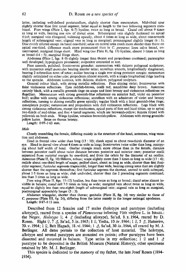
Closely resembling the female, differing mainly in the structure of the head, antennae, wing vena- tion and abdomen.
Head in frontal view wider than long (13 : 10); cheek equal to about two-thirds diameter of an eye. Head in dorsal view about 4 times as wide as long; frontovertex twice wider than long, occupy- ing about half width of head. Ocellar triangle much more obtuse than in the female, distance between posterior ocelli about twice distance between posterior and anterior ones; posterior ocelli separated from occipital margin by one-third, and from the orbits by the diameter of an ocellus. Antennae (Plate 11, fig. 10) filiform, robust; scape slightly more than 3 times as long as wide (13 : 4); radicle about one-third length of scape; pedicel short, about as long as wide, shorter than first funi- cular segment; funicular segments trapezoidal, longer than wide, bearing strong setae that are slightly shorter than the segments themselves; first funicular segment the shortest, second to sixth subequal, about 1.5 times as long as wide; club undivided, shorter than the 2 preceding segments combined, less than 3 times as long as wide.
Fore wing (Plate 11, figs. 11-12) hyaline, less than twice as long as broad; discal setae almost in- visible in balsam; costal cell 7.5 times as long as wide; marginal vein about twice as long as wide, equal to slightly less than one-eighth length of submarginal vein; stigmal vein as long as marginal, postmarginal appreciably longer (5 : 3).
Abdomen triangular, shorter than thorax; genitalia (Plate 11, fig. 14) very similar to those of C. purpurea (Plate 111, fig. 24), differing from the latter mainly in the longer aedeagal apodemes.
Length: 087-1.13 mm.
Described from 12 females and 17 males (holotype and paratypes (including allotype)), reared from a species of Planococcus infesting Vitis vinifera L. in ISRAEL: the Negev. Holotype 9, 4 8 (including allotype), Sa’ad, 8.x.1964, reared by D. Rosen. Eight Q, 7 8, Sa’ad, 5.ix. 1963; 1 9, Yesha, 1O.iv. 1964; 1 9, 5 8, Hatserim, 20.v.1964; 1 Q, Beit Hagadi, 18.vi. 1964; 1 8, Sa’ad, 30.ix.1964, all reared by M. J. Berlinger. All dates pertain to the collection of host material. The holotype, allotype and several paratypes are mounted on points ; other paratypes have been dissected and mounted in balsam. Type series in my collection; 1 $? and 1 8 paratype to be deposited in the British Museum (Natural History) ; other specimens retained by Mr. M. J. Berlinger.
This species is dedicated to the memory of my father, the late Josef Rosen (1894- 1954).
Clausenia Ishii from Israel 63
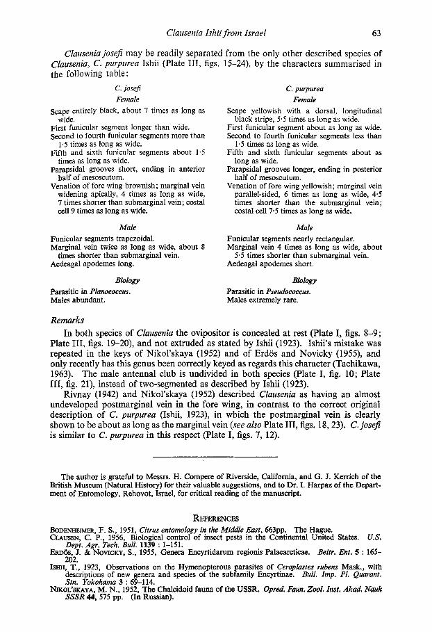
Clausenia josefi may be readily separated from the only other described species of clausenia, C . purpurea Ishii (Plate 111, figs. 15-24), by the characters summarised in the following table :
C. josefi Female
Scape entirely black, about 7 times as long as
First funicular segment longer than wide. Second to fourth funicular segments more than
1.5 times as long as wide. Fifth and sixth funicular segments about 1.5
times as long as wide. pwpsidal grooves short, ending in anterior
half of mesoscutum. Venation of fore wing brownish; marginal vein
widening apically, 4 times as long as wide, 7 times shorter than submarginal vein; costal cell 9 times as long as wide.
wide.
Male Funicular segments trapezoidal. Marginal vein twice as long as wide, about 8
Aedeagal apodemes long. times shorter than submarginal vein.
Biology Parasitic in Planococcus. Males abundant.
C. purpurea Female
Scape yellowish with a dorsal, longitudinal
First funicular segment about as long as wide. Second to fourth funicular segments less than
1.5 times as long as wide. Fifth and sixth funicular segments about as
long as wide. Parapsidal grooves longer, ending in posterior
half of mesoscutum. Venation of fore wing yellowish; marginal vein
parallel-sided, 6 times as long as wide, 4 3 times shorter than the submarginal vein; costal cell 7.5 times as long as wide.
black stripe, 5.5 times as long as wide.
Male Funicular segments nearly rectangular. Marginal vein 4 times as long as wide, about
Aedeagal apodemes short. 5 3 times shorter than submarginal vein.
Biology Parasitic in Pseudococcus. Males extremely rare.
Remarks In both species of Clausenia the ovipositor is concealed at rest (Plate I, figs. 8-9;
Plate 111, figs. 19-20), and not extruded as stated by Ishii (1923). Ishii’s mistake was repeated in the keys of Nikol’skaya (1952) and of Erdos and Novicky (1955), and only recently has this genus been correctly keyed as regards this character (Tachikawa, 1963). The male antenna1 club is undivided in both species (Plate I, fig. 10; Plate 111, fig. 21), instead of two-segmented as described by Ishii (1923).
Rivnay (1942) and Nikol’skaya (1952) described Cluusenia as having an almost undeveloped postmarginal vein in the fore wing, in contrast to the correct original description of C . purpurea (Ishii, 1923), in which the postmarginal vein is clearly shown to be about as long as the marginal vein (see also Plate 111, figs. 18,23). C. joseji is similar to C. purpureu in this respect (Plate I, figs. 7, 12).
The author is grateful to Messrs. H. Compere of Riverside, California, and G. J. Kerrich of the British Museum (Natural History) for their valuable suggestions, and to Dr. I. Harpaz of the Depart- ment of Entomology, Rehovot, Israel, for critical reading of the manuscript.
REFERENCES BODENHEIMER, F. S., 1951, Citrus entomology in the Middle East, 663pp. The Hague. CLAUSEN, C. P., 1956, Biological control of insect pests in the Continental United States. US.
Dept. Agr. Tech. Bull. 1139 : 1-151. ERDOS, J. & NOVICKY, S., 1955, Genera Encyrtidarum regionis Palaearcticae. Beitr. Ent. 5 : 165-
202. ISHII, T., 1923, Observations on the Hymenopterous parasites of Ceroplastes rubens Mask., with
descriptions of new genera and species of the subfamily Encyrtinae. Bull. Imp. PI. Quarant. Stn. Yokohama 3 : 69-114.
NIKOL’SKAYA, M. N., 1952, The Chalcidoid fauna of the USSR. Opred. Faun. Zool. Znst, Akad. Nauk SSSR 44,575 pp. (In Russian).
64 D. Rosen on a new species of Clausenia Isliii f rom Israel
RIVNAY, E., 1942, Clausenia purpurea Ishii, a parasite of Pseudococcus comstocki Kuw. introduced
- 1946, The status of Clauseniapurpurea Ishii and its competition with other parasites of Pseudo- Zbid. 30 : 11-19.
TACHIKAWA, T., 1963, Revisional studies on the Encyrtidae of Japan (Hymenoptera : Chalcidoidea).
into Palestine. Bull. SOC. Fouad ler Ent. 26 : 1-19.
coccus comstocki Kuw. in Palestine.
Mern. Ehime Univ. Sect. 6 (Agr.) 9 : 1-264.
PLATE I Clausenia josefi sp. n., female
FIG. 1.-Mandibles in frontal and lateral view. FIG. 2.-Palpus maxillaris. FIG. 3.-Palpus labialis. FIG. 4.-Antenna. FIG. 5.-Thorax and propodeum. FIG. 6.-Fore wing. FIG. 7.-Venation of fore wing. FIG. 8.-Abdomen, dorsal view. FIG. 9.-Abdomen, lateral view.
PLATE I1 Clausenia josefi sp. n., male
FIG. 10.-Antenna. FIG. 11 .-Fore wing. FIG. 12.-Venation of fore wing. FIG. 13.-Hind wing. FIG. 14.4enitalia.
PLATEIII Clausenia purpurea Ishii
FIG. 15.-Antenna, female. FIG. 16.-Thorax and propodeum, female. FIG. 17.-Fore wing, female. FIG. 18.-Venation of fore wing, female. FIG. 19.-Abdomen, dorsal view, female (at, abdominal tergite; pg, pygostylus; pt, paratergite;
FIG. 20.-Abdomen, lateral view, female. FIG. 21 .-Antenna, male. FIG. 22.-Fore wing, male. FIG. 23.-Venation of fore wing, male. FIG. %.-Genitalia, male.
sp, spiracle).