Embed Size (px)
Citation preview
A New Strategy for Modulating Chemotherapy-InducedAlopecia, Using PTH/PTHrP Receptor Agonist andAntagonist
Eva M.J. Peters,*² Kerstin Foitzik,*² Ralf Paus,² Swapna Ray,* and Michael F. Holick*1
*Department of Medicine, Boston University Medical Center, U.S.A.; ²Department of Dermatology, University Hospital Eppendorf, Hamburg,
Germany
Parathyroid hormone (PTH) related peptide(PTHrP) and the PTH/PTHrP receptor (PTH/PTHrP-R) show prominent cutaneous expression,where this signaling system may exert importantparacrine and/or autocrine functions, such as in hairgrowth control. Chemotherapy-induced alopecia ±one of the fundamental unsolved problems of clinicaloncology ± is driven in part by de®ned abnormalitiesin hair follicle cycling. We have therefore exploredthe therapeutic potential of a PTH/PTHrP-R agonistand two PTH/PTHrP-R antagonists in a mousemodel of cyclophosphamide-induced alopecia.Intraperitoneal administration of the agonist PTH(1±34) or the antagonists PTH(7±34) and PTHrP(7±34)signi®cantly altered the follicular response to cyclo-phosphamide in vivo. PTH(7±34) and PTHrP(7±34)shifted it towards a mild form of ``dystrophic ana-gen'', associated with a signi®cant reduction inapoptotic (TUNEL+) hair bulb cells, thus mitigating
the degree of follicle damage and retarding the onsetof cyclophosphamide-induced alopecia. PTH(1±34),in contrast, forced hair follicles into ``dystrophiccatagen'', associated with enhanced intrafollicularapoptosis. We had previously shown that an inducedshift in the follicular damage-response towards ``dys-trophic catagen'' mitigates cyclophosphamide-induced alopecia, whereas a shift towards ``dys-trophic catagen'' initially enhanced the hair loss, yetsubsequently promoted accelerated hair folliclerecovery. Therefore, this study in an established ani-mal model of chemotherapy-induced alopecia,which closely mimics human chemotherapy-inducedalopecia, strongly encourages the exploration ofPTH/PTHrP-R agonists and antagonists as noveltherapeutic agents in chemotherapy-induced alope-cia. Key words: cancer therapy/hair loss/parathyroidhormone/parathyroid hormone related peptide. J InvestDermatol 117:173±178, 2001
Parathyroid hormone related peptide (PTHrP), the majorcausative agent of malignancy-associated hypercalcemia,plays a role in regulating keratinocyte proliferation anddifferentiation (Merendino et al, 1986; Merryman et al,1993; Holick et al, 1994; Kaiser et al, 1994;
Wysolmerski et al, 1994; Kremer et al, 1996). In human and ratskin, PTHrP is produced by keratinocytes (Danks et al, 1989; Leeet al, 1995; Shin et al, 1997), whereas the PTH/PTHrP receptor(PTH/PTHrP-R) is expressed by ®broblasts (Hana®n et al, 1995;Lee et al, 1995). PTHrP synthesis and release from keratinocytes areenhanced under conditions in which keratinocyte differentiation isinduced, e.g., by ®broblast-derived growth factors (Kremer et al,1991; Grone et al, 1996). PTH/PTHrP-R agonists stimulateinsulin-like growth factor I production by ®broblasts (Wu et al,1987; Shin et al, 1997), which acts as a cell cycle progression factorin keratinocytes (Tsuboi et al, 1992; Ristow, 1996; Vogt et al,
1997). PTHrP may thus be involved in dermo-epidermal(epithelial±mesenchymal) cross-talk, acting as a paracrine factor,which promotes keratinocyte differentiation and regulates prolif-eration (Merryman et al, 1993; Holick et al, 1994; Kaiser et al,1994). PTH/PTHrP-R agonists can inhibit proliferation and canstimulate differentiation of keratinocytes (Holick et al, 1994; Kaiseret al, 1994), probably via an additional PTH/PTHrP-R subtypethat remains to be identi®ed (Henderson et al, 1992; Orloff et al,1992, 1995).
Increasing evidence suggests that PTH/PTHrP-R-mediatedsignaling is also involved in hair growth control, speci®cally inthe control of hair follicle cycling (Schilli et al, 1997). Humankeratin 14 promoter-PTHrP transgenic mice show a retardation ofor even lack hair follicle initiation (Wysolmerski et al, 1994). Whenthe synthetic PTH/PTHrP-R antagonist PTH(7±34) was givenintraperitoneally to hairless mice, it increased hair growth (Holicket al, 1994). PTH(7±34) also accelerated anagen development ininfantile C57BL/6 mice with normal hair growth patterns, andinhibited spontaneous hair follicle regression (catagen) in adolescentC57BL/6 mice (Schilli et al, 1997).
As abnormalities of hair follicle cycling are at the basis ofmost clinically important human hair growth disorders (Paus et al,1996; Paus and Cotsarelis, 1999), these hair-cycle-modulatoryproperties of a PTH/PTHrP-R antagonist encouraged us toexplore both antagonists and an agonist of the PTH/PTHrP-R in
0022-202X/01/$15.00 ´ Copyright # 2001 by The Society for Investigative Dermatology, Inc.
173
Manuscript received October 5, 2000; revised January 22, 2001;accepted for publication March 30, 2001.
Reprint requests to: Dr. M. F. Holick, Department of Medicine,Vitamin D, Skin and Bone Research Laboratory, Boston UniversityMedical Center, Boston, MA 02118. Email: [email protected]
Abbreviations: CYP, cyclophosphamide; HCS, hair cycle score; p.d.,post depilation; PTH, parathyroid hormone; PTHrP, parathyroid hormonerelated peptide; PTH/PTHrP-R, PTH/PTHrP receptor.
1Declared con¯ict of interest.

the chemotherapy-induced alopecia C57BL/6 mouse model forhair research (Paus et al, 1990, 1994a, b). Speci®cally, we wereinterested in determining whether PTH/PTHrP-R agonist orantagonist might be exploitable to manipulate chemotherapy-induced alopecia in these mice, which had been shown to closelymimic human chemotherapy-induced alopecia (Paus et al, 1994a,1996; Slominski et al, 1996).
Chemotherapy-induced alopecia remains one of the chiefunresolved clinical problems in oncologic practice, and asubstantial source of patient suffering from the psychologicallydevastating effects of sudden and often dramatic hair loss. Wetherefore reasoned that the hair-cycle-modulatory properties ofPTH/PTHrP-R-mediated signaling might offer a novel tool fortherapeutic hair growth manipulation in patients with chemo-therapy-induced alopecia, who show striking abnormality of hairfollicle cycling (Paus et al, 1994a, 1996; Paus and Cotsarelis,1999). Using this mouse model of chemotherapy-inducedalopecia, we had previously shown that the onset and degreeof alopecia and the hair regrowth after intraperitoneal treatmentwith cyclophosphamide (CYP) re¯ect varying degrees of follicledamage (Paus et al, 1994a; Maurer et al, 1997a), which can bestbe judged from the degree of pigmentary disturbances (``dys-trophy'') in the hair follicle (Slominski et al, 1996) and themassively upregulated apoptosis of hair bulb keratinocytes as aconsequence of chemotherapy (Lindner et al, 1997).Chemotherapy-damaged anagen hair follicles respond in twodistinct patterns. The so-called dystrophic anagen pathwayresults in a less severe degree of follicle damage that actuallyleads to a prolongation of the normal duration of anagen, but isassociated with a relatively slow, incomplete follicle recoveryand regrowth of normal hair shafts. The so-called dystrophiccatagen pathway results in maximal follicle damage, yet alsoallows the fastest recovery from chemotherapy-induced alopeciaand the fastest regrowth of normally pigmented hair, becausethe damaged anagen follicle is immediately deconstructed topermit the generation of a new, normally functioning anagenfollicle (Paus et al, 1994a, 1996).
We therefore hypothesized that a PTH/PTHrP-R agonistmight favor the dystrophic catagen pathway by upregulatingfollicle keratinocyte differentiation and/or apoptosis, whereas aPTH/PTHrP-R antagonist might favor the dystrophic anagenpathway by stimulating hair follicle keratinocyte proliferation,possibly as a consequence of antagonizing the inhibitory effectsof local PTH/PTHrP on keratinocyte proliferation. This wouldhave important conceptual implications for the design of newtherapeutic strategies to counteract human chemotherapy-induced alopecia (either by mitigating the degree of alopeciavia favoring dystrophic anagen, or by accelerating normal hairregrowth via stimulation of dystrophic catagen). In addition, thiswould provide additional insights into the biologic signi®canceof PTH/PTHrP-R-mediated signaling in hair follicle biology.Therefore, we investigated the effects of parenteral administra-tion of a PTH/PTHrP-R agonist [PTH(1±34)] and two PTH/
PTHrP-R antagonists [PTH(7±34) and PTHrP(7±34)] onCYP-induced alopecia, hair follicle cycling, and keratinocyteapoptosis in C57BL/6 mice after CYP treatment.
MATERIALS AND METHODS
Mice Six- to 9-wk-old, syngeneic, female C57BL/6 mice in thetelogen stage of the hair cycle were purchased from Jackson Laboratories(Bar Harbour, ME). The mice were housed in community cages with 12h light periods at animal facilities in the Boston University MedicalCenter, and were fed water and mouse chow ad libitum.
PTH and PTHrP analogs The PTH(1±34) human [PTH(1±34)],PTHrP(1±34) amide human [PTHrP(1±34)], PTHrP(7±34) amide human[PTHrP(7±34)] and Nle8,Tyr34-PTH(7±34) amide bovine [PTH(7±34)]were purchased from Bachem California (Torrance, CA).
Hair cycle induction and chemotherapy-induced alopeciamodel Anagen was induced in the back skin of mice in the telogenphase of the hair cycle (identi®ed by their homogeneously pink backskin color) by depilation. Depilation was accomplished by applying a 1:1melted wax/rosin mixture under anesthesia, as described previously (Pauset al, 1990). After hardening, the wax/rosin mixture was peeled off theskin, plucking out all telogen hair shafts, which induces the predictableand highly synchronized development of anagen follicles that aremorphologically indistinguishable from spontaneous anagen follicles(Chase, 1954; Slominski and Paus, 1993). On day 9 post depilation(p.d.), i.e., when all hair follicles in the depilated area had reached earlyanagen VI, a single dose of CYP (120 mg per g bodyweight; obtainedfrom Novartis, Basel, Switzerland) was administered intraperitoneally,which induces hair follicle dystrophy in all anagen hair follicles asdescribed previously (Paus et al, 1994a, 1996; Maurer et al, 1997a).
Pharmacologic manipulation of chemotherapy-induced alopeciaby PTH(7±34) or PTHrP(7±34) PTH(7±34), PTHrP(7±34), orPTH(1±34) (50 mg per kg bodyweight) was injected twice (at 8:00 a.m.and 12:00 p.m. Eastern Standard Time) intraperitoneally on days 6, 7, 8,10, 12, and 13 p.d. to a total of 23±29 mice per group in three separateexperiments that yielded highly comparable results. Control micereceived the vehicle or distilled H2O and CYP only. On day 9 p.d. micereceived three injections once every 2 h of one of the PTH/PTHrPanalogs, and a single additional injection of CYP (120 mg per kgbodyweight) 30 min after the second injection of the PTH/PTHrPanalog (Table I). All tested substances were dissolved in deionized H2O,aliquoted, lyophilized in glass tubes, and stored at ±80°C until needed, toguarantee peptide stability. Immediately prior to use, aliquoted portionswere dissolved in a total volume of deionized H2O and injectedintraperitoneally into each test mouse.
Mice were killed by cervical dislocation on days 13, 14, or 30 p.d.Back skin from the depilated skin area (reaching from the midlinebetween the ears to the insertion of the tail) was harvested perpendicularto the paravertebral line in order to obtain longitudinal hair folliclesections. Skin samples were immediately frozen in liquid nitrogen andembedded as described (Paus et al, 1994a; Lindner et al, 1997; Maureret al, 1997a).
Evaluation of chemotherapy-induced alopecia After the CYPinjection on day 9 p.d., the back skin of test and control animals wasexamined daily for signs of alopecia. In one of the three separateexperiments, six mice out of each group were kept alive until day 30
Table I. Treatment schedules for PTH and PTHrP analogs and CYP in depilated C57BL/6 mice
Tested compound Conc. tested Days of applicationPhotodocumentation/macroscopic evaluation Day of harvesting
PTH(7±34) 50 mg per kg bodyweight 6 7 8 9 10 11 12 7 8 9 1 1 1 1 1 13, 14 or 302x 2x 2x 3x 2x 2x 2x 0 1 2 3 4
PTHrP(7±34) 50 mg per kg bodyweight 6 7 8 9 10 12 1 7 8 9 1 1 1 1 1 13, 14 or 302x 2x 2x 3x 2x 2x 2x 0 1 2 3 4
PTH(1±34) 50 mg per kg bodyweight 6 7 8 9 10 12 13 7 8 9 1 1 1 1 1 13, 14 or 302x 2x 2x 3x 2x 2x 2x 0 1 2 3 4
CYP 120 mg per kg bodyweight 91x
174 PETERS ET AL THE JOURNAL OF INVESTIGATIVE DERMATOLOGY

p.d. so as to follow the course of alopecia and hair regrowth afterchemotherapy-induced alopecia (Paus et al, 1994). Mice werephotographed under standardized conditions up to twice daily from day7. The macroscopic data were derived from an analysis of micephotographed on day 14 p.d. Visual quanti®cation of hair loss wasperformed on photomicrographs taken daily. The length of the totaldepilated area and the length of the back skin in anagen (black skin,growing hair), as well as the length of the back skin with signs ofalopecia or total hair loss were measured in centimeters. The ratio ofanagen skin to total back skin for each mouse was determined. Thesenumbers were expressed in percent and pooled for each group to allowfor statistical analysis. These macroscopic observations were correlated byquantitative histomorphometry. Hair cycle staging was performed on10 mm skin sections at 2003 magni®cation under a Zeiss Axioscopemicroscope on test or control skin of all mice, and harvested on days 13or 14 p.d., as previously described (Paus et al, 1994a; Maurer et al,1997a, b). In short, a minimum of 20 randomly chosen hair folliclesfrom three skin regions (neck, mid, tail) of the mouse, comprising anaverage mixture of all hair cycle stages present in the paravertebraldepilated skin area (i.e., a minimum of 360 hair follicles per group andday p.d.), was evaluated, and the percent of anagen VI, early catagen,mid catagen, and late catagen hair follicles as well as the mean hair cyclestage (hair cycle score [HCS]: anagen VI, 100; early catagen, 200; midcatagen, 300; late catagen, 400) per mouse was calculated as describedpreviously (Paus et al, 1994a; Maurer et al, 1997a). Data were pooled,and the means 6 SEM were calculated. p-values were determined bythe Kruskal±Wallis test for unpaired samples.
Apoptosis assay To evaluate the number of apoptotic cells in the hairfollicle epithelium, we used a commercially available ¯uorescent TUNELkit (ApopTag, Oncor) in combination with counterstaining by Hoechst33342 dye (Sigma) as described previously (Lindner et al, 1997).Apoptotic cells were identi®ed as TUNEL+ cells with shrunken,intensively Hoechst 33342-stained nuclei, and fragmented chromatin. Asthere is prominent, spontaneous apoptosis in thymocytes of infantilemice, the same procedure was carried out on thymus cryosections of 4-wk-old mice as positive controls, which con®rmed the sensitivity andspeci®city of our TUNEL technique (Lindner et al, 1997).
Negative controls for the TUNEL staining were made by omittingterminal desoxynucleotidyl transferase. For quantitative histomorphome-try, TUNEL+ cells were counted in the hair bulb and epithelial strand(Bulfone-Paus et al, 1997; Lindner et al, 1998) of at least 10 randomlychosen hair follicles from three skin regions (neck, mid, tail) of themouse, comprising an average mixture of all hair cycle stages present inthe paravertebral depilated skin area derived from three randomlyselected mice per group, harvested on day 13 or day 14 p.d. (i.e., morethan 90 hair follicles were studied for each group and day p.d.). Data
were pooled, and the means 6 SEM were calculated. p-values weredetermined by the Kruskal±Wallis test for unpaired samples.
RESULTS
CYP-induced alopecia The mice that received the CYPshowed severe hair loss (Fig 1A). Signs of CYP-induced alopeciawere detected ®rst in the neck region of the control animals on day12 p.d., when hair shafts became easily pluckable in the neckregion, and proceeded towards the tail over the entire depilatedskin area. Alopecia in the control animals extended from the neckto the mid-back region between days 13 and 14 p.d. (Fig 1A).Histologically, this was accompanied by the induction of dystrophicanagen and dystrophic catagen (Fig 1B, C) as previously described(Paus et al, 1994a; Slominski et al, 1996).
Effect of PTHrP(7±34) and PTH(7±34) on retarding theonset of CYP-induced alopecia Administration of PTH(7±34)or PTHrP(7±34) intraperitoneally signi®cantly retarded thedevelopment of CYP-induced alopecia by 48% 6 4% and81% 6 4% (mean 6 SEM; p < 0.01), respectively (Fig 2). Muchless hair was lost in mice treated with PTHrP(7±34) (Fig 1D) andPTH(7±34) (data not shown) than in those treated with vehiclealone (Fig 1A) and the hair follicles were more robust and lessdystrophic (Fig 1E, F) compared with the control animals(Fig 1B, C). The average hair cycle score in CYP-treatedcontrol animals demonstrated that the hair follicles weretransformed from early catagen transgressing to mid-catagen,whereas mice treated with PTH(7±34) and PTHrP(7±34)maintained a hair cycle score around or below 200, whichindicated that most of the CYP-damaged hair follicles hadremained in anagen VI and early catagen (Fig 2A).
These differences became obvious when the percentage ofanagen VI follicles in the skin of test and control animals wascompared by quantitative histomorphometry. In control micetreated with CYP and vehicle, less than 20% of the hair folliclesover the whole back skin remained in anagen VI on day 13 p.d.(Fig 2B) and this number declined further on day 14 (data notshown). In contrast, between 30% and 40% of all back skin hairfollicles were in anagen VI on days 13 and 14 for the mice treatedwith PTHrP(7±34) (Fig 2B) or PTH(7±34) (data not shown).Thus, the PTH/PTHrP-R antagonists markedly inhibited CYP-induced dystrophic damage of the hair follicle.
Figure 1. Development of alopecia in CYP-treated mice. Representative pictures of micetreated with CYP on day 14 d.p. and controlvehicle (A) or PTHrP(7±34) (D) and histologicsections from a control mouse (B, C) and aPTHrP(7±34)-treated mouse (E, F). Allphotographs shown were taken prior to harvestingon day 14 p.d. Loose hair shafts were removedprior to photography by stroking the back skin ofeach mouse 10 times from the tail to the neckfollowed by 10 strokes from the neck to the tailwith the ¯at right hand covered by a plastic glove.(A) Animals treated with CYP (120 mg per kgbodyweight) only displayed 80% hair loss onaverage. The irregular orientation of small groupsof hair shafts in the upper back region occurswhen hair shafts are easily pluckable. (D) Animalstreated with CYP and PTHrP(7±34) (50 mg perkg bodyweight) displayed visible hair loss overapproximately 30% of their depilated skin area.(C) and (F) are higher magni®cations of hairfollicles in (B) and (E), respectively.
VOL. 117, NO. 2 AUGUST 2001 PTH ANALOGS FOR CHEMOTHERAPY ALOPECIA 175

Previously, it was shown that, when mice were evaluated for hairfollicle recovery after CYP injection, drugs that favored thedystrophic anagen pathway led to relatively slow and incompletefollicle recovery with retarded regrowth of normal pigmented hairshafts (Fig 3B). Mice treated with PTH(7±34) (Fig 3C) andPTHrP(7±34) (data not shown), however, had accelerated hairregrowth.
Consistent with the observation that PTH/PTHrP antagonistsinhibited chemotherapy-induced dystrophic hair follicle progres-sion, our evaluation of apoptotic TUNEL+ cells in the hair bulb onday 13 revealed a 26% 6 3% (mean 6 SEM; p < 0.01) reduction inthe animals treated with the PTH/PTHrP-R antagonist PTHrP(7±34) compared with the control mice treated only with CYP andcontrol vehicle.
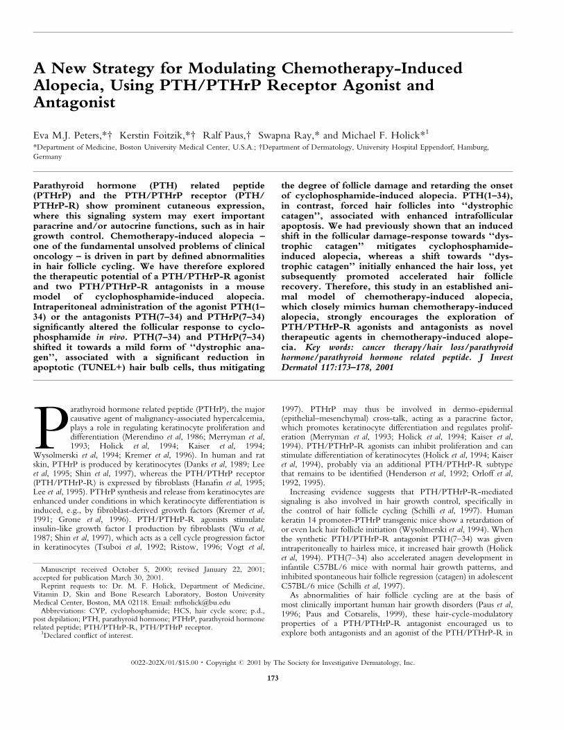
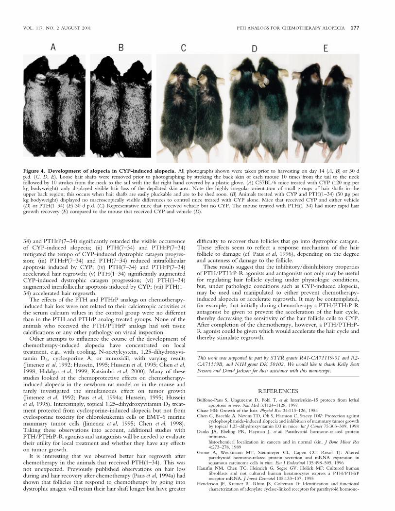
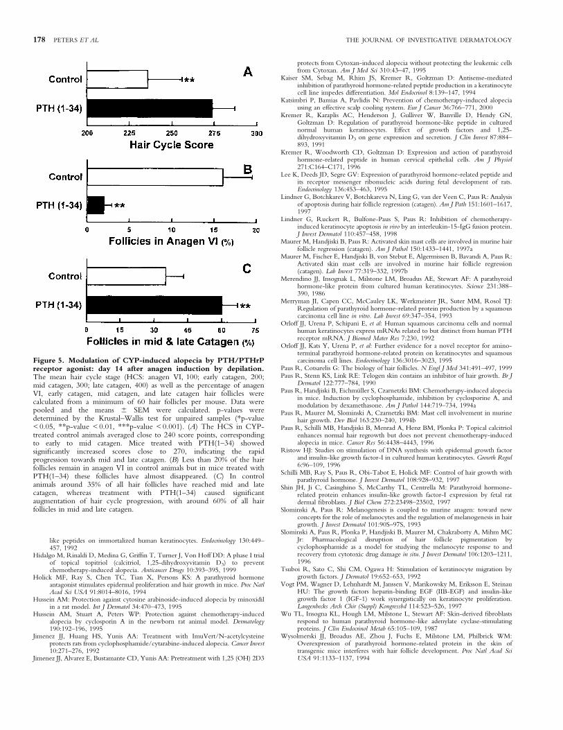
Effect of PTH(1±34) on accelerating CYP-inducedalopecia It was hypothesized that as PTH(1±34) enhanced hairfollicle keratinocyte maturation and differentiation, it wouldaccelerate the hair cycle and therefore make hair follicles moresensitive to chemotherapy. Macroscopic evaluation revealed thatthe hair loss in mice treated with PTH(1±34) was signi®cantly more(160% 6 4%; mean 6 SEM; p < 0.01) than in the control group(Fig 4). Quantitative histomorphometry revealed that PTH(1±34)signi®cantly augmented CYP-induced dystrophic catagenprogression as predicted. An analysis of the hair cycle scorerevealed that animals receiving PTH(1±34) had a score of 250±275,which indicated that the hair cycle progression was acceleratedtowards late catagen (Fig 5A). Therefore, treatment with PTH(1±34) signi®cantly accelerated the hair cycle progression towardscompletion of catagen development. By day 14 p.d. less than 2% ofthe hair follicles were in anagen VI in the PTH(1±34)-treatedanimals compared with 15% in the control mice (Fig 5B), and 60%of all hair follicles in the PTH(1±34) animals were in mid and latecatagen compared with 35% in the control group (Fig 5C).
Effect of PTH(1±34) on hair regrowth If the hair folliclekeratinocytes could be made to be more sensitive to CYP byaccelerating differentiation, this could lead to a more rapidrecovery. Mice that received PTH(1±34) showed faster regrowthof a normally pigmented fur coat than the control group treatedwith CYP and vehicle alone (Fig 4D, E).
An evaluation of the number of apoptotic cells in the hair folliclebulb from animals treated with PTH(1±34) showed a markedlyincreased number (120% 6 4%, mean 6 SEM; p < 0.001)compared with the control animals, consistent with the hypothesis.
CONCLUSION
Using the mouse model of chemotherapy-induced alopecia thatclosely mimics human chemotherapy-induced alopecia, we havedemonstrated for the ®rst time that PTH/PTHrP-R agonistPTH(1±34) and the PTH/PTHrP-R antagonists PTH(7±34) andPTHrP(7±34) can effectively manipulate the hair follicle responseto CYP-induced damage. We have demonstrated that (i) PTH(7±
Figure 3. C57BL/6 mice that received CYPand either vehicle (B), or PTH(7±34) (C) 30 dp.d. (A) Representative mice that did not receiveCYP. Note that the mice treated with PTH(7±34)or PTHrP(7±34) had more rapid hair growthrecovery compared with the mouse that receivedvehicle alone.
Figure 2. Modulation of CYP-induced alopecia by PTH/PTHrPreceptor antagonists, on day 13 after anagen induction bydepilation. The mean hair cycle stage (HCS: anagen VI, 100; earlycatagen, 200; mid catagen, 300; late catagen, 400), as well as thepercentage of anagen VI, early catagen, mid catagen, and late catagenhair follicles, were calculated from a minimum of 60 hair follicles permouse. Data were pooled and the means 6 SEM were calculated. p-values were determined by the Krustal±Wallis test for unpaired samples(*p-value < 0.05, **p-value < 0.01). (A) The HCS in CYP-treatedcontrol animals averaged 220, corresponding to early to mid catagen.Mice treated with PTHrP(7±34) or PTH(7±34) showed scores below200, which is representative for the transition from anagen to earlycatagen. (B) Less than 20% of the hair follicles remain in anagen incontrol animals. In contrast, PTH(7±34)- and PTHrP(7±34)-treated micemaintain around 35% of their follicles in anagen VI.
176 PETERS ET AL THE JOURNAL OF INVESTIGATIVE DERMATOLOGY
34) and PTHrP(7±34) signi®cantly retarded the visible occurrenceof CYP-induced alopecia; (ii) PTH(7±34) and PTHrP(7±34)mitigated the tempo of CYP-induced dystrophic catagen progres-sion; (iii) PTHrP(7±34) and PTH(7±34) reduced intrafollicularapoptosis induced by CYP; (iv) PTH(7±34) and PTHrP(7±34)accelerated hair regrowth; (v) PTH(1±34) signi®cantly augmentedCYP-induced dystrophic catagen progression; (vi) PTH(1±34)augmented intrafollicular apoptosis induced by CYP; (vii) PTH(1±34) accelerated hair regrowth.
The effects of the PTH and PTHrP analogs on chemotherapy-induced hair loss were not related to their calciotropic activities asthe serum calcium values in the control group were no differentthan in the PTH and PTHrP analog treated groups. None of theanimals who received the PTH/PTHrP analogs had soft tissuecalci®cations or any other pathology on visual inspection.
Other attempts to in¯uence the course of the development ofchemotherapy-induced alopecia have concentrated on localtreatment, e.g., with cooling, N-acetylcystein, 1,25-dihydroxyvi-tamin D3, cyclosporine A, or minoxidil, with varying results(Jimenez et al, 1992; Hussein, 1995; Hussein et al, 1995; Chen et al,1998; Hidalgo et al, 1999; Katsimbri et al, 2000). Many of thesestudies looked at the chemoprotective effects on chemotherapy-induced alopecia in the newborn rat model or in the mouse andrarely investigated the simultaneous effect on tumor growth(Jimenez et al, 1992; Paus et al, 1994a; Hussein, 1995; Husseinet al, 1995). Interestingly, topical 1,25-dihydroxyvitamin D3 treat-ment protected from cyclosporine-induced alopecia but not fromcyclosporine toxicity for chloroleukemia cells or EMT-6 murinemammary tumor cells (Jimenez et al, 1995; Chen et al, 1998).Taking these observations into account, additional studies withPTH/PTHrP-R agonists and antagonists will be needed to evaluatetheir utility for local treatment and whether they have any effectson tumor growth.
It is interesting that we observed better hair regrowth afterchemotherapy in the animals that received PTH(1±34). This wasnot unexpected. Previously published observations on hair lossduring and hair recovery after chemotherapy (Paus et al, 1994a) hadshown that follicles that respond to chemotherapy by going intodystrophic anagen will retain their hair shaft longer but have greater
dif®culty to recover than follicles that go into dystrophic catagen.These effects seem to re¯ect a response mechanism of the hairfollicle to damage (cf. Paus et al, 1996), depending on the degreeand acuteness of damage to the follicle.
These results suggest that the inhibitory/disinhibitory propertiesof PTH/PTHrP-R agonists and antagonists not only may be usefulfor regulating hair follicle cycling under physiologic conditions,but, under pathologic conditions such as CYP-induced alopecia,may be used and manipulated to either prevent chemotherapy-induced alopecia or accelerate regrowth. It may be contemplated,for example, that initially during chemotherapy a PTH/PTHrP-Rantagonist be given to prevent the acceleration of the hair cycle,thereby decreasing the sensitivity of the hair follicle cells to CYP.After completion of the chemotherapy, however, a PTH/PTHrP-R agonist could be given which would accelerate the hair cycle andthereby stimulate regrowth.
This work was supported in part by STTR grants R41-CA71119-01 and R2-
CA71119B, and N1H grant DK 50102. We would like to thank Kelly Scott
Persons and David Jackson for their assistance with this manuscript.
REFERENCES
Bulfone-Paus S, Ungureanu D, Pohl T, et al: Interleukin-15 protects from lethalapoptosis in vivo. Nat Med 3:1124±1128, 1997
Chase HB: Growth of the hair. Physiol Rev 34:113±126, 1954Chen G, Baechle A, Nevins TD, Oh S, Harmon C, Stacey DW: Protection against
cyclophosphamide-induced alopecia and inhibition of mammary tumor growthby topical 1,25-dihydroxyvitamin D3 in mice. Int J Cancer 75:303±309, 1998
Danks JA, Ebeling PR, Hayman J, et al: Parathyroid hormone-related proteinimmuno-histochemical localization in cancers and in normal skin. J Bone Miner Res4:273±278, 1989
Grone A, Weckmann MT, Steinmeyer CL, Capen CC, Rosol TJ: Alteredparathyroid hormone-related protein secretion and mRNA expression insquamous carcinoma cells in vitro. Eur J Endocrinol 135:498±505, 1996
Hana®n NM, Chen TC, Heinrich G, Segre GV, Holick MF: Cultured human®broblasts and not cultured human keratinocytes express a PTH/PTHrPreceptor mRNA. J Invest Dermatol 105:133±137, 1995
Henderson JE, Kremer R, Rhim JS, Goltzman D: Identi®cation and functionalcharacterization of adenylate cyclase-linked receptors for parathyroid hormone-
Figure 4. Development of alopecia in CYP-induced alopecia. All photographs shown were taken prior to harvesting on day 14 (A, B) or 30 dp.d. (C, D, E). Loose hair shafts were removed prior to photographing by stroking the back skin of each mouse 10 times from the tail to the neckfollowed by 10 strokes from the neck to the tail with the ¯at right hand covered by a plastic glove. (A) C57BL/6 mice treated with CYP (120 mg perkg bodyweight) only displayed visible hair loss of the depilated skin area. Note the highly irregular orientation of small groups of hair shafts in theupper back region; this occurs when hair shafts are easily pluckable and are to be shed soon. (B) Animals treated with CYP and PTH(1±34) (50 mg perkg bodyweight) displayed no macroscopically visible differences to control mice treated with CYP alone. Mice that received CYP and either vehicle(D) or PTH(1±34) (E) 30 d p.d. (C) Representative mice that received vehicle but no CYP. The mouse treated with PTH(1±34) had more rapid hairgrowth recovery (E) compared to the mouse that received CYP and vehicle (D).
VOL. 117, NO. 2 AUGUST 2001 PTH ANALOGS FOR CHEMOTHERAPY ALOPECIA 177
like peptides on immortalized human keratinocytes. Endocrinology 130:449±457, 1992
Hidalgo M, Rinaldi D, Medina G, Grif®n T, Turner J, Von Hoff DD: A phase I trialof topical topitriol (calcitriol, 1,25-dihydroxyvitamin D3) to preventchemotherapy-induced alopecia. Anticancer Drugs 10:393±395, 1999
Holick MF, Ray S, Chen TC, Tian X, Persons KS: A parathyroid hormoneantagonist stimulates epidermal proliferation and hair growth in mice. Proc NatlAcad Sci USA 91:8014±8016, 1994
Hussein AM: Protection against cytosine arabinoside-induced alopecia by minoxidilin a rat model. Int J Dermatol 34:470±473, 1995
Hussein AM, Stuart A, Peters WP: Protection against chemotherapy-inducedalopecia by cyclosporin A in the newborn rat animal model. Dermatology190:192±196, 1995
Jimenez JJ, Huang HS, Yunis AA: Treatment with ImuVert/N-acetylcysteineprotects rats from cyclophosphamide/cytarabine-induced alopecia. Cancer Invest10:271±276, 1992
Jimenez JJ, Alvarez E, Bustamante CD, Yunis AA: Pretreatment with 1,25 (OH) 2D3
protects from Cytoxan-induced alopecia without protecting the leukemic cellsfrom Cytoxan. Am J Med Sci 310:43±47, 1995
Kaiser SM, Sebag M, Rhim JS, Kremer R, Goltzman D: Antisense-mediatedinhibition of parathyroid hormone-related peptide production in a keratinocytecell line impedes differentiation. Mol Endocrinol 8:139±147, 1994
Katsimbri P, Bamias A, Pavlidis N: Prevention of chemotherapy-induced alopeciausing an effective scalp cooling system. Eur J Cancer 36:766±771, 2000
Kremer R, Karaplis AC, Henderson J, Gulliver W, Banville D, Hendy GN,Goltzman D: Regulation of parathyroid hormone-like peptide in culturednormal human keratinocytes. Effect of growth factors and 1,25-dihydroxyvitamin D3 on gene expression and secretion. J Clin Invest 87:884±893, 1991
Kremer R, Woodworth CD, Goltzman D: Expression and action of parathyroidhormone-related peptide in human cervical epithelial cells. Am J Physiol271:C164±C171, 1996
Lee K, Deeds JD, Segre GV: Expression of parathyroid hormone-related peptide andits receptor messenger ribonucleic acids during fetal development of rats.Endocrinology 136:453±463, 1995
Lindner G, Botchkarev V, Botchkareva N, Ling G, van der Veen C, Paus R: Analysisof apoptosis during hair follicle regression (catagen). Am J Path 151:1601±1617,1997
Lindner G, Ruckert R, Bulfone-Paus S, Paus R: Inhibition of chemotherapy-induced keratinocyte apoptosis in vivo by an interleukin-15-IgG fusion protein.J Invest Dermatol 110:457±458, 1998
Maurer M, Handjiski B, Paus R: Activated skin mast cells are involved in murine hairfollicle regression (catagen). Am J Pathol 150:1433±1441, 1997a
Maurer M, Fischer E, Handjiski B, von Stebut E, Algermissen B, Bavandi A, Paus R:Activated skin mast cells are involved in murine hair follicle regression(catagen). Lab Invest 77:319±332, 1997b
Merendino JJ, Insognak L, Milstone LM, Broadus AE, Stewart AF: A parathyroidhormone-like protein from cultured human keratinocytes. Science 231:388±390, 1986
Merryman JI, Capen CC, McCauley LK, Werkmeister JR, Suter MM, Rosol TJ:Regulation of parathyroid hormone-related protein production by a squamouscarcinoma cell line in vitro. Lab Invest 69:347±354, 1993
Orloff JJ, Urena P, Schipani E, et al: Human squamous carcinoma cells and normalhuman keratinocytes express mRNAs related to but distinct from human PTHreceptor mRNA. J Biomed Mater Res 7:230, 1992
Orloff JJ, Kats Y, Urena P, et al: Further evidence for a novel receptor for amino-terminal parathyroid hormone-related protein on keratinocytes and squamouscarcinoma cell lines. Endocrinology 136:3016±3023, 1995
Paus R, Cotsarelis G: The biology of hair follicles. N Engl J Med 341:491±497, 1999Paus R, Stenn KS, Link RE: Telogen skin contains an inhibitor of hair growth. Br J
Dermatol 122:777±784, 1990Paus R, Handjiski B, EichmuÈller S, Czarnetzki BM: Chemotherapy-induced alopecia
in mice. Induction by cyclophosphamide, inhibition by cyclosporine A, andmodulation by dexamethasone. Am J Pathol 144:719±734, 1994a
Paus R, Maurer M, Slominski A, Czarnetzki BM: Mast cell involvement in murinehair growth. Dev Biol 163:230±240, 1994b
Paus R, Schilli MB, Handjiski B, Menrad A, Henz BM, Plonka P: Topical calcitriolenhances normal hair regrowth but does not prevent chemotherapy-inducedalopecia in mice. Cancer Res 56:4438±4443, 1996
Ristow HJ: Studies on stimulation of DNA synthesis with epidermal growth factorand insulin-like growth factor-I in cultured human keratinocytes. Growth Regul6:96±109, 1996
Schilli MB, Ray S, Paus R, Obi-Tabot E, Holick MF: Control of hair growth withparathyroid hormone. J Invest Dermatol 108:928±932, 1997
Shin JH, Ji C, Casinghino S, McCarthy TL, Centrella M: Parathyroid hormone-related protein enhances insulin-like growth factor-I expression by fetal ratdermal ®broblasts. J Biol Chem 272:23498±23502, 1997
Slominski A, Paus R: Melanogenesis is coupled to murine anagen: toward newconcepts for the role of melanocytes and the regulation of melanogenesis in hairgrowth. J Invest Dermatol 101:90S±97S, 1993
Slominski A, Paus R, Plonka P, Handjiski B, Maurer M, Chakraborty A, Mihm MCJr: Pharmacological disruption of hair follicle pigmentation bycyclophosphamide as a model for studying the melanocyte response to andrecovery from cytotoxic drug damage in situ. J Invest Dermatol 106:1203±1211,1996
Tsuboi R, Sato C, Shi CM, Ogawa H: Stimulation of keratinocyte migration bygrowth factors. J Dermatol 19:652±653, 1992
Vogt PM, Wagner D, Lehnhardt M, Janssen V, Marikowsky M, Eriksson E, SteinauHU: The growth factors heparin-binding EGF (IIB-EGF) and insulin-likegrowth factor 1 (IGF-1) work synergistically on keratinocyte proliferation.Langenbecks Arch Chir (Suppl) Kongressbd 114:523±526, 1997
Wu TL, Insogna KL, Hough LM, Milstone L, Stewart AF: Skin-derived ®broblastsrespond to human parathyroid hormone-like adenylate cyclase-stimulatingproteins. J Clin Endocrinol Metab 65:105±109, 1987
Wysolmerski JJ, Broadus AE, Zhou J, Fuchs E, Milstone LM, Philbrick WM:Overexpression of parathyroid hormone-related protein in the skin oftransgenic mice interferes with hair follicle development. Proc Natl Acad SciUSA 91:1133±1137, 1994
Figure 5. Modulation of CYP-induced alopecia by PTH/PTHrPreceptor agonist: day 14 after anagen induction by depilation.The mean hair cycle stage (HCS: anagen VI, 100; early catagen, 200;mid catagen, 300; late catagen, 400) as well as the percentage of anagenVI, early catagen, mid catagen, and late catagen hair follicles werecalculated from a minimum of 60 hair follicles per mouse. Data werepooled and the means 6 SEM were calculated. p-values weredetermined by the Krustal±Wallis test for unpaired samples (*p-value< 0.05, **p-value < 0.01, ***p-value < 0.001). (A) The HCS in CYP-treated control animals averaged close to 240 score points, correspondingto early to mid catagen. Mice treated with PTH(1±34) showedsigni®cantly increased scores close to 270, indicating the rapidprogression towards mid and late catagen. (B) Less than 20% of the hairfollicles remain in anagen VI in control animals but in mice treated withPTH(1±34) these follicles have almost disappeared. (C) In controlanimals around 35% of all hair follicles have reached mid and latecatagen, whereas treatment with PTH(1±34) caused signi®cantaugmentation of hair cycle progression, with around 60% of all hairfollicles in mid and late catagen.
178 PETERS ET AL THE JOURNAL OF INVESTIGATIVE DERMATOLOGY