Embed Size (px)
Citation preview
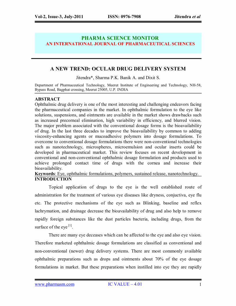
Vol-2, Issue-3, July-2011 ISSN: 0976-7908 Jitendra et al
www.pharmasm.com IC VALUE – 4.01
1
PHARMA SCIENCE MONITOR AN INTERNATIONAL JOURNAL OF PHARMACEUTICAL SCIENCES
A NEW TREND: OCULAR DRUG DELIVERY SYSTEM
Jitendra*, Sharma P.K. Banik A. and Dixit S.
Department of Pharmaceutical Technology, Meerut Institute of Engineering and Technology, NH-58, Bypass Road, Bagphat crossing, Meerut 25005, U.P. INDIA
ABSTRACT Ophthalmic drug delivery is one of the most interesting and challenging endeavors facing the pharmaceutical companies in the market. In ophthalmic formulation to the eye like solutions, suspensions, and ointments are available in the market shows drawbacks such as increased precorneal elimination, high variability in efficiency, and blurred vision. The major problem associated with the conventional dosage forms is the bioavailability of drug. In the last three decades to improve the bioavailability by common to adding viscosity-enhancing agents or mucoadhesive polymers into dosage formulations. To overcome to conventional dosage formulations there were non-conventional technologies such as nanotechnology, microspheres, microemulsion and ocular inserts could be developed in pharmaceutical market. This review focuses on recent development in conventional and non-conventional ophthalmic dosage formulation and products used to achieve prolonged contact time of drugs with the cornea and increase their bioavailability. Keywords: Eye, ophthalmic formulations, polymers, sustained release, nanotechnology. INTRODUCTION
Topical application of drugs to the eye is the well established route of
administration for the treatment of various eye diseases like dryness, conjuctiva, eye flu
etc. The protective mechanisms of the eye such as Blinking, baseline and reflex
lachrymation, and drainage decrease the bioavailability of drug and also help to remove
rapidly foreign substances like the dust particles bacteria, including drugs, from the
surface of the eye [1].
There are many eye deceases which can be affected to the eye and also eye vision.
Therefore marketed ophthalmic dosage formulations are classified as conventional and
non-conventional (newer) drug delivery systems. There are most commonly available
ophthalmic preparations such as drops and ointments about 70% of the eye dosage
formulations in market. But these preparations when instilled into eye they are rapidly

Vol-2, Issue-3, July-2011 ISSN: 0976-7908 Jitendra et al
www.pharmasm.com IC VALUE – 4.01
2
drained away from the ocular surface due to blincking, tear flow and lacrimal nasal
drainage of the eye. Only a small amount of drug is available for its therapeutic effect
resulting in frequent dosing application to the eye. So overcome to these problems newer
pharmaceutical ophthalmic formulation such as in-situ gel, nanoparticle, liposome,
nanosuspension, microemulsion, intophoresis and ocular inserts have been developed in
last three decades increase the bioavailability of the drug as a sustained and controlled
manner [2-9].
Anatomy and function of the eye
The eye is a spherical structure with a wall made up of three layers; the outer part
sclera, the middle parts choroid layer, Ciliary body and iris and the inner section nervous
tissue layer retina. The sclera is tough fibrous coating that protecting the inner tissues of
eye which is white except for the transparent area at the front, and the cornea allows light
to enter to the eye.
The choroid layer, situated situated in the sclera, contains many blood vessels that
modified at front of the eye as pigmented iris the coloured part of the eye ( blue, green,
brown, hazel, or grey) [10].
The structure of the cornea
The clear transparent bulge cornea situated at the front of the eye that conveys
images to the back of the nervous system. The adult cornea has a radius of approximately
7-8mm that covers about one-sixth of the total surface area of the eye ball that is a
vascular tissue to which provides nutrient and oxygen are supplied via lachrymal fluid
and aqueous humour as well as from blood vessels of the junction between the cornea
and sclera in fig.1[11].
Vol-2, Issue-3, July-2011 ISSN: 0976-7908 Jitendra et al
www.pharmasm.com IC VALUE – 4.01
3
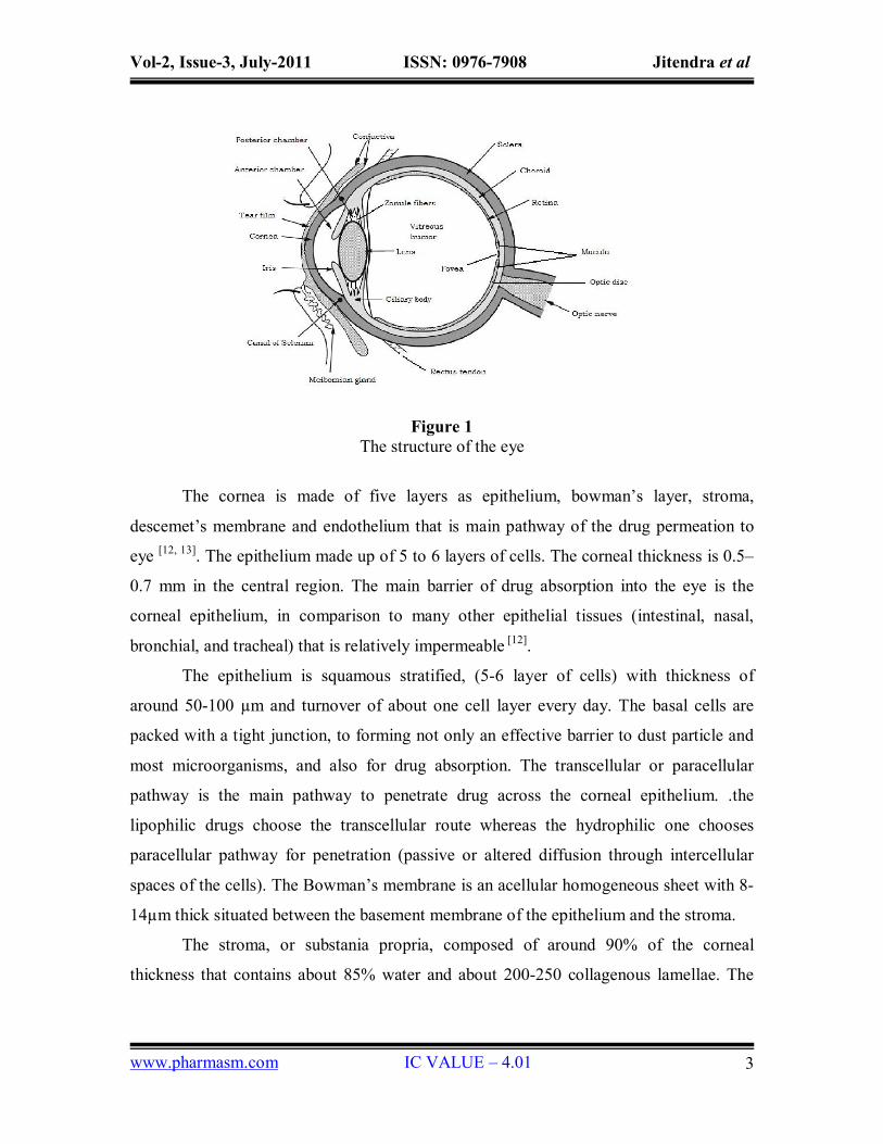
Figure 1 The structure of the eye
The cornea is made of five layers as epithelium, bowman’s layer, stroma,
descemet’s membrane and endothelium that is main pathway of the drug permeation to
eye [12, 13]. The epithelium made up of 5 to 6 layers of cells. The corneal thickness is 0.5–
0.7 mm in the central region. The main barrier of drug absorption into the eye is the
corneal epithelium, in comparison to many other epithelial tissues (intestinal, nasal,
bronchial, and tracheal) that is relatively impermeable [12].
The epithelium is squamous stratified, (5-6 layer of cells) with thickness of
around 50-100 µm and turnover of about one cell layer every day. The basal cells are
packed with a tight junction, to forming not only an effective barrier to dust particle and
most microorganisms, and also for drug absorption. The transcellular or paracellular
pathway is the main pathway to penetrate drug across the corneal epithelium. .the
lipophilic drugs choose the transcellular route whereas the hydrophilic one chooses
paracellular pathway for penetration (passive or altered diffusion through intercellular
spaces of the cells). The Bowman’s membrane is an acellular homogeneous sheet with 8-
14µm thick situated between the basement membrane of the epithelium and the stroma.
The stroma, or substania propria, composed of around 90% of the corneal
thickness that contains about 85% water and about 200-250 collagenous lamellae. The
Vol-2, Issue-3, July-2011 ISSN: 0976-7908 Jitendra et al
www.pharmasm.com IC VALUE – 4.01
4
lamellae provide physical strength while permitting optical transparency of the
membrane. The hydrophilic solutes diffuse through the stroma’s open structure..
The descemet’s membrane is secreted by the endothelium and lies between the stroma
and the endothelium [10, 11].
Conjunctiva
The conjunctiva protects the eye and also involved in the formation and
maintenance of the precorneal tear film. The conjunctiva is a thin transparent membrane
lies in the inner surface of the eyelids and that is reflected onto the globe. The
conjunctiva is made of an epithelium, a highly vascularised substantia propria, and a
submucosa. The bulbar epithelium contains 5 to 7 cell layers. The structure resembles a
pallisadeand not a pavemente corneal epithelium cells are connected by tight junctions,
which render the conjunctiva relatively impermeable. The molecules up to 20,000 Da can
cross the conjuctiva, while the cornea is restrict to molecules larger than 5000 Da. The
human conjunctiva is about 2 and 30 times more absorption of drugs than the cornea and
also proposed that loss of drug by this route is a major path for drug clearance.
The highest density of conjunctiva is due the presence of 1.5 million globlet cell
varying with age depended among the intersujects variability and age. The vernal
conjunctivitis and atopic kerato conjunctivitis occurs due to the great variation in goblet
cell density results only in a small difference in tear mucin concentration [12,13].
Nasolachrymal drainage system
Nasolachrymal drainage system consists of three parts; the secretory system, the
distributive system and the excretory system. The secretory portion is composed of the
lacrimal gland that secreted tears are spread over the ocular surface by the eyelids during
blinking. The secretory system is stimulated by blinking and temperature change due to
the tear evaporation and reflux secretors that have an efferent parasympathetic nerve
supply and secrete in response to physical and emotional state e.g. crying.
The distributive system consists of the eyelids and the tear meniscus around the
lid edges of the open eye, which spread tears over the ocular surface by blinking, thus
preventing dry areas from developing.
The excretory part of the Nasolachrymal drainage system consists of the
lachrymal puncta, the superior, inferior and common canaliculi; the lachrymal sac, and
Vol-2, Issue-3, July-2011 ISSN: 0976-7908 Jitendra et al
www.pharmasm.com IC VALUE – 4.01
5
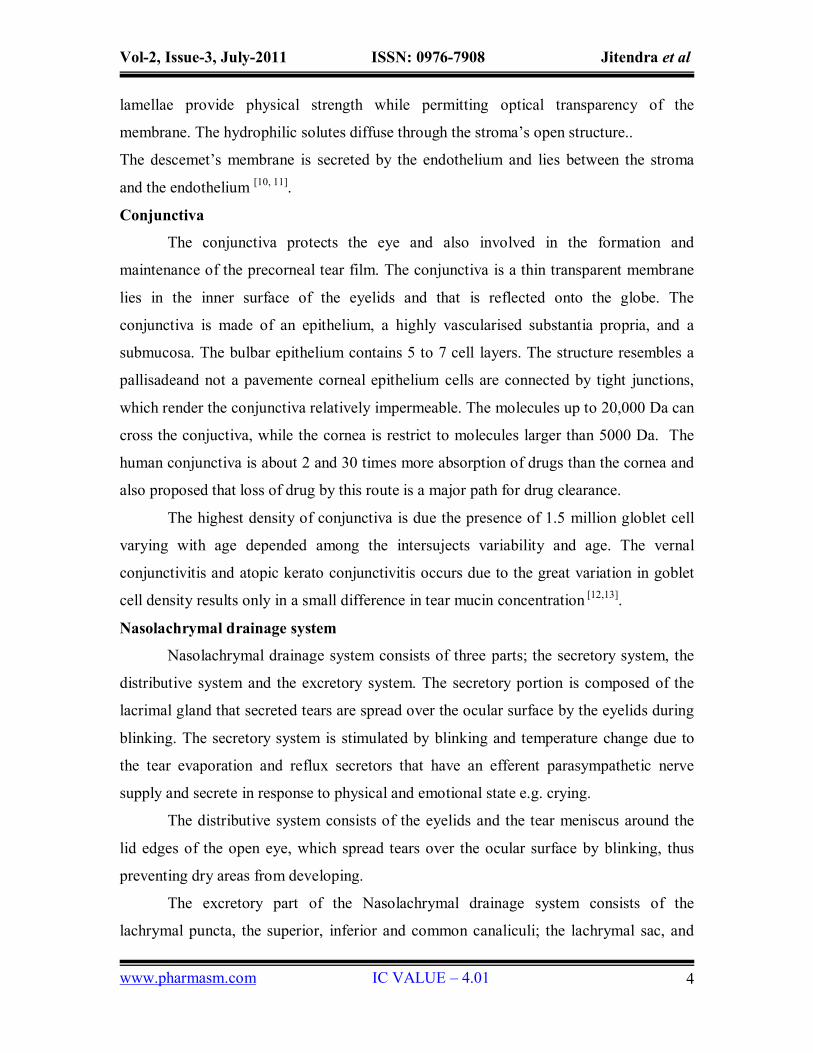
the nasochrymal duct. In humans, the two puncta are the openings of the lachrymal
canaliculi and are situated on an elevated area known as the lachrymal papilla. It is
thought that tears are largely absorbed by the mucous membrane that lines the ducts and
the lachrymal sac; only a small amount reaches the nasal passage [10, 13].
.
Figure 2 Schematic diagram of nasolachrymation drainage system
Tear film
A thin fluid layer is covered the exposed part of the eye called as precorneal tear
film. The film thickness is about 3–10 Am depending on the measurement method with
the resident volume approximately 10 µl. The osmolality of the tear fluid is approx. 310–
350 mOsm/kg in normal eyes and is maitained by the monovalent and divalent inorganic
ions present in fluid such as Na+, K+, Cl-, HCO3-, and proteins. The mean pH of normal
tears is about 7.4. Diurnal patterns of pH changes the pH of tear, which a general shift
from acid to alkaline during the day. The buffer capacity of the tears fluid is determined
by bicarbonate ions, proteins, and mucins [15, 16]. Tears exhibit a non-Newtonian
rheological behaviour with viscosity is about 3 mPas 12.The mean surface tension of tear
film value is about 44 mN/m [16].
Ocular absorption of drug
The common method of ocular drug delivery is topical administration of
ophthalmic dosage formulation drops into the lower cul-de-sac. Such drops are outflow
quickly due to the eye blinking reflux, and the precorneal region returns to maintain

Vol-2, Issue-3, July-2011 ISSN: 0976-7908 Jitendra et al
www.pharmasm.com IC VALUE – 4.01
6
resident volume of around 7µl. The available concentration of drug in precorneal fluid
provides the driving force for passive transport of drug across the cornea. However, the
epithelium is the predominant rate limiting barrier for hydrophilic drugs and where as
stroma is rate limiting step for most of the lipophilic drugs. Recent studies suggest that
the noncorneal routes of absorption are an across the sclera and conjunctiva have
significant role for drug molecules with poor corneal permeability. Studies with inulin,
timolol maleate, gentamicin, anesthetic and autonomic drug and PGF2α, PGF2α-1-methyl
ester, and PGF2α-1-isopropyl ester suggest that these drugs gain access through the non-
corneal route [17-21].
Thus the topically application of formulation to the surface of ocular are an
extremely complicated issue because of the numerous protective mechanisms of the eye
that protects the visual pathway from foreign materials. Design of modern ocular drug
delivery systems is based on the drug application pathways and absorption mechanism in
the eye and the overall ocular pharmacokinetic/pharmacodynamic profile. Thus for
efficient drug absorption require more prolonged contact time of dosage formulation to
within the eye.
Conventional ocular drug delivery system:-
The conventional ophthalmic drug delivery systems are used in today’s ocular
disease treatment and preventions are solutions, suspensions, ointments and Bioadhesive
polymer gel. In spite of significant criticisms over the efficacy and efficiency of these
conventional systems, such as limitation are such as bioavailability, sterility, dosing
administration. So these preparations are extensively used in a majority of commercial
products in pharmaceuticals market.
Aqueous solutions
Today most of the topical ophthalmic preparations are in the form of aqueous
solutions. A sterile homogeneous solution dosage form have many advantages over the
other dosage such as formulation, including the easily commercially capability produce
on large scale manufacture. There are various factors that must be consider during the
formulating aqueous solution includes selection of appropriate salt of the drug, solubility
in solvents, therapeutic systemic effect, ocular toxicology, pKa of formulation, and the
effect of pH of the formulation. Others stability parameters includes such as solubility,

Vol-2, Issue-3, July-2011 ISSN: 0976-7908 Jitendra et al
www.pharmasm.com IC VALUE – 4.01
7
tonicity, viscosity, buffering capacity, compatibility with formulation ingredients and
effect of packaging components, choice of appropriate preservative, ocular comfort and
dosing administration[1] .
The designing of experiments and parameters must be conducted to achieve the
optimum formulation. Corneal absorption enhancement can be achieved best by
increasing solution concentration and viscosity, increasing contact time of formulation in
the cornea film, appropriate pKa and offering optimal lipid solubility of drug [22].
Commonly added viscosity enhancer agents to improve ocular bioavailability, these
includes various synthetic polymers such as corboxymethyl cellulose, hydroxyl
methylcellulose, polyvinyl alcohol, hydroxypropyl methylcellulose, and carbomers.
Recently natural polymers have also been used to improve bioavailability of drugs.
Examples of these polymers are hyaluronic acid (HA), guar gum, xylloglucan
gum,Chitosan, gellan gum, pectin etc. The rheological characteristics of a polymer should
be implicated the such as no adverse effects, contact time of dosage formulation and
retention of dosage formulation ocular surface [23-24].
The solubility and stability, and corneal permeability of the drugs are depends on
pH of the formulation. A factor pH of the formulation is a best possible compromise
between stability and bioavailability of the drug. Ophthalmic solutions are should be
formulated in between pH range 4 to 8.0. If the pH range of the formulation is outside the
physiological pH range of eye, can be discomfort, irritation and also decrease the
bioavailability of the drug, due to the secretion of fluid and to aid in the restoration of
normal physiological conditions. The excessive tearing from the nasolacrymal gland also
results into rapidly flushing of the drug. So it necessary, proper choices of buffering
agents and buffer capacity are essential to optimize drug bioavailability as well as ocular
efficacy of drug [25].
The typical multitasking ophthalmic product containing antimicrobial
preservatives for prevention of administering microbiologically contaminated products
and is to prevent the patient. The main factors to be consider for selecting a preservative
for ophthalmic products are (a) a very low concentration should be effective at against a
broad spectrum of microorganisms (b) it should be inert and non toxic (c) form a
homogenous solution in the formulation (d) not reacts with the drug packaging

Vol-2, Issue-3, July-2011 ISSN: 0976-7908 Jitendra et al
www.pharmasm.com IC VALUE – 4.01
8
components (d) effective over the shelf life of products. The quaternary ammonium
compounds have good quality of good preservatives such as e.g., benzalkonium chloride,
parabens, chlorobutanol and 2-poly (ethylalcohol) and also new generation preservatives
such as Purite®.
The shelf life and expiration date of the product determined by stability of
ophthalmic products at various conditions. Products are analysing for physical, chemical
and microbiological parameters. Physical evaluation parameters for products are pH,
osmolality, viscosity, color and appearance of the product and Chemical parameters
include assays (UV, IR, TLC, HPLC) for the active and degradation product and
preservative/excipients content in the products[25,26] .
Aqueous ophthalmic solutions are generally manufactured by a process in which
the dissolution of the active and other inactive ingredients (excipients/additives) after
sterilization is achieved by application of heat or by sterile filtration. This prepared sterile
solution may further be then mixed with other components such as sterilized solutions of
viscosity – including agents and additives. The batch is made upto final volume with
additional sterile water [26].
Suspensions
Ophthalmic suspensions products is another part of the ocular drug delivery
system and have many distinct advantages over others formulation. Recently developed
drugs are generally hydrophobic poor solubility in water and aqueous medium.
Formulation offers a sterile, preserved, effective, stable and pharmaceutically elegant.
Ophthalmic suspensions are more complex and challenging if compare with to
ophthalmic (aqueous) solutions.
The formulation of a ophthalmic suspension many problem occured such as non-
homogeneity of the dosage form, settling of particles, cake formation, aggregation of the
suspended particles. The commercial ophthalmic products of should be effectively
preserved on storage. To study the surface tension properties, such as wetting, particle
size and interaction zeta potential, aggregation, sedimentation rate and rheological
characterization of the formulation. Above all criteria are necessory for formulating an
effective, elegant suspension ophthalmic formulation [6, 27].

Vol-2, Issue-3, July-2011 ISSN: 0976-7908 Jitendra et al
www.pharmasm.com IC VALUE – 4.01
9
Generally suspensions are kinetically stable at normal condition but
thermodynamically unstable systems. If suspension keep undisturbed for a long duration
of time, can be lead to aggregation of particles, sedimentation, settling of particles and
eventually forming caking.. But they are readily dispersible on hand shaking.
Flocculation state of suspension the particles are held together in a loose open structure
and the flocculated particles settle rapidly and for sediment. Relative properties of
flocculated and deflocculated particles in suspension
Deflocculated
Rate of sedimentation is slow because each particle settles separately due to the
particle size is minimal.
The sediment particle becomes very closely packed so that, a hard cake is formed
due to repulsive forces between particles are overcome then is difficult to
redisperse in media.
Generally suspensions have a pleasing appearance, due to the suspended material
in solution form. The supernatant also remains cloudy on settling is remains in
media.
Flocullated
Rate of sedimentation is high because particles settle as a floc, because a
collection of particles.
Particle size of the suspension has a key role in physical stability and
bioavailability. The rate of sedimentation, agglomeration and resuspendability of
suspensions are affected by particle size and bioavailability. In most ophthalmic
suspension, the average particle size is less than 10 μm. Mostly the particle achieved by
the efficient method is by dry milling and wet milling and other methods are also used of
particle size reduction include micro-pulverization, grinding, and controlled precipitation
[22, 26].
An ophthalmic suspension contains many inactive ingredients such as dispersing
and wetting agents, suspending agents, buffers and preservatives. Wetting agents are used
to decreases the contact angle between the solid surface and the wetting liquid. Generally
used wetting and solubilizing agents are Benzalkonium chloride, Benzethonium chloride,
Vol-2, Issue-3, July-2011 ISSN: 0976-7908 Jitendra et al
www.pharmasm.com IC VALUE – 4.01
10
Cetylpyridinium chloride, Nonoxynol 10, Octoxynol 9, Poloxamer, Polyoxyl 50 stearate,
Polyoxyl 20 cetostearyl ether, Polyoxyl 40 stearate.
Suspending agents are used to prevent sedimentation and affecting the rheological
behavior of a suspension. An ideal suspending agent should have to produce a structured
vehicle and it should be inert and non-toxic. Generally ophthalmic suspension used
suspending agents are includes cellulosic derivatives such as methyl cellulose, caboxy
methyl cellulose, and hydroxyl propyl methyl cellulose, synthetic polymers such as
carbomers, poloxamers, and polyvinyl alcohol. The selection of buffers and preservatives
for suspension ophthalmic solutions in almost same as aqueous except that they must also
be compatible with the flocculating systems [27].
An ophthalmic suspension difluprednate is an active ingredient that shows
superior antiinflammatory action and antiallergic action by local application for the
treatment and prevention of disorders of the eye, such as conjunctivitis (allergic, vernal,
blepharitis marginalis, catarrhal) and uveitis.(U.S. patent 5556848)[28]. There are various
ophthalmic solutions available in the market in table.1
Gel and Bioadhesive gel
There are many different marketed formulation adhesions depending upon the
environment of the eye for prolonging effectiveness of the drug at the site of the
administration (eye).
Adhesion as a process is defined as the ‘‘fixing” of two surfaces to one another
[29].Thus bioadhesion is the binding of a natural or synthetic polymer to a biological
substrate such as a mucous layer, the term mucoadhesion is often used 30. Mucoadhesion
has been mostly used to promote a simple way of achieving site of action drug delivery
by the incorporation of mucoadhesive hydrophilic polymers within ophthalmic
formulations along with the active pharmaceutical ingredient (API). The rationale behind
that is the formulation will be ‘held’ on a biological surface for localised drug delivery.
There are various theories wettability theory, electronic theory, fraction theory,
absorption and diffusion-interlocking theory of Bioadhesive force have been proposed in
this review reference [7].
The API from the formulation will be released close to the site of action with a
consequent incrment of bioavailability [31]. While mucoadhesive drug delivery systems

Vol-2, Issue-3, July-2011 ISSN: 0976-7908 Jitendra et al
www.pharmasm.com IC VALUE – 4.01
11
provides a means of enhancing retention time at defined sites of application, if systemic
uptake occurs the use of mucoadhesive polymers will not prove to effective distribution
of the API. Chitosan (CS) a cationic polysaccharide that has widely being used in
ophthalmic preparations [32]. The specific bioadhesiveness of CS to the ocular surface was
first observed in an ex-vivo study, in which the activity of radiolabelled CS was
measured by scintillation counting after addition to a freshly excised cornea and
exhaustive rinsing 29, 32. Electrostatic attraction is the major driving force for
mucoadhesion [33] .
Interactions of suitable mucoadhesive natural and synthetic polymers with mucins
were evaluated from biological substances. Interactions between the mucous layer and
the eye tissues, an increase in the precorneal residence time of the formulation was
observed. Some mucoadhesive polymers showed not only good potential to increase the
bioavailability of the drug applied, but also have a protective and healing properties to
epithelial cells such as Chitosan have a wound healing properties and antimicrobial
properties.The Carbomer polymeric gel base itself has been used successfully to treat
moderate to severe cases of dry eye such as Keratoconjuctivitis Sicca [32, 37].
Keratoconjunctivitis sicca (KCS), also called as dry eye syndrome (DES),is an
eye disease caused by decreased tear production or increased tear film evaporation
commonly found in humans and some animals. The choice of the polymer plays a
critical role in the release kinetics of the drugs from the dosage form [38].
Vol-2, Issue-3, July-2011 ISSN: 0976-7908 Jitendra et al
www.pharmasm.com IC VALUE – 4.01
12
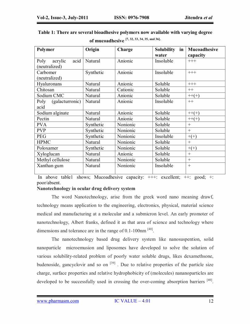
Table 1: There are several bioadhesive polymers now available with varying degree
of mucoadhesive [7, 32, 33, 34, 35, and 36].
Polymer Origin Charge Solubility in water
Mucoadhesive capacity
Poly acrylic acid (neutralized)
Natural Anionic Insoluble +++
Carbomer (neutralized)
Synthetic Anionic Insoluble +++
Hyaluronans Natural Anionic Soluble +++ Chitosan Natural Cationic Soluble ++ Sodium CMC Natural Anionic Soluble ++(+) Poly (galacturronic) acid
Natural Anionic Insoluble ++
Sodium alginate Natural Anionic Soluble ++(+) Pectin Natural Anionic Soluble ++(+) PVA Synthetic Nonionic Soluble + PVP Synthetic Nonionic Soluble + PEG Synthetic Nonionic Insoluble +(+) HPMC Natural Nonionic Soluble + Poloxamer Synthetic Nonionic Soluble +(+) Xyloglucan Natural Anionic Soluble + Methyl cellulose Natural Nonionic Soluble + Xanthan gum
Natural Nonionic Insoluble +
In above table1 shows; Mucoadhesive capacity: +++: excellent; ++: good; +: poor/absent. Nanotechnology in ocular drug delivery system
The word Nanotechnology, arise from the greek word nano meaning drawf,
technology means application to the engineering, electronics, physical, material science
medical and manufacturing at a molecular and a submicron level. An early promoter of
nanotechnology, Albert franks, defined it as that area of science and technology where
dimensions and tolerance are in the range of 0.1-100nm [40].
The nanotechnology based drug delivery system like nanosuspention, solid
nanoparticle microemusion and liposomes have developed to solve the solution of
various solubility-related problem of poorly water soluble drugs, likes dexamethsone,
budenoside, gancyclovir and so on [39] . Due to relative properties of the particle size
charge, surface properties and relative hydrophobicity of (molecules) nananoparticles are
developed to be successfully used in crossing the over-coming absorption barriers [40].

Vol-2, Issue-3, July-2011 ISSN: 0976-7908 Jitendra et al
www.pharmasm.com IC VALUE – 4.01
13
Furthermore, nanocarriers are critical in order to exploit the emerging in pharmaceutical
field of drug delivery systems and new gene therapies for the treatment of ocular
disorders and other alternatives for topical drug delivery involve the use of liposomes,
nanospheres, nanosuspension and nanoparticles and so on.
Different nanoparticles based drug delivery systems are
Microemulsion
Microemulsions were first described Hoar and Schulman. Microemulsion is a
dispersion of water and oil that formulated with surfactants and co-surfactants in order to
stabilize the surface tension of emulsion [41]. Microemulsions have a transparent
appearance, with thermodynamic stability and a small droplet size in the dispersed phase
(aqueous and nonaqueous phase) (<1.0µm). Microemusion are an interesting alternative
to ophthalmic formulation, due to their intrinsic properties and specific structure. They
can be easily prepared through emulsification method, easily sterilized, and are more
stable and have a high capacity for dissolving drugs [40, 42]. The ophthalmic o/w
Microemusion could be advantageous over other formulation, because the presence of
surfactants and co-surfactants increase the dug molecules permeability, thereby
increasing bioavailability of drugs. Due to, these systems act as penetration enhancers to
facilitate corneal drug delivery [43]. The in-vivo experiments and preliminary studies on
healthy volunteers have occurred a delayed effect and an increase in the bioavailability of
the drug. This mechanism is based on the adsorption of the nanodroplets representing the
internal phase of the microemulsions, which act as a reservoir of the drug on the cornea
and should decrease their drainage in limit [43, 44]. Indeed, in 2002 the FDA approved the
clinical use of an anionic emulsion containing cyclosporine A 0.05% (Restasis®,
Allergan) for the treatment of chronic dry eye. A similar formulation (anionic emulsion
containing difluprednate 0.05%, Durezol™, Sirion Terapeutics) has recently been
approved for the treatment of ocular inflammation. In the same field, a non-medicated
anionic emulsion for eye lubricating purposes, in patients suffering from moderate to
severe dry eye syndrome (Refresh Dry Eye Therapy®, Allergan), and two lipidic
emulsions, indicated for the restoration of the lipid layer of the lacrimal fluid (Lipimix™,
Tubilux Pharma, and Soothe XP® Emollient, Bausch and Lomb), have been launched in
the US and European markets.the cationic nanoemulsions have also made their way onto
Vol-2, Issue-3, July-2011 ISSN: 0976-7908 Jitendra et al
www.pharmasm.com IC VALUE – 4.01
14
the market. Namely, the product Cationorm® (Novagali Pharma, France) was launched in
the European market for the treatment of dry eye symptoms and two more products,
based upon the same technology and intended to deliver cyclosporine A, are currently
under registration or under clinical evaluation (Phase III).
Water-in-oil microemulsions (w/o ME) capable of undergoing a phase-transition
to lamellar liquid crystals (LC) or bicontinuous ME upon aqueous dilution were
formulated using Crodamol, sorbitan mono-laurate and polyoxyethylene 20 sorbitan
mono-oleate, an alkanol or alkanediol as cosurfactant and water. The hypothesis that
phase-transition of ME to LC may be induced by tears and serve to prolong precorneal
retention was tested. The ocular irritation potential of components and formulations was
assessed using a modified hen's egg chorioallantoic membrane test (HET-CAM) and the
preocular retention of selected formulations was investigated in rabbit eye using gamma
scintigraphy. Results showed that sorbitan mono-laurate, polyoxyethylene 20 sorbitan
mono-oleate and Crodamol ethyl oleate were non-irritant. However, all other
cosurfactants investigated were irritant and their irritation was dependent on their carbon
chain length. A w/o ME formulated without cosurfactant showed a protective effect when
a strong irritant (0.1 M NaOH) was used as the aqueous phase. Precorneal clearance
studies revealed that the retention of colloidal and coarse dispersed systems was
significantly greater than an aqueous solution with no significant difference between ME
systems (containing 5% and 10% water) as well as o/w emulsion containing 85% water.
Conversely, a LC system formulated without cosurfactant displayed a significantly
greater retention compared to other formulations [45].
Nanoparticles
Nanoparticles are the particle with a diameter of less than 1µm, containing of
various biodegradable materials, such as natural and synthetic polymer, liposomes, lipids,
phospholipids and even inorganic material. Biodegradable nanoparticles of polymers like
polylactides (PLAs), polycyanoacrylate, poly (d,l-lactides), natural polymers can be used
effectively for efficient drug delivery to the ocular tissues.
Pre-clinical Studies by Bourges et al. in rabbits shows that nanoparticles of
different size and electric charge, when injected into the vitreous, migrate through the
retinal layers and tend to accumulate in the retinal pigment epithelium (RPE) cells. They

Vol-2, Issue-3, July-2011 ISSN: 0976-7908 Jitendra et al
www.pharmasm.com IC VALUE – 4.01
15
found that presence of nanoparticles within the RPE cells up to four months after a single
intravitreous injection [46]. The movement of nanoparticles in the internal limiting
membrane (ILM) because of the modification of the vitreous interface structure
secondary to the presence of the PLA and poly (d,l-lactide-co-glycolide) (PLGA) [47].
The encapsulated nanospheres may also increases when in such bioavailability of
ophthalmic delivery. Recently, it reported that non-biodegradable polystyrene
nanospheres observed within the neuroretina [46]. Generally nanoparticles act at the
cellular level and followed to endocytosed/phagocytosed by cells, then resulting cell
internalization of the encapsulated drug. The surface charge and the binding of the drug
to the particles were found to be more important factors in the drug loading in case of
nanoparticles. Li et al. Proved, though being fully encapsulated in polybutyl cyano-
acrilate nanospheres, the drug progesterone not released properly, because of strong
interaction between the drug and the polymer [48].
The albumin nanoparticles was used to a very efficient ocular delivery system for
like CMV retinitis, they are biodegradable, non-toxic and have non-antigenic effects.
Since high content of charged amino acids, albumin nanoparticles allow the adsorption of
positively charged gancyclovir or negatively charged particles like oligonucleotides that
increased the bioavailability [49]. However nanoparticles of natural polymers which are
made up of like sodium alginate, chitosan, are very effective in intraocular penetration for
some specific drugs, because of contact time with corneal and conjunctival surfaces.
Studies results show the bioavailability of encapsulated indomethacin doubled when
Poly(epsilon-caprolacton) (PECL) nanoparticles were coated with Chitosan [50]. Greater
corneal penetration enhancement was occurred, when PECL nanoparticles were coated
with polyethylene glycol (PEG). All these studies lead us to believe that nanoparticles
have great potential ophthalmic delivery systems for ocular tissues [51].
Nanosuspensions
Nanosuspension contains of pure, hydrophobic drugs (poorly water soluble),
suspended in appropriate dispersion medium. Nanosuspension technology are utilised for
drug components that form crystals with high energy content molecule, which renders
them insoluble in either hydrophobic or hydrophilic media [52].
Vol-2, Issue-3, July-2011 ISSN: 0976-7908 Jitendra et al
www.pharmasm.com IC VALUE – 4.01
16
Although nanosuspensions offer advantages such as more residence time in a cul-
de-sac and avoidance of the high tonicity created by water-soluble drugs, their
performance depends on the intrinsic solubility of the drug in lachrymal fluids after
administration. Thus, the intrinsic solubility rate of the drug in lachrymal fluid controlled
its release and indrease ocular bioavailability. However, the intrinsic dissolution rate of
the drug after application will vary because of the constant inflow and outflow of
lachrymal fluids.. However, a nanosuspension, by their inherent ability to improve the
saturation solubility of the drug in media, also represents an ideal approach for
ophthalmic delivery of hydrophobic drugs in eye. Moreover, in earlier nanoparticulate
nature of the drug allows to prolonged residence (ocular surface) in the cul-de-sac, giving
sustained release of the drug. To achieve sustained release of the drug, nanosuspensions
can be incorporated or formulated with a suitable hydrogel or mucoadhesive base (in- situ
gel) or even in ocular inserts [53].
A recently approaches been developes desired release the drug is the formulation
formulated with polymeric nanosuspensions particles loaded with the drug. The
bioerodible as well as water soluble/permeable polymers could be used to sustain and
control the release of the medication. The nanosuspensions can be formulated by using
the quasi-emulsion and solvent diffusion method [54]. The using acrylate polymers such as
Eudragit RS 100 and Eudragit RL 100 in polymeric nanosuspensions of flurbiprofen and
ibuprofen have been successfully formulated, and these have been characterized for drug
loading, particle size, zeta potential, in-vitro drug release, ocular tolerability and in-vivo
biological performance in animal [55]. The flu is a non-steroidal anti-inflammatory drug
(NSAID) that using in inflammation and antagonizes papillary construction during intra-
ocular surgery. Since the flu-loaded Nanosuspension are formulated by the quasi-
emulsion solvent diffusion (QESD) method in which generally avoids using of toxic
chemical. They are proved to great potential for ophthalmic application [56].
Iontophoresis
Iontophoretic technique is used to depth penetration of topically applied drug
loaded nanoparticles Iontophoresis is a method for enhancing charged drug penetration
into anterior and posterior ocular structures, by using a low electric current. The
mechanisms of drug penetration are followed by iontophoresis of electrorepulsion,

Vol-2, Issue-3, July-2011 ISSN: 0976-7908 Jitendra et al
www.pharmasm.com IC VALUE – 4.01
17
elecroosmosis and current-induced tissue damage [8]. However, each drug has to be
evaluated for its penetration capacity and pharmacokinetically profile, due to different
physicochemical properties of the drug molecules. This novel approach of charged
nanoparticles iontophoresis can benefit from: (1) deep penetration, regardless of drug's
ionic activity strength and diffusion capacity in ocular tissues, (2) controlled and
sustained release of the drug for better therapeutic activity, (3) targeting to a specific
desired tissue and lacalised tissues [57].
Advantages
Increases the bioavailability and decreases the adverse effects.
iontophoresis of charged nanoparticles as drug carriers, providing a long duartion
therapeutic activity.
Topical ophthalmic preparation and easy to apply.
Good drug penetration to the anterior and posterior segments of the eye by
topically.
May combine to other drug delivery system.
Good acceptance by the patients [58, 59].
Ocular inserts
The ocular insert represents a significant advancement in the therapy of eye
disease. Ocular inserts are described as single, sterile, thin, and multilayered, drug-
impregnated, solid or semisolid consistency devices, whose size and shape are especially
designed for application in eye. A polymeric support is must for the ocular inserts which
may or may not contain the drug. The drug is later entrapped or dispersed or the drug
can be incorporated as a solution in the polymeric supports which have advantages as
they increases the residence of the drug in the eye so a sustained release dosage form
would be formulated . The body portion of the eye is sized in such a way so that it can
position in to position in lachrymal canaliculus of the eyelid. We can classify the inserts
on the basis of their solubility as insoluble, soluble, and bioerodible inserts. The drug
release from the inserts would take place by following three procedures 1) diffusion, 2)
osmosis, and 3) bioerosion [60, 61].
Ocular inserts serves as an alternative approach to overcome the problems which
were faced by other ocular drug delivery systems [61]. The commonly faced problems are

Vol-2, Issue-3, July-2011 ISSN: 0976-7908 Jitendra et al
www.pharmasm.com IC VALUE – 4.01
18
Disposition and elimination of a therapeutic agent depends on the physicochemical
properties and relevant ocular anatomy and physiology [62]. To design a successful
delivery system for ophthalmic use, it is required to have a complete knowledge about the
drug profile and other related constraints of the ocular route. Hornof et al., studies that
inserts based on thiolated poly(acrylic acid) were not soluble and had good cohesive
properties. In addition, a controlled release was achieved for the incorporated model drug
sodium fluorescein. In past In vivo studies done on human volunteers concludes that
inserts formulated using thiolatedpoly(acrylic acid) attains a fluorescein concentration
on the ocular surface for more than 8 h, while the concentration of fluorescein starts
decreasing rapidly after the application of aqueous eye drops or inserts based which were
formulated using on unmodified poly(acrylic acid) [63] . mostly inserts are well accepted
by the humans .so these studies concludes that ocular inserts made using thiolated
poly(acrylic acid) are positive approach towards newer solid devices for ocular drug
delivery [6].
Table 2: Current marketed ophthalmic implants
Registered name
Active substances
Implant size Marketing status
References
vitrasert® Ganciclovir Millimeter Clinical use www.bausch.com
retisert® Flucinolone acetonide
Tablet 3mmx 2mmx 5mm
Clinical use www.bausch.com
Medidur Flucinolone acetonide
Cylindrical tube 3.5 mm in length and 0.37 mm in diameter
Phase 3 www.psivida.com
Posurdex Dexamethasone Microsized implant Phase 3 www.retinalphysician.com
Ozurdex® Dexamethasone intravitreal implant) 0.7 mg
Clinical use www.allergan.com
Table 3: List of various ophthalmic marketed products in different formulation [64,65,66]
Brand name Drug Dosage form Use
Dichol Carbachol Sterile solution and prefilled syrings
in the treatment of glaucoma, also used in ophthalmic surgery.
Dilon sodium hyaluronate a sterile non use to protect eye tissues such as
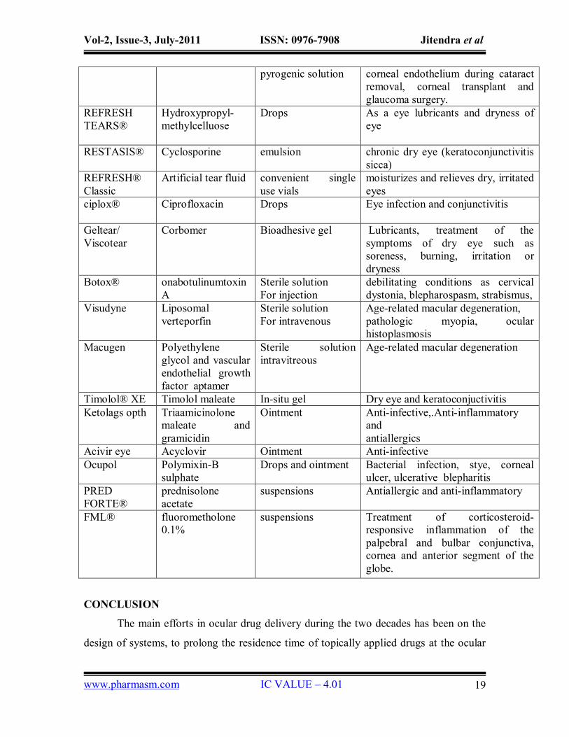
Vol-2, Issue-3, July-2011 ISSN: 0976-7908 Jitendra et al
www.pharmasm.com IC VALUE – 4.01
19
pyrogenic solution corneal endothelium during cataract removal, corneal transplant and glaucoma surgery.
REFRESH TEARS®
Hydroxypropyl-methylcelluose
Drops As a eye lubricants and dryness of eye
RESTASIS® Cyclosporine emulsion chronic dry eye (keratoconjunctivitis sicca)
REFRESH® Classic
Artificial tear fluid convenient single use vials
moisturizes and relieves dry, irritated eyes
ciplox® Ciprofloxacin Drops Eye infection and conjunctivitis
Geltear/ Viscotear
Corbomer Bioadhesive gel Lubricants, treatment of the symptoms of dry eye such as soreness, burning, irritation or dryness
Botox® onabotulinumtoxinA
Sterile solution For injection
debilitating conditions as cervical dystonia, blepharospasm, strabismus,
Visudyne Liposomal verteporfin
Sterile solution For intravenous
Age-related macular degeneration, pathologic myopia, ocular histoplasmosis
Macugen Polyethylene glycol and vascular endothelial growth factor aptamer
Sterile solution intravitreous
Age-related macular degeneration
Timolol® XE Timolol maleate In-situ gel Dry eye and keratoconjuctivitis Ketolags opth Triaamicinolone
maleate and gramicidin
Ointment Anti-infective,.Anti-inflammatory and antiallergics
Acivir eye Acyclovir Ointment Anti-infective Ocupol Polymixin-B
sulphate Drops and ointment Bacterial infection, stye, corneal
ulcer, ulcerative blepharitis PRED FORTE®
prednisolone acetate
suspensions Antiallergic and anti-inflammatory
FML® fluorometholone 0.1%
suspensions
Treatment of corticosteroid-responsive inflammation of the palpebral and bulbar conjunctiva, cornea and anterior segment of the globe.
CONCLUSION
The main efforts in ocular drug delivery during the two decades has been on the
design of systems, to prolong the residence time of topically applied drugs at the ocular
Vol-2, Issue-3, July-2011 ISSN: 0976-7908 Jitendra et al
www.pharmasm.com IC VALUE – 4.01
20
surface and conjuctival sac. The development of ophthalmic drug delivery systems is
easy because we can easily target the eye to treat ocular diseases as well as complicated
because the eye has specific characteristics such eye protecting mechanism, which make
ocular delivery systems extremely difficult. The most widely developed drug delivery
system is represented by the conventional and non-conventional ophthalmic formulations
to polymeric hydrogels, nanoparticle and nanosuspensions, microemulsions,
iontophorosis and ocular inserts. Currently, very few new ophthalmic drug delivery
systems have been commercialized in which them ocular inserts have been mostly used.
In future an ideal system should be able to achieve an effective drug concentration at the
target tissue for an extended period of time, while minimizing systemic exposure and the
system should be both comfortable and easy to use.
REFERENCES
1. Lee VHL, Robinson JR: Topical ocular drug delivery: recent developments and
future challenges. Journal of Ocular Pharmacology 1986; 2: 67–108.
2. Lang J C. Ocualar drug delivery conventional ocular formulation. Advanced drug
delivery review 1995;16:39-43.
3. K Basavaraj, Nanjawade, Manvi FV and Manjappa AS: In situ-forming
hydrogels for sustained ophthalmic drug delivery. Journal of Controlled Release
2007; 122: 119–134.
4. Sahoo KS, fahima SAD, kumar K: Nanotechnology in ocular drug delivery, Drug
delivery today 2008; 13: 144-151.
5. Weidener J: Mucoadhesive ocular inserts as an improved delivery vehicle for
ophthalmic indications. Drug Discovery Today 2003; 8: 906– 907.
6. Lang JC, Roehrs RTE and Jani R: Ophthalmic preparations, Edition 21, vol-1;
Lippincott Williams and Wilkins, 2005.
7. Andrews GP, Laverty TP and Jones DS: Mucoadhesive polymeric platforms for
controlled drug delivery Review article. European Journal of Pharmaceutics and
Biopharmaceutics 2009; 71: 505–518.
8. Alany RG, Rades T, Nicoll J, Tucker IG and Davies NM: W/O microemulsions
for ocular delivery: Evaluation of ocular irritation and precorneal retention.
Journal of Controlled Release 2006; 111: 145-152.

Vol-2, Issue-3, July-2011 ISSN: 0976-7908 Jitendra et al
www.pharmasm.com IC VALUE – 4.01
21
9. Binstock EE and Domb AJ: Iontophoresis: A non-invasive ocular drug delivery.
Journal of Controlled Release 2006; 110: 479–489.
10. Waugh A and Grant A: The special senses Ross and Wilson Anatomy and
physiology in Health and Illness, Churchill Livingstone 197-207.
11. Chien YW: Ocular drug delivery and delivery systems, special edition, 269-296.
12. Greaves JL and Wilson CG: Treatment of diseases of the eye with mucoadhesive
delivery systems. Advance Drug Delivery Review 1993; 11: 349– 383.
13. Robinson JC: Ocular anatomy and physiology relevant to ocular drug delivery, ,
Ophthalmic Drug Delivery Systems, New York, A.K. Mitra Edition, 1993, 29–
57.
14. Huang AJW, Tseng SCG and Kenyon KR: Paracellular permeability of corneal
and conjunctival epithelia. Investigation Ophthalmology of Visual Sciense 1989;
30 : 684– 689.
15. Haeringen NJV: Clinical biochemistry of tears. Survival Ophthalmology 1981; 5 :
84– 96.
16. Nagyova B and Tiffany JM: Components responsible for the surface tension of
human tears. Current Eye Research 1999; 19 : 4 – 11.
17. Ahmed L, Gokhale RD, Shah MV and Paton TF: Phytochemical determinants of
drug difussion across the conjunctiva, sclera and cornea. Journal of
Pharmaceutical Science 1987 : 76 : 583-587.
18. Bloomfield SE, Miyata T, Dunn MW, Bueser N, Stenza KH and Rubin AL:
Soluble gentamicin opthalmic insert as drug delivery system. Arch
Ophthalmology 1978: 96: 885-887.
19. Bito LZ, Baroody RA, Backerman A: The mechanism of peptidergic miosis: The
structural basis of miotic potency among biologically active polypeptides. Current
Eye Research 1982; 1: 559- 565,
20. Crawford K, Gabelt BT, Kaufman PL, Bito LZ: Effects of various anesthetic and
autonomic drugs on refraction in monkeys. Current Eye Research 1990; 9: 525-
532,.

Vol-2, Issue-3, July-2011 ISSN: 0976-7908 Jitendra et al
www.pharmasm.com IC VALUE – 4.01
22
21. Shell JW and and Baroody R.A: The ocular pharmacokinetics of eicosanoids and
their derivatives. 1. Comparison of ocular eicosanoid penetration and distribution
following the topical application of PGF2α, PGF2α-1-methyl ester, and PGF2α-1-
isopropyl ester. Experimental Eye Research 1987; 44: 217-226.
22. Bourlais CL, Acar L, Zia H and Sado PA, Ophthalmic drug delivery systems —
recent advances, Prostaglandin Retinal Eye Research 1998; 17 : 33–55.
23. Bhargava HN, Nicolai DW and Oza BJ: Topical suspensions Pharmaceutical
dosage forms: Disperse systems Marcel Dekker, vol. 2, 1996, 183–241.
24. Ludwig A, van ootengm M. influence of viscolyzers on the residence of
ophthalmic solution evaluated by slit lamp fluorometry. Pharmaceutical science
1992; 2: 81-87.
25. David NM, Farr SJ, Hadgraft J, Kellaway IW: Evalution of mucoadhesive
polymer in ocular drug delivery. I. Viscous solution. Pharmaceutical research
1991; 8: 1039-1043.
26. Ali Y, Beck R, Sport Process for manufacturing ophthalmic suspension, U.S.
Patent 6,071,904; 2000.
27. Kimura, Motoko, Morita, Yasushi, Ogawa, Takahiro, Terai, Tadashi. Ophthalmic
suspension containing diflupredonate United States; Patent 5556848.
28. Kinloch AJ: The science of adhesion. Journal of Material Science 1980; 15:
2141–2166.
29. Henriksen I, Green K, Smart J, Smistad G, and Karlsen J: Bioadhesion of
hydrated chitosans: an in vitro and in vivo study, International Journal of
Pharmaceutical science 1996; 45: 231–240.
30. Woodley J: Bioadhesion: new possibilities for drug administration?. Clinical
Pharmacokinetic 2001; 40: 77–84.
31. Alonso MJ and. Sanchez A: The potential of chitosan in ocular drug delivery.
journal. Pharmaceutics and Pharmacology 2003; 55: 1451–1463.
32. Lehr CM, Bouwstra JA, Schacht EH and Junginger HE: In vitro evaluation of
mucoadhesive properties of chitosan and some other natural polymers.
Inernational. Journal Pharmceutics 1992; 78: 43–48.
Vol-2, Issue-3, July-2011 ISSN: 0976-7908 Jitendra et al
www.pharmasm.com IC VALUE – 4.01
23
33. Ludwig A: The use of mucoadhesive polymers in ocular drug delivery. Advanced
Drug Delivery Reviews 2005; 57: 1595– 1639.
34. Patil SB, Murthy RSR, Mahajan HS, Wagh RD and Gattani SG. Mucoadhesive
polymers: Means of improving drug delivery. Pharma Times 2006 :38 : 25-28.
35. Vasant V. Ronade, Mannfred A. Hallonger: intranasal and ocular Drug delivery
system, CRC press pharmacology press, second edition, 2008, 267-280.
36. Uchegba IF and schtzeil AG: Polymer in drug delivery. Taylor and francis group
2007, 163-183.
37. Coviello T, Matricardi P, Marianecci C and Alhaique F: Polysaccharide hydrogels
for modified release formulations. Journal of Controlled Release Volume, 2007;
119: 5-24.
38. Seth A, and John S: keratoconjuctivitis sicca. Small Animal Ophthalmology
Secrets, 2002, 57
39. Sahoo SK and Labhasetwar V: Nanotech approaches to drug delivery and
imaging. Drug Discovery Today 2003; 8: 1112–1120.
40. Kayser O: The impact of nanobiotechnology on the development of new drug
delivery systems. Current Pharmceutical Biotechnology. 2005; 6 : 3–5.
41. Brigger: Nanoparticles in cancer therapy and diagnosis. Advance Drug Delivery
Review 2002; 54: 631–651.
42. Fialho SL: New vehicle based on a microemulsion for topical ocular
administration of Dexamethasone. Clinical Express Ophthalmology 2004; 32:
626–632.
43. Vandamme TF: Microemulsions as ocular drug delivery systems: recent
developments and future challenges, Progstaglandin Retinal. Eye Research 2002;
21: 15–34.
44. Lawrence MJ: Microemulsion-based media as novel drug delivery systems,
Advanced Drug Deliv. Rev. 2000; 45: pp. 89–121.
45. Ding S Tien W, Olejnik O. US Patent 1995; 5:474-979.
46. Alany RG, Rades T, Nicoll J, Tucker IG and Davies NM: W/O microemulsions
for ocular delivery: Evaluation of ocular irritation and precorneal retention,
Journal of Controlled Release 2006; 111: 145-152.
Vol-2, Issue-3, July-2011 ISSN: 0976-7908 Jitendra et al
www.pharmasm.com IC VALUE – 4.01
24
47. Bourges JL: Ocular drug delivery targeting the retina and retinal pigment
epithelium using polylactide nanoparticles. Investgation Ophthalmology in Visual
Science. 2003; 4: 3562–3569.
48. Sakurai E: Effect of particle size of polymeric nanospheres on intravitreal
kinetics. Ophthalmology Research 2001; 22: 31–36.
49. Lee VHL: Ocular drug delivery of progesterone using nanoparticles. Journal
Microencapsuational 1986; 3: 213–218.
50. Irache JM : Albumin nanoparticles for the intravitreal delivery of
anticytomegaloviral drugs, Mini Reviewon Medical Chemistry 2005; 5: 293–305.
51. Calvo P: Evaluation of cationic polymer-coated nanocapsules as ocular drug
carriers, International Journal of Pharmaceutics 1997; 153: 41–50.
52. Campos AMD: The effect of a PEG versus a chitosan coating on the interaction of
drug colloidal carriers with the ocular mucosa. European Journal
Pharmaceutical Science 2003: 20, 73–81.
53. O. Kayser et al., The impact of nanobiotechnology on the development of new
drug delivery systems, Curr. Pharm. Biotechnol. 6 (2005), pp. 3–5.
54. Pignatello R, Bucolo C, Spedalieri G, Maltese A, Puglisi G: Flurbiprofen-loaded
acrylate polymer nanosuspensions for ophthalmic application. Biomaterials 23:
3247–3255
55. Pignatello R., Bucolo C,and Puglisi G. Ocular tolerability of Eudragit RS 100 and
RL 100 nanosuspensions as carrier for ophthalmic controlled delivery. Journal
Pharmaceutical Science 91: 2636–2641
56. BeharCohen FF, Aouni AEl, and Gautier S: Transscleral Coulombcontrolled
iontophoresis of methylprednisolone into the rabbit eye: Influence of duration of
treatment, current intensity and drug concentration on ocular tissue and fluid
levels. Express Eye Research 2002; 74: 51–59.
57. Kalia YN, Naik A, Garrison J, Guy RH : Iontophoretic drug delivery. Advanced
Drug Delivery Reviews; 2004: 56 619– 658.
58. Dixit N. Bali V, Baboota S, Ahuja A, and Ali J: iontophoresis-an approach for
controlled drug delivery; a review. Current drug delivery, 2007; 4: 1-10.

Vol-2, Issue-3, July-2011 ISSN: 0976-7908 Jitendra et al
www.pharmasm.com IC VALUE – 4.01
25
59. Amo EMD and Urtti A: Current and future ophthalmic drug delivery systems a
shift to the posterior segments. Drug Delivery Today 2008; 13: 153-143.
60. Kumari A, Sharma PK and Garg VK: Ocular inserts — Advancement in therapy
of eye diseases. Journal Advance Pharmaceutical Technology Research 2010; 3:
87-96.
61. Neefe CW. Contact lens for ocular drug delivery. US Patent 1974; 3:786-812.
62. Gibaldi M, Perrier D. Pharmacokinetics. Marcel Dekker, New York, 2nd
edition,1993.
63. Hornof MD, Weyenberg W, Ludwig A, Schnurch AB: A mucoadhesive ocular
insert: development and in vivo evaluation in humans. Journal Controlled
Release 2003; 89 :419– 428.
64. www.mims-india.com date of citation at 03/11/2010.
65. www.allergan.com date of citation at 03/11/2010.
66. www.cimsasia.com date of citation at 03/11/2010.
For Correspondence: Mr. Jitendra Research Scholar (M. Pharm) Department of Pharmaceutical Technology, MIET NH-58, Bypass Road, Bagphat crossing, Meerut 25005, U.P. INDIA Mob. +919997300109 Email Id. [email protected]