Embed Size (px)
Citation preview
Zsltschrift f i lr
Parasitenkunde Parasilology Research
0 Springer-Verlag 1985
A new type of ciliated sensory receptor in the cercariae of Nicolla gal’ca (Trematoda)
Antoine Pariselle ‘9 * and Michelle Matricon-Gondran Laboratoire de Parasitologie Comparée, Université des Sciences et Techniques du Languedoc, Place E. Bataillon, F-34060 Montpellier Cedex, France Laboratoire d’Histologie et Cytologie des Invertébrés Marins, Université Pierre et Marie Curie, 12 rue Cuvier, F-75005 Paris, France
Abstract. Ciliated sensory receptors of a hitherto unknown type were found in the cercariae of Nicolla gallica (Trematoda, Coitocaecidae). They displayed a sheath or collar of tegumentary origin from which ’
stereocilia-like villi projected. Some of them might act as mechanorecep- tors.
Introduction Ciliated sensory receptors of the Platyhelminthes tegument are well docu- mented: they have been described in various Turbellaria, and in miracidia and cercariae of Trematodes and in Cestodes. Their structure is fairly invari- able, even .if the intrategumentary nerve ending may bear one or several cilia.
Trematode cercariae exhibit a specific distribution of these receptors or sensillae on their body. Study of the chaetotaxis of the cotylicercous cercaria in N . gallica (Pariselle and Lambert, in preparation) using conven- tional silver impregnation methods (Combes et al. 1976) and scanning elec- tron microscopy (SEM) revealed a new type of ciliated sensory receptor characterized by numerous villi.
Here, we analysed the fine structure of these receptors and their diversity by comparing data acquired from scanning electron microscope (SEM) and transmission electron microscopy (TEM).
Material and methods N. gallica cercariae (Trematoda, Coitocaecidae) were obtained from their host snails Tlzeodoxiu fluuiutilis, collected at Le Lez river springs near Montpellier (France).
* lfeient adZfeFs7- ORSTOM B e l 386,-Dakar Bel-Air, Senegal
Offprint requests to: A. Pariselle i ---
354 A. Pariselle and M. Matricon-Gondran
Cercariae already emitted or obtained by dissection of the snail were fixed by immersion in 2% glutaraIdehyde in 0.1 M cacodylate buffer, pH 7.4 for 1 h at 4" C, washed in 0.2 M cacodylate buffer, pH 7.4 for 12 h at 4" C and postfixed in 1.3% osmium tetroxide in 0.13 fil cacodylate buffer, pH 7.4, for 1 h at 4" C.
For SEM, samples were dehydrated, dried by the critical point method, metallised and observed with a JEOL JSM 35 microscope. For TEM observation, samples were dehydrated and embedded in Spurr resin. Ultrathin sections, contrasted by uranyl acetate and lead citrate, were observed with a Philips EM 300 microscope.
For comparison, two cercariae from closely related species of Coitocaecidae were studied by SEM only: one was found, also near Montpellier, in the Belgrandia gibba snail, and the other, in íhe Melanopsis praeinorsa snail from Morocco.
Results
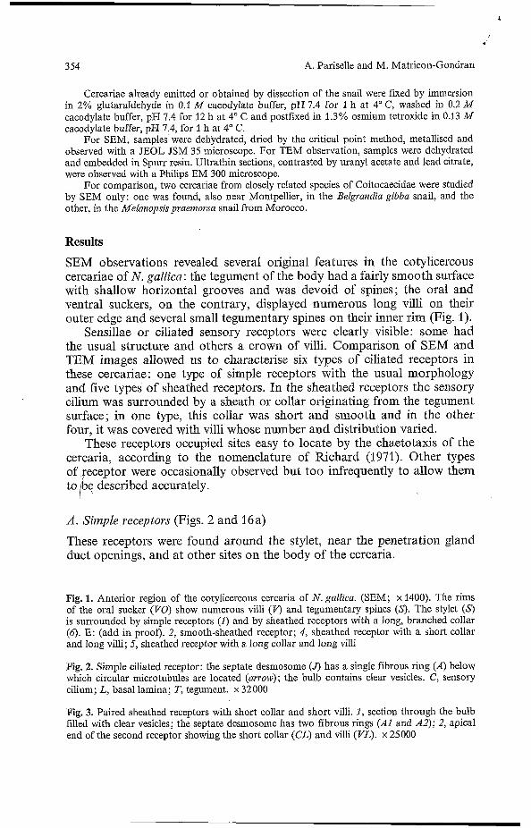
SEM observations revealed several original features in the cotylicercous cercariae of N . gallica : the tegument of the body had a fairly smooth surface with shallow horizontal grooves and was devoid of spines; the oral and ventral suckers, on the contrary, displayed numerous long villi on their outer edge and several small tegumentary spines on their inner rim (Fig. 1).
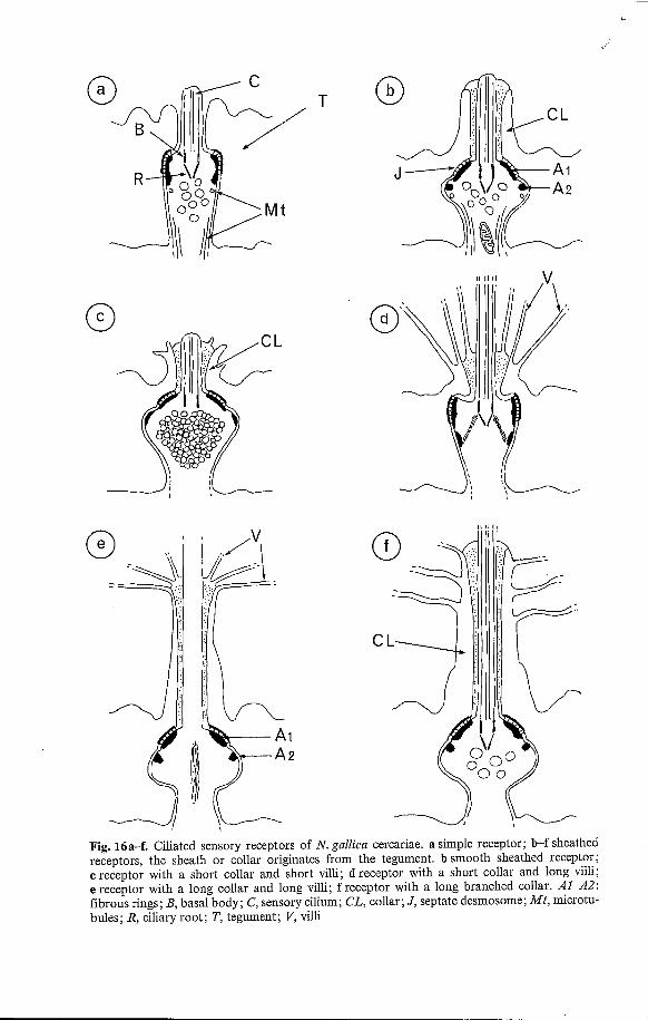
Sensillae or ciliated sensory receptors were clearly visible: some had the usual structure and others 'a crown of villi. Comparison of SEM and TEM images allowed us to characterise six types of ciliated receptors in these cercariae: one type of simple receptors with the usual morphology and five types of sheathed receptors. In the sheathed receptors the sensory cilium was surrounded by a sheath or collar originating from the tegument surface; in one type, this collar was short and smooth and in the other four, it was covered with villi whose number and distribution varied.
These receptors occupied sites easy to locate by the chaetotaxis of the cercaria, according to the nomenclature of Richard (1971). Other types of feceptor were occasionally observed but too infrequently to allow them to /be described accurately.
A . Simple receptors (Figs. 2 and 16a)
These receptors were found around the stylet, near the penetration gland duct openings, and at other sites on the body of the cercaria.
Fig. 1. Anterior region of the cotylicercous cercaria of N . gallica. (SEM; x 1400). The rims of the oral sucker (VO) show numerous villi (V) and tegumentary spines CS). The stylet (S) is surrounded by simple receptors ( I ) and by sheathed receptors with a long, branched collar (6). E: (add in proof). 2, smooth-sheathed receptor; 4, sheathed receptor with a short collar and long villi; 5, sheathed receptor with a long collar and long villi
Fig. 2. Simple ciliated receptor: the septate desmosome (4 has a single fibrous ring (A) below which circular microtubules are located (arrow) ; the bulb contains clear vesicles. C, sensory cilium; L, basal lamina; T, tegument. x 32000
Fig. 3. Paired sheathed receptors with short collar and short villi. 1, section through the bulb filled with clear vesicles; the septate desmosome has two fibrous rings (AI and A2); 2, apical end of the second receptor showing the short collar (CL) and villi (VL). x 25000
.bd
A new type of ciliated sensory receptor in the cercariae of Nicolla gallica (trematoda) 355
CL
356 A. Pariselle and M. Matricon-Gondran
The ciliated nerve ending was almost cylindrical since the nerve fibre did not broaden where it entered the tegument. The cilium was short and its basal body was extended by a conical ciliary root. The circular septate desmosome which bound the ciliated bulb to the tegument surface displayed fibrous thickenings of the cytoplasm along both adjacent plasma mem- branes. These thickenings appeared as a thin plaque on the tegumentary side, and as a thick ring, wider at its basal pole, in the receptor (Fig. 2). Axial microtubules occupied the periphery of the nerve fibre; one or two circular microtubules were visible below the fibrous ring underlining the septate junction. These receptors contained numerous vesicles of moderate electron density.
B. Sheathed receptors
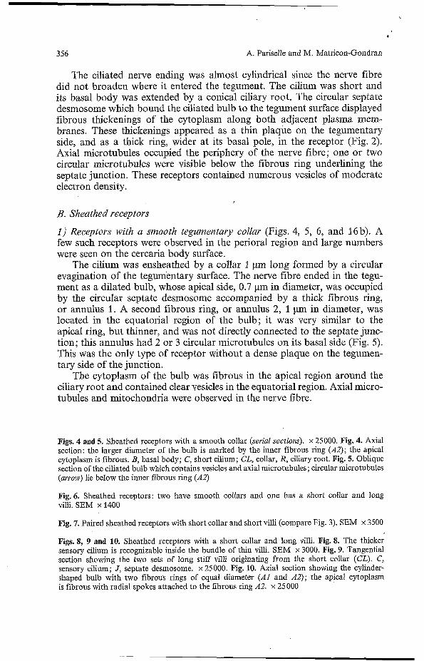
1) Receptors with a smooth tegumentary collar (Figs. 4, 5, 6, and 16b). A few such receptors were observed in the perioral region and large numbers were seen on the cercaria body surface.
The cilium was ensheathed by a collar 1 pm long formed by a circular evagination of the tegumentary surface. The nerve fibre ended in the tegu- ment as a dilated bulb, whose apical side, 0.7 pm in diameter, was occupied by the circular septate desmosome accompanied by a thick fibrous ring, or annulus 1. A second fibrous ring, or annulus 2, 1 pm in diameter, was located in the equatorial region of the bulb; it was very similar to the apical ring, but thinner, and was not directly connected to the septate junc- tion; this annulus had 2 or 3 circular microtubules on its basal side (Fig. 5). This was the only type of receptor without a dense plaque on the tegumen- tary side of the junction.
The cytoplasm of the bulb was fibrous in the apical region around the ciliary root and contained clear vesicles in the equatorial region. Axial micro- tubules and mitochondria were observed in the nerve fibre.
Figs. 4 and 5. Sheathed receptors with a smooth collar (serial sections). x 25000. Fig. 4. Axial section: the larger diameter of the bulb is marked by the inner fibrous ring (A2); the apical cytoplasm is fibrous. B, basal body; C, short cilium; CL, collar, R, ciliary root. Fig. 5. Oblique section of the ciliated bulb which contains vesicles and axial microtubules; circular microtubules (arrow) lie below the inner fibrous ring (A2)
Fig. 6. Sheathed receptors: two have smooth collars and one has a short collar and long villi. SEM x 1400
Fig. 7. Paired sheathed receptors with short collar and short villi (compare Fig. 3). SEM x 3500
Figs. 8, 9 and IO. Sheathed receptors with a short collar and long villi. Fig. 8. The thicker sensory cilium is recognizable inside the bundle of thin villi. SEM x 3000. Fig. 9. Tangential section showing the two sets of long stiff villi originating from the short collar (CL). C, sensory cilium; J, septate desmosome. x 25000. Fig. IO. Axial section showing the cylinder- shaped bulb with two fibrous rings of equal diameter (Ai' and A2); the apical cytoplasm is fibrous with radial spokes attached to the fibrous ring A2. x 25 O00

v
A new type of ciliated sensory receptor in the cercariae of Nicolla gallica (trematoda) 357
358 A. Pariselle and M. Matricon-Gondran
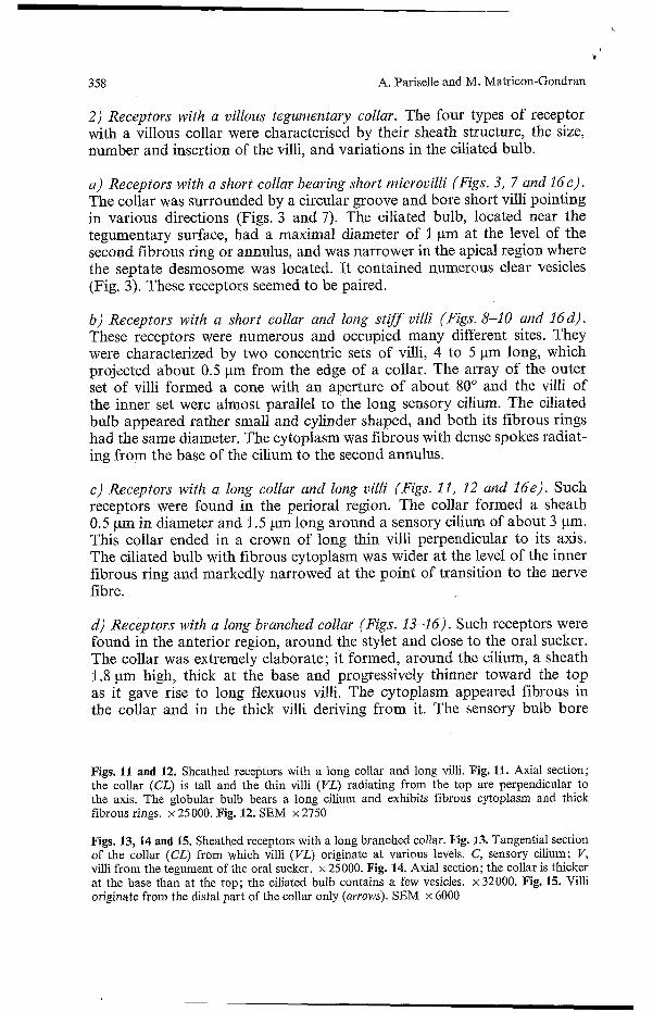
2 ) Receptors with a villous tegumentary collar. The four types of receptor with a villous collar were characterised by their sheath structure, the size, number and insertion of the villi, and variations in the ciliated bulb.
a ) Receptors with a short collar bearing short microvilli (Figs. 3, 7 and Ide) . The collar was surrounded by a circular groove and bore short villi pointing in various directions (Figs. 3 and 7). The ciliated bulb, located near the tegumentary surface, had a maximal diameter of 1 pm at the level of the second fibrous ring or annulus, and was narrower in the apical region where the septate desmosome was located. It contained numerous clear vesicles (Fig. 3). These receptors seemed to be paired.
b ) Receptors with a short collar and long stiff villi (Figs. 8-10 nnd 16d) . These receptors were numerous and occupied many different sites. They were characterized by two concentric sets of villi, 4 to 5 pm long, which projected about 0.5 pm from the edge of a collar. The array of the outer set of villi formed a cone with an aperture of about 80" and the villi of the inner set were almost parallel to the long sensory cilium. The ciliated bulb appeared rather small and cylinder shaped, and both its fibrous rings had the same diameter. The cytoplasm was fibrous with dense spokes radiat- ing from the base of the cilium to the second annulus.
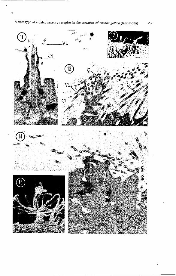
c ) Receptors with a long collar and long villi (Figs. I I , 12 and Ide). Such receptors were found in the perioral region. The collar formed a sheath 0.5 pm in diameter and 1.5 pm long around a sensory cilium of about 3 pm. This collar ended in a crown of long thin villi perpendicular to its axis. The ciliated bulb with fibrous cytoplasm was wider at the level of the inner fibrous ring and markedly narrowed at the point of transition to the nerve fibre.
d ) Receptors with a long branched collar (Figs. 13-16). Such receptors were found in the anterior region, around the stylet and close to the oral sucker. The collar was extremely elaborate; it formed, around the cilium, a sheath 1.8 pm high, thick at the base and progressively thinner toward the top as it gave rise to long flexuous villi. The cytoplasm appeared fibrous in the collar and in the thick villi deriving from it. The sensory bulb bore
Figs. 11 and 12. Sheathed receptors with a long collar and long villi. Fig. 11. Axial section; the collar (CL) is tall and the thin villi (VL) radiating from the top are perpendicular to the axis. The globular bulb bears a long cilium and exhibits fibrous cytoplasm and thick fibrous rings. x 25 000. Fig. 12. SEM x 2750
Figs. 13, 14 and 15. Sheathed receptors with a long branched collar. Fig. 13. Tangential section of the collar (CL) from which villi (VL) originate at various levels. C, sensory cilium; V, villi from the tegument of the oral sucker. x 25000. Fig. 14. Axial section; the collar is thicker at the base than at the top; the ciliated bulb contains a few vesicles. x 32000. Fig. 15. Villi originate from the distal part of the collar only (arrows). SEM x 6000
‘J
A new type of ciliated sensory receptor in the cercariae of Nicolla gallica (trematoda) 359
T
Fig. 1621-f. Ciliated sensory receptors of N . gallica cercariae. a simple receptor; b-f sheathed receptors, the sheath or collar originates from the tegument. b smooth sheathed receptor; c receptor with a short collar and short villi; d receptor with a short collar and long villi; e receptor with a long collar and long villi; f receptor with a long branched collar. Al-A2: fibrous rings; B, basal body; C, sensory cilium; CL, collar; J, septate desmosome; Mt, microtu- bules; R, ciliary root; T, tegument; V, villi
'U
A new type of ciliated sensory receptor in the cercariae of Nicolla gallica (trematoda) 361
a cilium longer than its sheath and contained clear vesicles. The septate desmosome was similar to that of the other sheathed receptors.
Cercariae from Belgvaiidia gibba had ciliary receptors very similar to those of N . gallica while Melariopsis praenzovsa cercariae had only simple and smooth-sheathed receptors.
Discussion
The general features of the ciliated sensory receptors of N . gallica cercariae are presented in Fig. 16. The structure of the cilia, basal bodies, and ciliary roots was fairly constant. Significant differences appeared in the cytoplasm which contained either clear vesicles or fibrous structures, or both. The essential differences were in the collar of tegumentary origin.
The definition of these various types of receptors could be questioned if the increasing structural complexity represented intermediate steps in the development of the most complex type of receptor only. However, this possibility can be ruled out for several reasons. The different types of recep- tors described here were found in cercariae still in the process of development in the sporocyst, as well as in free-swimming cercariae. Each type has a well-defined location in the chaetotaxis of the cercaria, as established by Pariselle and Lambert (in preparation). Moreover, it would be difficult to trace an evolution from a simpler type to a more elaborate one, as each receptor seems to have evolved in its own particular direction.
Simple ciliated receptors similar to those of N.gallica have been de- scribed in a number of Trematode cercariae (Chapman and Wilson 1970; Matricon-Gondran 1971; Kaie 1973; Bennet 1975; Edwards et al. 1977) and in Cestodes (Webb and Dawey 1974; Gabrion and Euzet-Sicard 1979).
Receptors in which a simple smooth collar is formed by the 'tegument and ensheaths the sensory cilium have been observed in the cercariae of Diylostoinum phoxini (Bibby and Rees 1971) and of Sclzistosovlza inansoni (Robson and Erasmus 1970; Ebrahimzadeh 1974). A larger more complex tegumentary sheath exists in receptors of the miracidium of Fasciola hepatica (Wilson 1970).
In some cases, the sensory bulb itself forms a collar around the cilium. In Monogeneans (Lyons 1972), the apical surface of the receptor is deeply embedded in a depression of the tegument; in fact the " collar" is an exten- sion of the bulb itself and connects it to the surface of the tegument. In Acoeleous Turbellaria, Bedini et al. (1973) found receptors with a fairly long collar; they also observed receptors with villi or stereocilia which pro- jected from the apical surface and were arranged in a circle around the sensory cilium or kinocilium. Both types of receptors may be present in other Turbellaria (Ehlers and Ehlers 1977).
To our knowledge no ciliated receptors ensheathed by a collar of tegu- mentary origin and covered with villi have been previously described in Platyhelminthes. We cannot at present explain how such elaborate structures are induced in the tegument, which is the non-nervous component of these receptors.
362 A. Pariselle and M. Matricon-Gondran
Structure of the annular septate desmosome Some authors apply the term “septate desmosome” to the circular junction connecting the ciliated bulb to the syncytial tegument in Platyhelminthes. However, we observed that in the cercariae examined here, the two types of cellular junctions which in other Invertebrates are usually separate were in fact superimposed. Here, we find a septate junction with an occluding function, as well as cytoplasmic filaments attached to the membrane, as in adhesive junctions: zonulae adherentes or desmosomes. The term “septate desmosome ” seems appropriate to the present situation, as this structure appears to carry out both adhesive and occluding functions.
The fibrous structures linked to the septate desmosome varied in size; they were thin or absent on the tegumentary side. In sheathed receptors, they formed two dense rings or annuli, of which the most apical was con- nected with the junctional area, as in most Trematodes (Morris and Thread- gold 1967; Matricon-Gondran 1971) and in Cestodes (Webb and Davey 1974; Blair and Burt 1976; Gabrion and Euzet-Sicard 1979). In the simple N . gallica receptors, the single dense ring with its basal thickening seemed to correspond to the double rings in sheathed receptors.
These fibrous structures may serve as points of attachment for the cyto- plasmic filaments surrounding the ciliary root and for the axial microtubules of the nerve fibre. We would stress the links observed between the circular microtubules and the fibrous ring in the equatorial region of several sheathed receptor bulbs; these skeletal structures may stabilise the shape of the bulb.
Functions of the sensory receptors of N . gallica From their structure and location ciliated sensory receptors of Platyhel- minthes are often considered to be chemoreceptors or mechanoreceptors ; this, however, lacks experimental confirmation.
In the present case, we noticed that in simple receptors and in sheathed receptors with a smooth collar, the bulb bears a rather short sensory cilium and contains numerous vesicles. However, in sheathed receptors whose col- lar bears long, stiff, stereocilia-like villi, the bulb has a long sensory cilium and a fibrous cytoplasm almost devoid of vesicles. Other sheathed receptors have intermediate features.
We believe the stereocilia-like villi on the collar of sheathed receptors could have a mechanoreceptive function. The rigid villi pointing in various directions would be sensitive to the slightest pressure, the movement being transmitted to the collar and then to the bulb. The images we obtained did not allow us to establish whether or not the fine structure of the axoneme was modified. In any case, the fibrous cytoplasm of the bulb is reminiscent of the tubular body, a structure which is both microtubular and fibrous and is observed in mechanoreceptors of insects (Matsumoto and Farley 1978). Consequently, such sheathed receptors may be sensitive to small water currents or to living organisms swimming in their immediate vicinity.
Y
A new type of ciliated sensory receptor in the cercariae of Nicolla gallica (trematoda) 363
This hypothesis is supported by cercarial behaviour: thus, according to Dollfus (1959) and Pariselle (unpublished data), the cotylicercous cercaria studied here does not swim but walks on supports like a span-worm or a leech, alternatively standing on its oral sucker and tail. When stopped, it stands on its secretion-rich tail stump; when touched by the slightest current, it stretches out and oscillates in various directions in search of a mobile support to which it can attach itself, such as its vector host, the small Crustacean Gaiharus.
Aclznowledgements. A. Pariselle is grateful to Mr. M. Luc to Dr. A. Lambert and to Dr. C. Gabrion for their encouragement and advice throughout this work.
References
Bedini C, Ferrero E, Lanfranchi A (1973) The ultrastructure of ciliary sensory cells in two Turbellaria Acoela. Tissue and Cell 5 : 359-372
Bennet CE (1975) Surface features, sensory structures and movement of the newly excysted juvenile Fusciola hepatica L. J Parasito1 61 : 886891
Bibby MC, Rees G (1971) The ultrastructure of the epidermis and associated structures in the metacercaria, cercaria and sporocyst of Diplostomilin plzoxini (Faust 1918). Z Parasitenk
Blair DG, Burt MDB (1976) Observations on the ultrastructure of papillae and associated sensilla on the scolex of Monoecocestus ainericaizus (Stiles 1895) (Cestoda : Anoplocephali- dae). Can J Zool 54: 802-806
Chapman HD, Wilson RA (1970) The distribution and fine structure of the integumentary pupillae of the cercaria of Hisinasthla secunda (Nicoll). Parasitology 61 : 21 9-227
Combes C, Bayssade-Dufour C, Cassone J (1976) Sur l’imprégnation et le montage des cercaires pour l’étude chétotaxique. Ann Parasit hum Comp 51 : 399-400
Dollfus RPh (1959) Recherches experimentales sur Nicolla gallica (R.Ph. Dollfus 1941) R.Ph. Dollfus, 1958, sa cercaire cotylicerque et sa métacercaire progénétique. Observations sur la famille des Coitocaecidae Y. Ozaki, 1928. sF. Coitocaecinae F. Poche, 1926, Trematoda Podocotyloidae et sur les cercaires cotylicerques d‘eau douce et marines. Ann Parasit hum Comp 34:595-622,35:65-117
Ebrahimzadeh A (1 974) Beitrag zur Feinstruktur des teguments der cercarien von Schistosoma mansoizi. Z Parasitenk 44: 117-132
Edwards HH, Nollen PM, Nadakavukaren MS (1977) Scanning and transmission electron microscopy of oral sucker papillae of Philophthalmus inegalurzu. Int J Parasito1 7 : 429-437
Ehlers U, Ehlers B (1977) Monociliary receptors in interstitial Proseriata and Neorhabdocoela (Turbellaria Neoophora). Zoomorphology 86: 197-222
Gabrion C, Euzet-Sicard S (1979) Etude du tégument et des récepteurs sensoriels du scolex d’un plérocercoide de Cestoda Tetraphyllidae à l’aide de la microscopie électronique. Ann Paras Hum Comp 54: 573-583
Kaie M (1 973) The host-parasite interface and associated structures of the cercaria and adult Neophasis Zagenforinis (Lebour 1910). Ophelia 12: 205-219
Lyons KM (1972) Sense organs of Monogeneans. Behavioral aspects of parasite transmission. Supp no 1 Zool J of Linn Soc 51 : 181-189
Matricon-Gondran M (1971) Etude ultrastructurale des récepteurs sensoriels tégumentaires de quelques Trématodes Digénétiques larvaires. Z Parasitenk 35 : 318-333
Matsumoto DE, Farley RD (1978) Comparison of the ultrastructure of stimulated and unstim- ulated mechanoreceptors in the taste hair of the blowfly Phaeizicia serricata. Tissue and Cell 10:63-76
37:169-186
364 A. Pariselle and M. Matricon-Gondran
Morris GP, Threadgold LT (1967) A presumed sensory structure associated with the tegument
Richard J (1971) La chétotaxie des cercaires. Valeur systématique et phylétique. Mem Mus
Robson RT, Erasmus DA (1970) The ultrastructure, based on stereoscan observations of the oral sucker of the cercaria of Schistosoma mansoni with special reference to penetration. Z Parasitenk 35: 76-86
Webb RA, Davey KG (1974) Ciliated sensory receptors of the unactivated metacestode of Hymenolepis microstoina. Tissue and Cell 6 ; 587-598
Wilson RA (1970) Fine structure of the nervous system and specialized nerve endings in the miracidium of Fasciola hepatica. Parasitology 60 : 39941 O
I of Schistosoma mansoni. J Parasito1 53 : 537-539
Nat Hist Nat série A 67:p 179 I I
Accepted December 3, 1984