Embed Size (px)
Citation preview
A new type of lordosis and vertebral body compression inGilthead sea bream, Sparus aurata L.: aetiology, anatomyand consequences for survival
M Loizides1, A N Georgiou1,2, S Somarakis3, P E Witten4,5 and G Koumoundouros1
1 Biology Department, University of Crete, Vasilica Vouton, Heraklio, Crete, Greece
2 Biology Department, University of Patras, Rio, Patras, Greece
3 Institute of Marine Biological Resources, Hellenic Centre for Marine Research, Thalassocosmos, Heraklion, Crete,
Greece
4 Evolutionary Developmental Biology, Biology Department, Ghent University, Ghent, Belgium
5 Institute of Veterinary Animal and Biomedical Science, Massey University, Palmerston North, New Zealand
Abstract
A new type of vertebral malformation is described,consisting of deformed cartilaginous neural andhaemal processes and the compression and fusionof vertebral bodies. The malformation is desig-nated as haemal vertebral compression and fusion(haemal VCF). We studied the aetiology of themalformations and described microanatomicalhistopathological alterations. The malformationswere detected during routine quality control inone of six monitored Gilthead sea bream popula-tions. Haemal VCF affected the posterior part ofthe vertebral column (haemal vertebrae). In 20%of the deformed specimens, haemal VCF wascombined with lordosis. At 35 dph (days post-hatching), early anatomical signs of the haemalVCF consisted of abnormal centrum mineraliza-tion, malformed cartilaginous neural and haemalprocesses and developing lordotic alterations. Thehistological examination of the deformed individ-uals revealed that haemal VCF is preceded bynotochord abnormalities. The frequency ofdeformed individuals was three times higher at 35than at 61 dph (50.3% vs. 17.2%, n = 157 andn = 250, respectively). No signs of repair orreversion of malformations have been observed.Thus, the steep decrease in deformities in older
animals suggests that haemal VCF is linked tohigh mortality rates. The results are discussed inrespect of the possible causative factors of haemalVCF.
Keywords: skeletal deformities, notochord abnor-malities, fish larvae.
Introduction
Skeletal deformities are a major problem for mar-ine finfish aquaculture, affecting survival, animalwelfare and product quality (reviewed by Koumo-undouros 2010; Boglione & Costa 2011; Cob-croft & Battaglene 2013). Although of lowerfrequency in the wild, skeletal deformities havebeen reported to develop in various fish popula-tions and have been suggested as significant indi-cators of environmental disturbances andpollution (Boglione et al. 2006; Koumoundouros2008; Diggles 2013).Concerning Mediterranean finfish aquaculture,
data from commercial hatcheries report mean fre-quencies of skeletal deformities that reached from7 to 20% during the last decade, for a totalannual production of approximately one billionjuveniles (Koumoundouros 2010). In advancedteleost fish, skeletal deformities develop especiallyin early life stages, during the larval rearing phase.Early malformations are believed to be linked tosuboptimal levels of a variety of abiotic (Polo,
Correspondence G Koumoundouros, Biology Department,
University of Crete, Vasilica Vouton, 70013 Heraklio, Crete,
Greece (e-mail: [email protected])
949� 2013
John Wiley & Sons Ltd
Journal of Fish Diseases 2014, 37, 949–957 doi:10.1111/jfd.12189
Yufera & Pascual 1991; Sfakianakis et al. 2006;Georgakopoulou et al. 2007, 2010; Cobcroft &Battaglene 2009) and nutritional factors (Fernan-dez et al. 2008; Mazurais et al. 2009; Darias et al.2010). In hatcheries, skeletal deformities developdue to gaps of knowledge about factors that arerequired to facilitate healthy skeletal development.Our knowledge is increasing (Boglione, Gavaia,Koumoundouros, Gisbert, Moren, Fontagn�e &Witten 2013a; Boglione et al. 2013b; Izquierdoet al. 2013), but uncontrolled fluctuations of vari-ous parameters in the complex larval rearingsystems can still cause early skeletal malforma-tions. A prerequisite for achieving healthy skeletaldevelopment is the early diagnosis of malforma-tions, a precise diagnosis of the pathological pro-cesses and consequently the identification of theoptimal rearing conditions for different species.Establishing procedures for early diagnosis willalso help to improve quality monitoring in com-mercial hatcheries (Koumoundouros 2010).Part of the solution relies on studies about the
ontogeny, anatomy and aetiology of skeletal defor-mities. Such studies have been suggested to besignificant for the effective hypothesis formationand experimental testing of the responsible causa-tive factors, as well as for quality monitoring inhatcheries (Koumoundouros 2010; Boglione et al.2013b). In the present study, we analyse a newtype of vertebral deformities in Gilthead seabream, Sparus aurata L.
Materials and methods
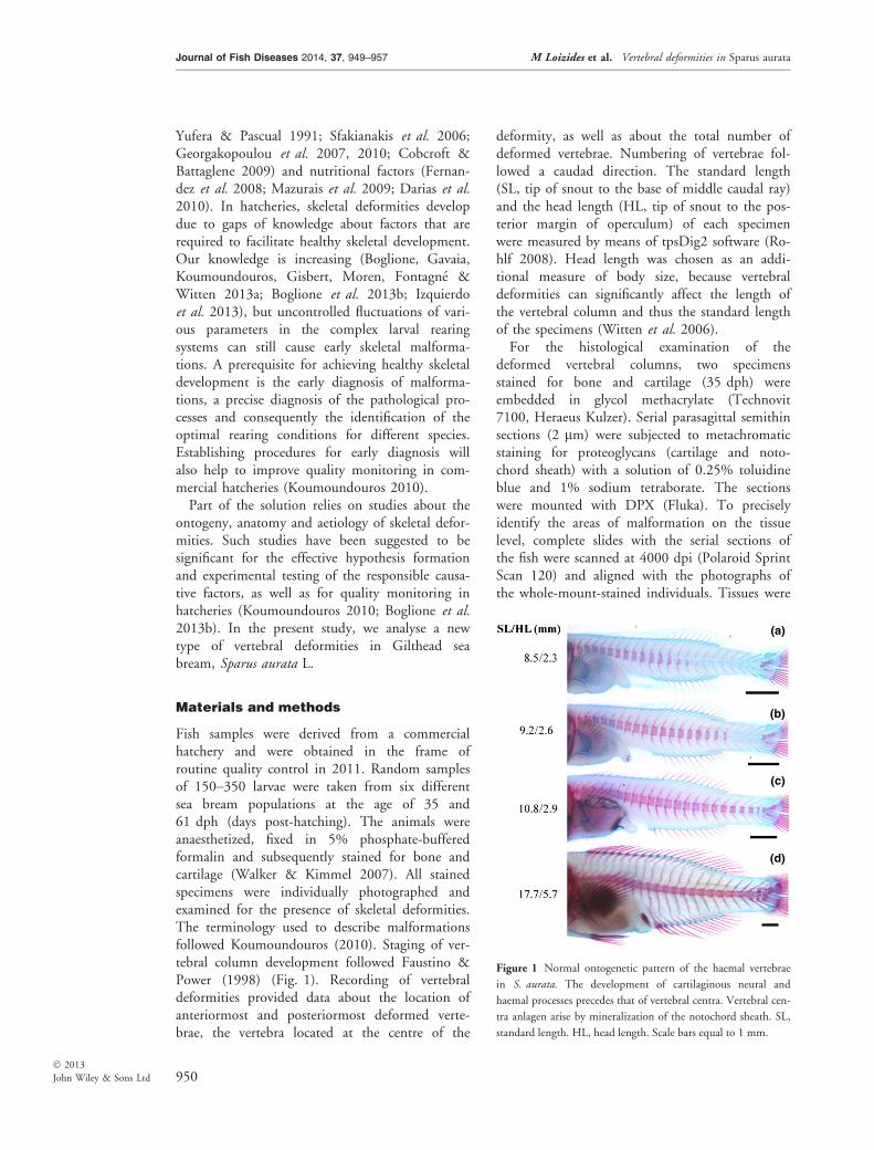
Fish samples were derived from a commercialhatchery and were obtained in the frame ofroutine quality control in 2011. Random samplesof 150–350 larvae were taken from six differentsea bream populations at the age of 35 and61 dph (days post-hatching). The animals wereanaesthetized, fixed in 5% phosphate-bufferedformalin and subsequently stained for bone andcartilage (Walker & Kimmel 2007). All stainedspecimens were individually photographed andexamined for the presence of skeletal deformities.The terminology used to describe malformationsfollowed Koumoundouros (2010). Staging of ver-tebral column development followed Faustino &Power (1998) (Fig. 1). Recording of vertebraldeformities provided data about the location ofanteriormost and posteriormost deformed verte-brae, the vertebra located at the centre of the
deformity, as well as about the total number ofdeformed vertebrae. Numbering of vertebrae fol-lowed a caudad direction. The standard length(SL, tip of snout to the base of middle caudal ray)and the head length (HL, tip of snout to the pos-terior margin of operculum) of each specimenwere measured by means of tpsDig2 software (Ro-hlf 2008). Head length was chosen as an addi-tional measure of body size, because vertebraldeformities can significantly affect the length ofthe vertebral column and thus the standard lengthof the specimens (Witten et al. 2006).For the histological examination of the
deformed vertebral columns, two specimensstained for bone and cartilage (35 dph) wereembedded in glycol methacrylate (Technovit7100, Heraeus Kulzer). Serial parasagittal semithinsections (2 lm) were subjected to metachromaticstaining for proteoglycans (cartilage and noto-chord sheath) with a solution of 0.25% toluidineblue and 1% sodium tetraborate. The sectionswere mounted with DPX (Fluka). To preciselyidentify the areas of malformation on the tissuelevel, complete slides with the serial sections ofthe fish were scanned at 4000 dpi (Polaroid SprintScan 120) and aligned with the photographs ofthe whole-mount-stained individuals. Tissues were
(a)
(b)
(c)
(d)
Figure 1 Normal ontogenetic pattern of the haemal vertebrae
in S. aurata. The development of cartilaginous neural and
haemal processes precedes that of vertebral centra. Vertebral cen-
tra anlagen arise by mineralization of the notochord sheath. SL,
standard length. HL, head length. Scale bars equal to 1 mm.
950
Journal of Fish Diseases 2014, 37, 949–957 M Loizides et al. Vertebral deformities in Sparus aurata
� 2013
John Wiley & Sons Ltd
subsequently analysed with a Carl Zeiss Axio Ima-ger Z microscope.All the six examined fish populations were
reared simultaneously in the facilities of the com-mercial hatchery, according to the standard meth-odology for the intensive larval rearing of seabream. The populations were established fromeggs that were obtained from the same broodstockduring two sequential spawns (three populationsfrom each spawn). In short, the autotrophic andlarval phases were performed in indoor tanks of12 m3 at an initial stocking density of 100ind L�1. Larvae were reared in the presence ofbackground phytoplankton (Chlorella sp.), withinitial feeding on enriched rotifers (5–30 dph),followed by gradual provision of Artemia instarI (19–24 dph) and enriched instar II nauplii(22–45 dph) and finally of inert commercial diets(>30 dph). During the entire rearing phase, thetanks were supplied with borehole sea water.Water temperature was 19.0–20.5 °C, salinity was35 g L�1 and oxygen saturation was 90–95%.Mann–Whitney U statistic was used to test the
significance of the differences in the meristic char-acters between different samples. G-test was usedto compare the frequency of the deformitiesbetween different samples (Sokal & Rohlf 1981).
Results
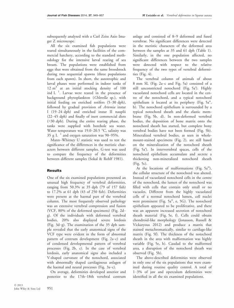
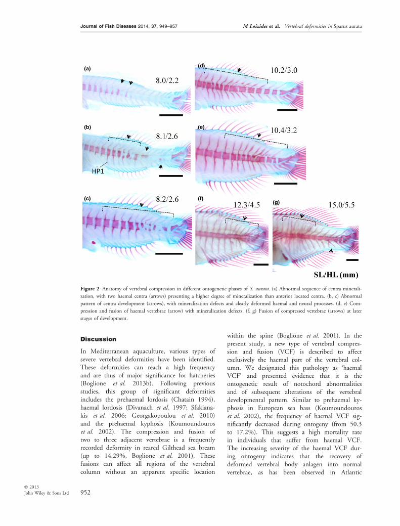
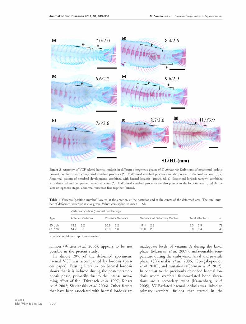
One of the six examined populations presented anunusual high frequency of vertebral deformities,ranging from 50.3% at 35 dph (79 of 157 fish)to 17.2% at 61 dph (43 of 250 fish). Deformitieswere present at the haemal part of the vertebralcolumn. The most frequently observed pathologywas an extensive vertebral compression and fusion(VCF, 80% of the deformed specimens) (Fig. 2d–g). Of the individuals with deformed vertebralbodies, 20% also displayed severe lordosis(Fig. 3d–g). The examination of the 35 dph sam-ple revealed that the early anatomical signs of theVCF type were evident in the form of abnormalpattern of centrum development (Fig. 2a–c) andof condensed developmental pattern of vertebralprocesses (Fig. 2b, c). In the case of vertebrallordosis, early anatomical signs also included aV-shaped curvature of the notochord, associatedwith abnormally shaped cartilaginous anlagen ofthe haemal and neural processes (Fig. 3a–c).On average, deformities developed anterior and
posterior to the 17th–18th vertebral centrum
anlage and consisted of 8–9 deformed and fusedvertebrae. No significant differences were detectedin the meristic characters of the deformed areabetween the samples at 35 and 61 dph (Table 1).Similarly, in the one population affected, nosignificant differences between the two sampleswere detected with respect to the relativefrequency of the two types of vertebral deformi-ties (Fig. 4).The vertebral column of animals of about
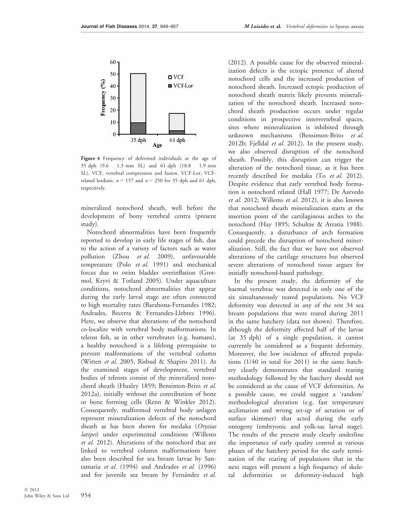
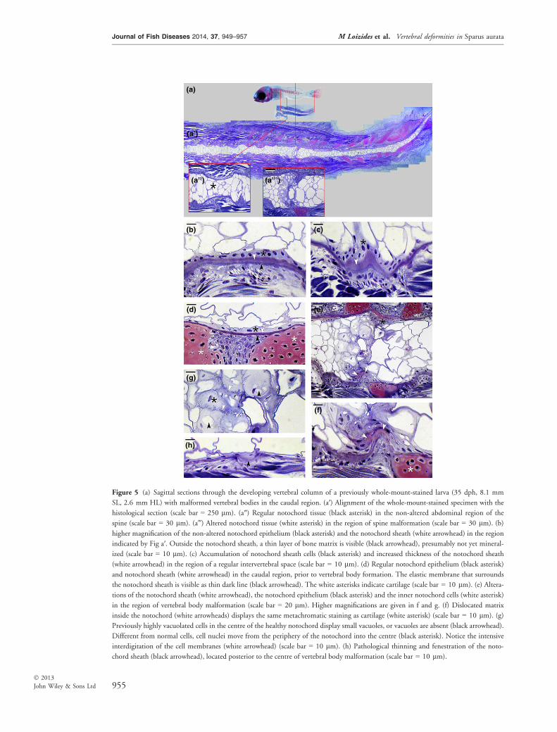
8 mm SL (Fig. 2a–c and Fig. 5a) consisted of astill unconstricted notochord (Fig. 5a′). Highlyvacuolated notochord cells are located in the cen-tre of the notochord, and a typical notochordepithelium is located at its periphery (Fig. 5a″,b). The notochord epithelium is surrounded by atypical notochord sheath and the elastic mem-brane (Fig. 5b, d). In non-deformed vertebralbodies, the deposition of bone matrix onto thenotochord sheath has started, but complete bonyvertebral bodies have not been formed (Fig. 5b).Mineralized vertebral bodies, as seen in whole-mount-stained specimens (Fig. 2), are still basedon the mineralization of the notochord sheath(Fig. 5a′). In intervertebral spaces, cells of thenotochord epithelium accumulate and secrete athickening non-mineralized notochord sheath(Fig. 5c).At the locations of malformations (Fig. 5a‴),
the cellular structure of the notochord was altered.Instead of vacuolated notochord cells in the centreof the notochord, the lumen of the notochord wasfilled with cells that contain only small or novacuoles. Different from the highly vacuolatedcells of a normal notochord lumen, cell nucleiwere prominent (Fig. 5a‴, e, 5G). The notochordepithelium appeared to be proliferative, and therewas an apparent increased secretion of notochordsheath material (Fig. 5e, f). Cells could obtainchondroid-like morphology (Jonasson, Russell &Vickaryous 2012) and produce a matrix thatstained metachromatically, similar to cartilage-likematrix (Fig. 5f). The thickness of the notochordsheath in the area with malformation was highlyvariable (Fig. 5e, h). Caudad to the malformedarea, a disruption of the notochord sheath wasobserved (Fig. 5h).The above-described deformities were observed
in only one of the six populations that were exam-ined during routine quality control. A total of1–3% of jaw and operculum deformities wereidentified in all the six examined populations.
951
Journal of Fish Diseases 2014, 37, 949–957 M Loizides et al. Vertebral deformities in Sparus aurata
� 2013
John Wiley & Sons Ltd
Discussion
In Mediterranean aquaculture, various types ofsevere vertebral deformities have been identified.These deformities can reach a high frequencyand are thus of major significance for hatcheries(Boglione et al. 2013b). Following previousstudies, this group of significant deformitiesincludes the prehaemal lordosis (Chatain 1994),haemal lordosis (Divanach et al. 1997; Sfakiana-kis et al. 2006; Georgakopoulou et al. 2010)and the prehaemal kyphosis (Koumoundouroset al. 2002). The compression and fusion oftwo to three adjacent vertebrae is a frequentlyrecorded deformity in reared Gilthead sea bream(up to 14.29%, Boglione et al. 2001). Thesefusions can affect all regions of the vertebralcolumn without an apparent specific location
within the spine (Boglione et al. 2001). In thepresent study, a new type of vertebral compres-sion and fusion (VCF) is described to affectexclusively the haemal part of the vertebral col-umn. We designated this pathology as ‘haemalVCF’ and presented evidence that it is theontogenetic result of notochord abnormalitiesand of subsequent alterations of the vertebraldevelopmental pattern. Similar to prehaemal ky-phosis in European sea bass (Koumoundouroset al. 2002), the frequency of haemal VCF sig-nificantly decreased during ontogeny (from 50.3to 17.2%). This suggests a high mortality ratein individuals that suffer from haemal VCF.The increasing severity of the haemal VCF dur-ing ontogeny indicates that the recovery ofdeformed vertebral body anlagen into normalvertebrae, as has been observed in Atlantic
(a)
(b)
(c)
(d)
(e)
(g)(f)
Figure 2 Anatomy of vertebral compression in different ontogenetic phases of S. aurata. (a) Abnormal sequence of centra minerali-
zation, with two haemal centra (arrows) presenting a higher degree of mineralization than anterior located centra. (b, c) Abnormal
pattern of centra development (arrows), with mineralization defects and clearly deformed haemal and neural processes. (d, e) Com-
pression and fusion of haemal vertebrae (arrow) with mineralization defects. (f, g) Fusion of compressed vertebrae (arrows) at later
stages of development.
952
Journal of Fish Diseases 2014, 37, 949–957 M Loizides et al. Vertebral deformities in Sparus aurata
� 2013
John Wiley & Sons Ltd
salmon (Witten et al. 2006), appears to be notpossible in the present study.In almost 20% of the deformed specimens,
haemal VCF was accompanied by lordosis (pres-ent paper). Existing literature on haemal lordosisshows that it is induced during the post-metamor-phosis phase, primarily due to the intense swim-ming effort of fish (Divanach et al. 1997; Kiharaet al. 2002; Sfakianakis et al. 2006). Other factorsthat have been associated with haemal lordosis are
inadequate levels of vitamin A during the larvalphase (Mazurais et al. 2009), unfavourable tem-perature during the embryonic, larval and juvenilephase (Sfakianakis et al. 2006; Georgakopoulouet al. 2010), and mutations (Gorman et al. 2012).In contrast to the previously described haemal lor-dosis where vertebral fusion-related bone altera-tions are a secondary event (Kranenbarg et al.2005), VCF-related haemal lordosis was linked toprimary vertebral fusions that started in the
(a)
(b)
(c)
(d)
(e)
(g)(f)
Figure 3 Anatomy of VCF-related haemal lordosis in different ontogenetic phases of S. aurata. (a) Early signs of notochord lordosis
(arrow), combined with compressed vertebral processes (*). Malformed vertebral processes are also present in the lordotic area. (b, c)
Abnormal pattern of vertebral development, combined with haemal lordosis (arrow). (d, e) Notochord lordosis (arrow), combined
with distorted and compressed vertebral centra (*). Malformed vertebral processes are also present in the lordotic area. (f, g) At the
later ontogenetic stages, abnormal vertebrae fuse together (arrow).
Table 1 Vertebra (position number) located at the anterior, at the posterior and at the centre of the deformed area. The total num-
ber of deformed vertebrae is also given. Values correspond to mean � SD
Age
Vertebra position (caudad numbering)
Total affected nAnterior Vertebra Posterior Vertebra Vertebra at Deformity Centre
35 dph 13.2 � 3.2 20.8 � 3.2 17.1 � 2.8 8.3 � 3.9 79
61 dph 14.2 � 3.1 22.0 � 1.8 18.0 � 2.3 8.8 � 3.4 43
n, number of deformed specimens examined.
953
Journal of Fish Diseases 2014, 37, 949–957 M Loizides et al. Vertebral deformities in Sparus aurata
� 2013
John Wiley & Sons Ltd
mineralized notochord sheath, well before thedevelopment of bony vertebral centra (presentstudy).Notochord abnormalities have been frequently
reported to develop in early life stages of fish, dueto the action of a variety of factors such as waterpollution (Zhou et al. 2009), unfavourabletemperature (Polo et al. 1991) and mechanicalforces due to swim bladder overinflation (Grot-mol, Kryvi & Totland 2005). Under aquacultureconditions, notochord abnormalities that appearduring the early larval stage are often connectedto high mortality rates (Barahona-Fernandes 1982;Andrades, Becerra & Fernandez-Llebrez 1996).Here, we observe that alterations of the notochordco-localize with vertebral body malformations. Inteleost fish, as in other vertebrates (e.g. humans),a healthy notochord is a lifelong prerequisite toprevent malformations of the vertebral column(Witten et al. 2005; Risbud & Shapiro 2011). Atthe examined stages of development, vertebralbodies of teleosts consist of the mineralized noto-chord sheath (Huxley 1859; Bensimon-Brito et al.2012a), initially without the contribution of boneor bone forming cells (Renn & Winkler 2012).Consequently, malformed vertebral body anlagenrepresent mineralization defects of the notochordsheath as has been shown for medaka (Oryziaslatipes) under experimental conditions (Willemset al. 2012). Alterations of the notochord that arelinked to vertebral column malformations havealso been described for sea bream larvae by San-tamaria et al. (1994) and Andrades et al. (1996)and for juvenile sea bream by Fern�andez et al.
(2012). A possible cause for the observed mineral-ization defects is the ectopic presence of alterednotochord cells and the increased production ofnotochord sheath. Increased ectopic production ofnotochord sheath matrix likely prevents minerali-zation of the notochord sheath. Increased noto-chord sheath production occurs under regularconditions in prospective intervertebral spaces,sites where mineralization is inhibited throughunknown mechanisms (Bensimon-Brito et al.2012b; Fjelldal et al. 2012). In the present study,we also observed disruption of the notochordsheath. Possibly, this disruption can trigger thealteration of the notochord tissue, as it has beenrecently described for medaka (To et al. 2012).Despite evidence that early vertebral body forma-tion is notochord related (Hall 1977; De Azevedoet al. 2012; Willems et al. 2012), it is also knownthat notochord sheath mineralization starts at theinsertion point of the cartilaginous arches to thenotochord (Hay 1895; Schultze & Arratia 1988).Consequently, a disturbance of arch formationcould precede the disruption of notochord miner-alization. Still, the fact that we have not observedalterations of the cartilage structures but observedsevere alterations of notochord tissue argues forinitially notochord-based pathology.In the present study, the deformity of the
haemal vertebrae was detected in only one of thesix simultaneously reared populations. No VCFdeformity was detected in any of the rest 34 seabream populations that were reared during 2011in the same hatchery (data not shown). Therefore,although the deformity affected half of the larvae(at 35 dph) of a single population, it cannotcurrently be considered as a frequent deformity.Moreover, the low incidence of affected popula-tions (1/40 in total for 2011) in the same hatch-ery clearly demonstrates that standard rearingmethodology followed by the hatchery should notbe considered as the cause of VCF deformities. Asa possible cause, we could suggest a ‘random’methodological alteration (e.g. fast temperatureacclimation and wrong set-up of aeration or ofsurface skimmer) that acted during the earlyontogeny (embryonic and yolk-sac larval stage).The results of the present study clearly underlinethe importance of early quality control at variousphases of the hatchery period for the early termi-nation of the rearing of populations that in thenext stages will present a high frequency of skele-tal deformities or deformity-induced high
Figure 4 Frequency of deformed individuals at the age of
35 dph (9.6 � 1.3 mm SL) and 61 dph (18.8 � 1.9 mm
SL). VCF, vertebral compression and fusion. VCF-Lor, VCF-
related lordosis. n = 157 and n = 250 for 35 dph and 61 dph,
respectively.
954
Journal of Fish Diseases 2014, 37, 949–957 M Loizides et al. Vertebral deformities in Sparus aurata
� 2013
John Wiley & Sons Ltd
(a)
(b) (c)
(d) (e)
(g)
(f)
(h)
(a’)
(a’’) (a’’’)
Figure 5 (a) Sagittal sections through the developing vertebral column of a previously whole-mount-stained larva (35 dph, 8.1 mm
SL, 2.6 mm HL) with malformed vertebral bodies in the caudal region. (a′) Alignment of the whole-mount-stained specimen with the
histological section (scale bar = 250 lm). (a″) Regular notochord tissue (black asterisk) in the non-altered abdominal region of the
spine (scale bar = 30 lm). (a‴) Altered notochord tissue (white asterisk) in the region of spine malformation (scale bar = 30 lm). (b)
higher magnification of the non-altered notochord epithelium (black asterisk) and the notochord sheath (white arrowhead) in the region
indicated by Fig a′. Outside the notochord sheath, a thin layer of bone matrix is visible (black arrowhead), presumably not yet mineral-
ized (scale bar = 10 lm). (c) Accumulation of notochord sheath cells (black asterisk) and increased thickness of the notochord sheath
(white arrowhead) in the region of a regular intervertebral space (scale bar = 10 lm). (d) Regular notochord epithelium (black asterisk)
and notochord sheath (white arrowhead) in the caudal region, prior to vertebral body formation. The elastic membrane that surrounds
the notochord sheath is visible as thin dark line (black arrowhead). The white asterisks indicate cartilage (scale bar = 10 lm). (e) Altera-
tions of the notochord sheath (white arrowhead), the notochord epithelium (black asterisk) and the inner notochord cells (white asterisk)
in the region of vertebral body malformation (scale bar = 20 lm). Higher magnifications are given in f and g. (f) Dislocated matrix
inside the notochord (white arrowheads) displays the same metachromatic staining as cartilage (white asterisk) (scale bar = 10 lm). (g)
Previously highly vacuolated cells in the centre of the healthy notochord display small vacuoles, or vacuoles are absent (black arrowhead).
Different from normal cells, cell nuclei move from the periphery of the notochord into the centre (black asterisk). Notice the intensive
interdigitation of the cell membranes (white arrowhead) (scale bar = 10 lm). (h) Pathological thinning and fenestration of the noto-
chord sheath (black arrowhead), located posterior to the centre of vertebral body malformation (scale bar = 10 lm).
955
Journal of Fish Diseases 2014, 37, 949–957 M Loizides et al. Vertebral deformities in Sparus aurata
� 2013
John Wiley & Sons Ltd
mortality rate. Our results demonstrate that noto-chord-related haemal lordosis could be identifiedduring the early larval stage, in the form of abnor-mal developmental patterns of the cartilaginousneural and haemal processes and a disturbedpattern of notochord centrum mineralization.
Acknowledgements
We thank Dr E. Schismenou for her help inembedding specimens and to Mieke Soenens forsectioning and staining. Also, we thank the man-agers of the hatchery for the provision of theinformation about rearing methodology. Thisstudy was funded by the program NSRF 2007-2013, ‘Competitiveness & Entrepreneurship’ (callCooperation I, Project No 09SYN-24-619) of theMinistry of Education, Lifelong Learning and Reli-gious Affairs, Greece. Mrs Georgiou A.N. holds ascholarship from European Union (EuropeanSocial Fund – ESF) and Greek national fundsthrough the Operational Program ‘Education andLifelong Learning’ of the National Strategic Refer-ence Framework (NSRF) – Research FundingProgram: Heracleitus II, Investing in knowledgesociety through the European Social Fund.
References
Andrades J.A., Becerra J. & Fernandez-Llebrez P. (1996)
Skeletal deformities in larval, juvenile and adult stages of
cultured gilthead sea bream (Sparus aurata L.). Aquaculture141, 1–11.
Barahona-Fernandes M.H. (1982) Body deformation in
hatchery reared European sea bass Dicentrarchus labrax (L.).Types, prevalence and effect on fish survival. Journal of FishBiology 21, 239–249.
Bensimon-Brito A., Cancela M.L., Huysseune A. & Witten
P.E. (2012a) Vestiges, rudiments and fusion events: the
zebrafish caudal fin endoskeleton in an evo-devo perspective.
Evolution & Development 14, 116–127.
Bensimon-Brito A., Cardeira J., Cancela M.L., Huysseune A.
& Witten P.E. (2012b) Distinct patterns of notochord
mineralization in zebrafish coincide with the localization of
Osteocalcin isoform 1 during early vertebral centra
formation. BMC Developmental Biology 12, 28.
Boglione C. & Costa C. (2011) Skeletal Deformities and
Juvenile Quality. In: Sparidae, pp. 233–294. Wiley-
Blackwell, Chichester, UK.
Boglione C., Gagliardi F., Scardi M. & Cataudella S.
(2001) Skeletal descriptors and quality assessment in larvae
and post-larvae of wild-caught and hatchery-reared
gilthead sea bream (Sparus aurata L. 1758). Aquaculture192, 1–22.
Boglione C., Costa C., Giganti M., Cecchetti M., Dato P.D.,
Scardi M. & Cataudella S. (2006) Biological monitoring of
wild thicklip grey mullet (Chelon labrosus), golden grey
mullet (Liza aurata), thinlip mullet (Liza ramada) andflathead mullet (Mugil cephalus) (Pisces: Mugilidae) from
different Adriatic sites: meristic counts and skeletal
anomalies. Ecological Indicators 6, 712–732.
Boglione C., Gavaia P., Koumoundouros G., Gisbert E.,
Moren M., Fontagne S. & Witten P.E. (2013a) A review on
skeletal anomalies in reared European larvae and juveniles.
Part 1: Normal and anomalous skeletogenic processes.
Reviews in Aquaculture 5, S99–S120.
Boglione C., Gisbert E., Gavaia P., Witten P.E., Moren M.,
Fontagn�e S. & Koumoundouros G. (2013b) Skeletal
anomalies in reared European fish larvae and juveniles. Part
2: main typologies, occurrences and causative factors.
Reviews in Aquaculture 5, S121–S167.
Chatain B. (1994) Abnormal swimbladder development and
lordosis in sea bass (Dicentrarchus labrax) and sea bream
(Sparus auratus). Aquaculture 119, 371–379.
Cobcroft J.M. & Battaglene S.C. (2009) Jaw malformation in
striped trumpeter Latris lineata larvae linked to walling
behaviour and tank colour. Aquaculture 289, 274–282.
Cobcroft J.M. & Battaglene S.C. (2013) Skeletal
malformations in Australian marine finfish hatcheries.
Aquaculture 396–399, 51–58.
Darias M.J., Mazurais D., Koumoundouros G., Glynatsi N.,
Christodoulopoulou S., Huelvan C., Desbruyeres E., Le Gall
M.M., Quazuguel P., Cahu C.L. & Zambonino-Infante J.L.
(2010) Dietary vitamin D(3) affects digestive system
ontogenesis and ossification in European sea bass (Dicentrachuslabrax, Linnaeus, 1758). Aquaculture 298, 300–307.
De Azevedo T.P., Witten P.E., Huysseune A., Bensimon-Brito
A., Winkler C., To T.T. & Palmeirim I. (2012)
Interrelationship and modularity of notochord and somites: a
comparative view on zebrafish and chicken vertebral body
development. Journal of Applied Ichthyology 28, 316–319.
Diggles B.K. (2013) Saddleback deformities in yellowfin
bream, Acanthopagrus australis (G€unther), from South East
Queensland. Journal of Fish Diseases 36, 521–527.
Divanach P., Papandroulakis N., Anastasiadis P.,
Koumoundouros G. & Kentouri M. (1997) Effect of water
currents on the development of skeletal deformities in sea bass
(Dicentrarchus labrax L.) with functional swimbladder during
postlarval and nursery phase. Aquaculture 156, 145–155.
Faustino M. & Power D.M. (1998) Development of
osteological structures in the sea bream: vertebral column
and caudal fin complex. Journal of Fish Biology 52, 11–22.
Fernandez I., Hontoria F., Ortiz-Delgado J.B., Kotzamanis Y.,
Estevez A., Zambonino-Infante J.L. & Gisbert E. (2008)
Larval performance and skeletal deformities in farmed
gilthead sea bream (Sparus aurata) fed with graded levels of
Vitamin A enriched rotifers (Brachionus plicatilis).Aquaculture 283, 102–115.
Fern�andez I., Ortiz-Delgado J.B., Sarasquete C. & Gisbert E.
(2012) Vitamin A effects on vertebral bone tissue
homeostasis in gilthead sea bream (Sparus aurata) juveniles.Journal of Applied Ichthyology 28, 419–426.
956
Journal of Fish Diseases 2014, 37, 949–957 M Loizides et al. Vertebral deformities in Sparus aurata
� 2013
John Wiley & Sons Ltd
Fjelldal P.G., Hansen T., Breck O., Ørnsrud R., Lock E.J.,
Waagbø R., Wargelius A. & Eckhard Witten P. (2012)
Vertebral deformities in farmed Atlantic salmon (Salmo salarL.) – etiology and pathology. Journal of Applied Ichthyology28, 433–440.
Georgakopoulou E., Angelopoulou A., Kaspiris P., Divanach
P. & Koumoundouros G. (2007) Temperature effects on
cranial deformities in European sea bass, Dicentrarchuslabrax (L.). Journal of Applied Ichthyology 23, 99–103.
Georgakopoulou E., Katharios P., Divanach P. &
Koumoundouros G. (2010) Effect of temperature on the
development of skeletal deformities in Gilthead seabream
(Sparus aurata Linnaeus, 1758). Aquaculture 308, 13–19.
Gorman K.F., Pohl K., Ali F., Bandwait K. & Breden F.
(2012) Model teleosts for the study of idiopathic-type spinal
curvatures: potential biomedical applications. Journal ofApplied Ichthyology 28, 353–359.
Grotmol S., Kryvi H. & Totland G.K. (2005) Deformation of
the notochord by pressure from the swim bladder may cause
malformation of the vertebral column in cultured Atlantic
cod Gadus morhua larvae: a case study. Diseases of AquaticOrganisms 65, 121–128.
Hall B.K. (1977) Chondrogenesis of the somitic mesoderm.
Advances in anatomy, embryology, and cell biology 53, 3–47.
Hay O.P. (1895) On the structure and development of thevertebral column of Amia [by] O. P. Hay, Chicago.
Huxley T.H. (1859) Observations on the Development of
some parts of the Skeleton of Fishes. Transactions of theMicroscopical Society & Journal 7, 33–46.
Izquierdo M.S., Scolamacchia M., Betancor M., Roo J.,
Caballero M.J., Terova G. & Witten P.E. (2013) Effects of
dietary DHA and a-tocopherol on bone development, early
mineralisation and oxidative stress in Sparus aurata (Linnaeus,1758) larvae. British Journal of Nutrition 109, 1796–1805.
Jonasson K.A., Russell A.P. & Vickaryous M.K. (2012)
Histology and histochemistry of the gekkotan notochord
and their bearing on the development of notochordal
cartilage. Journal of Morphology 273, 596–603.
Kihara M., Ogata S., Kawano N., Kubota I. & Yamaguchi R.
(2002) Lordosis induction in juvenile red sea bream, Pagrusmajor, by high swimming activity. Aquaculture 212, 149–158.
Koumoundouros G. (2008) First record of saddleback
syndrome in wild parrotfish Sparisoma cretense (L., 1758)(Perciformes, Scaridae). Journal of Fish Biology 72, 737–741.
Koumoundouros G. (2010) Morpho-anatomical abnormalities
in Mediterranean marine aquaculture. In: Recent Advances inAquaculture Research (ed. by G. Koumoundouros), pp.
125–148. Kerala, Transworld Research Network.
Koumoundouros G., Maingot E., Divanach P. & Kentouri M.
(2002) Kyphosis in reared sea bass (Dicentrarchus labrax L.):ontogeny and effects on mortality. Aquaculture 209, 49–58.
Kranenbarg S., Van Cleynenbreugel T., Schipper H. & Van
Leeuwen J. (2005) Adaptive bone formation in acellular
vertebrae of sea bass (Dicentrarchus labrax L.). Journal ofExperimental Biology 208, 3493–3502.
Mazurais D., Glynatsi N., Darias M.J., Christodoulopoulou S.,
Cahu C.L., Zambonino-Infante J.L. & Koumoundouros G.
(2009) Optimal levels of dietary vitamin A for reduced
deformity incidence during development of European sea
bass larvae (Dicentrarchus labrax) depend on malformation
type. Aquaculture 294, 262–270.
Polo A., Yufera M. & Pascual E. (1991) Effects of temperature
on egg and larval development on Sparus aurata L.
Aquaculture 92, 367–375.
Renn J. & Winkler C. (2012) Osterix:nlGFP transgenic
medaka identify regulatory roles for retinoic acid signaling
during osteoblast differentiation in vivo. Journal of AppliedIchthyology 28, 360–363.
Risbud M.V. & Shapiro I.M. (2011) Notochordal cells in the
adult intervertebral disc: new perspective on an old question.
Critical Reviews in Eukaryotic Gene Expression 21, 29–41.
Rohlf F.J. (2008) tpsDig, version 2.12. Department of Ecology and
Evolution, State University of New York, Stony Brook, NY.
Santamaria J.A., Andrades J.A., Herraez P., Fernandez-Llebrez
P. & Becerra J. (1994) Perinotochordal connective sheet of
gilthead sea bream larvae (Sparus aurata, L.) affected by axial
malformations: an histochemical and immunocytochemical
study. Anatomical Record 240, 248–254.
Schultze H.-P. & Arratia G. (1988) Reevaluation of the caudal
skeleton of some actinopterygian fishes: II. Hiodon, Elops,
and Albula. Journal of Morphology 195, 257–303.
Sfakianakis D.G., Georgakopoulou E., Papadakis I.E., Divanach
P., Kentouri M. & Koumoundouros G. (2006) Environmental
determinants of haemal lordosis in European sea bass,
Dicentrarchus labrax (Linnaeus, 1758). Aquaculture 254, 54–64.
Sokal R.R. & Rohlf F.J. (1981) Biometry: The Principles andPractice of Statistics in Biological Research. W.H, Freeman,
New York.
To T.T., Witten P.E., Renn J., Bhattacharya D., Huysseune A.
& Winkler C. (2012) Rankl-induced osteoclastogenesis leads
to loss of mineralization in a medaka osteoporosis model.
Development 139, 141–150.
Walker M.B. & Kimmel C.B. (2007) A two-color acid-free
cartilage and bone stain for zebrafish larvae. Biotechnic &Histochemistry 82, 23–28.
Willems B., B€uttner A., Huysseune A., Renn J., Witten P.E.
& Winkler C. (2012) Conditional ablation of osteoblasts in
medaka. Developmental Biology 364, 128–137.
Witten P.E., Gil-Martens L., Hall B.K., Huysseune A. &
Obach A. (2005) Compressed vertebrae in Atlantic salmon
Salmo salar: evidence for metaplastic chondrogenesis as a
skeletogenic response late in ontogeny. Diseases of AquaticOrganisms 64, 237–246.
Witten P.E., Obach A., Huysseune A. & Baeverfjord G.
(2006) Vertebrae fusion in Atlantic salmon (Salmo salar):development, aggravation and pathways of containment.
Aquaculture 258, 164–172.
Zhou S.L., Dong Q.X., Li S.N., Gu J.F., Wang X.X. & Zhu
G.N.A. (2009) Developmental toxicity of cartap on zebrafish
embryos. Aquatic Toxicology 95, 339–346.
Received: 29 June 2013Revision received: 28 August 2013Accepted: 30 August 2013
957
Journal of Fish Diseases 2014, 37, 949–957 M Loizides et al. Vertebral deformities in Sparus aurata
� 2013
John Wiley & Sons Ltd