Embed Size (px)
Citation preview
A new Ultrasound modality:US Elastography
A new Ultrasound modality:US Elastography
Joel ChabriaisJoel Chabriais
Why Elastography?Why Elastography?
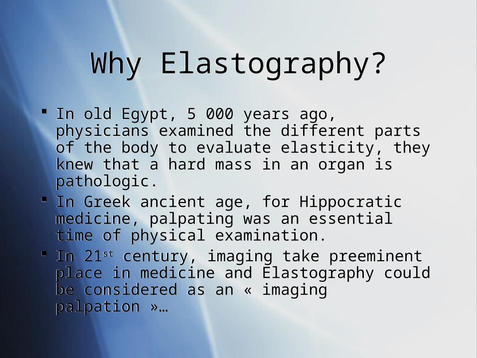
In old Egypt, 5 000 years ago, physicians examined the different parts of the body to evaluate elasticity, they knew that a hard mass in an organ is pathologic.
In Greek ancient age, for Hippocratic medicine, palpating was an essential time of physical examination.
In 21st century, imaging take preeminent place in medicine and Elastography could be considered as an « imaging palpation »…
What is Elastography?What is Elastography?
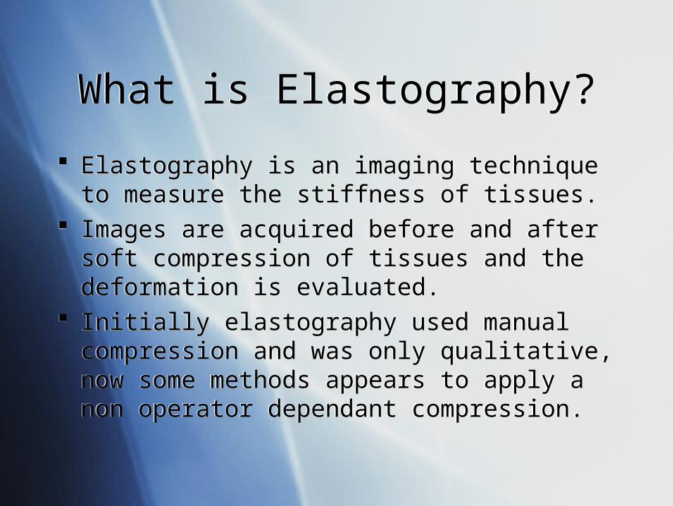
Elastography is an imaging technique to measure the stiffness of tissues.
Images are acquired before and after soft compression of tissues and the deformation is evaluated.
Initially elastography used manual compression and was only qualitative, now some methods appears to apply a non operator dependant compression.
Elastography and USElastography and US
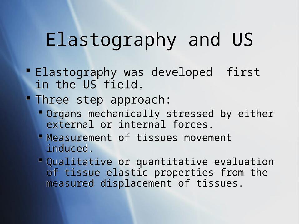
Elastography was developed first in the US field.
Three step approach: Organs mechanically stressed by either external
or internal forces. Measurement of tissues movement induced. Qualitative or quantitative evaluation of tissue
elastic properties from the measured displacement of tissues.
Elastography was developed first in the US field.
Three step approach: Organs mechanically stressed by either external
or internal forces. Measurement of tissues movement induced. Qualitative or quantitative evaluation of tissue
elastic properties from the measured displacement of tissues.
Several ApproachesSeveral Approaches
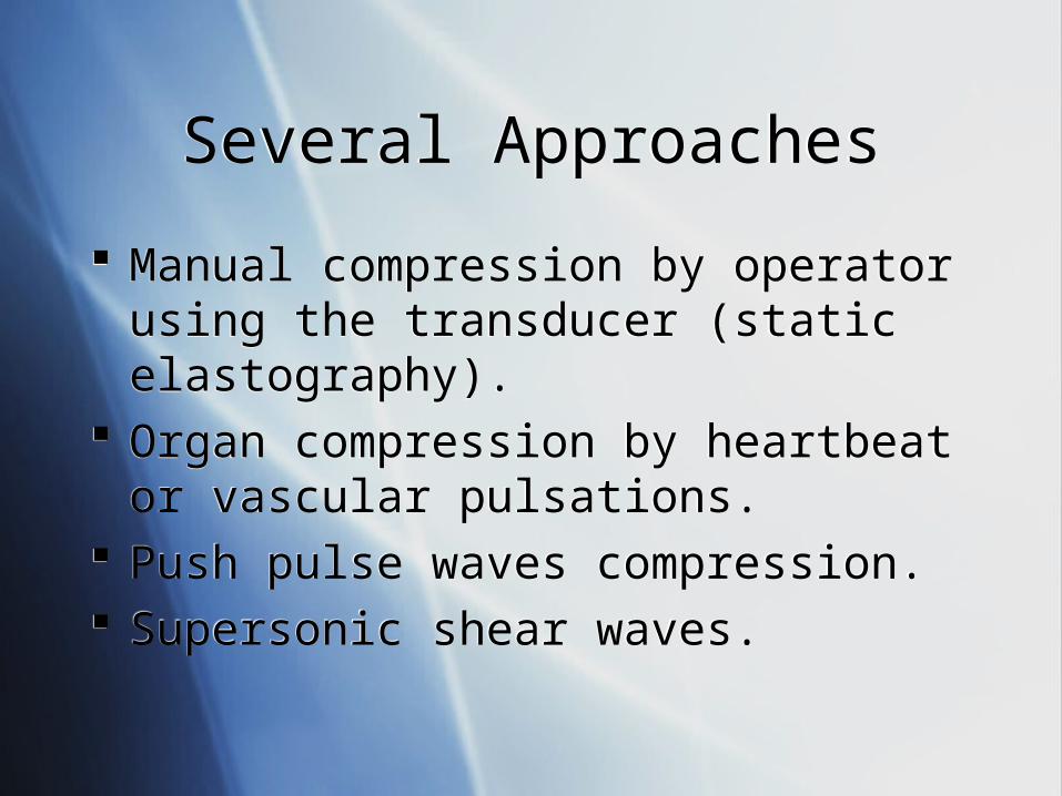
Manual compression by operator using the transducer (static elastography).
Organ compression by heartbeat or vascular pulsations.
Push pulse waves compression. Supersonic shear waves.
Manual compression by operator using the transducer (static elastography).
Organ compression by heartbeat or vascular pulsations.
Push pulse waves compression. Supersonic shear waves.

Axial and lateral Axial and lateral deformations deformations after an axial after an axial
constraintconstraint
Courtesy of Dr Anne Tardivon - Institut Curie - Paris
Static ElastographyStatic Elastography
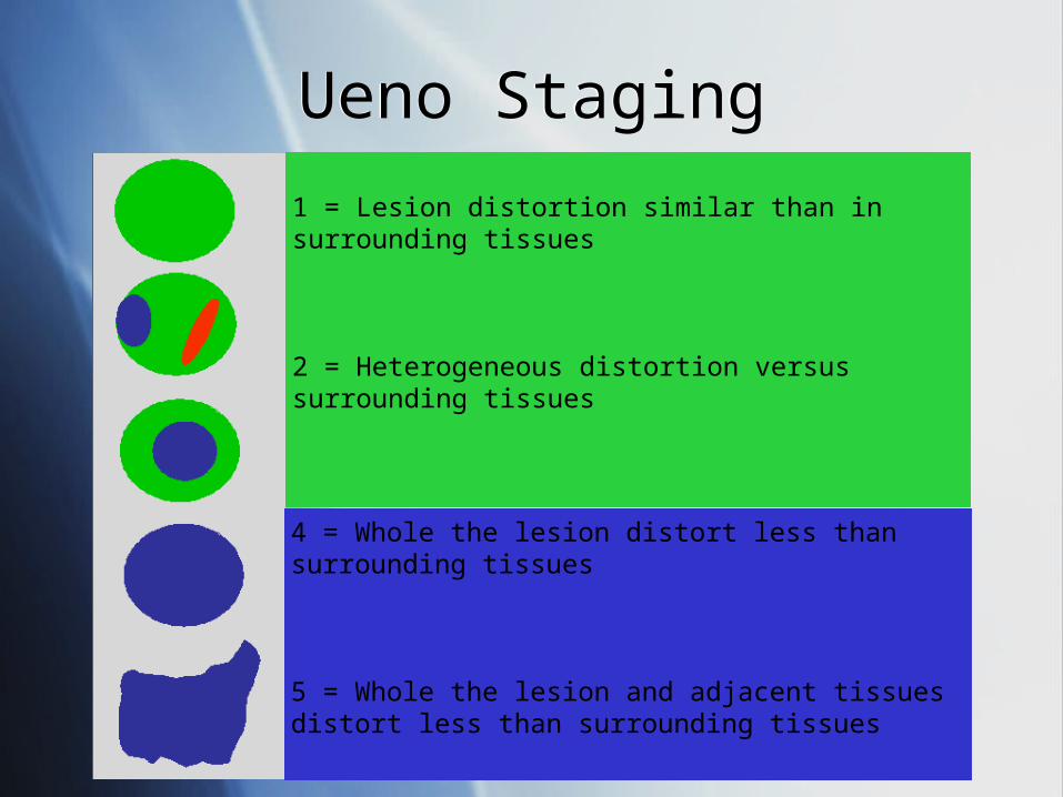
Ueno StagingUeno Staging
1 = Lesion distortion similar than in surrounding tissues
2 = Heterogeneous distortion versus surrounding tissues
3 = Lesion center distort less than surrounding tissues
4 = Whole the lesion distort less than surrounding tissues
5 = Whole the lesion and adjacent tissues distort less than surrounding tissues
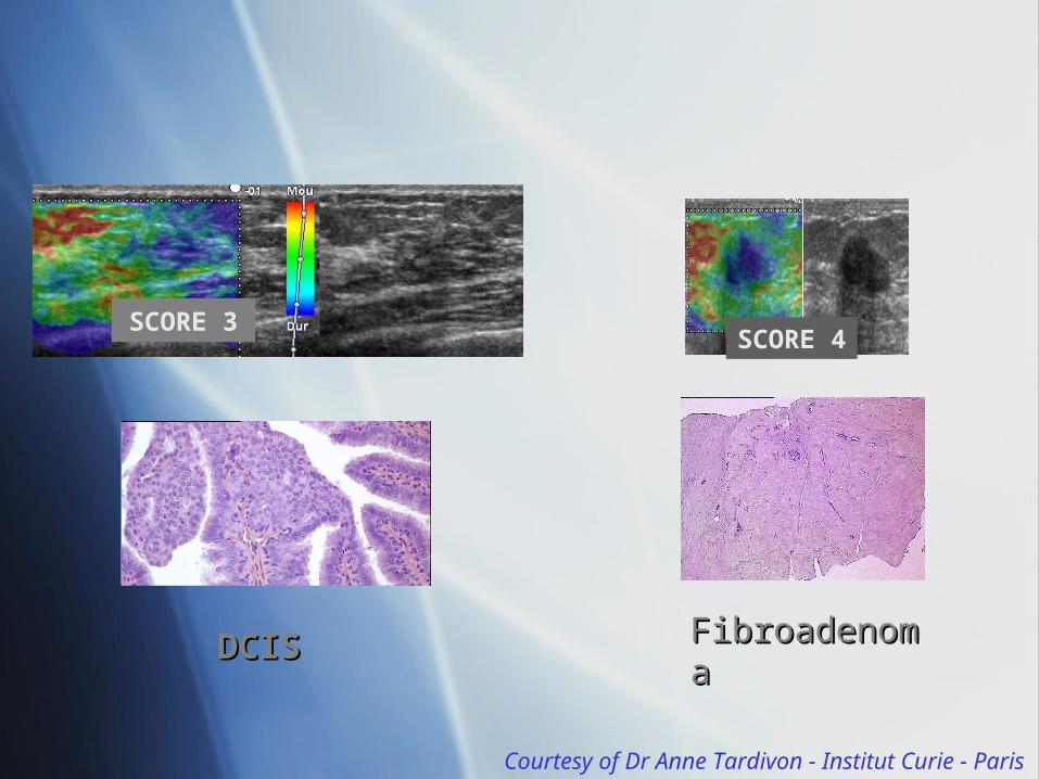
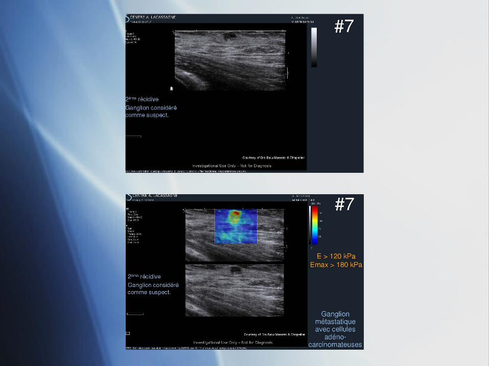
SCORE 3
DCISDCIS
SCORE 4
FibroadenomFibroadenomaa
Courtesy of Dr Anne Tardivon - Institut Curie - Paris
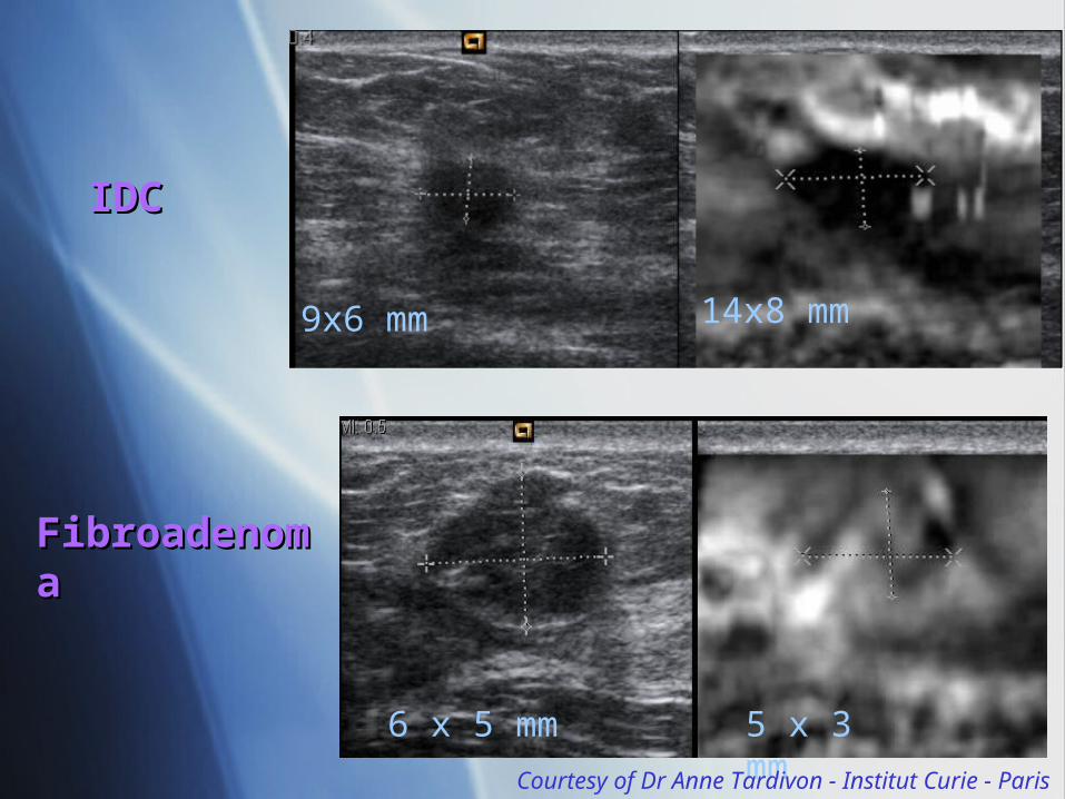
9x6 mm 14x8 mm
IDCIDC
6 x 5 mm 5 x 3 mm
FibroadenoFibroadenomama
Courtesy of Dr Anne Tardivon - Institut Curie - Paris
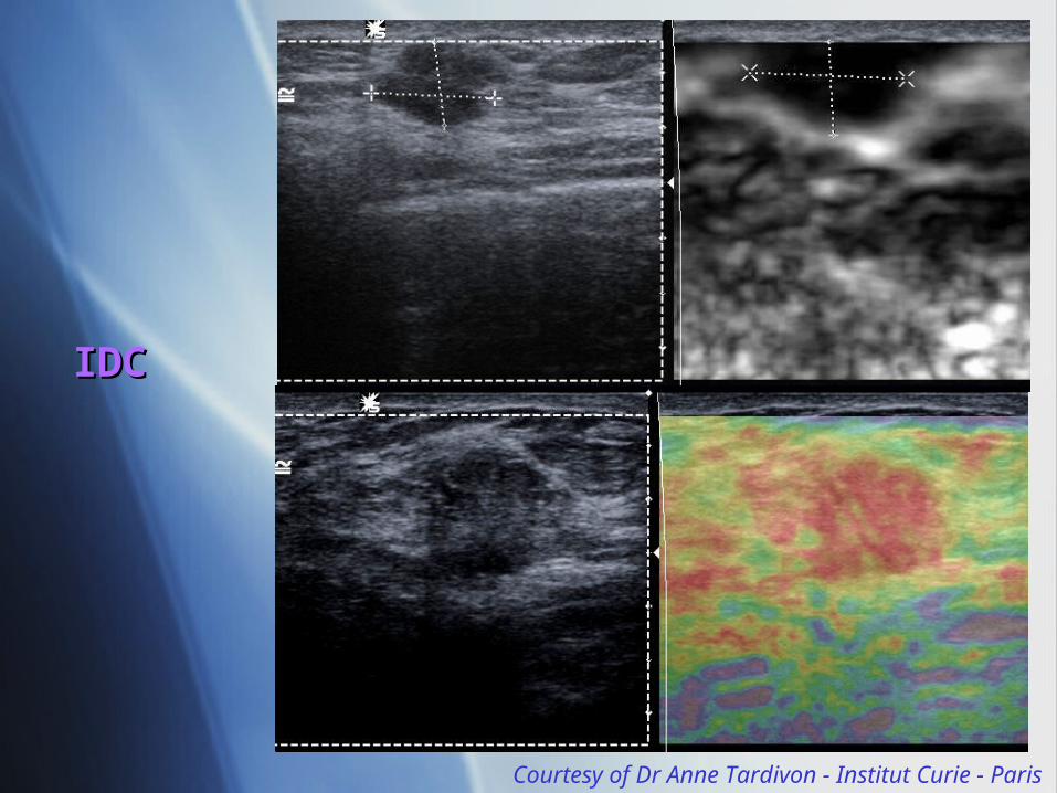
IDIDCC
Courtesy of Dr Anne Tardivon - Institut Curie - Paris
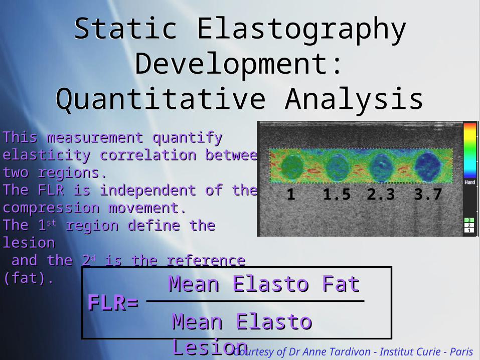
This measurement quantify This measurement quantify elasticity correlation between two elasticity correlation between two regions. regions. The FLR is independent of the The FLR is independent of the compression movement.compression movement.The 1The 1stst region define the lesion region define the lesion and the 2and the 2dd is the reference (fat). is the reference (fat).
a b c d
1 1.5 2.3 3.7
FLR=FLR=Mean Elasto FatMean Elasto Fat
Mean Elasto LesionMean Elasto LesionCourtesy of Dr Anne Tardivon - Institut Curie - Paris
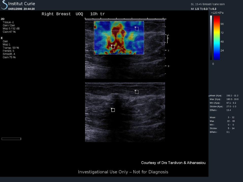
Static ElastographyDevelopment: Quantitative Analysis
Static ElastographyDevelopment: Quantitative Analysis
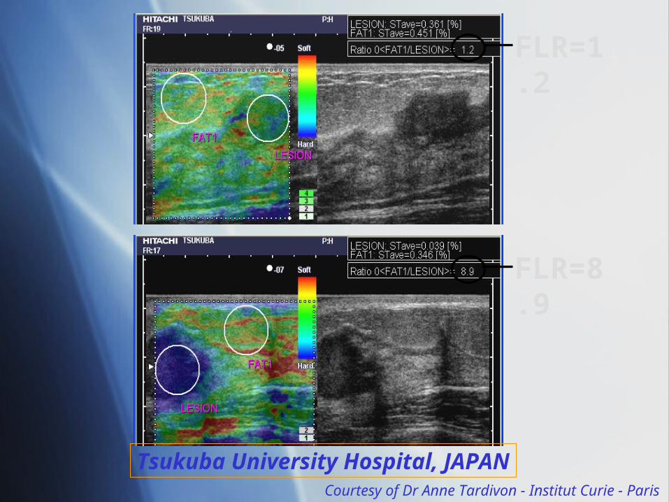
FLR=1.2
FLR=8.9
Tsukuba University Hospital, JAPANCourtesy of Dr Anne Tardivon - Institut Curie - Paris
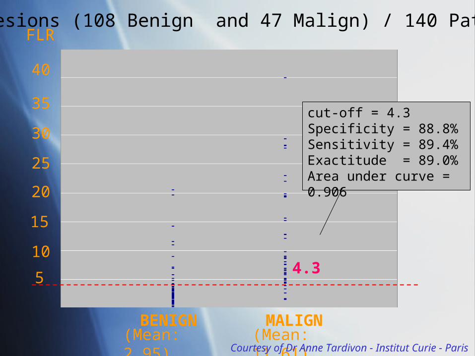
5
20
25
30
35
40
15
104.3
cut-off = 4.3 Specificity = 88.8%Sensitivity = 89.4%Exactitude = 89.0%Area under curve = 0.906
FLR
BENIGN MALIGN(Mean: 2.95) (Mean: 11.61)
155 lesions (108 Benign and 47 Malign) / 140 Patients
Courtesy of Dr Anne Tardivon - Institut Curie - Paris
Courtesy of Dr Anne Tardivon - Institut Curie - Paris
Elastography: improvementsElastography: improvements
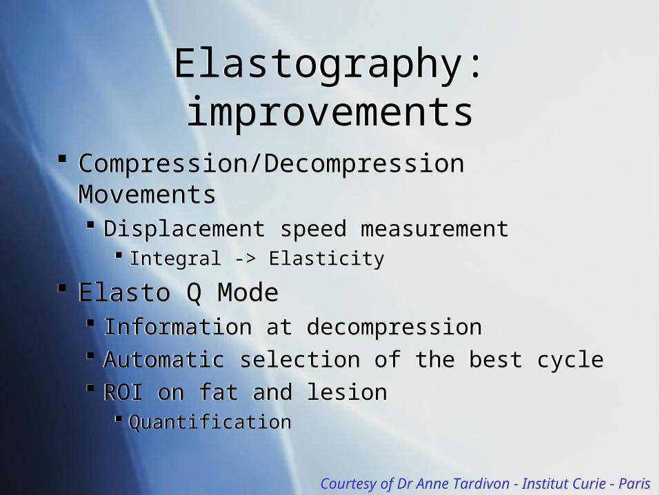
Compression/Decompression Movements Displacement speed measurement
Integral -> Elasticity
Elasto Q Mode Information at decompression Automatic selection of the best cycle ROI on fat and lesion
Quantification
Compression/Decompression Movements Displacement speed measurement
Integral -> Elasticity
Elasto Q Mode Information at decompression Automatic selection of the best cycle ROI on fat and lesion
Quantification

Courtesy of Dr Anne Tardivon - Institut Curie - Paris
4% -1% Agar-
Gelatin Elastic
phantom
~ 100 µs
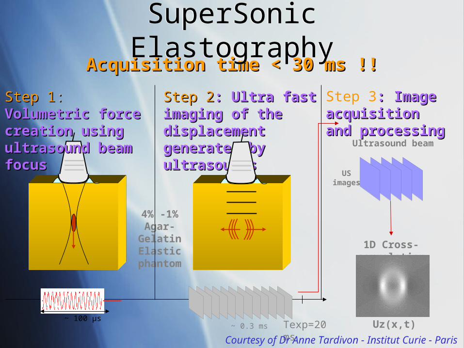
Step 1Step 1: : Volumetric Volumetric force creation force creation using ultrasound using ultrasound beam focusbeam focus
1D Cross-correlation
Step 3: Image : Image acquisition and acquisition and processingprocessing
Ultrasound beam
US image
s
Uz(x,t)
Step 2Step 2: Ultra fast : Ultra fast imaging of the imaging of the displacement displacement generated by generated by ultrasoundsultrasounds
Texp=20 ms
~ 0.3 ms
Acquisition time < 30 ms !!Acquisition time < 30 ms !!
Courtesy of Dr Anne Tardivon - Institut Curie - Paris
SuperSonic ElastographySuperSonic Elastography
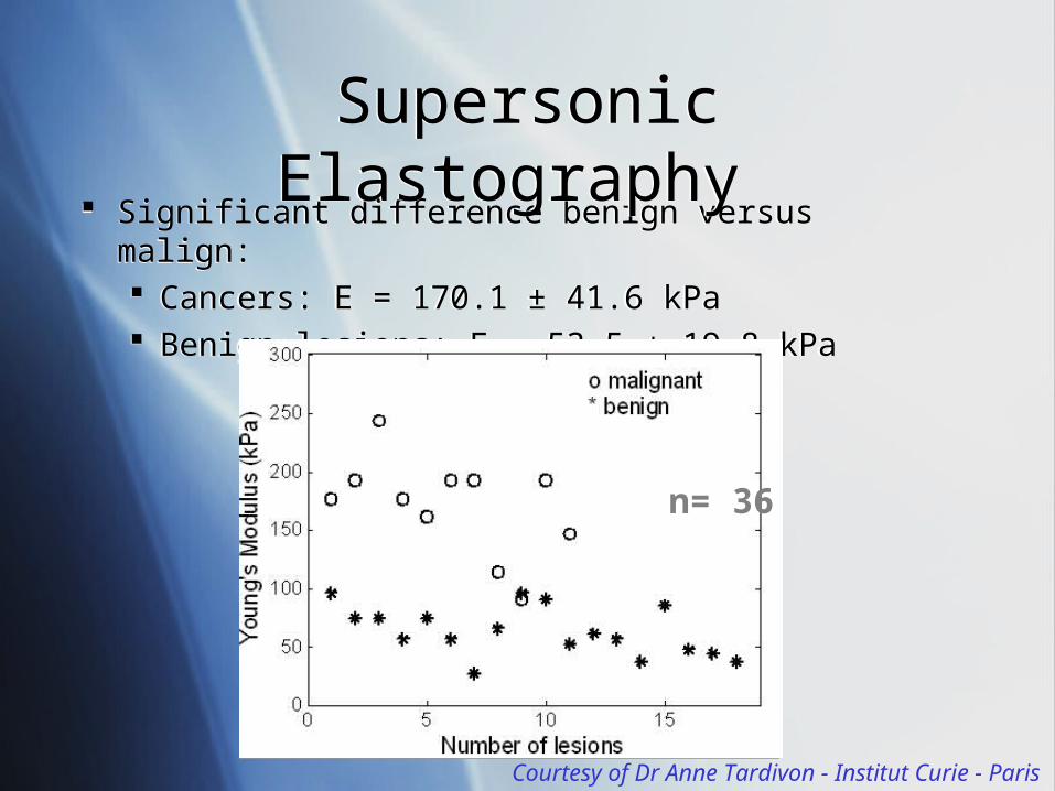
Significant difference benign versus malign: Cancers: E = 170.1 ± 41.6 kPa Benign lesions: E = 53.5 ± 19.8 kPa
Significant difference benign versus malign: Cancers: E = 170.1 ± 41.6 kPa Benign lesions: E = 53.5 ± 19.8 kPa
n= 36
Courtesy of Dr Anne Tardivon - Institut Curie - Paris
Supersonic Elastography Supersonic Elastography
µ (kPa)
µ (kPa)
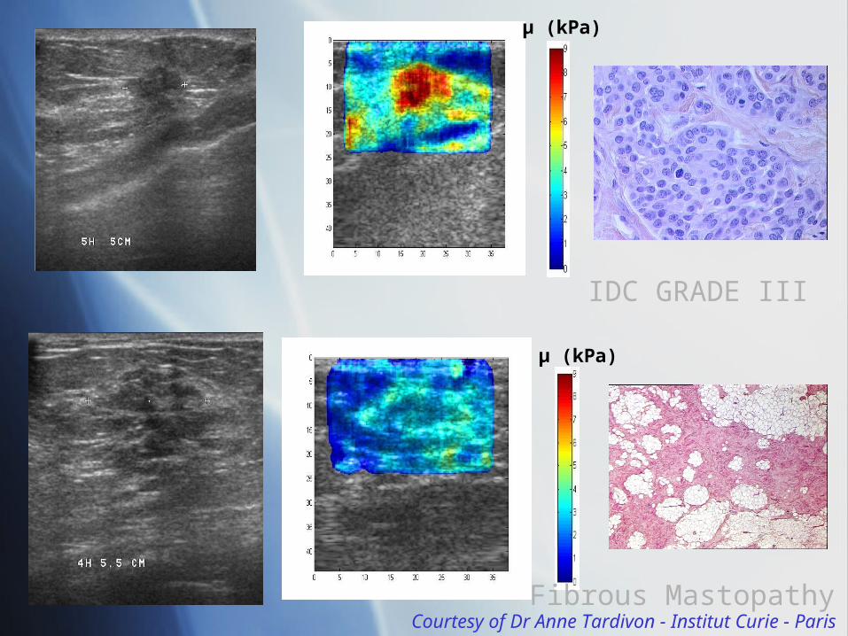
IDC GRADE III
Fibrous MastopathyCourtesy of Dr Anne Tardivon - Institut Curie - Paris
Fields of Application in MedicineFields of Application in Medicine
Breast Thyroid Liver Prostate …
Breast Thyroid Liver Prostate …
Elastography in DICOM: Why?Elastography in DICOM: Why?
Now, several vendors are coming on the market: Until 2008: 1 vendor had products for sale. 2008: 3 vendors. 2009: several announcements 6 vendors or more. … probably every US vendor at short term.
At this time, only secondary capture or US objects. All specific information to elastography are lost.
Now, several vendors are coming on the market: Until 2008: 1 vendor had products for sale. 2008: 3 vendors. 2009: several announcements 6 vendors or more. … probably every US vendor at short term.
At this time, only secondary capture or US objects. All specific information to elastography are lost.
Elastography in DICOM: Why?Elastography in DICOM: Why?
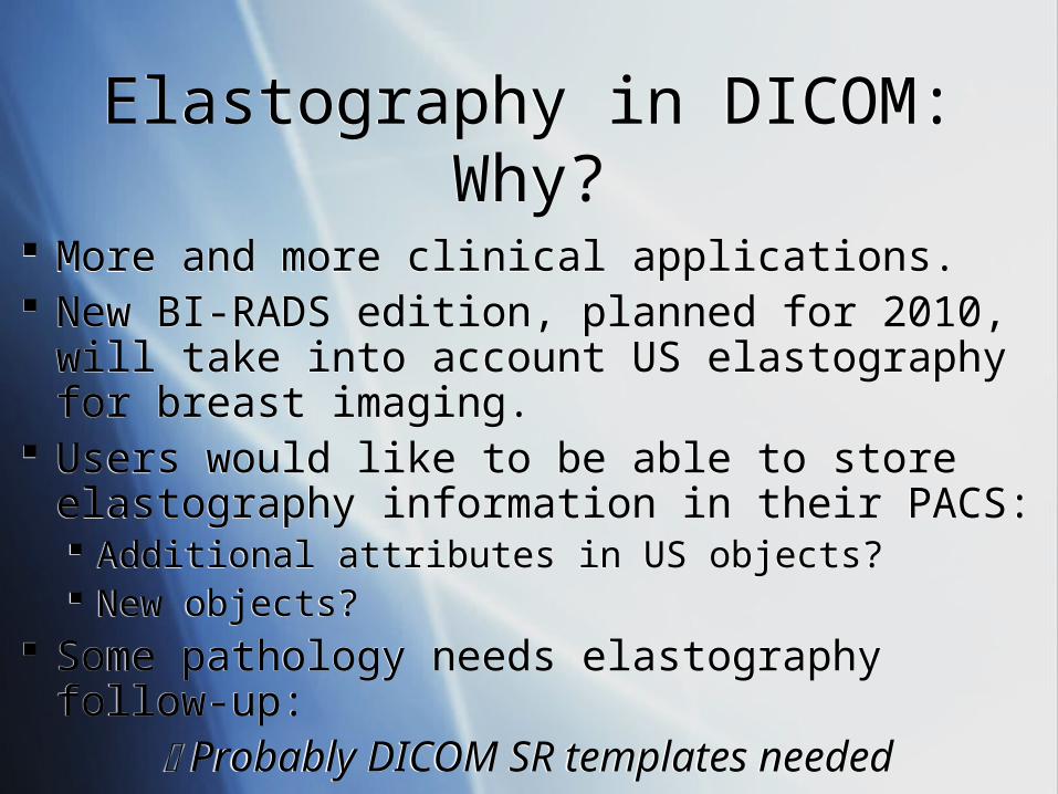
More and more clinical applications. New BI-RADS edition, planned for 2010, will take
into account US elastography for breast imaging. Users would like to be able to store elastography
information in their PACS: Additional attributes in US objects? New objects?
Some pathology needs elastography follow-up: Probably DICOM SR templates needed
Elastography in DICOM:Who is Concerned?
Elastography in DICOM:Who is Concerned?
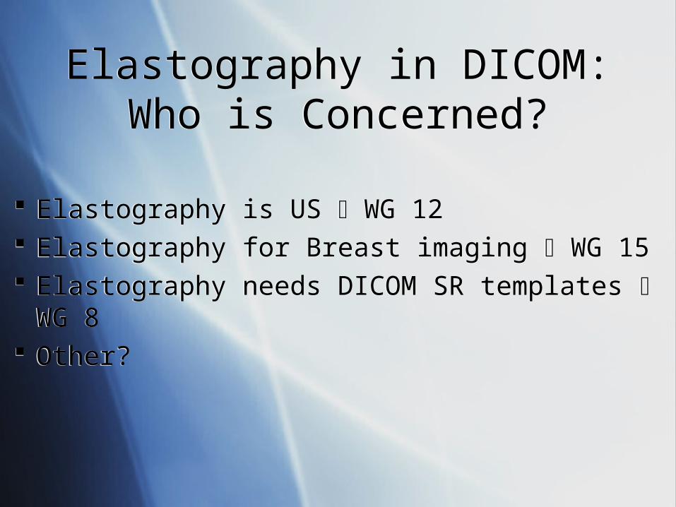
Elastography is US WG 12 Elastography for Breast imaging WG 15 Elastography needs DICOM SR templates WG
8 Other?
Elastography is US WG 12 Elastography for Breast imaging WG 15 Elastography needs DICOM SR templates WG
8 Other?

Elastography in DICOM:How To Go Further?
Elastography in DICOM:How To Go Further?
A motion to ask the concerned WG to investigate the domain to determine if further works needed?
Report to DICOM Standards Committee? …
A motion to ask the concerned WG to investigate the domain to determine if further works needed?
Report to DICOM Standards Committee? …
US ElastographyUS Elastography
And after?And after?
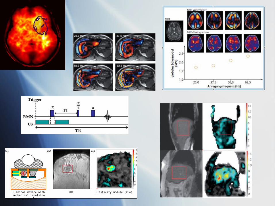
Clinical device with mechanical impulsion compression
MRI Elasticity module (kPa)
…MRE* is coming!!!…MRE* is coming!!!
But it is another story… ;-)But it is another story… ;-)
WG 16 wake-up…WG 16 wake-up…
*Magnetic Resonance Elastography


![Ultrasound elastography in neuromuscular and movement ......acoustic radiation force imaging (ARFI), and transient elastography (TE) [33]. 2.1. Ultrasound strain elastography Ultrasound](https://img.pdfslide.net/doc/110x75/5f02150f7e708231d4027b6b/ultrasound-elastography-in-neuromuscular-and-movement-acoustic-radiation.jpg)