Embed Size (px)
Citation preview

*For correspondence:
Competing interests: The
authors declare that no
competing interests exist.
Funding: See page 19
Received: 13 December 2019
Accepted: 21 May 2020
Published: 08 June 2020
Reviewing editor: Suzanne R
Pfeffer, Stanford University
School of Medicine, United
States
Copyright Cezanne et al. This
article is distributed under the
terms of the Creative Commons
Attribution License, which
permits unrestricted use and
redistribution provided that the
original author and source are
credited.
A non-linear system patterns Rab5GTPase on the membraneAlice Cezanne1, Janelle Lauer1, Anastasia Solomatina1,2,3, Ivo F Sbalzarini1,2,3,Marino Zerial1*
1Max-Planck Institute of Molecular Cell Biology and Genetics, Dresden, Germany;2Chair of Scientific Computing for Systems Biology, Faculty of Computer Science,Dresden, Germany; 3MOSAIC Group, Center for Systems Biology Dresden,Dresden, Germany
Abstract Proteins can self-organize into spatial patterns via non-linear dynamic interactions on
cellular membranes. Modelling and simulations have shown that small GTPases can generate
patterns by coupling guanine nucleotide exchange factors (GEF) to effectors, generating a positive
feedback of GTPase activation and membrane recruitment. Here, we reconstituted the patterning
of the small GTPase Rab5 and its GEF/effector complex Rabex5/Rabaptin5 on supported lipid
bilayers. We demonstrate a ‘handover’ of Rab5 from Rabex5 to Rabaptin5 upon nucleotide
exchange. A minimal system consisting of Rab5, RabGDI and a complex of full length Rabex5/
Rabaptin5 was necessary to pattern Rab5 into membrane domains. Rab5 patterning required a lipid
membrane composition mimicking that of early endosomes, with PI(3)P enhancing membrane
recruitment of Rab5 and acyl chain packing being critical for domain formation. The prevalence of
GEF/effector coupling in nature suggests a possible universal system for small GTPase patterning
involving both protein and lipid interactions.
IntroductionMembrane compartmentalization is of central importance for a variety of biological functions at mul-
tiple scales, from sub-cellular structures to multi-cellular organisms. Processes such as cell polariza-
tion, protein and lipid sorting within sub-cellular organelles or cell and tissue morphogenesis
depend on the emergence of patterns (Turing, 1952; Halatek et al., 2018). In Caenorhabditis ele-
gans, symmetry breaking of the plasma membrane is caused by PAR proteins that sort into distinct
anterior and posterior cortical domains and generate cell polarity (Kemphues et al., 1988;
Motegi and Seydoux, 2013). In budding yeast, the site of bud formation is marked by a single, dis-
crete domain of Cdc42 on the plasma membrane (PM) (Ziman et al., 1993; Chen et al., 1997;
Leberer et al., 1997). In xylem cells, ROP11 is organized into multiple domains on the PM where it
interacts with cortical microtubules to regulate cell wall architecture (Yang and Lavagi, 2012;
Oda and Fukuda, 2012). Membrane compartmentalization is not limited to the plasma membrane
but occurs also on cytoplasmic organelles. On early endosomes (EE), Rab5 exists in domains where
it regulates vesicle tethering and fusion (McBride et al., 1999; Sonnichsen et al., 2000;
Franke et al., 2019).
Cdc42, ROP11 and Rab5 are small GTPases, a class of molecules that play an important role in
symmetry breaking and membrane compartmentalization. Small GTPases use GTP/GDP binding to
act as an ON/OFF switch. The cycling between GTP and GDP-bound states is regulated by guanine
nucleotide exchange factors (GEFs) and GTPase activating proteins (GAPs) (Bos et al., 2007;
Cherfils and Zeghouf, 2013). Most small GTPases are post-translationally modified by lipid chains
which allow them to associate with membranes (Wang and Casey, 2016). The inactive GTPase forms
a high-affinity complex with guanine dissociation inhibitor (GDI), regulating membrane cycling
Cezanne et al. eLife 2020;9:e54434. DOI: https://doi.org/10.7554/eLife.54434 1 of 22
RESEARCH ARTICLE

(Sasaki et al., 1990; Ghomashchi et al., 1995; Cherfils and Zeghouf, 2013). Nucleotide exchange
to GTP or a GTP analogue prevents interaction with GDI and frees the GTPase to interact with the
membrane, where it can recruit effector proteins and mediate downstream activities (Wu et al.,
2010; Langemeyer et al., 2018). Upon hydrolysis of GTP to GDP the GTPase is once again available
for extraction from the membrane by GDI (Rak et al., 2004; Ghomashchi et al., 1995;
Pylypenko et al., 2006).
It has been proposed that small GTPase patterning can arise from the coupling of GEF activity
and effector binding (Horiuchi et al., 1997; Zerial and McBride, 2001). In this way, an active
GTPase can recruit its own GEF, creating a local, positive feedback loop of GTPase activation and
membrane recruitment. In general, self-organizing systems that form spatial patterns on membranes
often exhibit such non-linear dynamics of membrane recruitment and activation (Halatek et al.,
2018). The prevalence of GEF/effector coupling in small GTPase systems suggests that this may be
a general mechanism for symmetry breaking and spatial organization of GTPases (Goryachev and
Leda, 2019). The Rab5 GEF, Rabex5 is found in complex with the Rab5 effector Rabaptin5.
(Horiuchi et al., 1997). Similarly, the Cdc42 GEF Cdc24 is coupled to the effector Bem1
(Chenevert et al., 1992). Computational modelling revealed a Turing-type mechanism of pattern
formation by a minimal system composed of Cdc42, the Bem1/Cdc24 complex and GDI
(Goryachev and Pokhilko, 2008; Goryachev and Leda, 2017). In plants, the ROP11 GEF, ROP-
GEF4, forms a dimer that catalyzes nucleotide exchange but also interacts with the active ROP11
(Nagashima et al., 2018). We focus on Rab5, its GEF/effector complex Rabex5/Rabaptin5, and
RabGDI (hereafter referred to as GDI) in order to investigate general mechanisms for the spatial
organization of peripheral membrane proteins.
Rabex5/Rabaptin5 is one of the best characterized GEF/effector complexes in eukaryotes.
Rabex5 is a 57 kDa Vps9 domain containing GEF for Rab5 (Horiuchi et al., 1997; Delprato and
Lambright, 2007; Lauer et al., 2019). Rabaptin5 is a 99 kDa protein with multiple protein-protein
interaction sites that colocalizes with Rab5 on EE and is essential for endosome fusion
(Stenmark et al., 1995; Horiuchi et al., 1997). As Rabaptin5 forms a dimer in solution, the complex
is a tetramer of two Rabaptin5 and two Rabex5 subunits (Lauer et al., 2019). The interaction with
Rabaptin5 has been shown to increase Rabex5 GEF activity and produce structural rearrangements
in Rabex5 (Delprato et al., 2004; Delprato and Lambright, 2007; Lippe et al., 2001;
Horiuchi et al., 1997; Zhang et al., 2014; Lauer et al., 2019). By binding active Rab5, Rabaptin5
localizes the enhanced GEF activity of Rabex5 in the vicinity of active Rab5, thereby creating the
positive feedback loop. In addition, Rabex5 can be recruited to EE via binding to Ubiquitin via two
distinct Ubiquitin binding domains near the N-terminus (Penengo et al., 2006). Interestingly, Ubiqui-
tin binding enhances GEF activity toward Rab5 helping to initiate the positive feedback loop on
endosomes carrying ubiquitinated cargo (Lauer et al., 2019). Blumer et al., 2013 observed that arti-
ficially targeting Rabex5 to mitochondria resulted in Rab5 recruitment to these organelles, suggest-
ing that Rabex5 can be sufficient for localizing Rab5 to a membrane compartment. Rab5 associates
with the membrane by two 20-carbon geranylgeranyl chains attached at the C-terminus of the pro-
tein (Farnsworth et al., 1994). Molecular dynamics simulations showed that both cholesterol and PI
(3)P accumulate in the vicinity of Rab5, and predicted a direct interaction with PI(3)P mediated by an
Arg located in the flexible hypervariable region (HVR) between the C-terminal lipidation and the con-
served GTPase domain (Edler et al., 2017a).
Elucidating the precise mechanisms of self-organization of peripheral membrane proteins is criti-
cal to understanding endomembrane identity and functionality. We hypothesize that, similar to what
has been observed for Cdc42 in silico, Rab5, Rabex5/Rabaptin5 and GDI comprise a minimal system
that is capable of spatially organizing Rab5. We made use of in vitro reconstitution to test this
hypothesis and elucidate the contributions of individual components to membrane association and
organization. Our biochemical reconstitution system allowed for in-depth study of the biochemical
interactions underlying the self-organization of Rab5 and its interacting molecules on the membrane.
Cezanne et al. eLife 2020;9:e54434. DOI: https://doi.org/10.7554/eLife.54434 2 of 22
Research article Biochemistry and Chemical Biology

Results
Upon GDP/GTP exchange Rab5 is directly transferred from Rabex5 toRabaptin5 - a mechanistic basis for positive feedback of Rab5 activationTo directly test the positive feedback loop model, we investigated the structural rearrangements
occurring in Rab5 and Rabex5/Rabaptin5 in the course of nucleotide exchange by Hydrogen Deute-
rium Exchange Mass Spectrometry (HDX-MS). Rabex5/Rabaptin5 was first premixed with Rab5:GDP
and the resulting ternary complex diluted into deuterated buffer in the absence (Figure 1A top) or
presence (Figure 1A bottom) of GTPgS and incubated for 1, 5 and 15 min. In this way, we could
monitor structural rearrangement occurring in the early stages of the nucleotide exchange reaction.
Focusing first on Rab5, we could see evidence of nucleotide exchange from the dramatic stabiliza-
tion of Val24-Leu38, Leu130-Leu137 and Met160-Met168, encompassing the P-loop and parts of b
5, a4 and b 6 (Figure 1B and Figure 1—figure supplement 1A: dark blue), which, together, make
up most of the direct interaction sites with GTP. In addition, we saw stabilization of Gln60-Phe71,
parts of b 2 and b 3 (sky blue and pale green), consistent with the binding of Rabaptin5 (Zhu et al.,
2004). Indication of binding to Rabaptin5 was observed after only 1 min of reaction, thus providing
Figure 1. Rab5 backbone dynamics during nucleotide exchange. (A) Scheme of reaction. The ternary complex (Rab5/Rabex5/Rabaptin5) was incubated
in D2O for 1, 5 or 15 min in the presence or absence of GTPgS. (B) Crystal structure of Rab5:GTP (PDBID: 3MJH) pseudocolored to show differential
uptake of ternary complex (Rab5/Rabex5/Rabaptin5)±GTPgS (average of 1 min, 5 min and 15 min timepoints). The Mg2+ ion is shown as a sphere
(magenta) and GTPgS as a line structure. Color scheme: regions that are protected from exchange, i.e. stabilization, are colored with cool colors;
regions with enhanced exchange with warm colors; regions with no statistically different uptake are colored in grey; and regions with no peptide
coverage are white. (C) Deuterium incorporation over time in Rab5 b2 (aa 58–63, colored blue in B), in the ternary complex (Rab5/Rabex5/Rabaptin5)±
GTPgS (n = 3) (D) Differential deuterium incorporation in Rabaptin5 during the nucleotide exchange reaction. Two areas of protection (decrease in
deuterium uptake) correspond with the Rab5 binding sites.
The online version of this article includes the following figure supplement(s) for figure 1:
Figure supplement 1. Differential deuterium uptake of Rab5 and Rabaptin5 during nucleotide exchange.
Cezanne et al. eLife 2020;9:e54434. DOI: https://doi.org/10.7554/eLife.54434 3 of 22
Research article Biochemistry and Chemical Biology

evidence of a direct hand-off of active Rab5 from the Rabex5 catalytic domain to Rabaptin5 (See
Figure 1C). Interestingly, we also saw a destabilization of Ile177-Asp200, a5 (yellow), suggesting a
structural rearrangement of the C-terminal HVR. Figure 1D shows the alterations in deuterium
exchange for Rabaptin5 (See also Figure 1—figure supplement 1B). Since there is no available
structural model for Rabaptin5 the data are represented as a graph in which each peptide showing
statistically significant alterations in deuterium uptake is assigned a value for the percent alteration.
We saw stabilization in both of the regions known to bind Rab5, thus providing further evidence of
Rab5 binding to Rabaptin5 after the nucleotide exchange reaction. This provides a putative struc-
tural mechanism for positive feedback loop formation and the need to couple GEF and effector
activities. Next, we set out to test the hypothesis that such positive feedback is sufficient to induce
the recruitment and localized accumulation of membrane-bound Rab5.
Reconstituting Rab5 domain formation in vitroTo reconstitute Rab5 membrane recruitment and organization, we developed an in vitro system con-
sisting of recombinant proteins and synthetic membranes. The lipid composition of the synthetic
membrane was chosen based on the lipid composition of an enriched early endosomal fraction
determined in a previous study (Perini, 2012). Briefly, an enriched early endosomal fraction was pre-
pared as described (Horiuchi et al., 1997). Lipid extracts of this fraction were prepared in a final
solution of CHCl3:MeOH (1:2), as described by Kalvodova et al., 2009. The lipid extracts were then
subjected to quantitative lipid analysis as described by Sampaio et al., 2011. The results are broadly
in agreement with previous studies investigating the lipid composition of the plasma membrane
which is known to be similar to that of the early endosome (Casares et al., 2019). Lipids constituting
over 1 mol% of this lipid composition were utilized (EE, See Table 1). In order to test a wide number
of experimental conditions, we designed the following workflow: small unilamellar vesicles with the
EE-like lipid composition (EE-SUV) were deposited onto silica beads of 10 mm in diameter to form
membrane-coated beads (EE-MCB). EE-MCBs were incubated with recombinant proteins, some of
which were fluorescently tagged allowing us to monitor protein recruitment and spatial organization
using confocal microscopy. EE-MCBs were segmented and visualized as Mollweide map projections.
For visualization, the EE-MCBs are presented as equatorial slices in GFP/RFP and DiD channels, and
the reconstructed bead surface as a Mollweide map projection of the GFP/RFP signal (Mollweide
map projections of the DiD signal can be found in the corresponding supplementary figures).
EE-MCBs incubated with 10 nM GFP-Rab5/GDI showed membrane recruitment of GFP-Rab5 with
a random distribution (see Figure 2A and Figure 2—figure supplement 1A). The addition of GDI
and Rabex5/Rabaptin5-RFP in the presence GDP removed GFP-Rab5 from the membrane (see
Figure 2B and Figure 2—figure supplement 1B). However, the same reaction in the presence of
GTP produced a striking redistribution of GFP-Rab5 on the membrane into discrete clusters or
domains (see Figure 2C, and Figure 2—figure supplement 1C and Figure 2—Video 1). GDI was
provided in excess to allow for efficient flux of Rab5 between the membrane and soluble fraction
Table 1. Lipid compositions used in this study.
EE-MCB (mol%)
PC/PS/CH/PI(3)P/SM-MCB(mol%)
PC/PS/CH/PI(3)P/PlasmPE-MCB(mol%)
PC/PS/CH-MCB(mol%)
PC/PS-MCB (mol%)
Cholesterol 32.2 32.2 32.2 32.2 -
DOPC 16.6/15.6 39.1 38.8 51.7 84.9/83.9
Ethanolamineplasmalogen
12.9 - 12.9 - -
Sphingomyelin 12.6 12.6 - - -
GM3 9 - - - -
DOPS 6.1 15 15 15 15
DOPE 6.8 - - - -
Choline plasmalogen 3.6 - - - -
PI(3)P 0/1 1 1 0/1 0/1
DiD 0.1 0.1 0.1 0.1 0.1
Cezanne et al. eLife 2020;9:e54434. DOI: https://doi.org/10.7554/eLife.54434 4 of 22
Research article Biochemistry and Chemical Biology

Figure 2. Rab5 domains can be reconstituted in vitro. EE MCBs were incubated for 15 min at 23 ˚C with 10 nM GFP-Rab5/GDI A) and supplemented
with 1 mM GDI, 100 nM Rabex5/Rabaptin5-RFP and 1 mM GDP (B) or GTP (C). (D–F) GDI is necessary for Rab5 domain formation. EE MCBs were
incubated with 10 nM GFP-Rab5/GDI complex, 100 nM Rabex5/Rabaptin5 1 mM GTP and 0 nM (D), 100 nM (E) or 500 nM (F) GDI. Beads are presented
as equatorial slices in GFP and DiD channels (left) and a Mollweide projection of the GFP channel (right). Scale Bar = 10 mm. (G) Mean GFP-Rab5 signal
intensity outside of and within segmented domains in C) (See also Table 2) (p=<0.0001) (H) EE MCBs were at 23 ˚C with 10 nM GFP-Rab5/GDI 1 mM
GDI, 100 nM Rabex5/Rabaptin5 and 1 mM GTP and imaged in 1 min intervals for a total of 15 min. Graph presents mean GFP-Rab5 signal intensity
outside of and within segmented domains over time (n = 63).). (I) EE MCBs were incubated for 15 min at 23 ˚C with 10 nM GFP-Rab5/GDI 1 mM GDI,
100 nM Rabex5/Rabaptin5 and 1 mM GTP (panel 1; ‚t = 0‘) then bleached (panel 2; ‚FRAP‘) and imaged in 1 min intervals for a total of 15 min. Shown
here are stills from Figure 2—Video 2 (panels 1–3) and average intensity within segmented domains over time (panel 4; n = 27).
The online version of this article includes the following video, source data, and figure supplement(s) for figure 2:
Source data 1. Rab5 domains can be reconstituted in vitro.
Figure supplement 1. Rab5 domains can be reconstituted in vitro.
Figure 2—video 1. Rab5 domains can be reconstituted in vitro.
https://elifesciences.org/articles/54434#fig2video1
Figure 2—video 2. Rab5 domains recover after photobleaching.
https://elifesciences.org/articles/54434#fig2video2
Cezanne et al. eLife 2020;9:e54434. DOI: https://doi.org/10.7554/eLife.54434 5 of 22
Research article Biochemistry and Chemical Biology

and, although catalytically active at much lower GTPase:GEF ratios, Rabex5/Rabaptin5 was provided
in excess in order to allow for efficient binding between Rab5:GTP and Rabaptin5. Interestingly, the
formation of GFP-Rab5 domains required GDI in a concentration-dependent manner (see
Figure 2D–F and Figure 2—figure supplement 1D-F). GFP-Rab5 domains were segmented using
Squassh (Rizk et al., 2014) on the surface of the bead (see Figure 2—figure supplement 1G), and
the segmented structures were then characterized in terms of size and fluorescence intensity. Table 2
summarizes the characteristics of GFP-Rab5 domains from experiments shown in Figure 2. Domains
with a mean diameter of 1.32 mm were detected on MCBs incubated with GFP-Rab5/GDI, GDI,
Rabex5/Rabaptin5, and GTP but not GDP. They formed with a characteristic density of ~4.7
domains/EE-MCB and were rarely found adjacent to one another. A critical hallmark of the reconsti-
tuted domains is a marked increase in GFP-Rab5 signal within the segmented domain as compared
to the area outside (See Figure 2G). Comparison between GFP-Rab5 and DiD signals revealed that
the occasional apparent clusters of GFP-Rab5 in the absence of other factors (see Figure 2A and
Figure 2—figure supplement 1A) were due to membrane inhomogeneity characterized by lower
DiD signal, unlike GFP-Rab5 domains.
In order to understand how these domains form, we monitored EE-MCBs over time (See
Figure 2H, Figure 2—Video 2). Domains appear to be nucleated within the first minute of the reac-
tion (which we could not capture due to the imaging setup) and then grow linearly in intensity
until ~5 min after initiation of the reaction. After this point individual domains increase in GFP-Rab5
signal intensity slowly or not at all, suggesting that some domains reach saturation. Interestingly,
when whole MCBs were bleached domains recovered in the same locations after photobleaching
indicating that there is a constant exchange of GFP-Rab5 with solution (See Figure 2I, Figure 2—
Video 2).
Rabex5/Rabaptin5 is essential for Rab5 domain formation in vitroIn order to understand the mechanisms by which Rab5 domains form, we dissected the contribution
of each component of our reconstituted system. GDI delivers and extracts Rab5, as seen in Figure 2,
and is essential for domain formation. We observed that, similar to GDI, Rab5 domain formation
requires Rabex5/Rabaptin5 in a concentration-dependent manner (see Figure 3A–E, Figure 3—fig-
ure supplement 1A-E and Table 3, which summarizes the conditions shown in Figure 3A,B & C).
Next, we verified that the Rabex5/Rabaptin5 complex indeed localizes to the Rab5 domain. For
this, we used a fluorescent Rabex5/Rabaptin5-RFP complex and observed both enrichment of
Rabaptin5-RFP signal inside the domain (See Figure 4A) and colocalization with GFP-Rab5 (See
Figure 4B). Rabex5/Rabaptin5-RFP also showed some degree of membrane association in the
Table 2. Rab5 domains can be reconstituted in vitro.
EE MCBs were incubated for 15 min at 23 ˚C with 10 nM GFP-Rab5/GDI and supplemented with 1 mM GDI, 100 nM Rabex5/Rabap-
tin5-RFP and 1 mM GDP or GTP.
10 nM GFP-Rab5/GDI
10 nM GFP-Rab5/GDI,100 nM Rabex5/Rabaptin5,1 mM GDI, 1 mM GDP
10 nM GFP-Rab5/GDI,100 nM Rabex5/Rabaptin5,1 mM GDI, 1 mM GTP
# Domains 0 0 449
# Beads 30 44 96
Mean # Domains/Bead 0 0 4.7
Mean intensity/Bead (a.u.) 212.31 ± 67.04 128.40 ± 4.91 447.96 ± 403.41
Mean Standard Deviation/Bead 46.70 ± 21.26 0.36 ± 0.04 237.24 ± 225.54
Mean Intensity/Domain - - 1326.95 ± 1026.96
Mean Intensity/Outside 212.31 ± 67.04 128.40 ± 4.91 454.63 ± 364.79
Mean domain area, mm2 - - 1.74þ4:74
�1:00
Mean domain diameter, mm - - 1.32
The online version of this article includes the following source data for Table 2:
Source data 1. Rab5 domains can be reconstituted in vitro.
Cezanne et al. eLife 2020;9:e54434. DOI: https://doi.org/10.7554/eLife.54434 6 of 22
Research article Biochemistry and Chemical Biology

absence of other factors (See Figure 3—figure supplement 1F), however this was significantly lower
than the signal observed inside the GFP-Rab5 domains.
We next wanted to investigate whether the full Rabex5/Rabaptin5 complex was necessary for
domain formation (Figure 3F–J and Figure 3—figure supplement 1G-K). In the presence of Rab5,
GDI and GTP, neither full-length Rabex5 nor the Rabex5 catalytic domain (Rabex5CAT) alone were
sufficient to form domains (Figure 3F,G). Similarly, Rabaptin5 alone was not capable of forming
Figure 3. Rabex5/Rabaptin5 is essential for Rab5 domain formation in vitro. (A - E) Domain formation is dependent on concentration of Rabex5/
Rabaptin5. EE MCBs were incubated for 15 min at 23 ˚C with 10 nM GFP-Rab5/GDI, 1 mM GDI, 1 mM GTP and 0 nM (A), 50 nM (B), 100 nM (C), or 500
nM (D) Rabex5/Rabaptin5-RFP. (E) Mean GFP-Rab5 signal intensity outside of and within segmented domains as a function of Rabex5/Rabaptin5
concentration (50 nM Rabex5/Rabaptin5 p=0.001, n = 95; 100 nM Rabex5/Rabaptin5 p=<0.0001) See also Table 3) (F – J) Rabex5/Rabaptin5 cannot be
split into component parts and still form domains. EE MCBs were incubated for 15 min at 23 ˚C with 10 nM GFP-Rab5/GDI, 1 mM GDI, 1 mM GTP and
100 nM Rabex (F), 100 nM RabexCAT (G), 100 nM Rabaptin5 (H), 100 nM Rabex5CAT and Rabaptin5 (I), or 100 nM Rabex5/Rabaptin5 (J). Beads are
presented as equatorial slices in GFP and DiD channels (left) and a Mollweide projection of the GFP channel (right). Scale Bar = 10 mm.
The online version of this article includes the following figure supplement(s) for figure 3:
Figure supplement 1. Rabex5/Rabaptin5 is essential for Rab5 domain formation in vitro.
Cezanne et al. eLife 2020;9:e54434. DOI: https://doi.org/10.7554/eLife.54434 7 of 22
Research article Biochemistry and Chemical Biology

domains (Figure 3H). Unlike the full length Rabex5/Rabaptin5 complex, Rabex5CAT plus full-length
Rabaptin5 did not support Rab5 domain formation (compare Figure 3J and I). This suggests that
direct coupling of GEF activity and effector binding is essential for Rab5 domain formation.
Finally, we quantified the domain size distribution as a function of concentration of the compo-
nents in the reaction. Interestingly, neither the domain diameter nor the area differed significantly
Table 3. Domain formation is dependent on concentration of Rabex5/Rabaptin5.
EE MCBs were incubated for 15 min at 23 ˚C with 10 nM GFP-Rab5/GDI, 1 mM GDI, 1 mM GTP and 0 nM, 50 nM, 100 nM Rabex5/
Rabaptin5-RFP. Beads incubated with 10 nM GFP-Rab5/GDI, 1 mM GDI, 1 mM GTP and 500 nM Rabex5/Rabaptin5-RFP could not be
properly segmented due to the high GFP-Rab5 signal on the bead (See Figure 3D).
10 nM GFP-Rab5/GDI, 1 mM GDI, 1mM GTP
10 nM GFP-Rab5/GDI, 50 nM Rabex5/Rabaptin5,1 mM GDI, 1 mM GTP
10 nM GFP-Rab5/GDI, 100 nM Rabex5/Rabaptin5,1 mM GDI, 1 mM GTP
# Domains 0 96 90
# Beads 17 23 16
Mean # Domains/Bead 0 4.17 5.63
Mean intensity/Bead (a.u.)
132.95 ± 6.23 164.66 ± 24.13 946.76 ± 669.27
Mean StandardDeviation/Bead
13.14 ± 2.68 41.63 ± 17.87 526.77 ± 332.23
Mean Intensity/Domain - 282.58 ± 96.68 2767.14 ± 1039.34
Mean Intensity/Outside 132.95 ± 6.23 159.56 ± 18.33 856.22 ± 573.11
Mean domain area, mm2 - 1.71þ3:36
�0:951.97þ6:22
�1:26
Mean domain diameter,mm
- 1.31 1.40
The online version of this article includes the following source data for Table 3:
Source data 1. Domain formation is dependent on concentration of Rabex5/Rabaptin5.
Figure 4. Rabex5/Rabaptin5 localises to the reconstituted Rab5 domain. EE MCBs were incubated for 15 min at 23 ˚C with 10 nM GFP-Rab5/GDI, 1 mM
GDI, 1 mM GTP and 50 nM or 100 nM Rabex5/Rabaptin5-RFP (See Figure 3A–E). (A) Rabaptin5-RFP signal is enriched in domains. (50 nM Rabaex5/
Rabaptin5 p=0.001, n = 96; 100 nM Rabex5/Rabaptin5 p=0.0017, n = 90). Corresponding GFP enrichment in presented in Figure 3E. (B) Equatorial
slices and mollweide representations of GFP signal (top), RFP signal (bottom) and pixelwise GFP-RFP colocalization (bottom). Beads are presented as
equatorial slices (left) and Mollweide projections (right). Scale Bar = 10 mm.
Cezanne et al. eLife 2020;9:e54434. DOI: https://doi.org/10.7554/eLife.54434 8 of 22
Research article Biochemistry and Chemical Biology

when decreasing Rabex5/Rabaptin5 concentration, but the mean intensity of domains decreased
with decreasing concentration of Rabex5/Rabaptin5 (See Figure 3E and Table 3).
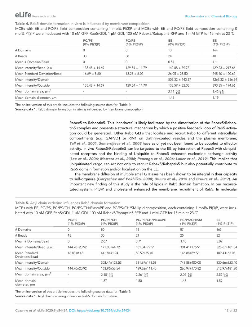
Rab5 domain formation is influenced by membrane compositionIn addition to protein-protein interactions, protein-lipid and lipid-lipid interactions also play a role in
Rab5 domain formation. The above experiments (Figures 2 and 3) were all conducted with the EE
lipid composition containing 1 mol% PI(3)P. The rearrangements in Rab5 during nucleotide exchange
reveal a destabilization of a5 that may alter membrane contacts or orientation of the protein with
respect to the membrane in the GDP- vs GTP-bound conformation (See Figure 1B). Previous work
using molecular dynamics simulations suggested an interaction between the Rab5 HVR and PI(3)P as
well as cholesterol (Edler and Stein, 2017b). To investigate the contribution of lipids, specifically PI
(3)P and cholesterol, to GFP-Rab5 domain formation, EE-MCBs as well as MCBs with a simple PC/PS
lipid composition were made with either 1 mol% or 0 mol% PI(3)P (PC/PS-MCB; See Table 1). Gera-
nylgeranylated GFP-Rab5 was recruited similarly to EE-MCBs and PC/PS-MCBs that included 1 mol
% PI(3)P (see Figure 5A,B E and Figure 5—figure supplement 1A B). However, recruitment of
GFP-Rab5 to both membranes lacking PI(3)P was greatly diminished (see Figure 5C–E and Fig-
ure 5—figure supplement 1C D). This suggests that the presence of PI(3)P enhances Rab5 recruit-
ment, either by facilitating the dissociation of Rab5 from GDI or by inhibiting the extraction of Rab5
by GDI. The presence of cholesterol (CH) appeared to also improve Rab5 recruitment to the simple
lipid composition, although to a lesser degree than PI(3)P (See Figure 5F; PC/PS/CH-MCB vs PC/PS-
MCB, See Table 1). Similar investigation of the contribution of cholesterol in the EE-like lipid compo-
sition was not possible in this system as membrane integrity was greatly compromised without cho-
lesterol (data not shown).
In order to determine whether these interactions have an effect on domain formation, the same
MCBs were incubated with Rab5/GDI, Rabex5/Rabaptin5, GDI and GTP. Strikingly, domain forma-
tion was most efficient on EE-MCBs with 1 mol% PI(3)P, less efficient on EE-MCBs with 0 mol% PI(3)
P and completely abolished on PC/PS membranes regardless of PI(3)P content (see Figure 6, Fig-
ure 6—figure supplement 1 and Table 4 which summarizes the conditions shown in Figure 6).
Domains formed on EE membranes in the absence of PI(3)P had a drastically reduced mean domain
intensity (508.32 ± 143.37) compared to domains formed in the presence of PI(3)P (mean domain
intensity 1269.32 ± 556.54) (See Figure 6E). Importantly, the membrane association of Rabex5/
Rabaptin5-RFP was not found to be similarly lipid composition-dependent (See Figure 3—figure
supplement 1F).
To investigate further which components of the EE lipid composition contribute to domain forma-
tion, the simple PC/PS lipid composition was sequentially modified to include the three most abun-
dant lipids in the EE lipid composition: cholesterol (32.3 mol%), sphingomyelin (SM; 12.6 mol%) and
ethanolamine plasmalogen (PlasmPE; 12.9 mol%) (See Figure 6F; Table 5). PI(3)P was included due
to the aforementioned effect on Rab5 recruitment. Interestingly, no domain formation could be
observed for PC/PS-MCBs while both PC/PS/CH-MCBs and PC/PS/CH/PlasmPE-MCBs showed infre-
quent and only low-intensity domains compared to EE-MCBs. Only PC/PS/CH/SM-MCBs were able
to recapitulate domains with a GFP signal intensity similar to that observed on EE-MCBs, although
the prevalence of domains on PC/PS/CH/SM-MCBs was still reduced. Staining EE-MCBs with C-laur-
dan in the absence of proteins and imaging by confocal microscopy, as described by Dodes Traian
et al., 2012 did not reveal the presence of macroscopic pre-existing liquid ordered (LO) domains
(data not shown).
The observation that Rab5 can be recruited efficiently to PC/PS/PI(3)P membranes but cannot be
organized into domains in the presence of Rabex5/Rabaptin5, excess GDI, and GTP suggest that
Rab5 interacts differently with the complex EE membrane that with a simple PC/PS membrane. Our
results demonstrate that PI(3)P enhances recruitment of Rab5 to MCBs and the presence of lipids
that contribute to acyl chain packing (cholesterol, sphingomyelin; Kaiser et al., 2009) are necessary
to drive Rab5 domain formation.
DiscussionGEF/effector coupling and the resulting positive feedback loop of GTPase activation and membrane
recruitment are common to many small GTPase systems and have been implicated in their spatial
Cezanne et al. eLife 2020;9:e54434. DOI: https://doi.org/10.7554/eLife.54434 9 of 22
Research article Biochemistry and Chemical Biology

patterning. In this study, we demonstrated that membrane recruitment and extraction (via GDI)
together with coupling of GEF and effector activities (via Rabex5/Rabaptin5) are sufficient to recon-
stitute domain organization of Rab5 in vitro. Geranylgeranylated Rab5 was observed to be recruited
to EE-like membranes from the Rab5/GDI complex. Whereas in the absence of other factors Rab5
was randomly distributed in the plane of the membrane, upon the addition of GDI, Rabex5/Rabap-
tin5 and GTP, it reorganized into discrete domains in a GTP-dependent manner. Key to Rab5
domain formation was the ‘handover’ of Rab5 from Rabex5 to Rabaptin5 and the lipid composition
Figure 5. Recruitment of geranylgeranylated GFP-Rab5 to EE and PC/PS bilayers is enhanced by PI(3)P. MCBs with PC/PS and EE lipid composition
containing 1 mol% PI(3)P (A) and B) respectively) and MCBs with PC/PS and EE lipid composition containing 0 mol% PI(3)P (C) and D) respectively) were
incubated with 10 nM GFP-Rab5/GDI for 15 min at 23 ˚C. Beads are presented as equatorial slices in GFP and DiD channels (left) and Mollweide
projection of the GFP channel (right). Scale Bar = 10 mm. (E) Mean equatorial GFP signal intensity in A–D). (p=<0.0001; n = 20) (F) MCBs with PC/PS and
PC/PS/CH lipid composition (0 mol% PI(3)P) incubated with 10 nM GFP-Rab5/GDI for 15 min at 23 ˚C. Graph presents mean equatorial GFP signal
intensity (p=0.005; n = 25). For both E) and F) GFP signal intensity is normalized to DiD signal intensity, however the same pattern can be seen in the
raw intensity values.
The online version of this article includes the following figure supplement(s) for figure 5:
Figure supplement 1. Recruitment of geranygeranylated GFP-Rab5 to EE and PC/PS bilayers is enhanced by PI(3)P.
Cezanne et al. eLife 2020;9:e54434. DOI: https://doi.org/10.7554/eLife.54434 10 of 22
Research article Biochemistry and Chemical Biology

of early endosomes, suggesting a hitherto unknown cooperativity between lipids and Rab-depen-
dent membrane self-organization.
Self-organizing systems that form spatial patterns on membranes often depend on non-linear
dynamics (Halatek et al., 2018). In our system, a key feature is the membrane recruitment and acti-
vation of Rab5, regulated by the Rabex5/Rabaptin5 complex. Neither GEF activity nor effector bind-
ing alone were capable of supporting domain formation unless physically coupled in a complex. We
found that, in the course of nucleotide exchange, newly activated Rab5 is released from Rabex5 and
immediately binds Rabaptin5 suggesting there is a direct delivery or ‘handover’ of Rab5 from
Figure 6. Rab5 domain formation in vitro is influenced by membrane composition. MCBs with PC/PS and EE lipid composition containing 1 mol% PI(3)
P (A) and B) respectively) and MCBs with PC/PS and EE lipid composition containing 0 mol% PI(3)P (C) and D) respectively) were incubated with 10 nM
GFP-Rab5/GDI, 1 mM GDI, 100 nM Rabex5/Rabaptin5-RFP and 1 mM GTP for 15 min at 23 ˚C. Beads are presented as equatorial slices in GFP and DiD
channels (left) and Mollweide projection of the GFP channel (right). Scale Bar = 10 mm. (E) Mean GFP-Rab5 signal intensity outside of and within
segmented domains in B) and D) (p=<0.0001) (See also Table 2). (F) Mean GFP-Rab5 signal intensity outside of and within segmented domains on
MCBs with PC/PS/CH/PlasmPE and PC/PS/CH/SM lipid composition containing 1 mol% PI(3)P (p=<0.0046) (See also Table 5).
The online version of this article includes the following figure supplement(s) for figure 6:
Figure supplement 1. Rab5 domain formation in vitro is influenced by membrane composition.
Cezanne et al. eLife 2020;9:e54434. DOI: https://doi.org/10.7554/eLife.54434 11 of 22
Research article Biochemistry and Chemical Biology
Rabex5 to Rabaptin5. This ‘handover’ is likely facilitated by the dimerization of the Rabex5/Rabap-
tin5 complex and presents a structural mechanism by which a positive feedback loop of Rab5 activa-
tion could be generated. Other Rab5 GEFs that localize and recruit Rab5 to different intracellular
compartments (e.g. GAPVD1 or RIN1 on clathrin-coated vesicles and the plasma membrane;
Tall et al., 2001; Semerdjieva et al., 2008 have as of yet not been found to be coupled to effector
activity. In vivo Rabex5/Rabaptin5 can be targeted to the EE by interaction of Rabex5 with ubiquiti-
nated receptors and the binding of Ubiquitin to Rabex5 enhances nucleotide exchange activity
(Lee et al., 2006; Mattera et al., 2006; Penengo et al., 2006; Lauer et al., 2019). This implies that
ubiquitinated cargo can act not only to recruit Rabex5/Rabaptin5 but also potentially contribute to
Rab5 domain formation and/or localization on the EE.
The membrane diffusion of multiple small GTPases has been shown to be integral in their capacity
to self-organize (Goryachev and Pokhilko, 2008; Bruurs et al., 2015 and Bruurs et al., 2017). An
important new finding of this study is the role of lipids in Rab5 domain formation. In our reconsti-
tuted system, PI(3)P and cholesterol enhanced the membrane recruitment of Rab5. In molecular
Table 4. Rab5 domain formation in vitro is influenced by membrane composition.
MCBs with EE and PC/PS lipid composition containing 1 mol% PI(3)P and MCBs with EE and PC/PS lipid composition containing 0
mol% PI(3)P were incubated with 10 nM GFP-Rab5/GDI, 1 mM GDI, 100 nM Rabex5/Rabaptin5-RFP and 1 mM GTP for 15 min at 23 ˚C.
PC/PS(0% PI(3)P)
PC/PS(1% PI(3)P)
EE(0% PI(3)P)
EE(1% PI(3)P)
# Domains 0 0 13 164
# Beads 33 38 24 40
Mean # Domains/Bead 0 0 0.54 4.1
Mean intensity/Bead (a.u.) 135.48 ± 14.69 129.54 ± 11.79 140.88 ± 39.73 429.23 ± 217.66
Mean Standard Deviation/Bead 16.69 ± 8.60 13.23 ± 6.02 26.05 ± 25.50 245.40 ± 120.62
Mean Intensity/Domain - - 508.32 ± 143.37 1269.32 ± 556.54
Mean Intensity/Outside 135.48 ± 14.69 129.54 ± 11.79 138.59 ± 32.05 393.35 ± 194.66
Mean domain area, mm2 - - 2.12þ4:76
�1:211.42þ2:67
�0:73
Mean domain diameter, mm - - 1.46 1.19
The online version of this article includes the following source data for Table 4:
Source data 1. Rab5 domain formation in vitro is influenced by membrane composition.
Table 5. Acyl chain ordering influences Rab5 domain formation.
MCBs with EE, PC/PS, PC/PS/CH, PC/PS/CH/PlasmPE and PC/PS/CH/SM lipid composition, each containing 1 mol% PI(3)P, were incu-
bated with 10 nM GFP-Rab5/GDI, 1 mM GDI, 100 nM Rabex5/Rabaptin5-RFP and 1 mM GTP for 15 min at 23 ˚C.
PC/PS(1% PI(3)P)
PC/PS/CH(1% PI(3)P)
PC/PS/CH/PlasmPE(1% PI(3)P)
PC/PS/CH/SM(1% PI(3)P)
EE(1% PI(3)P)
# Domains 0 80 78 87 163
# Beads 18 30 21 25 32
Mean # Domains/Bead 0 2.67 3.71 3.48 5.09
Mean intensity/Bead (a.u.) 144.70±20.92 171.03±64.72 181.34±79.51 301.41±175.91 525.67±181.34
Mean StandardDeviation/Bead
18.88±8.45 44.18±41.94 50.59±35.40 146.88±89.56 189.43±63.05
Mean Intensity/Domain - 303.44±129.53 381.67±178.58 743.88±400.00 830.66±323.40
Mean Intensity/Outside 144.70±20.92 163.96±53.54 139.62±111.45 265.97±170.82 512.97±181.20
Mean domain area, mm2 - 2.45þ5:35
�1:542.26þ4:20
�1:332.09þ5:00
�1:232.52þ3:43
�1:55
Mean domaindiameter, mm
- 1.57 1.50 1.45 1.59
The online version of this article includes the following source data for Table 5:
Source data 1. Acyl chain ordering influences Rab5 domain formation.
Cezanne et al. eLife 2020;9:e54434. DOI: https://doi.org/10.7554/eLife.54434 12 of 22
Research article Biochemistry and Chemical Biology

dynamics simulations, Edler et al., 2017a suggest a direct interaction between Rab5 and PI(3)P and
also observed accumulation of cholesterol in the proximity of Rab5. The requirement for PI(3)P has
important implications for the in vivo formation of Rab5 domains. On the EE, PI(3)P is mainly pro-
duced by the activity of the class II PI3K complex, Vps34/Vps15, which is regulated by a direct inter-
action between Rab5 and Vps15 (Christoforidis et al., 1999a; Christoforidis et al., 1999b;
Murray et al., 2002; Falasca and Maffucci, 2012). This suggests that Rab5 directly modifies the
local lipid environment to stabilize itself on the membrane, thus providing yet another level of posi-
tive feedback in vivo.
Similar to Rab5 recruitment, domain formation was lipid composition dependent and most
strongly observed on membranes containing the full EE lipid mixture. The observation that simple,
highly diffusive, PC/PS membranes do not support domain formation suggests that the EE lipid com-
position facilitates lateral lipid packing and protein-lipid interactions that are necessary for domain
formation. Lebrand et al., 2002 reported that cholesterol regulates the membrane association and
activity of Rab7 on late endosomes in vivo and decreases GDI extraction of Rab7 in vitro. The
requirement of cholesterol for stabilizing Rab5 on the membrane provides support to the idea that
lipid packing serves to adapt to the longer chain length of the geranylgeranyl anchor. Increasing the
complexity of the PC/PS lipid composition by adding cholesterol allowed for the formation of few
domains of low GFP signal intensity. The addition of sphingomyelin but not ethanolamine plasmalo-
gen was sufficient to produce domains with a high GFP-Rab5 signal intensity, as observed with the
EE lipid composition. Other than being the next most abundant lipids (after cholesterol) in the EE
lipid composition, both sphingomyelin and ethanolamine plasmalogen increase the rigidity of the
membrane. This increased rigidity occurs via the saturated acyl chains of sphingomyelin and the
small headgroup of the ethanolamine plasmalogen, two very different mechanisms that both reduce
diffusivity of the membrane. The observation that sphingomyelin was necessary for domain forma-
tion with intensity values comparable to the EE lipid mixture, but ethanolamine plasmalogen was
not, suggests that it is not a reduction in global membrane diffusivity that enables domain formation,
but the presence of saturated acyl chains and capacity for lateral lipid packing (Kaiser et al., 2009).
This may allow for dense packing of geranylgeranyl chains and stabilize a nascent domain by locally
reducing the diffusion of Rab5. Further, the destabilization of a5, which extends into the HVR,
observed in Rab5 by HDX-MS may alter the conformation of membrane-bound Rab5 upon nucleo-
tide exchange. In molecular dynamics simulations, Edler and Stein, 2017b observed a rotation
within the membrane of Rab5:GTP with respect to Rab5:GDP, that not only exposes the effector
binding site but also suggests that Rab5 makes different membrane contacts depending on its
nucleotide state. Further molecular dynamics simulations showed that this nucleotide state-depen-
dent orientation, as well as correct insertion of the geranylgeranyl anchors into the lipid bilayer, is
only supported by an EE-like membrane, containing PI(3)P, cholesterol, sphingomyelin and charged
lipids (Edler and Stein, 2017b; Munzberg and Stein, 2019). However, the EE-lipid composition
alone did not yield macroscopic LO domains. Rab5 domain formation therefore requires the synergy
between the Rab5 minimal machinery and lateral lipid packing. We suggest that the EE lipid compo-
sition supports Rab5 domain formation in our in vitro system through a combination of 1) direct
interactions between Rab5 and PI(3)P enhancing recruitment to the membrane, 2) cholesterol stabi-
lizing the geranylgeranyl anchor insertions to support a nucleotide-dependent orientation of Rab5
relative to the membrane, and 3) the presence of saturated lipids allowing for dense packing of ger-
anylgeranyl chains, contributing to domain stabilization and growth.
The non-linearity of the nucleotide cycle coupled to specific lipid interactions make small GTPases
widespread regulators of membrane self-organization. K-Ras for example, has long been known to
cluster and alter the local lipid environment, e.g. by forming nanoclusters of PI(4,5)P2 on the PM
(Zhou et al., 2017). However, with the same design, different GTPase systems can form one (e.g.
Cdc42) or multiple domains (e.g. ROP11, Rab5). Our in vitro system recapitulates the formation of
multiple Rab5 domains on the same membrane. In the reconstituted system, Rab5 domains were
formed with a characteristic density of ~4.7 domains/EE-MCB surface and a mean area of 1.74 mm2
(which would be estimated to contain up to ~10,000 molecules of Rab5). Chiou et al., 2018 propose
that coexistence of multiple GTPase domains can arise if the density of active GTPase in the domain
reaches a ‘saturation’ point. This would slow competition between domains, allowing multiple
domains to exist simultaneously, and could occur via multiple biologically relevant mechanisms (e.g.
local depletion of components or strong negative feedback). In our reconstituted system, we indeed
Cezanne et al. eLife 2020;9:e54434. DOI: https://doi.org/10.7554/eLife.54434 13 of 22
Research article Biochemistry and Chemical Biology
saw both characteristic spacing of domains and saturation of GFP-Rab5 signal, indicating that such a
‘saturation’ point can be reached. From the domain intensity we could observe two phases in
domain growth, an initial phase characterized by rapid increase in GFP signal intensity over time,
and a second phase characterized by slow increase or even saturation in signal intensity. We suggest
that fast growth is dominated by reorganization of the local lipid environment and rapid recruitment
of proteins from solution. Upon depletion of the critical components from the local membrane,
domains stabilize and reach a second, slow-growing or saturated phase. We suggest that in this
phase, domains reach dynamic equilibrium where domain size has stabilized but the domain contin-
ues to exchange proteins with the soluble pool, as suggested by the observation that domains
recover in the same location after photobleaching. It may therefore be the interaction with the lipid
membrane that stabilizes and determines the size of the domains obtained in our system. Further, it
is apparent during purification that recombinant Rab5 dimerizes at high concentrations and this
dimerization is enhanced by geranylgeranylation (data not shown). Given that domains create a
locally high concentration of protein, Rab5 dimerization may also contribute to stabilization of a
Rab5 domain. How domain growth is regulated and by what means biological systems can produce
a variety of spatial patterns based on common design principles has been the subject of multiple
recent in silico models and simulations (Chiou et al., 2018; Halatek et al., 2018; Jacobs et al.,
2019). For Turing-type reaction-diffusion systems, theory can predict regions in the parameter space
where the system either can form dynamically stable patterns or support oscillatory behavior such as
travelling waves. In our reconstituted system, we did not observe such oscillatory behavior. This may
either be a limitation of the experimental set-up (e.g. too low resolution in time and space) or an
intrinsic property of the system we investigated. The design principles of biochemical oscillators
require delayed negative feedback (Novak and Tyson, 2008) which might not be achieved by the
linear GTP to GDP hydrolysis and GDI extraction reconstituted in our reactions. Further adaptation
of the system (e.g. the addition of GAP activity) may allow for wave-like behavior and remains for
future investigation. Our results imply that the specific interaction of proteins with lipids in the mem-
brane must also be considered in in silico studies of pattern formation.
Herein, we reconstituted a minimal system for the formation of Rab5 GTPase domains in vitro
and demonstrated that both GEF/effector coupling and lipid interactions contribute to the self-orga-
nization of Rab5 on the membrane, where the lipid composition plays an important role beyond that
of a solvent for lipidated proteins. This appears to be a universal system deploying small GTPases to
pattern membranes from mono-cellular to multi-cellular organisms.
Materials and methods
Key resources table
Reagent type(species) or resource Designation Source or reference Identifiers Additional information
RecombinantDNA reagent
pOEM-1 N-His Oxford ExpressionTechnologies,MPI-CBG PEP facility
vector, NotI and AscIsites used for ligation
RecombinantDNA reagent
pOEM-1 N-GST Oxford ExpressionTechnologies,MPI-CBG PEP facility
vector, NotI and AscIsites used for ligation
RecombinantDNA reagent
pOEM-1 N-His-eGFP Oxford ExpressionTechnologies,MPI-CBG PEP facility
vector, NotI and AscIsites used for ligation
RecombinantDNA reagent
pOEM-1 C-His-tagRFP Oxford ExpressionTechnologies,MPI-CBG PEP facility
vector, NotI and AscIsites used for ligation
Transfectedconstruct(Homo sapiens)
Rab5a This paper In vector pOEM-1 N-His-eGFP
Transfectedconstruct(Bos taurus)
Rabex5 (pOEM-1 N-His) Lauer et al., 2019 In vector pOEM-1 N-His
Continued on next page
Cezanne et al. eLife 2020;9:e54434. DOI: https://doi.org/10.7554/eLife.54434 14 of 22
Research article Biochemistry and Chemical Biology

Continued
Reagent type(species) or resource Designation Source or reference Identifiers Additional information
Transfectedconstruct(Bos taurus)
Rabex5CAT (pOEM-1 N-His) Lauer et al., 2019 In vector pOEM-1 N-His
Transfectedconstruct(Homo sapiens)
Rabaptin5 (pOEM-1 N-GST) Lauer et al., 2019 In vector pOEM-1 N-GST
Transfectedconstruct(Homo sapiens)
Rabaptin5-RFP-6xHis This paper pOEM-1 C-His-tagRFP
Transfectedconstruct(Homo sapiens)
GDIA (pOEM-1 N-His) This paper In vector pOEM-1 N-His
Commercialassay or kit
Silica Beads (10 mm) Corpuscular C-SIO-10.0 10 mm standardmicrospheres formicroscopy
Commercialassay or kit
Ni-NTA Agarose Qiagen
Commercialassay or kit
GlutathioneSepharose 4B Resion
GE
Commercialassay or kit
BCA assay Thermo Scientific 23225
Other GTP Sigma 10106399001
Other Cholesterol (ovine wool) Avanti 700000
Other 18:1 (D9-Cis) PC (DOPC)(1,2-dioleoyl-sn-glycero-3-phosphocholine)
Avanti 850375
Other C18(Plasm)�18:1 PC(1-(1Z-octadecenyl)�2-oleoyl-sn-glycero-3-phosphocholine)
Avanti 852467
Other Sphingomyelin (Egg, Chicken) Avanti 860061
Other GM3 Ganglioside(Milk, Bovine-Ammonium Salt)
Avanti 860058
Other 18:1 PS (DOPS) (1,2-dioleoyl-sn-glycero-3-phospho-L-serine (sodium salt))
Avanti 840035
Other 18:1 (D9-Cis) PE (DOPE)(1,2-dioleoyl-sn-glycero-3-phosphoethanolamine)
Avanti 850725
Other C18(Plasm)�18:1 PE(1-(1Z-octadecenyl)�2-oleoyl-sn-glycero-3-phosphoethanolamine)
Avanti 852758
Other Phosphatidylinositol 3-phosphatediC16 (PI(3)P diC16)
Echelon P-3016
Other DiD [DiIC18(5);1,1’-dioctadecyl-3,3,3’,3’-tetramethylindodicar-bocyanine, 4-chlorobenzenesulfonate salt]
Thermo Fischer D7757
CloningRab5, Rabex5, Rabaptin5, and GDI were cloned into pOEM series vectors (Oxford Expression Tech-
nologies), modified to contain a Human Rhino Virus (HRV) 3C cleavable tag at either the Nor C-ter-
minus, followed by a protease cleavage site (Not1 at N-terminus, Asc1 at C-terminus) for insect (SF9)
cell expression. Cleavable tags consisted of either 6x-Histidine (6xHis), for Rab5 and Rabex5, or Glu-
thathione S-Transferase (GST), for GDI and Rabaptin5. In order to monitor membrane association
and organization, fluorescent Rab5 and Rabaptin5 constructs were created. The proteins were
Cezanne et al. eLife 2020;9:e54434. DOI: https://doi.org/10.7554/eLife.54434 15 of 22
Research article Biochemistry and Chemical Biology

cloned into SF9 expression vectors containing either an N or C-terminal fluorescent tag (GFP or RFP)
attached to the protein by a 13 amino acid flexible linker (N-terminal linker: GSAGSAAGSGAAA;
C-terminal: linker: GAPGSAGSAAGSG). As the addition of a fluorescent tag to a protein always car-
ries the risk of altering protein behavior by interfering with protein folding, fluorescent proteins were
compared to non-fluorescent constructs known to fold properly by Hydrogen Deuterium Exchange
Mass Spectrometry (HDX-MS) and discarded if they showed any aberrant dynamics. The following
constructs were used in this study: 6xHis-GFP-Rab5, GST-GDI, GST-Rabaptin5, Rabex5-6xHis, RFP-
Rabaptin5, 6xHis-RabexCAT.
Protein expression and purificationSF9 cells were grown in ESF921 media (Expression Systems) and co-transfected with linearised viral
genome and expression plasmid. P1 and P2 virus was generated per manufacturers protocol and
yield was optimised by expression screens and infection time course experiments. The P2 virus was
used to infect SF9 cells (grown to a density of 1 million cells/ml) at 1% (v/v). Rabex5/Rabaptin5 and
geranylgeranylatedRab5/GDI complexes were produced by co-infection. Cells were harvested after
30–40 hr by spinning in a tabletop centrifuge at 500 g for 10 min. Cell pellets were resuspended in
Standard Buffer (20 mM Tris pH7.5, 150 mM NaCl, 5 mM MgCl2, 0.5 mM TCEP; STD) supplemented
with DNAse one and protease inhibitor cocktail (chymostatin 6 mg/mL, leupeptin 0.5 mg/mL, anti-
pain-HCl 10 mg/mL, aprotinin 2 mg/mL, pepstatin 0.7 mg/mL, APMSF 10 mg/mL). Pellets were flash
frozen and stored at �80�C. All subsequent steps performed at 4�C or on ice. Cells were thawed on
ice and lysed by sonication (previously frozen SF9 cell pellets were not sonicated as freeze-thawing
is sufficient for lysis). Cell lysates were spun with a JA 25.50 rotor at 22500 rpm for 20 min at 4�C.
Histidine-tagged proteins were bound to Ni-NTA Agarose resin (1L of culture = 1 mL resin) in the
presence of 20 mM Imidazole. Resin was washed with STD buffer supplemented with 20 mM Imidaz-
ole. Proteins were eluted using 200 mM Imidazole only followed by Histidine-tag cleavage during
overnight dialysis with 3C protease. GST tagged proteins were bound to Glutathione Sepharose
resin (GS-4B, GE Healthcare) for 2 hr at 4�C, washed with Standard Buffer and cleaved from resin
overnight with a GST-3C protease. Rabex5/Rabaptin5 and Rab5/GDI complexes were purified by
both His- and GST-tag affinity purification to obtain pure complex. Size Exclusion Chromatography
was performed in STD on a Superdex200 Increase 10/30. Concentrations were determined by a
bicinchoninic acid protein Assay (Pierce BCA Protein Assay Kit, ThermoFischer) and purity was
assessed by SDS-PAGE followed by colloidal Coomassie staining. Proteins were aliquoted, flash fro-
zen in liquid nitrogen and stored at �80�C.
Liposome preparationThe lipids listed below were purchased and resuspended in either CHCl3, CHCl3:MeOH (2:1 for
GM3) or CHCl3:MeOH:H2O (1:2:0.8 for PI(3)P) as per manufacturer’s instructions and stored at
�20�C. To form liposomes, lipids were mixed together and the solvent was evaporated under a
stream of nitrogen. Residual solvent was removed by drying under vacuum overnight in a desiccator.
Lipids were rehydrated for at 37�C in SLB Buffer (20 mM TRIS, 150 mM NaCl) and vortexed to form
a stock solution of 1 mM lipid. Small unilamellar vesicles (SUV) were prepared by freeze-thaw cycles
(10x snap freezing and thawing at 37�C). Vesicles were stored at �20�C and sized by sonication
before each application. Size distribution of liposome preparations was assessed by Dynamic Light
Scattering using a Zetasizer Nano ZSP Malvern.
EE lipid compositionDOPC(1,2-dioleoyl-sn-glycero-3-phosphocholine):DOPS(1,2-dioleoyl-sn-glycero-3-phospho-L-serine):
DOPE(1,2-dioleoyl-sn-glycero-3-phosphoethanolamine):Sphingomyelin:Cholesterol:ethanolamine
plasmalogen (1-(1Z-octadecenyl)�2-oleoyl-sn-glycero-3-phosphoethanolamine):choline plasmalogen
(1-(1Z-octadecenyl)�2-oleoyl-sn-glycero-3-phosphocholine):GM3: PI(3P) (diC16 Phosphatidylinositol
3-phosphate): DiD [DiIC18(5); 1,1’-dioctadecyl-3,3,3’,3’-tetramethylindodicar-bocyanine, 4-chloro-
benzenesulfonate salt] (13.8:6.1:6.8:12.6:32.3:12.9:3.6:9:1:0.1) (See Table 1).
Cezanne et al. eLife 2020;9:e54434. DOI: https://doi.org/10.7554/eLife.54434 16 of 22
Research article Biochemistry and Chemical Biology

PC/PS lipid compositionDOPC(1,2-dioleoyl-sn-glycero-3-phosphocholine): DOPS(1,2-dioleoyl-sn-glycero-3-phospho-L-ser-
ine): Phosphatidylinositol 3-phosphate (PI(3P)diC16): DiD [DiIC18(5); 1,1’-dioctadecyl-3,3,3’,3’-tetra-
methylindodicar-bocyanine, 4-chlorobenzenesulfonate salt] (83.95:15:1:0.1) (See Table 1).
MCB preparationSilica beads (10 mm standard microspheres for microscopy) were coated with a supported lipid
bilayer as described (Neumann et al., 2013) with minor modifications to ensure a tight lipid mem-
brane. Beads were incubated with either 800 mM NaCl and 250 mM EE liposomes or 375 mM PC/PS
liposomes (Z average diameter 100–120 nm by SLS) for 15 min RT on a rotator wheel. MCBs were
washed with 1ml H20 and 2 � 1 mL Standard Buffer, centrifuging at 2000rpm for 1 min in a tabletop
centrifuge. Membrane integrity was assessed at different time points and after increasing centrifuga-
tion steps. MCBs were found to be robust at 13000 rpm washing steps and up to 4 hr at RT. MCBs
were consequently used within 3 hr of formation. The formation protocol was adapted for PC/PS
membranes in order to produce MCBs with similar amounts of membrane as compared to EE-MCBs
in order to make direct comparisons of GFP-Rab5 recruitment.
Confocal microscopyMicroscopy experiments were performed on either Nikon TiE (manual imaging, for high resolution
and 3D reconstructions) or Cell Voyager 7000S (CV7000S) (automated imaging for time lapse experi-
ments). For manual imaging in Nikon TiE, reactions were prepared in an 8-well NuncTM Lab-TekTM
Chamber Slide for imaging. Images were acquired with a Nikon TiE equipped with a 100x/1.45NA
Plan Apochromat, DIC oil immersion objective, Yokogawa CSU-X1 scan head and Andor DU-897
back-illuminated CCD. Images were acquired with 80 ms exposure at l 488, 561 and 660 with the
following laser intensities: 15% 488; 5% 561; and 2% 660. For automated imaging, reactions were
prepared in a Greiner Square bottom 384 well plate. Images were acquired with Cell Voyager 7000S
(CV7000S) equipped with a 60x/1.2NA water immersion objective at 30% 488 and 660 laser. Color
and illumination corrections were applied though CV7000S software. Imaging support by M. Sto€ter
(TDS, MPI-CBG).
Image analysisIntensity quantifications at MCB equators were performed manually in FIJI (Schindelin et al., 2012).
Beads were segmented manually and intensity values along the surface were extracted by determin-
ing line profiles 10 pixels wide along the surface of the bead as defined by DiD signal. Intensity in
488 and 561 was normalized to the intensity in the 660 channel in a pixelwise manner to account for
potential differences in membrane amount between beads and lipid compositions. Box and whiskers
plots show median (line), 25/75 quartiles (box boundaries), and min/max values (error bars).
Unpaired t-tests were performed to test statistical significance.
To quantify size, intensity, and number of domains on MCBs, a novel image analysis pipeline was
developed. The pipeline consists of the following steps, illustrated in Figure 2—figure supplement
1G:
1. The membrane surface wass extracted by selecting all pixels in the upper 0.5% intensity per-centile for the membrane channel. The sphere center and radius were fitted using a linearleast-squares solver with the normal residuals as cost function. Next, particles were distributedon the fitted sphere on a latitude-longitude mesh with 3
� resolution in both azimuthal andpolar directions. The particles were thereafter extended from the surface into a narrow bandaround it by replicating the particles in the radial direction with 1 pixel spacing until a distanceof 5 pixels from the surface. Next, GFP-Rab5 intensity values were interpolated from pixels toparticles in the narrow band using moment-conserving interpolation schemes (Mona-ghan, 1985). Finally, the particle intensity values were maximum-projected in the radialdirection.
2. Uneven background in the tangent space of beads was corrected using a ‘rolling ball’ algo-rithm, with a radius of 2 mm (Sternberg, 1983).
3. Rab5 domains were segmented using a globally optimal model-based method Squassh(Rizk et al., 2014). The segmentation was applied on pixels after replacing all pixel values withintensities obtained by interpolating back from the particles to pixels to yield a clean pixel
Cezanne et al. eLife 2020;9:e54434. DOI: https://doi.org/10.7554/eLife.54434 17 of 22
Research article Biochemistry and Chemical Biology
image with denoised and background-corrected intensities. All segmentations were performedusing the following parameters in Squassh: regularization parameter 0.35 and minimum objectintensity 0.3. In addition, sub-pixel segmentation with 4-fold oversampling was enabled.
4. A marching cubes algorithm (Lorensen and Cline, 1987) was used to construct a triangulatedmesh of the surface of each segmented domain. Next, the triangulated mesh was used to mapthe segmentation to the particles in the narrow band. Each particle within the triangulatedmesh was orthogonally projected to the surface. For domain size estimation, the projectedareas of all in-surface particles belonging to each domain were summed.
Spatial representation of correlationCorrelation maps were created by computing the normalized mean deviation product (nMDP) as a
measure of correlation between the corresponding pair of particles with intensities according to the
formula:
nMDP¼Ai� �Að Þ Bi� �Bð Þ
Amax � �Að Þ Bmax � �Bð Þ
Ai and Bi – intensity of the given particle on the bead A or bead B�A and �B – average intensity of the bead A or bead B
Amax and Bmax – maximum intensity of the bead A or bead B
Hydrogen Deuterium Exchange-Mass Spectrometry (HDX-MS)HDX-MS was performed essentially as previously described (He et al., 2015; Mayne et al., 2011;
Walters et al., 2012). Proteins (1 uM) are diluted 6:4 with 8M urea, 1% trifluoroacetic acid, passed
over an immobilized pepsin column (2.1 mm x 30 mm, ThermoFisher Scientific) in 0.1% trifluoroace-
tic acid at 15 ˚C. Peptides are captured on a reversed-phase C8 cartridge, desalted and separated
by a Zorbax 300 SB-C18 column (Agilent) at 1 ˚C using a 5–40% acetonitrile gradient containing
0.1% formic acid over 10 min and electrosprayed directly into an Orbitrap mass spectrometer (LTQ-
Orbitrap XL, ThermoFisher Scientific) with a T-piece split flow setup (1:400). Data were collected in
profile mode with source parameters: spray voltage 3.4kV, capillary voltage 40V, tube lens 170V,
capillary temperature 170 ˚C. MS/MS CID fragment ions were detected in centroid mode with an
AGC target value of 104. CID fragmentation was 35% normalized collision energy (NCE) for 30 ms at
Q of 0.25. HCD fragmentation NCE was 35 eV. Peptides were identified using Mascot (Matrix Sci-
ence) and manually verified to remove ambiguous peptides. For measurement of deuterium uptake,
10 uM protein is diluted 1:9 in Rab5 buffer prepared with deuterated solvent. Samples were incu-
bated for varying times at 22 ˚C followed by the aforementioned digestion, desalting, separation
and mass spectrometry steps. The intensity weighted average m/z value of a peptide’s isotopic
envelope is compared plus and minus deuteration using the HDX workbench software platform. Indi-
vidual peptides are verified by manual inspection. Data are visualized using Pymol. Deuterium
uptake is normalized for back-exchange when necessary by comparing deuterium uptake to a sam-
ple incubated in 6M urea in deuterated buffer for 12–18 hr at room temperature and processed as
indicated above.
AcknowledgementsWe warmly thank David H Murray for training in lipid techniques, discussions and support during the
initial stages of the project. We also thank Yannis Kalaidzidis, Robert Ernst, Unal Coskun and Ste-
phan Grill for their helpful discussions and suggestions, as well as Martin Stoter for help with the
timelapse imaging. We would also like to thank the following Services and Facilities of the Max
Planck Institute of Molecular Cell Biology and Genetics for their support: Light Microscopy Facility
(LMF), Technology Development Studio (TDS) and the Protein Expression Purification and Character-
ization (PEPC) Facility. This work was financially supported by the Max Planck Society (MPG) and the
Deutsche Forschungsgemeinschaft (DFG, German Research Foundation) – 1) TRR 83 (grant no.
112927078, TP23 M Zerial) and 2) under Germany´s Excellence Strategy – EXC-2068–390729961–
Cluster of Excellence Physics of Life of TU Dresden.
Cezanne et al. eLife 2020;9:e54434. DOI: https://doi.org/10.7554/eLife.54434 18 of 22
Research article Biochemistry and Chemical Biology

Additional information
Funding
Funder Grant reference number Author
Deutsche Forschungsge-meinschaft
TRR 83 112927078 Marino Zerial
Deutsche Forschungsge-meinschaft
TRR 83 TP23 Marino Zerial
Max-Planck-Gesellschaft Marino Zerial
The funders had no role in study design, data collection and interpretation, or the
decision to submit the work for publication.
Author contributions
Alice Cezanne, Conceptualization, Data curation, Formal analysis, Investigation, Methodology, Writ-
ing - original draft, Project administration, Writing - review and editing; Janelle Lauer, Conceptualiza-
tion, Data curation, Supervision, Methodology, Writing - original draft, Project administration,
Writing - review and editing; Anastasia Solomatina, Resources, Software, Formal analysis, Visualiza-
tion, Methodology, Writing - original draft, Writing - review and editing; Ivo F Sbalzarini, Conceptu-
alization, Supervision, Methodology, Writing - review and editing; Marino Zerial, Conceptualization,
Supervision, Funding acquisition, Methodology, Project administration, Writing - review and editing
Author ORCIDs
Alice Cezanne http://orcid.org/0000-0002-6319-9235
Janelle Lauer http://orcid.org/0000-0003-1412-6766
Marino Zerial https://orcid.org/0000-0002-7490-4235
Decision letter and Author response
Decision letter https://doi.org/10.7554/eLife.54434.sa1
Author response https://doi.org/10.7554/eLife.54434.sa2
Additional files
Supplementary files. Transparent reporting form
Data availability
All data generated or analysed during this study are included in the manuscript and supporting files.
Source data files have been provided for Tables 1, 2 and 3.
ReferencesBlumer J, Rey J, Dehmelt L, Mazel T, Wu YW, Bastiaens P, Goody RS, Itzen A. 2013. RabGEFs are a majordeterminant for specific rab membrane targeting. The Journal of Cell Biology 200:287–300. DOI: https://doi.org/10.1083/jcb.201209113, PMID: 23382462
Bos JL, Rehmann H, Wittinghofer A. 2007. GEFs and GAPs: critical elements in the control of small G proteins.Cell 129:865–877. DOI: https://doi.org/10.1016/j.cell.2007.05.018, PMID: 17540168
Bruurs LJ, Donker L, Zwakenberg S, Zwartkruis FJ, Begthel H, Knisely AS, Posthuma G, van de Graaf SF,Paulusma CC, Bos JL. 2015. ATP8B1-mediated spatial organization of Cdc42 signaling maintains singularityduring enterocyte polarization. Journal of Cell Biology 210:1055–1063. DOI: https://doi.org/10.1083/jcb.201505118, PMID: 26416959
Bruurs LJM, Zwakenberg S, van der Net MC, Zwartkruis FJ, Bos JL. 2017. A Two-Tiered mechanism enableslocalized Cdc42 signaling during enterocyte polarization. Molecular and Cellular Biology 37:e00547.DOI: https://doi.org/10.1128/MCB.00547-16, PMID: 28069739
Cezanne et al. eLife 2020;9:e54434. DOI: https://doi.org/10.7554/eLife.54434 19 of 22
Research article Biochemistry and Chemical Biology

Casares D, Escriba PV, Rossello CA. 2019. Membrane lipid composition: effect on membrane and organellestructure, function and compartmentalization and therapeutic avenues. International Journal of MolecularSciences 20:2167. DOI: https://doi.org/10.3390/ijms20092167
Chen G-C, Kim Y-J, Chan CSM. 1997. The Cdc42 GTPase-associated proteins Gic1 and Gic2 are required forpolarized cell growth in Saccharomyces cerevisiae. Genes & Development 11:2958–2971. DOI: https://doi.org/10.1101/gad.11.22.2958
Chenevert J, Corrado K, Bender A, Pringle J, Herskowitz I. 1992. A yeast gene (BEM1) necessary for cellpolarization whose product contains two SH3 domains. Nature 356:77–79. DOI: https://doi.org/10.1038/356077a0
Cherfils J, Zeghouf M. 2013. Regulation of small GTPases by GEFs, GAPs, and GDIs. Physiological Reviews 93:269–309. DOI: https://doi.org/10.1152/physrev.00003.2012, PMID: 23303910
Chiou JG, Ramirez SA, Elston TC, Witelski TP, Schaeffer DG, Lew DJ. 2018. Principles that govern competition orco-existence in Rho-GTPase driven polarization. PLOS Computational Biology 14:e1006095. DOI: https://doi.org/10.1371/journal.pcbi.1006095, PMID: 29649212
Christoforidis S, McBride HM, Burgoyne RD, Zerial M. 1999a. The Rab5 effector EEA1 is a core component ofendosome docking. Nature 397:621–625. DOI: https://doi.org/10.1038/17618, PMID: 10050856
Christoforidis S, Miaczynska M, Ashman K, Wilm M, Zhao L, Yip SC, Waterfield MD, Backer JM, Zerial M. 1999b.Phosphatidylinositol-3-OH kinases are Rab5 effectors. Nature Cell Biology 1:249–252. DOI: https://doi.org/10.1038/12075, PMID: 10559924
Delprato A, Merithew E, Lambright DG. 2004. Structure, exchange determinants, and Family-Wide rab specificityof the tandem helical bundle and Vps9 domains of Rabex-5. Cell 118:607–617. DOI: https://doi.org/10.1016/j.cell.2004.08.009
Delprato A, Lambright DG. 2007. Structural basis for rab GTPase activation by VPS9 domain exchange factors.Nature Structural & Molecular Biology 14:406–412. DOI: https://doi.org/10.1038/nsmb1232
Dodes Traian MM, Gonzalez Flecha FL, Levi V. 2012. Imaging lipid lateral organization in membranes withC-laurdan in a confocal microscope. Journal of Lipid Research 53:609–616. DOI: https://doi.org/10.1194/jlr.D021311, PMID: 22184757
Edler E, Schulze E, Stein M. 2017a. Membrane localization and dynamics of geranylgeranylated Rab5hypervariable region. Biochimica Et Biophysica Acta (BBA) - Biomembranes 1859:1335–1349. DOI: https://doi.org/10.1016/j.bbamem.2017.04.021
Edler E, Stein M. 2017b. Probing the druggability of membrane-bound Rab5 by molecular dynamics simulations.Journal of Enzyme Inhibition and Medicinal Chemistry 32:434–443. DOI: https://doi.org/10.1080/14756366.2016.1260564, PMID: 28090783
Falasca M, Maffucci T. 2012. Regulation and cellular functions of class II phosphoinositide 3-kinases. BiochemicalJournal 443:587–601. DOI: https://doi.org/10.1042/BJ20120008, PMID: 22507127
Farnsworth CC, Seabra MC, Ericsson LH, Gelb MH, Glomset JA. 1994. Rab geranylgeranyl transferase catalyzesthe geranylgeranylation of adjacent cysteines in the small GTPases Rab1A, Rab3A, and Rab5A. PNAS 91:11963–11967. DOI: https://doi.org/10.1073/pnas.91.25.11963, PMID: 7991565
Franke C, Repnik U, Segeletz S, Brouilly N, Kalaidzidis Y, Verbavatz JM, Zerial M. 2019. Correlative single-molecule localization microscopy and electron tomography reveals endosome nanoscale domains. Traffic 20:601–617. DOI: https://doi.org/10.1111/tra.12671, PMID: 31206952
Ghomashchi F, Zhang X, Liu L, Gelb MH. 1995. Binding of prenylated and polybasic peptides to membranes:affinities and intervesicle exchange. Biochemistry 34:11910–11918. DOI: https://doi.org/10.1021/bi00037a032,PMID: 7547927
Goryachev AB, Leda M. 2017. Many roads to symmetry breaking: molecular mechanisms and theoretical modelsof yeast cell polarity. Molecular Biology of the Cell 28:370–380. DOI: https://doi.org/10.1091/mbc.e16-10-0739, PMID: 28137950
Goryachev AB, Leda M. 2019. Autoactivation of small gtpases by the gef–effector positive feedback modules[version 1; peer review: 2 approved]. F1000Research 8:1676. DOI: https://doi.org/10.12688/f1000research.20003.1
Goryachev AB, Pokhilko AV. 2008. Dynamics of Cdc42 network embodies a Turing-type mechanism of yeast cellpolarity. FEBS Letters 582:1437–1443. DOI: https://doi.org/10.1016/j.febslet.2008.03.029, PMID: 18381072
Halatek J, Brauns F, Frey E. 2018. Self-organization principles of intracellular pattern formation. PhilosophicalTransactions of the Royal Society B: Biological Sciences 373:20170107. DOI: https://doi.org/10.1098/rstb.2017.0107
He W, Bai G, Zhou H, Wei N, White NM, Lauer J, Liu H, Shi Y, Dumitru CD, Lettieri K, Shubayev V, Jordanova A,Guergueltcheva V, Griffin PR, Burgess RW, Pfaff SL, Yang XL. 2015. CMT2D neuropathy is linked to theneomorphic binding activity of glycyl-tRNA synthetase. Nature 526:710–714. DOI: https://doi.org/10.1038/nature15510, PMID: 26503042
Horiuchi H, Lippe R, McBride HM, Rubino M, Woodman P, Stenmark H, Rybin V, Wilm M, Ashman K, Mann M,Zerial M. 1997. A novel Rab5 GDP/GTP exchange factor complexed to Rabaptin-5 links nucleotide exchange toeffector recruitment and function. Cell 90:1149–1159. DOI: https://doi.org/10.1016/S0092-8674(00)80380-3,PMID: 9323142
Jacobs B, Molenaar J, Deinum EE. 2019. Small GTPase patterning: how to stabilise cluster coexistence. PLOSONE 14:e0213188. DOI: https://doi.org/10.1371/journal.pone.0213188, PMID: 30845201
Cezanne et al. eLife 2020;9:e54434. DOI: https://doi.org/10.7554/eLife.54434 20 of 22
Research article Biochemistry and Chemical Biology

Kaiser HJ, Lingwood D, Levental I, Sampaio JL, Kalvodova L, Rajendran L, Simons K. 2009. Order of lipid phasesin model and plasma membranes. PNAS 106:16645–16650. DOI: https://doi.org/10.1073/pnas.0908987106,PMID: 19805351
Kalvodova L, Sampaio JL, Cordo S, Ejsing CS, Shevchenko A, Simons K. 2009. The lipidomes of vesicularstomatitis virus, semliki forest virus, and the host plasma membrane analyzed by quantitative shotgun massspectrometry. Journal of Virology 83:7996–8003. DOI: https://doi.org/10.1128/JVI.00635-09, PMID: 19474104
Kemphues KJ, Priess JR, Morton DG, Cheng NS. 1988. Identification of genes required for cytoplasmiclocalization in early C. elegans embryos. Cell 52:311–320. DOI: https://doi.org/10.1016/S0092-8674(88)80024-2, PMID: 3345562
Langemeyer L, Perz A, Kummel D, Ungermann C. 2018. A guanine nucleotide exchange factor (GEF) limits rabGTPase-driven membrane fusion. Journal of Biological Chemistry 293:731–739. DOI: https://doi.org/10.1074/jbc.M117.812941, PMID: 29184002
Lauer J, Segeletz S, Cezanne A, Guaitoli G, Raimondi F, Gentzel M, Alva V, Habeck M, Kalaidzidis Y, Ueffing M,Lupas AN, Gloeckner CJ, Zerial M. 2019. Auto-regulation of Rab5 GEF activity in Rabex5 by allosteric structuralchanges, catalytic core dynamics and ubiquitin binding. eLife 8:e46302. DOI: https://doi.org/10.7554/eLife.46302, PMID: 31718772
Leberer E, Wu C, Leeuw T, Fourest-Lieuvin A, Segall JE, Thomas DY. 1997. Functional characterization of theCdc42p binding domain of yeast Ste20p protein kinase. The EMBO Journal 16:83–97. DOI: https://doi.org/10.1093/emboj/16.1.83, PMID: 9009270
Lebrand C, Corti M, Goodson H, Cosson P, Cavalli V, Mayran N, Faure J, Gruenberg J. 2002. Late endosomemotility depends on lipids via the small GTPase Rab7. The EMBO Journal 21:1289–1300. DOI: https://doi.org/10.1093/emboj/21.6.1289, PMID: 11889035
Lee S, Tsai YC, Mattera R, Smith WJ, Kostelansky MS, Weissman AM, Bonifacino JS, Hurley JH. 2006. Structuralbasis for ubiquitin recognition and autoubiquitination by Rabex-5. Nature Structural & Molecular Biology 13:264–271. DOI: https://doi.org/10.1038/nsmb1064
Lippe R, Horiuchi H, Runge A, Zerial M. 2001. Expression, purification, and characterization of Rab5 effectorcomplex, rabaptin-5/rabex-5. Methods in Enzymology 329:132–145. DOI: https://doi.org/10.1016/s0076-6879(01)29074-0, PMID: 11210529
Lorensen WE, Cline HE. 1987. Marching cubes: a high resolution 3D surface construction algorithm. ACMSIGGRAPH Computer Graphics 21:163–169. DOI: https://doi.org/10.1145/37402.37422
Mattera R, Tsai YC, Weissman AM, Bonifacino JS. 2006. The Rab5 guanine nucleotide exchange factor Rabex-5binds ubiquitin (Ub) and functions as a ub ligase through an atypical Ub-interacting motif and a zinc fingerdomain. Journal of Biological Chemistry 281:6874–6883. DOI: https://doi.org/10.1074/jbc.M509939200,PMID: 16407276
Mayne L, Kan ZY, Chetty PS, Ricciuti A, Walters BT, Englander SW. 2011. Many overlapping peptides for proteinhydrogen exchange experiments by the fragment separation-mass spectrometry method. Journal of theAmerican Society for Mass Spectrometry 22:1898–1905. DOI: https://doi.org/10.1007/s13361-011-0235-4,PMID: 21952777
McBride HM, Rybin V, Murphy C, Giner A, Teasdale R, Zerial M. 1999. Oligomeric complexes link Rab5 effectorswith NSF and drive membrane fusion via interactions between EEA1 and syntaxin 13. Cell 98:377–386.DOI: https://doi.org/10.1016/S0092-8674(00)81966-2
Monaghan JJ. 1985. Extrapolating B splines for interpolation. Journal of Computational Physics 60:253–262.DOI: https://doi.org/10.1016/0021-9991(85)90006-3
Motegi F, Seydoux G. 2013. The PAR network: redundancy and robustness in a symmetry-breaking system.Philosophical Transactions of the Royal Society B: Biological Sciences 368:20130010. DOI: https://doi.org/10.1098/rstb.2013.0010
Munzberg E, Stein M. 2019. Structure and dynamics of mono- vs. doubly lipidated Rab5 in membranes.International Journal of Molecular Sciences 20:4773. DOI: https://doi.org/10.3390/ijms20194773
Murray JT, Panaretou C, Stenmark H, Miaczynska M, Backer JM. 2002. Role of Rab5 in the recruitment ofhVps34/p150 to the early endosome. Traffic 3:416–427. DOI: https://doi.org/10.1034/j.1600-0854.2002.30605.x, PMID: 12010460
Nagashima Y, Tsugawa S, Mochizuki A, Sasaki T, Fukuda H, Oda Y. 2018. A Rho-based reaction-diffusion systemgoverns cell wall patterning in metaxylem vessels. Scientific Reports 8:11542. DOI: https://doi.org/10.1038/s41598-018-29543-y
Neumann S, Pucadyil TJ, Schmid SL. 2013. Analyzing membrane remodeling and fission using supported bilayerswith excess membrane reservoir. Nature Protocols 8:213–222. DOI: https://doi.org/10.1038/nprot.2012.152,PMID: 23288321
Novak B, Tyson JJ. 2008. Design principles of biochemical oscillators. Nature Reviews Molecular Cell Biology 9:981–991. DOI: https://doi.org/10.1038/nrm2530, PMID: 18971947
Oda Y, Fukuda H. 2012. Initiation of cell wall pattern by a rho- and microtubule-driven symmetry breaking.Science 337:1333–1336. DOI: https://doi.org/10.1126/science.1222597, PMID: 22984069
Penengo L, Mapelli M, Murachelli AG, Confalonieri S, Magri L, Musacchio A, Di Fiore PP, Polo S, Schneider TR.2006. Crystal structure of the ubiquitin binding domains of rabex-5 reveals two modes of interaction withubiquitin. Cell 124:1183–1195. DOI: https://doi.org/10.1016/j.cell.2006.02.020, PMID: 16499958
Perini E. 2012. In vitro reconstitution of the molecular mechanisms of vesicle tethering and membrane fusion.Dresden, Germany, MPI-CBG.
Cezanne et al. eLife 2020;9:e54434. DOI: https://doi.org/10.7554/eLife.54434 21 of 22
Research article Biochemistry and Chemical Biology

Pylypenko O, Rak A, Durek T, Kushnir S, Dursina BE, Thomae NH, Constantinescu AT, Brunsveld L, Watzke A,Waldmann H, Goody RS, Alexandrov K. 2006. Structure of doubly prenylated Ypt1:gdi complex and themechanism of GDI-mediated rab recycling. The EMBO Journal 25:13–23. DOI: https://doi.org/10.1038/sj.emboj.7600921, PMID: 16395334
Rak A, Pylypenko O, Niculae A, Pyatkov K, Goody RS, Alexandrov K. 2004. Structure of the Rab7:rep-1 complex:insights into the mechanism of rab prenylation and choroideremia disease. Cell 117:749–760. DOI: https://doi.org/10.1016/j.cell.2004.05.017, PMID: 15186776
Rizk A, Paul G, Incardona P, Bugarski M, Mansouri M, Niemann A, Ziegler U, Berger P, Sbalzarini IF. 2014.Segmentation and quantification of subcellular structures in fluorescence microscopy images using squassh.Nature Protocols 9:586–596. DOI: https://doi.org/10.1038/nprot.2014.037, PMID: 24525752
Sampaio JL, Gerl MJ, Klose C, Ejsing CS, Beug H, Simons K, Shevchenko A. 2011. Membrane lipidome of anepithelial cell line. PNAS 108:1903–1907. DOI: https://doi.org/10.1073/pnas.1019267108, PMID: 21245337
Sasaki T, Kikuchi A, Araki S, Hata Y, Isomura M, Kuroda S, Takai Y. 1990. Purification and characterization frombovine brain cytosol of a protein that inhibits the dissociation of GDP from and the subsequent binding of GTPto smg p25A, a ras p21-like GTP-binding protein. The Journal of Biological Chemistry 265:2333–2337.PMID: 2105320
Schindelin J, Arganda-Carreras I, Frise E, Kaynig V, Longair M, Pietzsch T, Preibisch S, Rueden C, Saalfeld S,Schmid B, Tinevez JY, White DJ, Hartenstein V, Eliceiri K, Tomancak P, Cardona A. 2012. Fiji: an open-sourceplatform for biological-image analysis. Nature Methods 9:676–682. DOI: https://doi.org/10.1038/nmeth.2019,PMID: 22743772
Semerdjieva S, Shortt B, Maxwell E, Singh S, Fonarev P, Hansen J, Schiavo G, Grant BD, Smythe E. 2008.Coordinated regulation of AP2 uncoating from clathrin-coated vesicles by rab5 and hRME-6. Journal of CellBiology 183:499–511. DOI: https://doi.org/10.1083/jcb.200806016, PMID: 18981233
Sonnichsen B, De Renzis S, Nielsen E, Rietdorf J, Zerial M. 2000. Distinct membrane domains on endosomes inthe recycling pathway visualized by multicolor imaging of Rab4, Rab5, and Rab11. Journal of Cell Biology 149:901–914. DOI: https://doi.org/10.1083/jcb.149.4.901, PMID: 10811830
Stenmark H, Vitale G, Ullrich O, Zerial M. 1995. Rabaptin-5 is a direct effector of the small GTPase Rab5 inendocytic membrane fusion. Cell 83:423–432. DOI: https://doi.org/10.1016/0092-8674(95)90120-5, PMID:8521472
Sternberg SR. 1983. Biomedical image processing. Computer 16:22–34. DOI: https://doi.org/10.1109/MC.1983.1654163
Tall GG, Barbieri MA, Stahl PD, Horazdovsky BF. 2001. Ras-activated endocytosis is mediated by the Rab5guanine nucleotide exchange activity of RIN1. Developmental Cell 1:73–82. DOI: https://doi.org/10.1016/S1534-5807(01)00008-9, PMID: 11703925
Turing A. 1952. The chemical basis of morphogenesis. Phil. Trans. R. Soc. Lond. B 237:37–71.Walters BT, Ricciuti A, Mayne L, Englander SW. 2012. Minimizing back exchange in the hydrogen exchange-massspectrometry experiment. Journal of the American Society for Mass Spectrometry 23:2132–2139. DOI: https://doi.org/10.1007/s13361-012-0476-x, PMID: 22965280
Wang M, Casey PJ. 2016. Protein prenylation: unique fats make their mark on biology. Nature Reviews MolecularCell Biology 17:110–122. DOI: https://doi.org/10.1038/nrm.2015.11, PMID: 26790532
Wu YW, Oesterlin LK, Tan KT, Waldmann H, Alexandrov K, Goody RS. 2010. Membrane targeting mechanism ofrab GTPases elucidated by semisynthetic protein probes. Nature Chemical Biology 6:534–540. DOI: https://doi.org/10.1038/nchembio.386, PMID: 20512138
Yang Z, Lavagi I. 2012. Spatial control of plasma membrane domains: rop GTPase-based symmetry breaking.Current Opinion in Plant Biology 15:601–607. DOI: https://doi.org/10.1016/j.pbi.2012.10.004, PMID: 23177207
Zerial M, McBride H. 2001. Rab proteins as membrane organizers. Nature Reviews Molecular Cell Biology 2:107–117. DOI: https://doi.org/10.1038/35052055, PMID: 11252952
Zhang Z, Zhang T, Wang S, Gong Z, Tang C, Chen J, Ding J. 2014. Molecular mechanism for Rabex-5 GEFactivation by Rabaptin-5. eLife 3:e02687. DOI: https://doi.org/10.7554/eLife.02687
Zhou Y, Prakash P, Liang H, Cho KJ, Gorfe AA, Hancock JF. 2017. Lipid-Sorting specificity encoded in K-Rasmembrane anchor regulates signal output. Cell 168:239–251. DOI: https://doi.org/10.1016/j.cell.2016.11.059,PMID: 28041850
Zhu G, Zhai P, Liu J, Terzyan S, Li G, Zhang XC. 2004. Structural basis of Rab5-Rabaptin5 interaction inendocytosis. Nature Structural & Molecular Biology 11:975–983. DOI: https://doi.org/10.1038/nsmb832,PMID: 15378032
Ziman M, Preuss D, Mulholland J, O’Brien JM, Botstein D, Johnson DI. 1993. Subcellular localization of Cdc42p,a Saccharomyces cerevisiae GTP-binding protein involved in the control of cell polarity. Molecular Biology ofthe Cell 4:1307–1316. DOI: https://doi.org/10.1091/mbc.4.12.1307, PMID: 8167411
Cezanne et al. eLife 2020;9:e54434. DOI: https://doi.org/10.7554/eLife.54434 22 of 22
Research article Biochemistry and Chemical Biology

![VIPP1 Involved in Chloroplast Membrane Integrity Has GTPase … · VIPP1 Involved in Chloroplast Membrane Integrity Has GTPase Activity in Vitro1[OPEN] Norikazu Ohnishi,2 Lingang](https://img.pdfslide.net/doc/110x75/601595bc46ef9c1a1123436e/vipp1-involved-in-chloroplast-membrane-integrity-has-gtpase-vipp1-involved-in-chloroplast.jpg)