Embed Size (px)
Citation preview

A Novel Family of Lectins Evolutionarily Related to ClassV Chitinases: An Example of Neofunctionalizationin Legumes1[W][OA]
Els J.M. Van Damme*, Raphael Culerrier, Annick Barre, Richard Alvarez,Pierre Rouge, and Willy J. Peumans
Department of Molecular Biotechnology, Laboratory of Biochemistry and Glycobiology, Ghent University,9000 Gent, Belgium (E.J.M.V.D., W.J.P.); Surfaces Cellulaires et Signalisation chez les Vegetaux, Unite Mixtede Recherche, Centre National de la Recherche Scientifique, Universite Paul Sabatier 5546, Pole deBiotechnologies Vegetales, 31326 Castanet-Tolosan, France (R.C., A.B., P.R.); and Department ofBiochemistry and Molecular Biology, University of Oklahoma, Health Sciences Center,Oklahoma City, Oklahoma 73104 (R.A.)
A lectin has been identified in black locust (Robinia pseudoacacia) bark that shares approximately 50% sequence identity withplant class V chitinases but is essentially devoid of chitinase activity. Specificity studies indicated that the black locustchitinase-related agglutinin (RobpsCRA) preferentially binds to high-mannose N-glycans comprising the proximal pentasac-charide core structure. Closely related orthologs of RobpsCRA could be identified in the legumes Glycine max, Medicagotruncatula, and Lotus japonicus but in no other plant species, suggesting that this novel lectin family most probably evolved inan ancient legume species or possibly an earlier ancestor. This identification of RobpsCRA not only illustrates neofunction-alization in plants, but also provides firm evidence that plants are capable of developing a sugar-binding domain from anexisting structural scaffold with a different activity and accordingly sheds new light on the molecular evolution of plant lectins.
Flowering plants express a whole battery of carbo-hydrate-binding proteins commonly known as lectinsor agglutinins. Despite the apparent heterogeneity inmolecular structure and sugar specificity, virtually allknown plant lectins can be classified into seven fam-ilies of structurally and evolutionarily related proteins(Van Damme et al., 1998, 2004). Taking into account theobvious differences in both the overall fold and struc-ture of the carbohydrate-binding sites, it seems likelythat each of these seven sugar-binding domains is thefinal result of a unique evolutionary pathway. Severalmodern plant lectins belong to protein families with anobvious prokaryotic origin. For example, proteins shar-ing reasonable sequence similarity with plant lectinscomprising a ricin B domain (cd00161; pfam00652) or
GNA domains (cd00028; pfam01453) have been iden-tified in bacteria as well as in various nonplant eukary-otes (for a quick overview, see the National Center forBiotechnology Information [NCBI] conserved domains[http://www.ncbi.nlm.nih.gov/Structure/cdd] and thePfam Protein Families database [http://www.sanger.ac.uk/Software/Pfam]). Other plant lectins have nocounterparts in prokaryotes but are clearly related tohomologous proteins or protein domains found inanimals, fungi, or some lower eukaryotes. Heveindomains (cd00035; pfam00187), for instance, are notconfined to plants but are quite common in fungi,indicating that this carbohydrate-binding unit wasalready present in an early common eukaryotic ances-tor. The same applies to the legume lectin domain(pfam00139), which is classified in the same proteinsuperfamily as the animal and fungal vesicular inte-gral membrane protein 36 (VIP36) and the endoplasmicreticulum-Golgi-intermediate compartment 53-kD pro-tein (ERGIC-53). For jacalin-related lectins (pfam01419),the situation is less clear. Recent reports claimed thatthe zymogen granule membrane protein 16 found inmouse, rat, and a few other vertebrates, as well as thelectin from the mushroom Grifola frondosa, belong tothe jacalin family (Nagata et al., 2005). However, theresidual sequence identities are low, suggesting that,even if a common ancestral domain occurred in anearly eukaryote, parallel evolution eventually led todistantly related modern animal and fungal homologsof the plant jacalins. No homologous proteins or corre-sponding genes have hitherto been identified outside
1 This work was supported by the Fund for Scientific Research-Flanders (project no. G.0201.04) and the Research Council of GhentUniversity. The glycan array analysis was conducted by the Protein-Glycan Interaction Core H of the Consortium for Functional Glyco-mics, funded by the National Institute of General Medical Sciences(grant no. GM62116).
* Corresponding author; e-mail [email protected]; fax32–92646219.
The author responsible for distribution of materials integral to thefindings presented in this article in accordance with the policydescribed in the Instructions for Authors (www.plantphysiol.org) is:Els J.M. Van Damme ([email protected]).
[W] The online version of this article contains Web-only data.[OA] Open Access articles can be viewed online without a sub-
scription.www.plantphysiol.org/cgi/doi/10.1104/pp.106.087981
662 Plant Physiology, June 2007, Vol. 144, pp. 662–672, www.plantphysiol.org � 2006 American Society of Plant Biologists www.plantphysiol.orgon April 10, 2019 - Published by Downloaded from
Copyright © 2007 American Society of Plant Biologists. All rights reserved.
the plant kingdom for the amaranthins (pfam07468)and Cucurbitaceae phloem lectins. Although no de-finitive conclusions can be drawn on the basis of cur-rently available data, it is tempting to speculate thatthe latter carbohydrate-binding domains evolved inplants. One of the major problems in unraveling theunderlying mechanisms is the fact that no potentialtemplate protein or peptide can be traced.
Here we report the identification of a novel family ofplant lectins that is structurally and evolutionaryclosely related to proteins that were originally de-scribed as class V chitinases, but, according to thegenerally accepted CAZY classification system (http://afmb.cnrs-mrs.fr/CAZY/index.html), are placed in theglycoside hydrolase 18 (GH18) family (Henrissat andBairoch, 1993), which is an ancient chitinase familyfound in all kingdoms from bacteria to fungi, animals,and plants. Only a relatively small number of plantGH18 chitinases have previously been identified. Mostplant chitinases are classified, indeed, in the GH19family, which so far has been found only in higherplants. GH18 and GH19 plant chitinases not onlydiffer in sequence, but also in hydrolytic mechanismsbecause they operate with retention and inversion,respectively, of the anomeric configuration (Iseli et al.,1996). According to available sequence data, none ofthe plant GH18 enzymes comprises a putative chitin-binding domain in addition to the canonical catalyticdomain. Cloning and characterization of the purifiedprotein revealed that the bark of black locust (Robiniapseudoacacia) contains a lectin that shares high sequenceidentity with class V chitinases but is essentially de-void of chitinase activity. Closely related expressedorthologs and/or corresponding genes were found inseveral other legumes but could not be identifiedoutside the family Fabaceae, indicating that the novellectin might have arisen in an evolutionary recent pastin an ancestor of modern legumes. The identificationof the novel black locust agglutinin provides evidencethat plants are capable of developing a domain withspecific sugar-binding activity from a structural scaf-fold found in an existing protein and, accordingly,provides a well-defined example of neofunctionaliza-tion. Similar conversion of a chitinase into a lectinhas been reported in mammalian systems. However,although both plants and mammals used a homologouschitinase as a structural scaffold, there are importantdifferences in the conversion of a carbohydrate-modifying into a carbohydrate-binding protein.
RESULTS
Bark of Black Locust Contains a Lectin Unrelated to
Legume Lectins That Shares Sequence Similarity withTobacco Class V Chitinase
Analysis of black locust bark extracts depleted fromthe legume lectin-type agglutinins black locust barklectin I (RPbAI; Van Damme et al., 1995b) and the self-
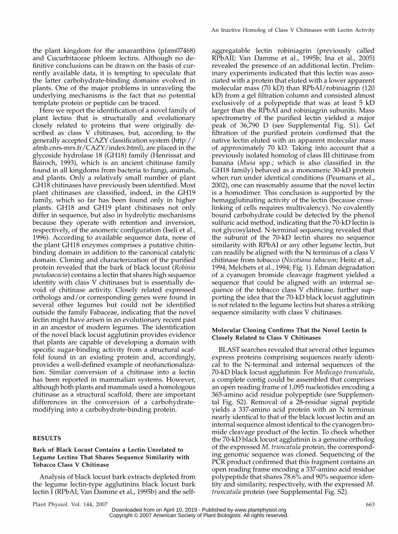
aggregatable lectin robiniagrin (previously calledRPbAII; Van Damme et al., 1995b; Ina et al., 2005)revealed the presence of an additional lectin. Prelim-inary experiments indicated that this lectin was asso-ciated with a protein that eluted with a lower apparentmolecular mass (70 kD) than RPbAI/robiniagrin (120kD) from a gel filtration column and consisted almostexclusively of a polypeptide that was at least 5 kDlarger than the RPbAI and robiniagrin subunits. Massspectrometry of the purified lectin yielded a majorpeak of 36,790 D (see Supplemental Fig. S1). Gelfiltration of the purified protein confirmed that thenative lectin eluted with an apparent molecular massof approximately 70 kD. Taking into account that apreviously isolated homolog of class III chitinase frombanana (Musa spp.; which is also classified in theGH18 family) behaved as a monomeric 30-kD proteinwhen run under identical conditions (Peumans et al.,2002), one can reasonably assume that the novel lectinis a homodimer. This conclusion is supported by thehemagglutinating activity of the lectin (because cross-linking of cells requires multivalency). No covalentlybound carbohydrate could be detected by the phenolsulfuric acid method, indicating that the 70-kD lectin isnot glycosylated. N-terminal sequencing revealed thatthe subunit of the 70-kD lectin shares no sequencesimilarity with RPbAI or any other legume lectin, butcan readily be aligned with the N terminus of a class Vchitinase from tobacco (Nicotiana tabacum; Heitz et al.,1994; Melchers et al., 1994; Fig. 1). Edman degradationof a cyanogen bromide cleavage fragment yielded asequence that could be aligned with an internal se-quence of the tobacco class V chitinase, further sup-porting the idea that the 70-kD black locust agglutininis not related to the legume lectins but shares a strikingsequence similarity with class V chitinases.
Molecular Cloning Confirms That the Novel Lectin IsClosely Related to Class V Chitinases
BLAST searches revealed that several other legumesexpress proteins comprising sequences nearly identi-cal to the N-terminal and internal sequences of the70-kD black locust agglutinin. For Medicago truncatula,a complete contig could be assembled that comprisesan open reading frame of 1,095 nucleotides encoding a365-amino acid residue polypeptide (see Supplemen-tal Fig. S2). Removal of a 28-residue signal peptideyields a 337-amino acid protein with an N terminusnearly identical to that of the black locust lectin and aninternal sequence almost identical to the cyanogen bro-mide cleavage product of the lectin. To check whetherthe 70-kD black locust agglutinin is a genuine orthologof the expressed M. truncatula protein, the correspond-ing genomic sequence was cloned. Sequencing of thePCR product confirmed that this fragment contains anopen reading frame encoding a 337-amino acid residuepolypeptide that shares 78.6% and 90% sequence iden-tity and similarity, respectively, with the expressed M.truncatula protein (see Supplemental Fig. S2).
An Inactive Homolog of Class V Chitinases with Lectin Activity
Plant Physiol. Vol. 144, 2007 663 www.plantphysiol.orgon April 10, 2019 - Published by Downloaded from
Copyright © 2007 American Society of Plant Biologists. All rights reserved.
BLASTp searches using the deduced sequence as aquery revealed that the black locust agglutinin yieldedclass V chitinases from Arabidopsis (Arabidopsis thali-ana; At4g19810) and tobacco (CAA54374; NtChi) asbest matches. The new black locust agglutinin sharesapproximately 54% identity and 80% similarity, re-spectively, with both At4g19810 and CAA54374 (Fig.1), leaving no doubt that it is a structural homolog ofclass V chitinases. Accordingly, the protein will furtherbe referred to as black locust chitinase-related aggluti-nin (RobpsCRA). Although RobpsCRA is undoubtedlya homolog of a class V chitinase, there is apparently amajor difference for what concerns the molecular struc-ture of the native proteins because RobpsCRA is a homo-dimer, whereas all class V chitinases are monomericproteins. This suggests that, unlike class V chitinases,the RobpsCRA subunits contain some structural fea-tures that promote dimerization and hence allowformation of a divalent carbohydrate-binding proteinthat behaves as a genuine agglutinin.
Molecular cloning also yielded additional infor-mation about the biosynthesis and processing of
RobpsCRA. On the analogy of the M. truncatulaortholog, one can reasonably assume that RobpsCRAis synthesized with a signal peptide and follows thesecretory pathway. The calculated molecular mass ofthe protein (36,747.9 D) is nearly identical to thatmeasured by matrix-assisted laser-desorption ioniza-tion (MALDI)-time-of-flight (TOF) mass spectrometry(36,790 D). Taking into account that RobpsCRA is notglycosylated, it seems that no posttranslational pro-cessing takes place.
RobpsCRA Exhibits No Chitinase Activity But Is a
Genuine Lectin
Unlike class I chitinases, class V chitinases fromplants do not possess a genuine chitin-binding domaincorresponding to a hevein domain. Accordingly, theagglutinating activity of RobpsCRA cannot be as-cribed to the presence of a genuine or modified heveindomain.
A number of control experiments were set up to ruleout the possibility that the observed agglutination
Figure 1. Sequence alignment of RobpsCRA and the most closely related homologous proteins identified thus far. NtChi(CAA54374) is a catalytically active class V chitinase isolated from tobacco. At4g19810 is an expressed ortholog of Arabidopsis,but the protein has not yet been isolated and assayed for chitinase activity. In the top row, the N-terminal sequence of the nativelectin (Lec-Nter) and a cyanogen bromide fragment (Lec-CNBr) are aligned with the tobacco chitinase. Identical andhomologous residues are indicated by black and white boxes, respectively. Residues involved in the catalytic cleavage of chitinare indicated by black triangles. The Ser residue specifically involved in the catalytic activity of family 18 chitinases (Synstadet al., 2004) is indicated by a black square.
Van Damme et al.
664 Plant Physiol. Vol. 144, 2007 www.plantphysiol.orgon April 10, 2019 - Published by Downloaded from
Copyright © 2007 American Society of Plant Biologists. All rights reserved.

activity might be due to contamination by the legumelectin-type bark lectin RPbAI. First, SDS-PAGE, usingincreasing amounts of the purified protein, yielded noadditional protein band in the 29- to 32-kD range.Second, the chromatograms of the automated Edmandegradation were indicative of a single sequence. Third,gel filtration experiments confirmed that the agglutina-tion activity coeluted with RobpsCRA and hence cannotbe ascribed to the larger tetrameric legume lectin-typebark lectins. Fourth, western-blot analysis indicatedthat RobpsCRA does not show any cross-reactivitywith antibodies raised against RPbAI. Moreover, as isdemonstrated below, the specificity of RobpsCRAdoes not match that of RPbAI and RPbAII.
Because RobpsCRA shares high sequence identitywith class V chitinases, the possible enzymatic activityof the protein was checked. Concentrated solutions ofthe protein (final concentration 2 mg/mL) were incu-bated with carboxymethyl-chitin-Remazol-Brilliant-Violet 5R at different pH values ranging between 4.0and 7.0. Even upon incubation for 72 h, no acid-solublefragments were generated, indicating that RobpsCRAis devoid of chitinase activity. It should be mentionedhere that two genuine class V chitinases isolated fromtobacco leaves inoculated with Tobacco mosaic virusexhibited readily measurable catalytic activity whenassayed with the same substrate (Melchers et al., 1994).
Agglutination assays with animal red blood cells dem-onstrated that RobpsCRA is a genuine lectin. Trypsin-treated human erythrocytes (type A) were agglutinatedat a lectin concentration of approximately 20 mg/mL.Hapten inhibition assays indicated that the agglutina-tion activity of RobpsCRA is not affected by anysimple sugar. Chito-oligosaccharides with chain lengthsup to 4 GlcNAc units also could not prevent aggluti-nation, indicating that the lectin activity of RobpsCRAdoes not rely on binding to chitin-like compounds. Onlysome animal glycoproteins, like thyroglobulin, inhibitedthe agglutination of human erythrocytes by RobpsCRA.Although indicative, the results of these preliminaryinhibition assays did not allow any conclusion to bedrawn with respect to the carbohydrate-binding spec-ificity of the lectin. Therefore, more appropriate tech-niques, based on direct measurements of lectin-glycaninteractions, were employed to unravel the fine spec-ificity of RobpsCRA.
RobpsCRA Specifically Binds High-Man N-Glycans
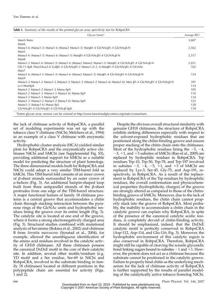
Glycan array analysis revealed that RobpsCRAbinds exclusively to some, but not all, high-Man-typeN-glycans. As shown in Table I, lectin reacted moststrongly with Man5-9mix, which is a mixture of high-Man N-glycans differing in the number of Man residuesand the nature of the bonds between the individualMan units. Besides the Man5-9mix, RobpsCRA alsoreacted well with individual high-Man N-glycans.Man6, Man5, and Man8 were approximately 30%less reactive than the Man5-9mix, whereas Man7 andMan9 were roughly 5 times less active than the mix-
ture (Table I). None of the oligomannosides testedshowed any reactivity. The same applies to chitotriose.These findings clearly indicate that the specificity ofRobpsCRA is directed toward the core pentasaccha-ride of N-glycans.
Although the results of glycan array screening ex-periments are only semiquantitative, they clearly dem-onstrate that the specificity of the lectin is directedtoward high-Man N-glycans comprising the core pen-tasaccharide of N-glycans. Therefore, it is important torealize that the results of glycan arrays are based on adirect binding assay and, accordingly, give a fairlygood idea of the relative affinity of the lectin for a verylarge set of glycans. At present, no conclusions can bedrawn with respect to the affinity of RobpsCRA for theN-glycans. The figures obtained with the different high-Man N-glycans are relatively low (,4,000 relative fluo-rescence units [RFU]) as compared to other lectins (upto .50,000 RFU). However, these low values mightpartly be due to poor coupling of the fluorochrome tothe lectin.
It should be emphasized here that the specificity ofRobpsCRA differs from that of the previously de-scribed legume lectin-type bark agglutinins. The ag-glutination activity of RobpsCRA cannot be inhibited,indeed, by any simple sugar, whereas RPbAI and theself-aggregatable lectin lose their hemagglutinatingactivity in the presence of GalNAc and GlcNAc/Man,respectively. Moreover, RPbAI-type isolectins interactstrongly with complex-type N-glycans, but are nonre-active toward high-Man N-glycans (Van Damme et al.,1995b). This difference in specificity confirms that theobserved lectin activity of RobpsCRA is not due tocontamination by another bark lectin.
Molecular Modeling of RobpsCRA and TobaccoClass V Chitinase
To find possible clues for the obvious lack of chitinaseactivity, the overall fold and structure of RobpsCRAwas tentatively determined by molecular modeling.Because previously no structure was available for plantclass V chitinase, the model was built using the coor-dinates of a human chitotriosidase (hMChi), which, ofall resolved GH18 proteins, shares the highest sequenceidentity/similarity with RobpsCRA (Fusetti et al.,2002). Sequence alignments indicated that RobpsCRAand hMChi share 33% and 51% identity/similarity,respectively, over a 305-amino acid residue overlap(spanning residues 17–322 of RobpsCRA; Fig. 2), whichis reasonably high considering the relatively low overallsequence identity/similarity within the GH18 family.Moreover, it should be emphasized here that, in spiteof the apparent low overall sequence identity, theresidues involved in binding of the substrate, as wellas those involved in the catalytic reaction, are mark-edly conserved between all members of the GH18.Therefore, hMChi can be considered a suitable modelto predict the structure of RobpsCRA. Because themodeling studies were primarily intended to explain
An Inactive Homolog of Class V Chitinases with Lectin Activity
Plant Physiol. Vol. 144, 2007 665 www.plantphysiol.orgon April 10, 2019 - Published by Downloaded from
Copyright © 2007 American Society of Plant Biologists. All rights reserved.
the lack of chitinase activity of RobpsCRA, a parallelset of modeling experiments was set up with thetobacco class V chitinase (NtChi; Melchers et al., 1994)as an example of a class V chitinase with enzymaticactivity.
Hydrophobic cluster analysis (HCA) yielded similarplots for RobpsCRA and the enzymatically active chi-tinases NtChi and hMChi (see Supplemental Fig. S3),providing additional support for hMChi as a suitablemodel for predicting the structure of plant homologs.The three-dimensional models built for RobpsCRA andNtChi could adopt a very similar TIM-barrel fold ashMChi. This TIM-barrel fold consists of an inner crownof b-sheet strands surrounded by an outer crown ofa-helices (Fig. 3). An additional hairpin-shaped loopbuilt from three antiparallel strands of the b-sheetprotrudes from one edge of the TIM-barrel structure.A major functional feature of family 18 chitinase pro-teins is a central groove that accommodates a chitinchain through stacking interaction between the pyra-nose rings of the GlcNAc units and hydrophobic res-idues lining the groove over its entire length (Fig. 3).The catalytic site is located at one end of the groove,where it forms a strong electronegatively charged area(Fig. 3). Structural studies combined with mutationalanalysis of hevamine (Bokma et al., 2002) and chitinaseB from Serratia marcescens (Synstad et al., 2004), forexample, allowed the unambiguous identification ofthe amino acid residues involved in the catalytic activ-ity of GH18 chitinases. All these chitinases possessthe canonical DxDxE motif in the core of their catalyticsite. In addition, several other motifs/residues (e.g.YD motif and a Ser residue, Ser-69 in NtChi andRobpsCRA, involved in the substrate binding in fam-ily 18 chitinases) located at different positions in thepolypeptide chain are essential for activity (Figs.1 and 2).
Despite the obvious overall structural similarity withgenuine GH18 chitinases, the structure of RobpsCRAexhibits striking differences especially with respect tothe solvent-exposed hydrophobic residues that arepositioned along the chitin-binding groove and ensureproper stacking of the chitin chain onto the chitinases.Most of the hydrophobic residues lining the 25, 24,23, 11, and 13 subsites of hMChi (Rao et al., 2005) arereplaced by hydrophilic residues in RobpsCRA. Trpresidues Trp-10, Trp-50, Trp-78, and Trp-197 involvedin subsites 23, 24, 25, 11, and 13 of hMChi arereplaced by Lys-3, Ser-45, Gly-75, and Asp-191, re-spectively, in RobpsCRA. As a result of the replace-ment in RobpsCRA of the Trp residues by hydrophilicresidues, the overall conformation and physicochem-ical properties (hydrophilicity, charges) of the grooveare strongly altered as compared to those of the chitin-binding groove of hMChi. Moreover, due to the lack ofhydrophobic residues, the chitin chain cannot prop-erly stack into the groove of RobpsCRA. Most proba-bly, the inability to accommodate a chitin chain in thecatalytic groove can explain why RobpsCRA, in spiteof the presence of the canonical catalytic acidic resi-dues, is completely devoid of chitin-binding activity.It should be emphasized, indeed, that the DxDxEcatalytic motif is perfectly conserved in RobpsCRA(Asp-112, Asp-114, and Glu-116; Fig. 3). Moreover, thehydrophobic environment of this catalytic region isalso conserved in RobpsCRA. Therefore, RobpsCRAmight still be capable of cleaving the scissile glycosidicbond linking sugars bound to subsites 21 and 11. How-ever, the protein does not act as a chitinase because thesubstrate cannot be positioned in the catalytic groove.Failure to properly bind chitin as the underlying mech-anism for the lack of chitinase activity of RobpsCRAis further supported by the results of parallel model-ing of the catalytically active tobacco homolog NtChi.
Table I. Summary of the results of the printed glycan array specificity test for RobpsCRA
Glycan Namea Average RFU
Man5-9mix 3,807Man6Mana1-6 (Mana1-3) Mana1-6 (Mana2 Mana1-3) Manb1-4 GlcNAcb1-4 GlcNAcb-N 2,562Man5Mana1-6 (Mana1-3) Mana1-6 (Mana1-3) Manb1-4 GlcNAcb1-4 GlcNAcb-N 2,517Man8Mana1-2 Mana1-6 (Mana1-3) Mana1-6 (Mana2 Mana2 Mana1-3) Manb1-4 GlcNAcb1-4 GlcNAcb-N 2,031OS-11-Sp8 (Neu5Aca2-6 Galb1-4 GlcNAcb1-2 Mana1-)2-3, 6-Manb1-4 GlcNAcb1-4 GlcNAc 762Man7Mana1-6 (Mana1-2 Mana1-3) Mana1-6 (Mana2 Mana1-3) Manb1-4 GlcNAcb1-4 GlcNAcb-N 724Man9Mana1-2 Mana1-2 Mana1-3 [Mana1-2 Mana1-3 (Mana1-2 Mana1-6) Mana1-6] Man b1-4 GlcNAcb1-4 GlcNAcb-N 391a-D-ManSp8 114Mana1-2 Mana1-2 Mana1-3 Mana-Sp9 105Mana1-2 Mana1-3 (Mana1-2 Mana1-6) Mana-Sp9 110Mana1-2 Mana1-3 Mana-Sp9 134Mana1-3 (Mana1-2 Mana1-2 Mana1-6) Mana-Sp9 122Mana1-3 (Mana1-6) Mana-Sp3 130GlcNAcb1-4 GlcNAcb1-4 GlcNAcb-Sp8 118
aEntire glycan array version can be viewed at http://www.functionalglycomics.org/static/consortium.
Van Damme et al.
666 Plant Physiol. Vol. 144, 2007 www.plantphysiol.orgon April 10, 2019 - Published by Downloaded from
Copyright © 2007 American Society of Plant Biologists. All rights reserved.

Unlike in RobpsCRA, most of the hydrophobic resi-dues found in the groove of hMChi are conserved inNtChi. Only Trp-10 and Trp-78 (of hMChi) are re-placed by Lys-4 and Gly-74, respectively, in NtChi(Fig. 3F). Accordingly, the groove of NtChi is fully ca-pable of properly positioning a chitin chain for cleav-age by the catalytic motif Asp-111, Leu-112, Asp-113,Trp-114, Glu-115.
Expressed Orthologs of RobpsCRA Are Common in
Legumes But Not Found in Other Plants
As already mentioned above, several other legumesexpress closely related orthologs of RobpsCRA. Com-plete or nearly complete contigs could be assembledfor M. truncatula, Glycine max, and Lotus japonicus. Allfour proteins share 62.5% identity and 82.4% similar-ity, respectively, within a 301-amino acid residue over-lap (see Supplemental Fig. S2).
In addition, a previously described, but only par-tially characterized, 67-kD homodimeric lectin frombean (Phaseolus vulgaris) seeds (Ye et al., 2001) alsomight represent a RobpsCRA ortholog. However, no
genuine orthologs of RobpsCRA could be identified inprotein, expressed sequence tag, or genomic databasesof any other plant species. Although no definitiveconclusions can be drawn from the available sequencedata, it is evident that RobpsCRA-type lectins are farless widespread among flowering plants than class Vchitinases and most likely are confined to a relativelysmall taxonomic group. Within the Fabaceae, RobpsCRAorthologs occur in at least four different tribes ofthe subfamily Papilionoideae (black locust, G. max,M. truncatula, and L. japonicus belong to the Robinieae,Phaseoleae, Trifolieae, and Loteae, respectively), indi-cating that they are not confined to a small taxon of theFabaceae.
DISCUSSION
Biochemical analyses and molecular cloning dem-onstrated that a minor lectin from the bark of blacklocust is a catalytically inactive homolog of class Vchitinases. The lectin behaves as a genuine hemagglu-tinin and specifically binds, albeit with a relatively low
Figure 2. Alignment of the amino acid sequences of RobpsCRA, tobacco class V chitinase NtChi, human chitotriosidase hMChi,and the chitinase-related murine protein Ym1. Identical and homologous residues are indicated by black and white boxes,respectively. Residues involved in the catalytic cleavage of chitin are indicated by black triangles, and residues lining up thecatalytic groove of genuine chitinases are indicated by black circles (hydrophobic residues) and asterisks (hydrophilic residues).The Ser residue specifically involved in the catalytic activity of family 18 chitinases (Synstad et al., 2004) is indicated by a blacksquare.
An Inactive Homolog of Class V Chitinases with Lectin Activity
Plant Physiol. Vol. 144, 2007 667 www.plantphysiol.orgon April 10, 2019 - Published by Downloaded from
Copyright © 2007 American Society of Plant Biologists. All rights reserved.

Figure 3. Molecular modeling of RobpsCRA and related proteins. A and B, Ribbon diagrams of the modeled NtChi (A) andRobpsCRA (B) proteins. Acidic residues involved in the catalytic cleavage of chitin are indicated by stick representation (pink).
Van Damme et al.
668 Plant Physiol. Vol. 144, 2007 www.plantphysiol.orgon April 10, 2019 - Published by Downloaded from
Copyright © 2007 American Society of Plant Biologists. All rights reserved.

affinity, to high-Man N-glycans. Activity assays usingdye-labeled substrates indicated that the protein isdevoid of chitinase activity. Molecular modeling andsequence comparisons indicated that the apparent lackof catalytic activity most probably has to be ascribed tothe protein’s inability to accommodate the chitin sub-strate in its catalytic groove as a consequence of anextensive replacement of hydrophobic by hydrophilicamino acids.
Although there is no doubt that RobpsCRA is struc-turally and evolutionarily related to class V chitinases,the available sequence data are insufficient to trace thedetails of the conversion of a plant chitinase into alectin. However, even in the absence of full details, onecan reasonably assume that RobpsCRA evolved from aGH18 chitinase and not the other way around. GH18represents an ancient chitinase family because it isfound in all kingdoms from bacteria to fungi, animals,and plants (Iseli et al., 1996). Moreover, sequence dataclearly indicate that all GH18 chitinases, includingthose from plants, have a common ancestor. This,taken together with the fact that GH18 chitinases orcorresponding genes have been found in numerousplant species, implies that the GH18 structural scaffoldis quite common in higher plants. Considering theapparent confinement of RobpsCRA orthologs to thelegume family, it is tempting to speculate that a cata-lytically active chitinase from an ancient legume speciesor possibly an earlier ancestor served as a structuralscaffold for the development of a small family ofcarbohydrate-binding proteins. This evolutionary pro-cess most likely involved gene duplication followed byneofunctionalization. The RobpsCRA orthologs repre-sent a documented example of how plants managed todevelop a domain with a specific sugar-binding activityfrom a functionally unrelated protein in general or anenzyme in particular. Therefore, it should be empha-sized that the novel lectin no longer recognizes thesubstrate of the original hydrolase (in casu chitin orchito-oligosaccharides) but a structurally unrelatedglycan (namely, high-Man N-glycans). Interestingly, asimilar conversion of a glycosyl hydrolase into acarbohydrate-binding, but catalytically inactive, ho-molog also occurred in higher animals. A so-calledeosinophil chemotactic cytokine has been identified inmouse (Chang et al., 2001) and human (Boot et al.,1995) that shares approximately 50% sequence identitywith the respective conspecific chitotriosidase but isdevoid of chitinase activity. Instead, the mouse protein(referred to as ECF-L or secretory protein Ym1) is alectin with binding specificity to glucosamine and
heparin/heparan sulfate (Chang et al., 2001; Sun et al.,2001). Although there is certainly a parallel betweenthe conversion of a chitinase into a lectin in animalsand plants, there are two major differences. First, incontrast to RobpsCRA, the canonical DxDxE motif isreplaced in Ym1 by a motif in which two catalyticacidic residues are substituted (Asn-115, Leu-116,Asp-117, Trp-118, Gln-119; Tsai et al., 2004; Fig. 2).Second, Ym1 binds glucosamine and heparin/heparansulfate, whereas RobpsCRA interacts exclusively withhigh-Man N-glycans.
Identification of RobpsCRA orthologs as lectins notonly adds a novel carbohydrate-binding domain tothe existing list of documented plant lectin familiesbut also illustrates that plants are capable of devel-oping sugar-binding domains from an existing struc-tural scaffold with different activity. At present, thebinding affinity of RobpsCRA is relatively low. How-ever, RobpsCRA might just be an intermediate in anevolutionary pathway that eventually will yield lec-tins with a high affinity. Even if there is no evidence ofwhether analogous evolutionary mechanisms mighthave given rise to other carbohydrate-binding do-mains that are confined to plants, the discovery ofa lectin ortholog of class V chitinases puts the evolu-tion of plant lectins in a novel perspective. In addi-tion, legume RobpsCRA orthologs represent a novelexample of well-defined neofunctionalization inplants.
It is also worth noting in this context that at least twodifferent cases have been reported of neofunctionali-zation-related evolutionary events whereby plantsused the structural scaffold of a lectin domain for thedevelopment of a protein that lost sugar-binding ac-tivity but acquired totally different biological activity.Curculin from Curculigo latifolia fruits is a homolog ofthe GNA-related lectins that possesses no sugar-bindingactivity but has sweet-tasting properties (Haradaet al., 1994). Seeds of several Phaseolus species containstructural homologs of the bean lectin that have nocarbohydrate-binding activity but are potent a-amylaseinhibitors or insecticidal proteins (called arcelins;Mirkov et al., 1994). It should be emphasized thatthese lectin homologs are not just binding-defectivemutants but proteins with well-defined biological activ-ity. Other binding-defective lectins have been identi-fied in the bark of the legume tree Cladrastis lutea andSambucus nigra, but the respective legume lectin andtype 2 ribosome-inactivating protein homologs haveno known biological activity other than a presumedstorage function (Van Damme et al., 1995a, 1997).
Figure 3. (Continued.)Hydrophobic (orange) and hydrophilic (blue) residues lining up the catalytic groove of chitinases are in stick representation. Cand D, Mapping of electrostatic potentials on the molecular surface of NtChi (C) and RobpsCRA (D). Acidic residues responsiblefor the electronegative character of the binding groove of chitinases are labeled in white. E to G, Enlarged ribbon diagrams of thecatalytic groove of hMChi (E), NtChi (F), and RobpsCRA (G). Catalytic residues (pink) and residues lining up the catalytic groove(orange for hydrophobic and blue for hydrophilic residues) are in stick representation.
An Inactive Homolog of Class V Chitinases with Lectin Activity
Plant Physiol. Vol. 144, 2007 669 www.plantphysiol.orgon April 10, 2019 - Published by Downloaded from
Copyright © 2007 American Society of Plant Biologists. All rights reserved.
MATERIALS AND METHODS
Plant Material
Bark was stripped from the stems of 4- to 5-year-old black locust (Robinia
pseudoacacia) trees at the end of the winter (beginning of April). All trees were
from the same clone (growing in the garden of W. Peumans). The inner bark
was collected using a knife, taking care to remove the outer corky bark tissue,
cut in small pieces, and stored at 220�C until use.
Isolation of RobpsCRA
Because RobpsCRA is only a very minor bark protein and has a relatively
low affinity for carbohydrates, the lectin could not be purified from crude or
partially purified bark extracts by affinity chromatography. Therefore, a
purification scheme was developed based on a combination of conventional
protein purification techniques and affinity chromatography. In a first step, a
partially purified protein fraction was isolated by cation ion-exchange chro-
matography. This protein fraction was in a second step depleted from the
major bark lectin (RPAbI) by affinity chromatography on immobilized Gal and
concentrated by hydrophobic interaction chromatography. Further purifica-
tion was achieved by gel filtration and affinity chromatography on immobi-
lized thyroglobulin. Gal and thyroglobulin were coupled to Sepharose 4B by
the divinylsulfon method.
Extraction and Removal of the Major LegumeLectin-Type Bark Agglutinins
Batches of 1 kg of frozen bark were transferred into 5 L of a solution of
20 mM acetic acid and, after thawing, homogenized in a blender. The homogenate
was passed through a sieve (mesh size approximately 1.5 mm) and centri-
fuged (3,000g; 10 min). To the supernatant, solid CaCl2 was added (1 g/L) and
the pH adjusted to 9.0 with 1 N NaOH. After standing overnight in the cold
(2�C–4�C), the precipitate was removed by centrifugation (3,000g; 10 min). The
cleared extract was adjusted to pH 3.2, centrifuged (3,000g; 10 min), and
filtered through filter paper. The filtrate was diluted with an equal volume of
water and applied onto a column (5 cm 3 10 cm; approximately 200-mL bed
volume) of S Fast Flow (Amersham Biosciences) and preequilibrated with
20 mM acetic acid. Loading was continued until the column was 70% saturated
with protein. Then the column was washed with 50 mM sodium formate (pH
3.8) until the A280 fell below 0.1 and the bound proteins eluted with 500 mL
0.2 M NaCl in 0.1 M Tris-HCl (pH 8.7). After regeneration of the column, ion-
exchange chromatography was repeated with new batches of the crude
extract. The eluates of the different runs were pooled, adjusted to 1.8 M
ammonium sulfate with solid salt, and centrifuged (9,000g for 15 min).
Aliquots of the supernatant (equivalent to approximately 10,000 A280 units)
were applied onto a column (5 cm 3 15 cm; approximately 300-mL bed
volume) of Gal-Sepharose 4B. Under these conditions, the major legume
lectin-type bark agglutinin (RPAbI) was quantitatively retained on the col-
umn. After passing the protein fraction, the column was washed with a
solution of 1.8 M ammonium sulfate (adjusted to pH 7.5 with 1 N HCl) until the
A280 fell below 0.1. The run-through and wash fractions were collected and
pooled. After washing, the lectin was desorbed with 20 mM unbuffered 1,3-
diaminopropane and the Gal-Sepharose 4B column regenerated for the next
run. The pass-through and wash fractions of the different runs were pooled
and rechromatographed on the same column Gal-Sepharose 4B to remove any
remaining RPAbI.
Purification of RobpsCRA from theRPAbI-Depleted Fraction
The RPAbI-depleted fraction was applied onto a column (5 cm 3 5 cm;
approximately 100-mL bed volume) of phenyl-Sepharose preequilibrated
with 1.8 M ammonium sulfate. After loading, the column was washed with
1.8 M ammonium sulfate until the A280 fell below 0.01 and the bound proteins
eluted with 0.1 M Tris-HCl (pH 10.0). Fractions with an A280 . 3 were pooled,
adjusted to pH 7.5 with 1 N HCl, and applied in 25-mL aliquots on a column
(5 cm 3 50 cm; approximately 1,000-mL bed volume) of Sephacryl 200
(Pharmacia) equilibrated with 0.2 M NaCl in 20 mM Tris-HCl (pH 7.5). Gel
filtration yielded two major peaks. Agglutination assays and SDS-PAGE
indicated that the first peak contained predominantly RobpsCRA. The frac-
tions eluting in the first peak of the different runs were pooled, concentrated
on a small column (1.5 cm 3 10 cm; approximately 14-mL bed volume) of
phenyl-Sepharose, and rechromatographed on Sephacryl 200 using a longer
column (2.5 cm 3 70 cm; approximately 350-mL bed volume). The peak
fractions, which consisted almost exclusively of RobpsCRA were pooled,
brought at 1.5 M ammonium sulfate with solid salt, and applied onto a column
(2.5 cm 3 10 cm; approximately 50-mL bed volume) of thyroglobulin-
Sepharose 4B. After loading, the column was washed with 1.5 M ammonium
sulfate (adjusted to pH 7.5 with HCl) until the A280 fell below 0.01 and the
lectin desorbed with 0.1 M Tris-HCl (pH 10.0). The affinity-purified lectin was
dialyzed against water or an appropriate buffer and stored at 220�C until use.
The overall yield of RobpsCRA was approximately 10 mg/kg bark tissue.
Starting from the same extracts, about 2 g RPAbI were recovered per kilogram
of bark tissue. This implies that RobpsCRA is roughly 200 times less abundant
than the major bark lectin.
Analytical Methods
RobpsCRA was analyzed by SDS-PAGE using 15% (w/v) acrylamide
gradient gels. For N-terminal amino acid sequencing, purified RobpsCRA was
separated by SDS-PAGE, electroblotted on a polyvinylidene difluoride mem-
brane, and sequenced on an Applied Biosystems model 477A protein se-
quencer interfaced with an Applied Biosystems model 120A online analyzer.
Cyanogen bromide cleavage of RobpsCRA (2 mg) was done in 0.1 mL of 70%
formic acid containing 10 mg of cyanogen bromide. After incubation for 15 h
at 37�C (in the dark), peptides were recovered by evaporation under vacuum,
separated by SDS-PAGE, and electroblotted on a polyvinylidene difluoride
membrane for subsequent sequencing.
Total neutral sugar was determined by the phenol/H2SO4 method
(Dubois et al., 1956), with D-Glc as a standard. Analytical gel filtration was per-
formed on a Pharmacia Superose 12 column (Amersham Biosciences) using
phosphate-buffered saline as running buffer at a flow rate of 20 mL/h.
For MALDI-TOF mass spectrometry, samples (0.75 mL) of a 0.5 mg/mL
solution of RobpsCRA in 50 mM phosphate buffer (pH 7.5) containing 50 mM
NaCl were cocrystallized on the MALDI plate with 0.75 mL of a 0.6 mM
3,5-dimethoxy-4-hydroxy cinnamic acid (sinapinic acid) solution made up in
50% (w/v) aqueous azidonitrile. Desorption and ionization of crystallized
samples were carried out on a Voyager-DE STR (Perspective Biosystems) mass
spectrometer in positive linear mode using an accelerating voltage of 25 kV, a
grid voltage of 93%, and an extraction delay time of 800 ns. Acquisition mass
was performed between 4,000 and 50,000 D using a mixture of proteins of
known molecular mass for internal calibration.
PCR Amplification of Sequence Encoding RobpsCRA
Genomic DNA was isolated from seeds of black locust using the FastDNA
spin kit in an automatic homogenizer (FastPrep instrument; MP Biomedicals
and Qbiogene) following the manufacturer’s recommendations. DNA frag-
ments encoding RobpsCRA were amplified using degenerate PCR primers
derived from the N-terminal sequence of RobpsCRA and the C-terminal
sequence of MedtrCRA (see Supplemental Fig. S4). The reaction mixture for
amplification of genomic sequences contained 10 mM Tris-HCl (pH 8.3), 50 mM
KCl, 1.5 mM MgCl2, 100 mg/L gelatin, 0.4 mM of each dNTP, 2.5 units of Taq
polymerase (Invitrogen), 5 mL of cDNA, and 20 mL of the appropriate primer
mixtures (5 mM), in a 25-mL reaction volume. After denaturation of the DNA
for 5 min at 95�C, amplification was performed for 30 cycles through a regime
of 15-s template denaturation at 92�C, followed by 30-s primer annealing at
45�C to 50�C and 1-min primer extension at 72�C. The PCR fragments were
cloned in TOPO pCR2.1-TOPO cloning vector using the TOPO cloning kit
from Invitrogen. Plasmids were isolated from purified single colonies on a
miniprep scale using the alkaline lysis method (Mierendorf and Pfeffer, 1987)
and sequenced by the dideoxy method (Sanger et al., 1977).
Hemagglutination Activity
Agglutination assays were carried out in small glass tubes or in the wells of
96 U-welled microtiter plates in a final volume of 50 mL containing 40 mL of a
1% (v/v) suspension of trypsin-treated human erythrocytes and 10 mL of
lectin solution. Agglutination was monitored visually after 1 h of incubation at
room temperature. To determine the specific agglutinating activity, the lectin
was serially diluted with 2-fold increments and the dilution end point
determined.
Van Damme et al.
670 Plant Physiol. Vol. 144, 2007 www.plantphysiol.orgon April 10, 2019 - Published by Downloaded from
Copyright © 2007 American Society of Plant Biologists. All rights reserved.

Glycan Array Screening
The specificity of RobpsCRA was screened on the consortium printed array
(PA V1) as previously described (Blixt et al., 2005). Briefly, an aliquot of
RobpsCRA-Alexa 488 (200 mg/mL) was applied in a volume of 50 mL binding
buffer onto the preprinted slide, cover-slipped, and incubated protected from
light for 1 h. The slide was washed successively in wash buffer, wash buffer
minus Tween 20, and deionized water, and then dried under a stream of
nitrogen. The slide was read on a ScanArray Express microarray scanner and
the image analyzed using ScanArray Express software (Perkin-Elmer Life
Sciences).
Retrieval of Sequences
Sequences encoding RobpsCRA orthologs were retrieved by BLAST
searches. First, a complete contig was assembled for Medicago truncatula (see
Supplemental Fig. S4) using the experimentally determined N-terminal and
internal amino acid sequence of RobpsCRA. Next, the deduced amino acid
sequence of RobpsCRA was used as a query for BLASTp and tBLASTn
searches.
Molecular Modeling
Multiple amino acid sequence alignments were carried out with ClustalX
(Thompson et al., 1997) and displayed with ESPript (Gouet et al., 1999) using
the structural Risler’s matrix for homologous residues (Risler et al., 1998).
HCA (Gaboriaud et al., 1987) was carried out to delineate the structurally
conserved secondary structural features (strands of b-sheet and stretches of
a-helix) along the amino acid sequences of the RobpsCRA, the class V
chitinase from tobacco (Nicotiana tabacum; NtChi; Melchers et al., 1994), the
murine secretory protein Ym1 (Sun et al., 2001), and the human chitotriosidase
hMChi (Fusetti et al., 2002). HCA plots were generated using the HCA server
(http://www.lmcp.jussieu.fr).
Homology modeling of RobpsCRA and the genuine class V chitinase
NtChi from tobacco (Melchers et al., 1994) was performed on a Silicon
Graphics O2 10000 workstation, using the programs InsightII, Homology, and
Discover (Accelrys). The atomic coordinates of the human chitotriosidase
hMChi (RCSB Protein Data Bank, code 1WB0; Fusetti et al., 2002; Rao et al.,
2005) were used to build a three-dimensional model. Steric conflicts resulting
from the replacement or the deletion of some residues in the modeled
domains were corrected during the model-building procedure using the
rotamer library (Ponder and Richards, 1987) and the search algorithm of the
Homology program (Mas et al., 1992) to maintain proper side-chain orienta-
tion. Energy minimization of the final models was carried out by 200/300
cycles of steepest descent using Discover3. The program TurboFrodo (Roussel
and Cambillau, 1989) was run to draw the Ramachandran plot and to perform
the superposition of the models. PROCHECK (Laskowski et al., 1993) was
used to assess the geometric quality of the three-dimensional models.
Electrostatic potentials were calculated and displayed with GRASP using
the parse3 parameters (Nicholls et al., 1991). The solvent probe radius used for
molecular surfaces was 1.4 A and a standard 2.0 A Stern layer was used to
exclude ions from the molecular surface (Gilson and Honing, 1987). The inner
and outer dielectric constants applied to the protein and the solvent were
respectively fixed at 4.0 and 80.0 and the calculations were performed keeping
a salt concentration of 0.145 M. The surfaces occupied by hydrophobic (Leu,
Ile, Val, Met, Phe, Trp, and Tyr included) residues on the molecular surface of
the modeled domains were calculated with GRASP. Cartoons were drawn
with PyMol (http://www.delanoscientific.com).
Sequence data from this article can be found in the GenBank/EMBL data
libraries under accession number EF152992.
Supplemental Data
The following materials are available in the online version of this article.
Supplemental Figure S1. Mass spectra of purified RobpsCRA.
Supplemental Figure S2. Sequence alignment of RobpsCRA and ortho-
logs from G. max, M. truncatula, and L. japonicus.
Supplemental Figure S3. HCA plots of human chitotriosidase hMChi,
murine secretory protein Ym1, NtChi, and RobpsCRA.
Supplemental Figure S4. Nucleotide sequence of the contig encoding
MedtrCRA (the RobpsCRA ortholog from M. truncatula).
ACKNOWLEDGMENT
We are deeply indebted to Angela Lee of Core H of the Consortium for
Functional Glycomics for expert technical assistance.
Received August 4, 2006; accepted November 4, 2006; published November
10, 2006.
LITERATURE CITED
Blixt O, Head S, Mondala T, Scanlan C, Huflejt ME, Alvarez R, Bryan MC,
Fazio F, Calarese D, Stevens J, et al (2005) Printed covalent glycan array
for ligand profiling of diverse glycan binding proteins. Proc Natl Acad
Sci USA 101: 17033–17038
Bokma E, Rozeboom HJ, Sibbald M, Dijkstra BW, Beintema JJ (2002)
Expression and characterization of active site mutants of hevamine, a
chitinase from the rubber tree Hevea brasiliensis. Eur J Biochem 269:
893–901
Boot RG, Renkema GH, Strijland A, van Zonneveld AJ, Aerts JM (1995)
Cloning of a cDNA encoding chitotriosidase, a human chitinase pro-
duced by macrophages. J Biol Chem 270: 26252–26256
Chang NC, Hung SI, Hwa KY, Kato I, Chen JE, Liu CH, Chang AC (2001)
A macrophage protein, Ym1, transiently expressed during inflammation
is a novel mammalian lectin. J Biol Chem 276: 17497–17505
Dubois M, Gilles KA, Hamilton JK, Rebers PA, Smith F (1956) Colori-
metric method for determination of sugar and related substances. Anal
Chem 28: 350–356
Fusetti F, von Moeller H, Houston D, Rozeboom HJ, Dijkstra BW, Boot
RG, Aerts JM, van Aalten DM (2002) Structure of human chitotriosi-
dase. Implications for specific inhibitor design and function of mam-
malian chitinase-like lectins. J Biol Chem 277: 25537–25544
Gaboriaud C, Bissery V, Benchetrit T, Mornon JP (1987) Hydrophobic
cluster analysis: an efficient new way to compare and analyse amino
acid sequences. FEBS Lett 224: 149–155
Gilson MK, Honing BH (1987) Calculation of electrostatic potential in an
enzyme active site. Nature 330: 84–86
Gouet P, Courcelle E, Stuart DI, Metoz F (1999) ESPript: analysis of
multiple sequence alignments in PostScript. Bioinformatics 15: 305–308
Harada S, Otani H, Maeda S, Kai Y, Kasai N, Kurihara Y (1994) Crystal-
lization and preliminary X-ray diffraction studies of curculin. A new
type of sweet protein having taste-modifying action. J Mol Biol 238:
286–287
Heitz T, Segond S, Kauffmann S, Geoffroy P, Prasad V, Brunner F, Fritig
B, Legrand M (1994) Molecular characterization of a novel tobacco
pathogenesis-related (PR) protein: a new plant chitinase/lysozyme. Mol
Gen Genet 245: 246–254
Henrissat B, Bairoch A (1993) New families in the classification of glycosyl
hydrolases based on amino acid sequence similarities. Biochem J 293:
781–788
Ina C, Sano K, Yamamoto-Takahashi M, Matsushita-Oikawa H, Takekawa
H, Takehara Y, Ueda H, Ogawa H (2005) Screening for and purifica-
tion of novel self-aggregatable lectins reveal a new functional lectin
group in the bark of leguminous trees. Biochim Biophys Acta 1726:
21–27
Iseli B, Armand S, Boller T, Neuhaus JM, Henrissat B (1996) Plant
chitinases use two different hydrolytic mechanisms. FEBS Lett 382:
186–188
Laskowski RA, MacArthur MW, Moss DS, Thornton JM (1993) PRO-
CHECK: a program to check the stereochemistry of protein structures.
J Appl Crystallogr 26: 283–291
Mas MT, Smith KC, Yarmush DL, Aisaka K, Fine RM (1992) Modeling the
anti-CEA antibody combining site by homology and conformational
search. Proteins Struct Funct Genet 14: 483–498
Melchers LS, Apotheker-de Groot M, van der Knaap JA, Ponstein AS,
Sela-Buurlage MB, Bol JF, Cornelissen BJ, van den Elzen PJ, Linthorst
HJ (1994) A new class of tobacco chitinases homologous to bacterial exo-
chitinases displays antifungal activity. Plant J 5: 469–480
An Inactive Homolog of Class V Chitinases with Lectin Activity
Plant Physiol. Vol. 144, 2007 671 www.plantphysiol.orgon April 10, 2019 - Published by Downloaded from
Copyright © 2007 American Society of Plant Biologists. All rights reserved.
Mierendorf RC, Pfeffer D (1987) Direct sequencing of denatured plasmid
DNA. Methods Enzymol 152: 556–562
Mirkov TE, Wahlstrom JM, Hagiwara K, Finardi-Filho F, Kjemtrup S,
Chrispeels MJ (1994) Evolutionary relationships among proteins in the
phytohemagglutinin-arcelin-alpha-amylase inhibitor family of the com-
mon bean and its relatives. Plant Mol Biol 26: 1103–1113
Nagata Y, Yamashita M, Honda H, Akabane J, Uehara K, Saito A, Sumisa
F, Nishibori K, Oodaira Y (2005) Characterization, occurrence, and
molecular cloning of a lectin from Grifola frondosa: jacalin-related lectin
of fungal origin. Biosci Biotechnol Biochem 69: 2374–2380
Nicholls A, Sharp KA, Honig B (1991) Protein folding and association:
insights from the interfacial and thermodynamic properties of hydro-
carbons. Proteins Struct Funct Genet 11: 281–296
Peumans WJ, Proost P, Swennen RL, Van Damme EJM (2002) The
abundant class III chitinase homolog in young developing banana fruits
behaves as a transient vegetative storage protein and most probably
serves as an important supply of amino acids for the synthesis of
ripening-associated proteins. Plant Physiol 130: 1063–1072
Ponder JW, Richards FM (1987) Tertiary templates for proteins. Use of
packing criteria in the enumeration of allowed sequences for different
structural classes. J Mol Biol 193: 775–791
Rao FV, Houston DR, Boot RG, Aerts JMFG, Hodkinson M, Adams DJ,
Shiomi K, Omura S, Van Aalten DMF (2005) Specificity and affinity of
natural product cyclopentapeptide inhibitors against A. fumigatus, hu-
man, and bacterial chitinases. Chem Biol 12: 65–76
Risler J-L, Delorme MO, Delacroix H, Henaut A (1998) Amino acid
substitutions in structurally related proteins. A pattern recognition
approach. Determination of a new and efficient scoring matrix. J Mol
Biol 204: 1019–1029
Roussel A, Cambillau C (1989) TURBO-FRODO. In Silicon Graphics
Committee, ed, Silicon Graphics Geometry Partners Directory. Silicon
Graphics, Mountain View, CA
Sanger F, Nicklen S, Coulson AR (1977) DNA sequencing with chain-
terminating inhibitors. Proc Natl Acad Sci USA 74: 5463–5467
Sun YJ, Chang NC, Hung SI, Chang AC, Chou CC, Hsiao CD (2001) The
crystal structure of a novel mammalian lectin, Ym1, suggests a saccha-
ride binding site. J Biol Chem 276: 17507–17514
Synstad B, Gaseidnes S, Van Aalten DM, Vriend G, Nielsen JE, Eijsink
VG (2004) Mutational and computational analysis of the role of con-
served residues in the active site of a family 18 chitinase. Eur J Biochem
271: 253–262
Tsai ML, Liaw SH, Chang NC (2004) The crystal structure of Ym1 at 1.31 A
resolution. J Struct Biol 148: 290–296
Thompson JD, Gibson TJ, Plewniak F, Jeanmougin F, Higgins DG (1997)
The CLUSTAL-X windows interface: flexible strategies for multiple
sequence alignment aided by quality analysis tool. Nucleic Acids Res 15:
4876–4882
Van Damme EJM, Barre A, Bemer V, Rouge P, Van Leuven F, Peumans WJ
(1995a) A lectin and a lectin-related protein are the two most prominent
proteins in the bark of yellow wood (Cladrastis lutea). Plant Mol Biol 29:
579–598
Van Damme EJM, Barre A, Rouge P, Peumans WJ (2004) Cytoplasmic/
nuclear plant lectins: a new story. Trends Plant Sci 9: 484–489
Van Damme EJM, Barre A, Rouge P, Van Leuven F, Peumans WJ (1997)
Isolation and molecular cloning of a novel type 2 ribosome-inactivating
protein with an inactive B chain from elderberry (Sambucus nigra) bark.
J Biol Chem 272: 8353–8360
Van Damme EJM, Barre A, Smeets K, Torrekens S, Van Leuven F, Rouge P,
Peumans WJ (1995b) The bark of Robinia pseudoacacia contains a com-
plex mixture of lectins. Characterization of the proteins and the cDNA
clones. Plant Physiol 107: 833–843
Van Damme EJM, Peumans WJ, Barre A, Rouge P (1998) Plant lectins: a
composite of several distinct families of structurally and evolutionary
related proteins with diverse biological roles. CRC Crit Rev Plant Sci 17:
575–692
Ye XY, Ng TB, Tsang PW, Wang J (2001) Isolation of a homodimeric lectin
with antifungal and antiviral activities from red kidney bean (Phaseolus
vulgaris) seeds. J Protein Chem 20: 367–375
Van Damme et al.
672 Plant Physiol. Vol. 144, 2007 www.plantphysiol.orgon April 10, 2019 - Published by Downloaded from
Copyright © 2007 American Society of Plant Biologists. All rights reserved.
![Legume Lectins: Proteins with Diverse Applications · Legume Lectins: Proteins with Diverse Applications Irlanda Lagarda-Diaz 1, ... [30]. The specificity of legume lectins for some](https://img.pdfslide.net/doc/110x75/5fc6b4c426138432574b638e/legume-lectins-proteins-with-diverse-applications-legume-lectins-proteins-with.jpg)