Embed Size (px)
Citation preview

A Novel Glycolipid Biosurfactant ConfersGrazing Resistance upon Pantoeaananatis BRT175 against the SocialAmoeba Dictyostelium discoideum
Derek D. N. Smith,a Arvin Nickzad,b Eric Déziel,b John Stavrinidesa
Department of Biology, University of Regina, Regina, Saskatchewan, Canadaa; INRS-Institut Armand-Frappier,Laval, Québec, Canadab
ABSTRACT Pantoea is a versatile genus of bacteria with both plant- and animal-pathogenic strains, some of which have been suggested to cause human infections.There is, however, limited knowledge on the potential determinants used for hostassociation and pathogenesis in animal systems. In this study, we used the modelhost Dictyostelium discoideum to show that isolates of Pantoea ananatis exhibit dif-ferential grazing susceptibility, with some being resistant to grazing by the amoe-bae. We carried out a high-throughput genetic screen of one grazing-resistant iso-late, P. ananatis BRT175, using the D. discoideum pathosystem to identify genesresponsible for the resistance phenotype. Among the 26 candidate genes involvedin grazing resistance, we identified rhlA and rhlB, which we show are involved in thebiosynthesis of a biosurfactant that enables swarming motility in P. ananatis BRT175.Using liquid chromatography-mass spectrometry (LC-MS), the biosurfactant wasshown to be a glycolipid with monohexose-C10-C10 as the primary congener. Weshow that this novel glycolipid biosurfactant is cytotoxic to the amoebae and is ca-pable of compromising cellular integrity, leading to cell lysis. The production of thisbiosurfactant may be important for bacterial survival in the environment and couldcontribute to the establishment of opportunistic infections.
IMPORTANCE The genetic factors used for host interaction by the opportunistichuman pathogen Pantoea ananatis are largely unknown. We identified two genesthat are important for the production of a biosurfactant that confers grazing resis-tance against the social amoeba Dictyostelium discoideum. We show that the biosur-factant, which exhibits cytotoxicity toward the amoebae, is a glycolipid that incorpo-rates a hexose rather than rhamnose. The production of this biosurfactant mayconfer a competitive advantage in the environment and could potentially contributeto the establishment of opportunistic infections.
KEYWORDS: biosurfactants, Pantoea, Dictyostelium, opportunistic infections
Pantoea is a genus of Gram-negative bacilli in the Enterobacteriaceae and a closerelative of human-pathogenic genera Klebsiella and Enterobacter. Pantoea includes
epiphytic and pathogenic members that have been identified throughout the environ-ment, including clinical settings. Infections caused by members of Pantoea are knownto occur primarily in immunocompromised individuals with preexisting conditions andcan result in abscesses, pneumonia, or bacteremia (1–6); however, healthy individualsmay be infected through penetrative injury by plant vegetation, which can result incutaneous infections or develop into septic arthritis (6, 7). More concerning are theclinical cases of fatal bacteremia in neonates caused by contaminated parenteralnutrition as well as bacteremia in immunocompromised adults (1, 3–6, 8). Manymembers of Pantoea, such as Pantoea ananatis, while considered common environ-
Received 26 November 2015 Accepted 1December 2015 Published 20 January 2016
Citation Smith DDN, Nickzad A, Déziel E,Stavrinides J. 2016. A novel glycolipidbiosurfactant confers grazing resistance uponPantoea ananatis BRT175 against the socialamoeba Dictyostelium discoideum. mSphere 1(1):e00075-15. doi:10.1128/mSphere.00075-15.
Editor Katherine McMahon, University ofWisconsin
Copyright © 2016 Smith et al. This is an open-access article distributed under the terms ofthe Creative Commons Attribution 4.0International license.
Address correspondence to John Stavrinides,[email protected].
RESEARCH ARTICLEApplied and Environmental Science
crossmark
Volume 1 Issue 1 e00075-15 msphere.asm.org 1
on May 17, 2020 by guest
http://msphere.asm
.org/D
ownloaded from

mental microbes, have been suggested to be opportunists with human-pathogenicpotential (9, 10).
P. ananatis was originally isolated from pineapple as the causative agent of fruitletbrown rot (11). Many isolates have also been found to cause disease in a wide range ofplant species but are also reported to colonize humans opportunistically (9). A studyexamining virulence potential showed that a P. ananatis pineapple isolate was espe-cially virulent in an embryonated hen egg model compared to five clinical Pantoeaagglomerans isolates (12). Similarly, quantitative growth assays have shown that closelyrelated isolates of P. ananatis can vary greatly in their growth potential in plant andinsect model hosts (13), suggesting the presence of specific genetic factors thatmediate host association (14–16). This has been explored further in a comparativegenomics analysis of eight P. ananatis genomes, which determined that isolates readilyexchange genetic factors involved in host- and niche-specific colonization (17). Candi-date disease factors were suggested to include a putative adhesin, multiple type VIsecretion systems, and even type I fimbriae (17, 18). The presence of animal cell-specifictype III secretion systems has also been noted in several strains, although theirinvolvement in host association and specificity is still unclear (19, 20). There is currentlylimited functional information on these and other genetic factors in this species thatmay contribute to opportunism and human pathogenicity.
Model pathosystems are excellent tools for identifying candidate disease factors inpathogenic bacteria. Dictyostelium discoideum has been used as a model host forhost-pathogen interactions due to its tractability, its similarities to mammalian cells inits cellular response to virulence factors, and its ability to phagocytose bacteria (21–25).Resistance to D. discoideum grazing can be used to identify isolates with possiblevirulence potential (26). Pseudomonas aeruginosa and Burkholderia pseudomallei mu-tants that are attenuated for virulence in the D. discoideum model are also attenuatedin more complex model hosts like fruit flies and mice (22, 25). The D. discoideumpathosystem has been used to identify and assess the involvement of a variety ofanimal-specific virulence factors in host association, including the type VI secretionsystem, the type III secretion system, and various cytotoxins (25, 27–30). In this study,we used D. discoideum as a model host to screen for genetic factors that may enableP. ananatis to exploit animal hosts. One strain, P. ananatis BRT175, which was shown tohave a grazing-resistant phenotype, was subjected to a genetic screen to identify thegenetic determinants involved in resisting D. discoideum feeding. We show that twogenes, rhlA and rhlB, are involved in producing a novel biosurfactant that is cytotoxicto D. discoideum.
RESULTSQualitative assay identifies grazing-resistant P. ananatis isolates. The grazingresistance of 10 phylogenetically characterized P. ananatis isolates (13) was evaluatedusing a qualitative assay in which the plaque formation and sporulation on Esche-richia coli B/r, the standard food source for D. discoideum, were used as a reference(Table 1). Grazing resistance was variable between the P. ananatis isolates and rangedfrom minimal plaque formation to full plaque formation and sporulation. P. ananatisBRT175 was the most resistant isolate, with minimal plaque formation and no sporu-lation when challenged with 10,000, 1,000, or 100 D. discoideum cells on modified SM(MSM) agar (Fig. 1). Similarly, P. ananatis M232A, LMG20103, and 15320 also showed agrazing-resistant phenotype with no sporulation. P. ananatis B7, BRT98, and 26SR6showed an intermediate phenotype with large plaque formation occurring even whenonly 100 D. discoideum cells were applied. P. ananatis Cit30-11, LMG5342, and 17671were completely susceptible to D. discoideum grazing, with large plaques forming andsporulation occurring in all treatments. Interestingly, the sister taxon isolate toP. ananatis BRT175, P. ananatis 17671, was one of the least grazing-resistant isolates,with plaque and spore formation being evident throughout the lawn. Grazing resis-tance and susceptibility did not appear correlated with phylogenetic relatedness within
Smith et al.
Volume 1 Issue 1 e00075-15 msphere.asm.org 2
on May 17, 2020 by guest
http://msphere.asm
.org/D
ownloaded from
the P. ananatis species group (13), as isolates at both extremes of grazing resistance areclosely related.
Transposon mutagenesis identifies multiple genetic factors linked to graz-ing resistance. To identify the genetic factors involved in grazing resistance, thegrazing-resistant P. ananatis BRT175 was selected for mutagenesis and a high-throughput genetic screen was initiated for D. discoideum grazing susceptibility. A totalof 3,789 mutants of P. ananatis BRT175 were screened, and 36 wells were identified ascontaining a grazing-susceptible mutant, as indicated by large plaque formation and/orsporulation. These 36 mutants were shown to represent 26 candidate genes thatinfluence bacterial grazing resistance. Representative mutants from each of the 26candidate genes were tested for growth deficiencies, with many of the mutantsshowing little or no defect in growth rate or growth density relative to the wild type(see Fig. S1 in the supplemental material). These 26 genes included quorum-sensing-related autoinducer synthase homologs of phzI and eanI, as well as purine andpyrimidine biosynthesis genes guaB and pyrC, pyrD, and carB (Table 2). Also identifiedwas the dsbB gene, which is involved in a two-component system that forms disulfidebonds in periplasmic proteins (31–33), as well as the thioredoxin system componenttrxB, which plays an important role in replication during intracellular growth (34).
The screen also identified rhlA and rhlB as being important for grazing resistance.The rhlA and rhlB genes, which were recovered once and four times, respectively(Table 2), are adjacent genes in the P. ananatis BRT175 genome. Homologs of these rhlAand rhlB genes encode enzymes responsible for the biosynthesis of rhamnolipids inPseudomonas aeruginosa and a few Burkholderia species (35). The transposon insertionin the rhlA::Tn5 mutant (mutant 28C8) occurred near nucleotide 385 of the 837-bp openreading frame, which would induce a polar mutation if the genes are coregulated andin a putative operon (36, 37). The transposon insertion in the rhlB::Tn5 mutant (mutant32C5) occurred near nucleotide 531 of the 1,173-bp open reading frame. Both rhlmutants had severe impairment in grazing resistance, with amoebal sporulation beingevident after 7 days of grazing compared to no sporulation and minimal plaqueformation on wild-type P. ananatis BRT175. The rhlA::Tn5 mutant was even moresusceptible, showing complete grazing susceptibility with as few as 500 amoebae beingsufficient for sporulation, whereas 2,000 or more amoebae were necessary for sporu-lation on the rhlB::Tn5 mutant (Fig. 2A).
rhlA is involved in biosynthesis of a glycolipid biosurfactant. To establish theinvolvement of a biosurfactant, we measured and compared surface tension and
TABLE 1 Bacterial and eukaryotic strains used in this study
Species and strain Selection Source or reference
Pantoea ananatis15320 Ricea
17671 Ricea
26SR6 Maize leafb
B7 Rifampin Maizeb
BRT175 Rifampin Strawberryc
BRT98 Rifampin Strawberryb
Cit30-11 Rifampin Navel orange leafb
M232A Maizeb
LMG20103 Eucalyptusd
LMG5342 Human woundd
Escherichia coliHB101 (RK600) Chloramphenicol 74VPE42(pBSL118) Kanamycin, ampicillin 37B/r DBS0305924 (75)
Dictyostelium discoideum AX2-214 Streptomycin, ampicillin DBS0235534 (75)aInternational Collection of Microorganisms from Plants.bSteven Lindow, University of California, Berkeley.cGwyn Beattie, Iowa State University.dTeresa Coutinho, University of Pretoria.
P. ananatis Grazing Resistance
Volume 1 Issue 1 e00075-15 msphere.asm.org 3
on May 17, 2020 by guest
http://msphere.asm
.org/D
ownloaded from

emulsification activities of culture extracts from P. ananatis BRT175 and the rhlA::Tn5mutant. Wild-type culture extracts were found to reduce the surface tension of waterfrom 72 to 40 mN/m, whereas the rhlA::Tn5 culture extracts could reduce the watersurface tension only to 62 mN/m. Accordingly, the mutant extracts had almost noemulsification activity compared to the wild type (Fig. 3). These results suggested theproduction of an extracellular biosurfactant and the involvement of the rhlA product inits synthesis.
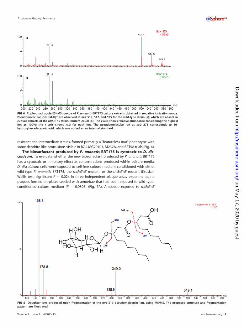
To determine if the biosurfactant was a rhamnolipid, we analyzed the extracts fromthe wild type and the rhlA::Tn5 cultures using liquid chromatography-mass spectrom-etry (LC-MS) protocols developed for rhamnolipids. Wild-type extracts had three peakswith m/z values of 519, 547, and 575, which were absent from the rhlA::Tn5 cultureextracts (Fig. 4), but none of these corresponded to any of the approximately 60different known rhamnolipid congeners (38). To investigate this unexpected result, weused tandem mass spectrometry (MS/MS) fragmentation to further analyze the m/z 519pseudomolecular ion, the most abundant molecule requiring rhlA for its biosynthesis.This biosurfactant was determined to be a monohexose-C10-C10, although the exactnature of the sugar moiety is still undetermined (Fig. 5). The two other identifiedmasses (m/z 547 and m/z 575) appear to correspond to congeners containing hydroxy-dodecanoic (C12) and hydroxytetradecanoic (C14) acid moieties as side chain fatty acids.Further investigation will be required to fully identify the exact nature of these newbiosurfactants.
FIG 1 Qualitative assay of resistance of P. ananatis isolates to D. discoideum grazing. Ten thousand,1,000, and 100 Dictyostelium discoideum AX2-214 cells were applied to dried bacterial lawns andmonitored for 7 days for plaque formation and sporulation. E. coli B/r is used as the food source forD. discoideum and was included as a reference. The buffer SorC was used for the no-amoeba control.
Smith et al.
Volume 1 Issue 1 e00075-15 msphere.asm.org 4
on May 17, 2020 by guest
http://msphere.asm
.org/D
ownloaded from

rhlA but not rhlB is essential for full swarming motility. Because rhamnolipidscan aid in swarming motility in some species (39–41), we compared the swarmingproficiency of the rhl mutants relative to that of the wild type. Wild-type P. ananatisBRT175 exhibits a characteristic dendritic pattern on 0.5% modified SM agar (Fig. 2Band C), whereas the rhlA::Tn5 mutant is deficient in swarming on 0.5% modified SM agarand 0.5% modified M9 agar (Fig. 2B and C). The rhlB::Tn5 mutant was still capable ofswarming on both medium types (Fig. 2B and C). The three other rhlB::Tn5 mutants(25E4, 24H12, and 40C2), each of which appeared to have a unique transposoninsertion site, also retained swarming activity on 0.5% modified SM agar. To determinewhether swarming motility was an indicator for grazing resistance, all isolates in theP. ananatis group were tested for swarming proficiency. Grazing-susceptible P. ananatis
TABLE 2 Locations of Tn5 transposon gene insertions in grazing-susceptible mutantsd
Grazing phenotype and locusa Gene Predicted function Swarming Mutant designation(s)
Grazing susceptibleL585_12500 ompR Transcriptional regulator Y 28H12L585_05285 cpxA Histidine kinase Y 35D8, 18A11L585_21390 phzR/luxR sdiA [Intergenic space-upstream]
transcriptional regulatorY 10E5
L585_04860 ptsN PTS sugar transporter subunit IIA Y 19E7L585_22810 nagC/nagD Transcriptional regulator (polar
mutation of nagC)Y 26A5
L585_16630 dsbB Disulfide bond formation protein Y 26H9L585_21750 serC 3-Phosphoserine/
phosphohydroxythreonineaminotransferase
Y 28G12
L585_20040 srmB RNA helicase N 36D8L585_10945 yagG [Intergenic space-upstream]
uncharacterized member of theGPH family of galactose-pentose-hexuronide transporters(symporter)
Y 31C1
L585_00065 motA Flagellar motor protein N 36E1L585_00075 flhD [Intergenic space-upstream]
transcriptional regulator andflagellar apparatus operon
N 42A7, 42H1b
L585_10950 eanI Acylhomoserine lactone synthase Y 22A12Enhanced grazing susceptible
L585_10305 Hypothetical protein (rrf2 domain,badM-like transcriptionalregulator)
Y 16A5, 16A2,b 16A1,b 16H12b
L585_04880 arcB [Intergenic space-downstream]aerobic respiration control sensorprotein
Y 24A2, 24A5c
L585_16795 prc Carboxy-terminal protease Y 14G9L585_21810 trxB Thioredoxin reductase Y 39D5L585_03130 rhlB Rhamnosyltransferase I, subunit B Y 32C5, 24H12, 25E4, 40C2L585_21385 phzI Autoinducer synthase Y 25E1, 26F4b
L585_03125 rhlA Rhamnosyltransferase I, subunit A N 28C8L585_04515 pyrB Aspartate carbamoyltransferase
catalytic subunitY 36A4
L585_03535 carB Carbamoyl phosphate synthaselarge subunit
Y 24G12
L585_19770 guaB IMP dehydrogenase Y 4H11L585_21595 pyrD Dihydroorotate dehydrogenase Y 28H8L585_04660 pnp Polynucleotide phosphorylase/
polyadenylaseN 30D8
L585_15360 prfC Peptide chain release factor 3 N 8F6L585_21170 pyrC Dihydroorotase Y 35F10
aMutants exhibiting plaque formation were scored as grazing susceptible, whereas mutants in which 4 out of 8 test wells exhibited spore formation were classified asenhanced grazing susceptible. The NCBI accession number for the draft annotated whole-genome shotgun sequence is ASJH01000000.
bMutant not tested for swarming capability.cMutant did not swarm.dAbbreviations: PTS, phosphotransferase; Y, yes; N, no.
P. ananatis Grazing Resistance
Volume 1 Issue 1 e00075-15 msphere.asm.org 5
on May 17, 2020 by guest
http://msphere.asm
.org/D
ownloaded from

Cit30-11 and LMG5342 and E. coli B/r showed no motility (Fig. 6), whereas P. ananatis17671, a grazing-susceptible isolate, had a different motility phenotype, as it appearedto move mostly linearly from the point of inoculation. P. ananatis 17671 did not formdendrites, nor did it form what is commonly referred to as a “featureless mat” (42),which is one of the motility patterns formed by some swarming bacteria. P. ananatisLMG20103, M232A, B7, 15320, BRT98, and 26SR6, which represent both grazing-
FIG 2 Grazing and swarming phenotypes of Pantoea ananatis BRT175 biosurfactant mutants. The predicted operonis shown at the top for each strain, with a red “X” indicating the disrupted gene. (A) Grazing of P. ananatis BRT175 atdifferent cell densities (250 to 8,000 cells) of D. discoideum AX2-214 after a 7-day incubation. WT, wild type. (B)Swarming after a 24-h incubation on 0.5% modified SM agar. (C) Swarming after 48 h on 0.5% modified M9 agar.
FIG 3 Emulsification activity of culture extracts of P. ananatis BRT175 strains against n-hexadecane.The wild-type (WT) extract exhibits emulsifying activity, whereas the extract from the rhlA::Tn5(mutant 28C8) culture lacks any emulsifying activity, with the height of the hydrophobic phase beingidentical to the negative control (reagent-grade water).
Smith et al.
Volume 1 Issue 1 e00075-15 msphere.asm.org 6
on May 17, 2020 by guest
http://msphere.asm
.org/D
ownloaded from
resistant and intermediate strains, formed primarily a “featureless mat” phenotype withsome dendrite-like protrusions visible in B7, LMG20103, M232A, and BRT98 trials (Fig. 6).
The biosurfactant produced by P. ananatis BRT175 is cytotoxic to D. dis-coideum. To evaluate whether the new biosurfactant produced by P. ananatis BRT175has a cytotoxic or inhibitory effect at concentrations produced within culture media,D. discoideum cells were exposed to cell-free culture medium conditioned with eitherwild-type P. ananatis BRT175, the rhlA::Tn5 mutant, or the rhlB::Tn5 mutant (Kruskal-Wallis test, significant P � 0.02). In three independent plaque assay experiments, noplaques formed on plates seeded with amoebae that had been exposed to wild-type-conditioned culture medium (P � 0.0305) (Fig. 7A). Amoebae exposed to rhlA::Tn5
FIG 4 Triple-quadrupole ESI-MS spectra of P. ananatis BRT175 culture extracts obtained in negative ionization mode.Pseudomolecular ions [M-H]� are observed at m/z 519, 547, and 575 for the wild-type strain (a), which are absent inculture extracts of the rhlA::Tn5 strain (mutant 28C8) (b). The y axis shows relative abundance considering the highestion as 100%; the x axis shows m/z for each ion. The pseudomolecular ion at m/z 271 corresponds to 16-hydroxyhexadecanoic acid, which was added as an internal standard.
FIG 5 Daughter ions produced upon fragmentation of the m/z 519 pseudomolecular ion, using MS/MS. The proposed structure and fragmentationpattern are illustrated.
P. ananatis Grazing Resistance
Volume 1 Issue 1 e00075-15 msphere.asm.org 7
on May 17, 2020 by guest
http://msphere.asm
.org/D
ownloaded from

strain-conditioned medium formed plaques similar to the number seen in the control(P � 0.2728). Interestingly, the rhlB::Tn5 strain-conditioned medium also did not allowfor plaque formation (P � 0.0305). Amoeba cell viability assays using trypan bluestaining were consistent with these results (Kruskal-Wallis test, P � 0.04), with exposureto the wild-type-conditioned culture medium leaving mostly cellular debris (P �
0.0064) (Fig. 7B). Cells exposed to the rhlB::Tn5 strain-conditioned medium were still
FIG 6 Swarming motility patterns of wild-type P. ananatis strains and E. coli B/r. All images represent themotility patterns observed after incubation for 24 h at 21°C following a single point inoculation on 0.5%modified SM agar. B7-A and B7-B represent two different swarming phenotypes of P. ananatis B7.
FIG 7 Mean plaque and cell viability counts after exposure to conditioned culture medium. Thefigure was generated in R using ggplot2 (72, 73). (A) Plaque-forming units were enumerated after25 min of room-temperature exposure to medium conditioned with either wild-type P. ananatisBRT175, the rhlA::Tn5 strain (mutant 28C8), or the rhlB::Tn5 strain (mutant 32C5). Each data pointrepresents the mean from three independent experiments with the standard error reported. Theasterisk denotes statistical significance using Dunn’s test (P � 0.0305). (B) D. discoideum AX2-214 cellswere enumerated after exposure to medium conditioned with either wild-type P. ananatis BRT175, therhlA::Tn5 strain (mutant 28C8), or the rhlB::Tn5 strain (mutant 32C5) for 20 min at room temperature. Eachdata point represents the mean from three independent experiments with the standard error reported.The asterisk denotes statistical significance using Dunn’s test (P � 0.0064).
Smith et al.
Volume 1 Issue 1 e00075-15 msphere.asm.org 8
on May 17, 2020 by guest
http://msphere.asm
.org/D
ownloaded from

largely intact, but many cells showed trypan blue uptake, indicating loss of membraneintegrity (P � 0.1065); there was also no significant difference between the rhlA::Tn5strain-conditioned medium and the control (P � 0.4549).
rhlA and rhlB are distributed among isolates of P. ananatis. A survey of thedistribution of the P. ananatis BRT175 rhlA and rhlB genes using BLAST identified theclosest homologs in several other sequenced isolates of P. ananatis, including P. anana-tis LMG20103 (43), LMG5342 (44), AJ13355, and PA13, as well as in unpublished draftgenomes of P. ananatis 15320, BRT98, 26SR6, Cit30-11, and 17671. Homologs were alsoidentified in Pantoea stewartii subsp. stewartii DC283, which is a sister species toP. ananatis (13). Independent genealogies of the two genes constructed using repre-sentative sequences from a variety of species show partial congruence of the majorclades, with monophyly of the P. ananatis-P. stewartii homologs; however, the mostrecent common ancestor of the P. ananatis rhlA gene is shared with Lonsdalea quercina,whereas the P. ananatis rhlB gene is more closely related to the Serratia marcescenshomolog (Fig. 8). In the Serratia and Dickeya lineages, the rhlA and rhlB genes are notadjacent to each other as in the P. ananatis isolates.
DISCUSSION
This work explored the genetic factors responsible for grazing resistance and/orvirulence of P. ananatis in a D. discoideum pathosystem. The qualitative grazing assayidentified four out of 10 isolates as having a resistant phenotype (limited plaqueformation), three as having intermediate phenotypes (plaque formation but no sporu-lation), and three as having a susceptible phenotype (plaque and spore formation)(Fig. 1). Sister isolates P. ananatis BRT175 and P. ananatis 17671 showed phenotypes on
FIG 8 Phylogenetic trees of RhlA and RhlB homologs. Radial (unrooted) neighbor-joining phylogenies of represen-tative RhlA (A) and RhlB (B) homologs, constructed using the Jones-Taylor-Thornton amino acid model, withcomplete gap deletion. Only bootstrap values greater than 70% are shown. In addition to the P. ananatis strains usedin this study, other representative taxa included Burkholderia cenocepacia H111 (CDN64069), Burkholderia glumaeBGR1 (ACR31008), Burkholderia pseudomallei 1106a (ABN94800), Dickeya dadantii 3937 (YP_003882762), Lonsdaleaquercina (WP_026742016), Pantoea stewartii subsp. stewartii DC283 (EHU02365), Pseudomonas aeruginosa PAO1(NP_252169), and Serratia marcescens FGI94 (AGB82844).
P. ananatis Grazing Resistance
Volume 1 Issue 1 e00075-15 msphere.asm.org 9
on May 17, 2020 by guest
http://msphere.asm
.org/D
ownloaded from

the two extremes of the grazing resistance spectrum, supporting the presence ofspecific genetic factors that contribute to this phenotype. It has been shown that thereare differences in the flexible genomic complement of P. ananatis isolates, such that thespecies appears to have an open genome (17), which facilitates the introduction ofsubstantial genetic variability and the acquisition of isolate-specific virulence traits. Inthe case of grazing resistance, the method by which such factors confer the grazing-resistant phenotype may be through one of several mechanisms. The plaque formationwith no sporulation, for example, may suggest that the bacteria interfere with theD. discoideum life cycle, as shown with Salmonella enterica subsp. Typhimurium (45).S. enterica inhibits the D. discoideum starvation response through the type III secretionsystem (45), thereby preventing sporulation. We were unable to identify any type IIIsecretion system in P. ananatis BRT175 that might function analogously (46). However,isolates within the P. ananatis group may use other secretion systems, which are knownto be responsible for producing cytotoxic factors by some bacteria (18, 47). It is alsopossible that bacteria produce antifeeding factors, such that the amoebae starve todeath. Serratia marcescens produces serrawettin W2, a surfactant that causes Caeno-rhabditis elegans to avoid feeding on the bacteria (48). Another mechanism to explainthe grazing phenotype is the direct killing of the predatory amoebae. A wide variety oftoxins and virulence factors are activated in a density-dependent manner throughquorum sensing regulation in a number of pathogenic bacteria (49–51), which maydirectly target the amoebae. Several of the 26 candidate loci implicated in grazingresistance were genes involved in biosynthetic pathways that had been shown previ-ously to be involved in virulence in humans and animal model systems (Table 2).
Of the candidate genes identified, the rhamnolipid biosynthesis genes rhlA and rhlBwere of particular interest due to their involvement in the production of rhamnolipidsand their importance in the virulence of the opportunistic human pathogens P. aerugi-nosa and Burkholderia spp. (52–54). Typically, rhlA and rhlB are in an operon (55, 56),and this may be the case for P. ananatis BRT175 based on the collinearity and closeproximity of the two coding regions in the genome sequence (46). In other systems,these genes direct the production of rhamnolipids, which act as wetting agents thatenable swarming motility but which also have cytotoxic properties (40, 53). RhlA isnecessary for the synthesis of 3-(3-hydroxyalkanoyloxy)alkanoic acids (HAA) (40). RhlB isthe rhamnosyltransferase that transfers a dTDP-L-rhamnose to HAA to form a monor-hamnolipid, while RhlC can transfer an additional rhamnose sugar to create a dirham-nolipid (40, 55, 57). Interestingly, these glycolipidic biosurfactants have been reportedonly anecdotally in cultures of Enterobacteriaceae (38). In P. ananatis BRT175, theproducts of rhlA and rhlB are responsible for the biosynthesis of a novel, hexose-basedsurfactant. The lipid moieties primarily consist of hydroxydecanoic (C10) acid, much likethe rhamnolipids produced by P. aeruginosa (58), but other congeners that likelyconsisted of hydroxydodecanoic (C12) or hydroxytetradecanoic (C14) acid were de-tected. This glycolipid also exhibits emulsification and surface tension reduction prop-erties, which are known characteristics of rhamnolipid biosurfactants (38).
The culture medium conditioned by the growth of P. ananatis BRT175 was cytotoxicbased on our observations in a plaque assay and trypan blue staining of exposed cells.The cytotoxicity of the wild-type-conditioned medium is consistent with previous workin P. aeruginosa that showed rapid lysis of D. discoideum cells (30) and polymorpho-nuclear leukocytes (53) after exposure to rhamnolipid extracts. While we did notobserve plaque formation after exposure to rhlB::Tn5 strain supernatant, trypan bluestaining revealed compromised cells, suggesting the retention of some level of cyto-toxicity likely due to the production of the fatty acid component of the biosurfactantby RhlA (Fig. 2A). In P. aeruginosa, the HAA precursor still possesses some surfactantproperties (39, 40), which also explains the capacity of the rhlB::Tn5 strain to still swarm,albeit differentially from wild type (Fig. 2B and C). Rhamnolipid-free and HAA-dependent P. aeruginosa swarming can lead to a reduced swarming zone (40) andaltered motility patterns (39). Another study examining the influence of rhamnolipids
Smith et al.
Volume 1 Issue 1 e00075-15 msphere.asm.org 10
on May 17, 2020 by guest
http://msphere.asm
.org/D
ownloaded from

and HAA on swarming motility patterns found that HAA can inhibit dendritic tendrilformation and can cause repulsion of cells away from concentrations of HAA (59).
Our analysis of the distribution of the rhlA and rhlB homologs revealed that thesetwo genes are common to all P. ananatis strains analyzed. The qualitative differences inthe grazing resistance phenotypes between P. ananatis BRT175, P. ananatis BRT98,P. ananatis LMG20103, P. ananatis LMG5342, P. ananatis Cit30-11, P. ananatis 26SR6, P.ananatis 17671, and P. ananatis 15320 are not attributable solely to the presence ofrhlA-rhlB genes, as all 8 genomes carry the genes (Fig. 8). This suggests that differencesin regulation, other unidentified genetic factors, or combinations thereof are involved inthe P. ananatis-D. discoideum interaction. Nonetheless, the persistence of these genesin the P. ananatis lineage suggests that they play an important role in the biology ofthis species. Furthermore, the presence of these genes in the sister species P. stewartiicould suggest acquisition by the P. ananatis-P. stewartii common ancestor, althoughadditional P. stewartii isolates will need to be surveyed to test this hypothesis. Theorigin of acquisition is not entirely clear, particularly given that the closest homolog ofthe P. ananatis-P. stewartii rhlA gene is that from the enteric bacterium L. quercina,whereas the P. ananatis-P. stewartii rhlB gene is closest to the homolog from a differententeric bacterium, S. marcescens (Fig. 8). Because the homologs are not adjacent in theSerratia genome, this could indicate that P. ananatis rhlA and rhlB have come fromdifferent sources. Alternatively, the Pantoea homologs may have been transferred tosome of the other enteric organisms individually, although there are insufficientrepresentative sequences from this group to determine whether this is the case.
The evolutionary arms race between microorganisms competing for the sameenvironmental niche is thought to be an evolutionary driver for opportunistic patho-gens (60–62). This predatory relationship of environmental amoebae with bacteria, forexample, may select for traits in prey that confer resistance to phagocytosis but whichhave the potential for exaptation as virulence factors that function against the cells ofthe innate immune system (30, 53, 61, 63). Such determinants may enable those sameisolates to exploit additional hosts, effectively expanding their host range to includeanimals and humans. Unraveling the key genetic factors that enable strains of Pantoeaspp. to colonize different hosts will provide important insight into the pathogeniccapabilities of this highly versatile group.
MATERIALS AND METHODSCell growth and culturing. Overnight cultures of Pantoea and Escherichia coli were grown at 30°C and37°C, respectively, in Miller LB broth (BD, Franklin Lakes, NJ) with shaking at 220 rpm under appropriateantibiotic selection and conditions (Table 1) (50 �g/ml kanamycin, 150 �g/ml ampicillin, 50 �g/mlrifampin, 38 �g/ml chloramphenicol). Dictyostelium discoideum AX2-214 cultures were incubated at 21°Cand maintained in petri dishes containing 10 ml HLF1 (Formedium, Hunstanton, United Kingdom) HL5medium with vitamins and microelements, supplemented with 13.5 g/liter glucose, 300 �g/ml strepto-mycin, and 150 �g/ml ampicillin. Cells were passaged every 4 to 6 days following a 100-fold dilution, andfresh stocks were prepared axenically from spores after 3 to 4 weeks of passaging (64). Spores wereharvested from SM/5 plates (2 g/liter glucose, 2 g/liter Bacto peptone [BD], 0.2 g/liter yeast extract [BD],0.2 g/liter MgSO4·7H2O, 1.9 g/liter KH2PO4, 1.0 g/liter K2HPO4, 1.5% agar [BD], pH 6.5) with E. coli B/r asa food source (64). Cultures for downstream assays were prepared via a 100- to 200-fold dilution ofconfluent cells in Erlenmeyer flasks containing HL5 at no more than 20% of the maximum volume. Thecells were incubated at room temperature and shaken at 180 rpm. Cells were harvested during log-phasegrowth (typically 3 � 106 to 6 � 106 cells/ml) by centrifuging 15 to 50 ml of cell culture three times at500 � g for 5 min and resuspending the pellet each time in Sorensen’s buffer with calcium (SorC)(2 g/liter KH2HPO4, 0.29 g/liter Na2HPO4, 50 �M CaCl2, pH 6.0). The final pellet was resuspended in 1 mlof SorC. Cells were enumerated in a hemocytometer and diluted to appropriate concentrations fordownstream applications.
Qualitative screen for grazing-resistant isolates. A collection of Pantoea isolates (13) was evalu-ated for grazing resistance using a modified qualitative D. discoideum assay (26). Cells from an overnightPantoea culture were centrifuged at 10,000 � g for 5 min and resuspended in an equal volume of SorCtwice. To ensure consistency of inoculations across qualitative grazing plates, bacterial cell densities werestandardized to a final optical density at 600 nm (OD600) of 1.0 in SorC by taking absorbance readingsfrom 200-�l samples using a BioTek Epoch microplate spectrophotometer (BioTek, Winooski, VT) in96-well plates (Greiner Bio-One, model 655180; Monroe, NC). Fifty microliters of each bacterial suspen-sion was spotted and allowed to dry onto 24-well plates containing 1.5 ml modified SM agar per well (65)(10 g/liter glucose, 10 g/liter Bacto peptone [BD], 1 g/liter yeast extract [BD], 1 g/liter MgSO4·7H2O, 1.9g/liter KH2PO4, 0.6 g/liter K2HPO4, 1.5% agar [BD], pH 6.5). Suspensions of D. discoideum were pipetted
P. ananatis Grazing Resistance
Volume 1 Issue 1 e00075-15 msphere.asm.org 11
on May 17, 2020 by guest
http://msphere.asm
.org/D
ownloaded from

in 5-�l aliquots onto the center of each well at 10,000, 1,000, and 100 total cells. Escherichia coli B/r, whichis used as the food source for D. discoideum, served as a control. Isolates were identified as grazingresistant if plaque formation was reduced or absent and sporulation did not occur after exposure to10,000 D. discoideum cells after 7 days of incubation at 21°C (in darkness). The experiment consisted oftwo replicates, and experiments were repeated twice with similar results.
Genetic screening and mutant characterization. Mutagenesis was carried out using a triparentalmating approach, whereby the RK600 helper plasmid was used to introduce the pBSL118 plasmid,carrying a mini-Tn5 transposon (kanamycin resistance), into P. ananatis BRT175 (rifampin resistance)(Table 1). Aliquots of each overnight culture (1 ml) were pelleted and resuspended in 100 �l LB andmixed in a 1:1:1 ratio. This triparental mixture (100 �l) was spotted on LB agar plates and incubatedovernight at 30°C. A portion of the bacterial lawn was then scraped from the plate with the edge of asterile spreader and spread on an LB plate containing rifampin and kanamycin. Following incubation for24 to 48 h at 30°C, individual transposon mutants were picked and grown overnight at 30°C with shakingat 150 rpm in 96-well plates containing 100 �l modified SM broth (recipe as described above, withoutagar, pH 6.0 to 6.4) per well. The following day, 5 �l from each well was plated on a corresponding96-well modified SM agar plate, allowed to dry, and overlaid with 2 �l of D. discoideum at 500 cells/�l.Plates were monitored for up to 7 days for wells containing susceptible mutants, which were charac-terized by enhanced plaque formation and/or sporulation. Susceptible mutants were retested in repli-cates of 8 for confirmation of the phenotype (Table 2).
The location of the transposon insertion in each mutant was determined by inverse PCR (66). Briefly,DNA was extracted from each mutant using the E.Z.N.A bacterial DNA kit (Omega Bio-Tek, Norcross, GA)per the manufacturer’s directions. About 1 to 2 �g of genomic DNA was digested for 2 h at 37°C usingHincII or PstI (New England Biolabs, Whitby, Ontario, Canada) in a 20-�l reaction volume using 20 unitsof enzyme and according to the manufacturer’s instructions. Ten microliters of heat-inactivated digestwas added to 3 �l T4 DNA ligase (New England Biolabs), 167 �l double-distilled water (ddH2O), and 20 �l10� T4 buffer to be incubated for 16 h at 16°C. Ligations were purified using the E.Z.N.A Cycle Pure kit(Omega Bio-Tek) and eluted into a 30-�l volume. Typically, 5 �l of purified ligation had sufficienttemplate to be amplified in a 25- to 50-�l PCR mixture using EconoTaq DNA polymerase (Lucigen Corp.,Middleton, WI) per the manufacturer’s instructions, using the primers NPT�772 (5=-TTCGCAGCGCATCGCCTTCTATC-3=) and NPT-41 (5=-AGCCGAATAGCCTCTCCACCCAAG-3=). Cycling conditions were one de-naturation cycle of 94°C for 120 s, followed by 40 cycles of 94°C for 30 s, 63°C for 30 s, and 72°C for 240 s,and one polymerization cycle of 72°C for 300 s. Amplification was verified by gel electrophoresis, andsingle amplicons were excised and gel purified from any reactions with multiple amplicons using theE.Z.N.A gel extraction kit (Omega Bio-Tek) per the manufacturer’s instructions. Amplicons were se-quenced by Génome Québec (Montréal, Québec, Canada). Insertion sites were confirmed by BLASTanalysis against the annotated P. ananatis BRT175 genome (46).
rhlA-rhlB mutant phenotypic characterization assays. To determine the relative differences ingrazing susceptibility caused by the interruption of rhlA and rhlB, cultures of P. ananatis BRT175, therhlA::Tn5 strain (mutant 28C8), and the rhlB::Tn5 strain (mutant 32C5) were grown overnight at 30°C inmodified SM broth. An 0.5-ml sample of each strain culture was added to 5 ml of fresh modified SM brothand incubated for another 40 min at 30°C to dilute the cultures and increase the proportion of live cells.Cell densities were standardized to an OD600 of ~0.125 from 200-�l samples using the plate spectro-photometer in 96-well plates (as described above). D. discoideum cells were diluted half-fold eight timesstarting at 1,600 cells/�l. A 12.5-�l volume of bacteria was spotted on modified SM agar and allowed todry. Five microliters of each of the dilution series of D. discoideum cells was then placed on separate spotsin order of decreasing concentration. Experiments were performed twice with two replicates each.
To create conditioned culture medium for the cytotoxicity assays, P. ananatis BRT175, P. ananatisBRT175 rhlA::Tn5 (mutant 28C8), and P. ananatis BRT175 rhlB::Tn5 (mutant 32C5) were each grown at 21°Cfor 6 days with shaking at 220 rpm in modified M9 medium (2 mM MgSO4, 20 g/liter glucose, 0.1 mMCaCl2, 1/5 dilution of 5� M9 salts [64 g/liter Na2HPO4, 15 g/liter KH2PO4, 2.5 g/liter NaCl, 13.78 g/literNaNO3]). Sodium nitrate was substituted for ammonium chloride as it has been shown to be a preferrednitrogen source in Pseudomonas aeruginosa for surfactant production (67). Filtration was performed toretain products exported by the bacteria while excluding bacterial cells. Cultures were centrifuged, thebacterial pellet was discarded, and supernatants were filter sterilized using 0.2- to 0.22-�m polyethersulfone (PES) bottle top filters. The supernatants and control media were adjusted to pH 6.52 � 0.02 andresterilized using 0.2- to 0.22-�m PES syringe filters to standardize pH values for downstream assays.Supernatants were kept at 4°C between experiments.
These supernatants were then used in PFU assays to assess D. discoideum cell viability after exposureto conditioned culture medium. Overnight cultures of E. coli B/r were centrifuged twice for 2 min at12,000 � g and resuspended in 1 ml SorC each time. The final E. coli suspension was then diluted to anOD600 of ~0.1 (200 �l, 96-well plate). The D. discoideum cells (harvested as described above) were dilutedto 15 cells/�l in SorC. Aliquots of 90 �l of modified M9 culture medium conditioned by P. ananatisBRT175, P. ananatis BRT175 rhlA::Tn5 (mutant 28C8), or P. ananatis BRT175 rhlB::Tn5 (mutant 32C5) orunconditioned culture medium as the control were prepared. Ten microliters of amoebae was thenadded to the 90 �l of medium and incubated for 25 min at 21°C. Three hundred microliters of theprepared E. coli suspension was added after incubation. The entire volume was pipetted on a 60-mmpetri dish containing SM/5 medium and dried while on a rotary shaker to help evenly distribute theliquid. The plates were incubated at 21°C, and plaque counts were taken after 3 to 4 days. Statisticalcomparisons were performed using the Kruskal-Wallis test and Dunn’s test (68) for pairwise comparisons
Smith et al.
Volume 1 Issue 1 e00075-15 msphere.asm.org 12
on May 17, 2020 by guest
http://msphere.asm
.org/D
ownloaded from

of the mean plaque counts from three independent experiments between the control and eachtreatment.
Cell viability was assessed using a trypan blue viability assay. This was performed by resuspendingpelleted D. discoideum cells in 125 �l of filtered conditioned culture medium from either P. ananatisBRT175, P. ananatis BRT175 rhlA::Tn5, or P. ananatis BRT175 rhlB::Tn5, with unconditioned modified M9medium as a control. A stock of D. discoideum cells was prepared at ~4,000 cells/�l for creating 250-�laliquots for each replicate. The cells were centrifuged for 5 min at 500 � g, and the supernatant wasdiscarded. D. discoideum cells were resuspended and exposed to the conditioned culture medium andcontrol for a minimum of 20 min at room temperature. One hundred twenty-five microliters of 0.4%trypan blue was added, and total and unstained cells were enumerated by counting 25 of the 0.040-mm2
squares on a hemocytometer. Each treatment was performed in triplicate. Most cells in the wild-type-conditioned culture medium treatment could not be enumerated as they were reduced to cellular debris.Statistical comparisons were performed using the Kruskal-Wallis test and Dunn’s test (68) for pairwisecomparisons of the mean viability counts from three independent experiments between the control andeach treatment.
Swarming trials were performed by inoculating a single colony in the center of a modified SM agarplate containing 0.5% (wt/vol) agar and incubating it at 21°C for 24 h. P. ananatis BRT175 mutants weremonitored for up to 48 h for swarming. Swarming assays were also performed on modified M9 agarplates containing 0.5% (wt/vol) agar by centrifuging cultures grown at 30°C and resuspending thebacterial pellet in the medium to an OD600 of ~0.4 (200 �l, 96-well plate). Ten microliters of concentratedculture was applied to the center of the agar plates and incubated at 21°C for 48 h. All images in thisstudy were captured using an Epson Perfection V330 photo scanner at 600 to 1,200 dots per inch (DPI).
Biosurfactant production and extraction. To characterize the structure of the biosurfactant, solventextractions were performed on culture medium conditioned with either P. ananatis BRT175 or P. ananatisBRT175 rhlA::Tn5 (mutant 28C8). Cultures were grown in 200 ml MSM supplemented with 20 g/literglucose and 2 g/liter NaNO3 in 1-liter Erlenmeyer flasks and incubated at 30°C with shaking at 250 rpm.MSM had the following composition: 2.5 g/liter KH2PO4, 1.5 g/liter K2HPO4, 0.1 g/liter CaCl2·2H2O,0.4 g/liter MgSO4·7H2O, 2 ml/liter trace element solution (TES), pH adjusted to 7. The composition of TESwas 2 g/liter FeSO4·7H2O, 1.5 g/liter MnSO4·H2O, 0.6 g/liter (NH4)6Mo7O24·4H2O, 1.4 g/liter ZnSO4·7H2O,1.2 g/liter CoCl2·6H2O, 1.2 g/liter CuSO4·5H2O, and 2 g/liter sodium citrate·2H2O.
After 6 days of incubation, the cells were pelleted by centrifugation at 8,000 � g for 15 min, and theresulting supernatant was acidified with 1 N HCl to a pH of 2 to 3. The supernatant was then extractedtwice with equal volumes of ethyl acetate. The organic fractions were then pooled and rotary evaporated,which yielded a crude extract containing the biosurfactant product. The final crude extract from 200 mlof culture was dissolved in 25 ml of MilliQ water to give an 8-fold-concentrated extract solution, whichwas subsequently used for further analysis.
Liquid chromatography-mass spectrometry analyses. The culture extracts of P. ananatis BRT175and P. ananatis BRT175 rhlA::Tn5 (mutant 28C8) were dissolved in methanol, and 16-hydroxyhexadecanoicacid was added as an internal standard. Samples were analyzed by high-performance liquid chroma-tography (HPLC; Waters 2795, Mississauga, Ontario, Canada) equipped with a 6.4- by 150-mm AgilentZorbax Eclipse XDB-C8 reverse-phase column (particle size, 5 mm) using a water-acetonitrile gradientwith a constant 2 mmol · liter�1 concentration of ammonium acetate (56). The detector was a massspectrometer (Quattro Premier XE; Waters). Analyses were performed in negative electrospray ionization(ESI�), supplemented by the multiple-reaction monitoring (MRM) mode (69).
Surface tension and emulsification assays. The surface tension of the extract solutions of P. anana-tis BRT175 and P. ananatis BRT175 rhlA::Tn5 (mutant 28C8) strains was measured by the du Noüy ringmethod using a Fisher tensiometer model 20 (Fisher Scientific, Pittsburgh, PA). The instrument wascalibrated against water, and measurements were performed in triplicate at room temperature. Theemulsifying activity of culture extracts was tested against n-hexadecane. Three-milliliter aliquots ofextract solutions of P. ananatis BRT175 and P. ananatis BRT175 rhlA::Tn5 (mutant 28C8) were mixed with2 ml of n-hexadecane and vortexed at high speed for 2 min. The emulsion was observed after letting thetubes stand at room temperature for 60 min. The lipophilic dye Sudan black was added to then-hexadecane to increase contrast.
Phylogenetic analyses. Homologs of the P. ananatis BRT175 rhlA and rhlB genes were identifiedusing BLAST against both complete and draft genome sequences available at NCBI. Alignments weremade using ClustalX2 with default parameters using iteration after each alignment step. Neighbor-joining phylogenies were constructed using MEGA6 (70) with the Jones-Taylor-Thornton amino acidmodel, complete gap deletion, and 500 bootstrap replicates.
Growth comparisons of P. ananatis BRT175 mutants. P. ananatis BRT175 and Tn5 mutants wereinoculated into 10 ml of modified SM broth in a 15-ml conical tube, which was then tightly sealed andincubated overnight at 30°C without shaking (71). Overnight cultures were standardized to an OD600 of~0.2 (300 �l, 96-well plate). Of the standardized culture, 50 �l was added to 250 �l of modified SM brothin a 96-well plate to be grown at 30°C for 18 h with continuous medium shaking, and OD600 readingswere taken every 20 min in a BioTek Synergy HT plate reader. Each experiment was performed threetimes independently with 6 replicates per strain. Nine mutants were tested per plate, with P. ananatisBRT175 being used as a reference control for each set of mutants. Only one mutant per affected genewas tested (Table 2). Figure S1 in the supplemental material was generated in R using ggplot2 (72, 73).
Nucleotide sequence accession numbers. The rhlA and rhlB genes from P. ananatis 15320, BRT98,Cit30-11, 26SR6, and 17671 have been deposited in GenBank under accession numbers KM819089,KM819090, KM819091, KM819092, KT455465, KT455466, KT455467, KT455468, KT455469, and KT455470.
P. ananatis Grazing Resistance
Volume 1 Issue 1 e00075-15 msphere.asm.org 13
on May 17, 2020 by guest
http://msphere.asm
.org/D
ownloaded from

SUPPLEMENTAL MATERIALSupplemental material for this article may be found at http://dx.doi.org/10.1128/mSphere.00075-15.
Figure S1, PDF file, 628 KB.
ACKNOWLEDGMENTSWe acknowledge the kind contributions of Steven Lindow, Teresa Coutinho, GwynBeattie, and the International Collection of Microorganisms from Plants. We thankAndrew Cameron, Tzu-Chiao Chao, Dae-Yeon Suh, Lucas Robinson, Alyssa Walterson,and Morgan Kirzinger for their valuable input and feedback on the manuscript. Wethank the dictyBase organization for being an invaluable resource and for providing theD. discoideum strain used in this work.
This work was funded by Discovery Grants from the Natural Sciences and Engineer-ing Research Council of Canada to J.S. and E.D., a Leaders Opportunity Fund (no. 28591)from the Canada Foundation for Innovation to J.S., and the University of Regina Facultyof Science. D.D.N.S. was supported by a Canadian Institutes of Health Research Master’sAward and University of Regina Faculty of Graduate Studies and Research scholarships.E.D. holds the Canada Research Chair in Sociomicrobiology.
FUNDING INFORMATIONGovernment of Canada | Natural Sciences and Engineering Research Council of Canada(NSERC) provided funding to John Stavrinides and Eric Déziel. Government of Canada| Canadian Institutes of Health Research (CIHR) provided funding to Derek D. N. Smith.Canada Foundation for Innovation (CFI) provided funding to John Stavrinides.
REFERENCES1. Bergman KA, Arends JP, Schölvinck EH. 2007. Pantoea agglomerans
septicemia in three newborn infants. Pediatr Infect Dis J 26:453– 454.http://dx.doi.org/10.1097/01.inf.0000261200.83869.92.
2. Shubov A, Jagannathan P, Chin-Hong PV. 2011. Pantoea agglomeranspneumonia in a heart-lung transplant recipient: case report and a reviewof an emerging pathogen in immunocompromised hosts. Transpl InfectDis 13:536 –539. http://dx.doi.org/10.1111/j.1399-3062.2011.00630.x.
3. De Baere T, Verhelst R, Labit C, Verschraegen G, Wauters G, ClaeysG, Vaneechoutte M. 2004. Bacteremic infection with Pantoea ananatis.J Clin Microbiol 42:4393– 4395. http://dx.doi.org/10.1128/JCM.42.9.4393-4395.2004.
4. Lalas KM, Erichsen D. 2010. Sporadic Pantoea agglomerans bacteremiain a near-term female: case report and review of literature. Jpn J InfectDis 63:290 –291.
5. Cheng A, Liu C, Tsai H, Hsu M, Yang C, Huang Y, Liao C, Hsueh P.2013. Bacteremia caused by Pantoea agglomerans at a medical center inTaiwan, 2000 –2010. J Microbiol Immunol Infect 46:187–194. http://dx.doi.org/10.1016/j.jmii.2012.05.005.
6. Cruz AT, Cazacu AC, Allen CH. 2007. Pantoea agglomerans, a plantpathogen causing human disease. J Clin Microbiol 45:1989 –1992. http://dx.doi.org/10.1128/JCM.00632-07.
7. De Champs C, Le Seaux S, Dubost JJ, Boisgard S, Sauvezie B, SirotJ. 2000. Isolation of Pantoea agglomerans in two cases of septic mono-arthritis after plant thorn and wood sliver injuries. J Clin Microbiol38:460 – 461.
8. Habsah H, Zeehaida M, Van Rostenberghe H, Noraida R, Wan PauziWI, Fatimah I, Rosliza AR, Nik Sharimah NY, Maimunah H. 2005. Anoutbreak of Pantoea spp. in a neonatal intensive care unit secondary tocontaminated parenteral nutrition. J Hosp Infect 61:213–218. http://dx.doi.org/10.1016/j.jhin.2005.01.004.
9. Coutinho TA, Venter SN. 2009. Pantoea ananatis: an unconventionalplant pathogen. Mol Plant Pathol 10:325–335. http://dx.doi.org/10.1111/j.1364-3703.2009.00542.x.
10. Walterson AM, Stavrinides J. 2015. Pantoea: insights into a highlyversatile and diverse genus within the Enterobacteriaceae. FEMS Micro-biol Rev 39:968 –984. http://dx.doi.org/10.1093/femsre/fuv027.
11. Serrano F. 1928. Bacterial fruitlet brown rot on pineapple in the Phil-ippines. Philipp J Sci 36:271–305.
12. Völksch B, Thon S, Jacobsen ID, Gube M. 2009. Polyphasic study of
plant- and clinic-associated Pantoea agglomerans strains reveals indis-tinguishable virulence potential. Infect Genet Evol 9:1381–1391. http://dx.doi.org/10.1016/j.meegid.2009.09.016.
13. Nadarasah G, Stavrinides J. 2014. Quantitative evaluation of the host-colonizing capabilities of the enteric bacterium Pantoea using plant andinsect hosts. Microbiology 160:602– 615. http://dx.doi.org/10.1099/mic.0.073452-0.
14. Kirzinger MWB, Nadarasah G, Stavrinides J. 2011. Insights into cross-kingdom plant pathogenic bacteria. Genes (Basel) 2:980 –997. http://dx.doi.org/10.3390/genes2040980.
15. Nadarasah G, Stavrinides J. 2011. Insects as alternative hosts forphytopathogenic bacteria. FEMS Microbiol Rev 35:555–575. http://dx.doi.org/10.1111/j.1574-6976.2011.00264.x.
16. Stavrinides J, No A, Ochman H. 2010. A single genetic locus in thephytopathogen Pantoea stewartii enables gut colonization and patho-genicity in an insect host. Environ Microbiol 12:147–155. http://dx.doi.org/10.1111/j.1462-2920.2009.02056.x.
17. De Maayer P, Chan WY, Rubagotti E, Venter SN, Toth IK, Birch PR,Coutinho TA. 2014. Analysis of the Pantoea ananatis pan-genomereveals factors underlying its ability to colonize and interact with plant,insect and vertebrate hosts. BMC Genomics 15:404. http://dx.doi.org/10.1186/1471-2164-15-404.
18. Shyntum D, Venter S, Moleleki L, Toth I, Coutinho T. 2014. Compar-ative genomics of type VI secretion systems in strains of Pantoea anana-tis from different environments. BMC Genomics 15:163. http://dx.doi.org/10.1186/1471-2164-15-163.
19. Kirzinger MWB, Stavrinides J. 2012. Host specificity determinants as agenetic continuum. Trends Microbiol 20:88 –93. http://dx.doi.org/10.1016/j.tim.2011.11.006.
20. Kirzinger MWB, Butz CJ, Stavrinides J. 2015. Inheritance of Pantoeatype III secretion systems through both vertical and horizontal transfer.Mol Genet Genomics 290:2075–2088. http://dx.doi.org/10.1007/s00438-015-1062-2.
21. Cosson P, Soldati T. 2008. Eat, kill or die: when amoeba meets bacteria.Curr Opin Microbiol 11:271–276. http://dx.doi.org/10.1016/j.mib.2008.05.005.
22. Hasselbring BM, Patel MK, Schell MA. 2011. Dictyostelium discoideumas a model system for identification of Burkholderia pseudomallei viru-
Smith et al.
Volume 1 Issue 1 e00075-15 msphere.asm.org 14
on May 17, 2020 by guest
http://msphere.asm
.org/D
ownloaded from

lence factors. Infect Immun 79:2079 –2088. http://dx.doi.org/10.1128/IAI.01233-10.
23. Pan Y, Lin T, Hsu C, Wang J. 2011. Use of a Dictyostelium model forisolation of genetic loci associated with phagocytosis and virulence inKlebsiella pneumoniae. Infect Immun 79:997–1006. http://dx.doi.org/10.1128/IAI.00906-10.
24. Lelong E, Marchetti A, Simon M, Burns JL, van Delden C, Köhler T,Cosson P. 2011. Evolution of Pseudomonas aeruginosa virulence ininfected patients revealed in a Dictyostelium discoideum host model. ClinMicrobiol Infect 17:1415–1420. http://dx.doi.org/10.1111/j.1469-0691.2010.03431.x.
25. Alibaud L, Köhler T, Coudray A, Prigent-Combaret C, Bergeret E,Perrin J, Benghezal M, Reimmann C, Gauthier Y, Van Delden C,Attree I, Fauvarque M, Cosson P. 2008. Pseudomonas aeruginosa vir-ulence genes identified in a Dictyostelium host model. Cell Microbiol10:729 –740. http://dx.doi.org/10.1111/j.1462-5822.2007.01080.x.
26. Froquet R, Lelong E, Marchetti A, Cosson P. 2008. Dictyosteliumdiscoideum: a model host to measure bacterial virulence. Nat Protoc4:25–30. http://dx.doi.org/10.1038/nprot.2008.212.
27. Pukatzki S, Kessin RH, Mekalanos JJ. 2002. The human pathogenPseudomonas aeruginosa utilizes conserved virulence pathways to infectthe social amoeba Dictyostelium discoideum. Proc Natl Acad Sci U S A99:3159 –3164. http://dx.doi.org/10.1073/pnas.052704399.
28. MacIntyre DL, Miyata ST, Kitaoka M, Pukatzki S. 2010. The Vibriocholerae type VI secretion system displays antimicrobial properties. ProcNatl Acad Sci U S A 107:19520 –19524. http://dx.doi.org/10.1073/pnas.1012931107.
29. Sperandio D, Decoin V, Latour X, Mijouin L, Hillion M, FeuilloleyMGJ, Orange N, Merieau A. 2012. Virulence of the Pseudomonas fluo-rescens clinical strain MFN1032 towards Dictyostelium discoideum andmacrophages in relation with type III secretion system. BMC Microbiol12:223. http://dx.doi.org/10.1186/1471-2180-12-223.
30. Cosson P, Zulianello L, Join-Lambert O, Faurisson F, Gebbie L,Benghezal M, Van Delden C, Curty LK, Köhler T. 2002. Pseudomonasaeruginosa virulence analyzed in a Dictyostelium discoideum host system.J Bacteriol 184:3027–3033. http://dx.doi.org/10.1128/JB.184.11.3027-3033.2002.
31. Totsika M, Heras B, Wurpel DJ, Schembri MA. 2009. Characterizationof two homologous disulfide bond systems involved in virulence factorbiogenesis in uropathogenic Escherichia coli CFT073. J Bacteriol 191:3901–3908. http://dx.doi.org/10.1128/JB.00143-09.
32. Qin A, Scott DW, Mann BJ. 2008. Francisella tularensis subsp. tularensisSchu S4 disulfide bond formation protein B, but not an RND-type effluxpump, is required for virulence. Infect Immun 76:3086 –3092. http://dx.doi.org/10.1128/IAI.00363-08.
33. Denoncin K, Collet J. 2013. Disulfide bond formation in the bacterialperiplasm: major achievements and challenges ahead. Antioxid RedoxSignal 19:63–71. http://dx.doi.org/10.1089/ars.2012.4864.
34. Bjur E, Eriksson-Ygberg S, Åslund F, Rhen M. 2006. Thioredoxin 1promotes intracellular replication and virulence of Salmonella entericaserovar Typhimurium. Infect Immun 74:5140 –5151. http://dx.doi.org/10.1128/IAI.00449-06.
35. Abdel-Mawgoud AM, Hausmann R, Lépine F, Müller MM, Déziel E.2011. Rhamnolipids: detection, analysis, biosynthesis, genetic regulation,and bioengineering of production, p 13–55. In Soberón-Chávez G (ed),Biosurfactants: from genes to applications. Springer, Berlin, Germany.
36. Alexeyev MF, Shokolenko IN, Croughan TP. 1995. Improvedantibiotic-resistance gene cassettes and omega elements for Escherichiacoli vector construction and in vitro deletion/insertion mutagenesis.Gene 160:63– 67. http://dx.doi.org/10.1016/0378-1119(95)00108-I.
37. Alexeyev MF, Shokolenko IN, Croughan TP. 1995. New mini-Tn5derivatives for insertion mutagenesis and genetic engineering in gram-negative bacteria. Can J Microbiol 41:1053–1055. http://dx.doi.org/10.1139/m95-147.
38. Abdel-Mawgoud AM, Lépine F, Déziel E. 2010. Rhamnolipids: diversityof structures, microbial origins and roles. Appl Microbiol Biotechnol86:1323–1336. http://dx.doi.org/10.1007/s00253-010-2498-2.
39. Caiazza NC, Shanks RMQ, O’Toole GA. 2005. Rhamnolipids modulateswarming motility patterns of Pseudomonas aeruginosa. J Bacteriol 187:7351–7361. http://dx.doi.org/10.1128/JB.187.21.7351-7361.2005.
40. Déziel E, Lépine F, Milot S, Villemur R. 2003. rhlA is required for theproduction of a novel biosurfactant promoting swarming motility inPseudomonas aeruginosa: 3-(3-hydroxyalkanoyloxy)alkanoic acids
(HAAs), the precursors of rhamnolipids. Microbiology 149:2005–2013.http://dx.doi.org/10.1099/mic.0.26154-0.
41. Nickzad A, Lépine F, Déziel E. 2015. Quorum sensing controls swarm-ing motility of Burkholderia glumae through regulation of rhamnolipids.P L o S O n e 1 0 : e 0 1 2 8 5 0 9 . h t t p : / / d x . d o i . o r g / 1 0 . 1 3 7 1 /journal.pone.0128509.
42. Kearns DB. 2010. A field guide to bacterial swarming motility. Nat RevMicrobiol 8:634 – 644. http://dx.doi.org/10.1038/nrmicro2405.
43. De Maayer P, Chan WY, Venter SN, Toth IK, Birch PRJ, Joubert F,Coutinho TA. 2010. Genome sequence of Pantoea ananatis LMG20103,the causative agent of eucalyptus blight and dieback. J Bacteriol 192:2936 –2937. http://dx.doi.org/10.1128/JB.00060-10.
44. De Maayer P, Chan WY, Rezzonico F, Bühlmann A, Venter SN, BlomJ, Goesmann A, Frey JE, Smits THM, Duffy B, Coutinho TA. 2012.Complete genome sequence of clinical isolate Pantoea ananatis LMG5342. J Bacteriol 194:1615–1616. http://dx.doi.org/10.1128/JB.06715-11.
45. Sillo A, Matthias J, Konertz R, Bozzaro S, Eichinger L. 2011. Salmo-nella typhimurium is pathogenic for Dictyostelium cells and subverts thestarvation response. Cell Microbiol 13:1793–1811. http://dx.doi.org/10.1111/j.1462-5822.2011.01662.x.
46. Smith DDN, Kirzinger MWB, Stavrinides J. 2013. Draft genome se-quence of the antibiotic-producing epiphytic isolate Pantoea ananatisBRT175. Genome Announc 1:e00902-13. http://dx.doi.org/10.1128/genomeA.00902-13.
47. Pukatzki S, Ma AT, Sturtevant D, Krastins B, Sarracino D, Nelson WC,Heidelberg JF, Mekalanos JJ. 2006. Identification of a conserved bac-terial protein secretion system in Vibrio cholerae using the Dictyosteliumhost model system. Proc Natl Acad Sci U S A 103:1528 –1533. http://dx.doi.org/10.1073/pnas.0510322103.
48. Pradel E, Zhang Y, Pujol N, Matsuyama T, Bargmann CI, Ewbank JJ.2007. Detection and avoidance of a natural product from the pathogenicbacterium Serratia marcescens by Caenorhabditis elegans. Proc Natl AcadSci U S A 104:2295–2300. http://dx.doi.org/10.1073/pnas.0610281104.
49. Antunes LCM, Ferreira RBR, Buckner MMC, Finlay BB. 2010. Quorumsensing in bacterial virulence. Microbiology 156:2271–2282. http://dx.doi.org/10.1099/mic.0.038794-0.
50. Pearson JP, Pesci EC, Iglewski BH. 1997. Roles of Pseudomonas aerugi-nosa las and rhl quorum-sensing systems in control of elastase andrhamnolipid biosynthesis genes. J Bacteriol 179:5756 –5767.
51. Deep A, Chaudhary U, Gupta V. 2011. Quorum sensing and bacterialpathogenicity: from molecules to disease. J Lab Physicians 3:4 –11.http://dx.doi.org/10.4103/0974-2727.78553.
52. Zulianello L, Canard C, Köhler T, Caille D, Lacroix J, Meda P. 2006.Rhamnolipids are virulence factors that promote early infiltration ofprimary human airway epithelia by Pseudomonas aeruginosa. InfectImmun 74:3134 –3147. http://dx.doi.org/10.1128/IAI.01772-05.
53. Jensen PO, Bjarnsholt T, Phipps R, Rasmussen TB, Calum H, Christ-offersen L, Moser C, Williams P, Pressler T, Givskov M, Høiby N. 2007.Rapid necrotic killing of polymorphonuclear leukocytes is caused byquorum-sensing-controlled production of rhamnolipid by Pseudomonasaeruginosa. Microbiology 153:1329 –1338. http://dx.doi.org/10.1099/mic.0.2006/003863-0.
54. Häussler S, Rohde M, von Neuhoff N, Nimtz M, Steinmetz I. 2003.Structural and functional cellular changes induced by Burkholderia pseu-domallei rhamnolipid. Infect Immun 71:2970 –2975. http://dx.doi.org/10.1128/IAI.71.5.2970-2975.2003.
55. Ochsner UA, Fiechter A, Reiser J. 1994. Isolation, characterization, andexpression in Escherichia coli of the Pseudomonas aeruginosa rhlAB genesencoding a rhamnosyltransferase involved in rhamnolipid biosurfactantsynthesis. J Biol Chem 269:19787–19795.
56. Dubeau D, Déziel E, Woods DE, Lépine F. 2009. Burkholderia thailan-densis harbors two identical rhl gene clusters responsible for the bio-synthesis of rhamnolipids. BMC Microbiol 9:263. http://dx.doi.org/10.1186/1471-2180-9-263.
57. Rahim R, Ochsner UA, Olvera C, Graninger M, Messner P, Lam JS,Soberón-Chávez G. 2001. Cloning and functional characterization of thePseudomonas aeruginosa rhlC gene that encodes rhamnosyltransferase 2,an enzyme responsible for di-rhamnolipid biosynthesis. Mol Microbiol 40:708–718. http://dx.doi.org/10.1046/j.1365-2958.2001.02420.x.
58. Déziel E, Lépine F, Milot S, Villemur R. 2000. Mass spectrometrymonitoring of rhamnolipids from a growing culture of Pseudomonasaeruginosa strain 57rp. Biochim Biophys Acta 1485:145–152. http://dx.doi.org/10.1016/S1388-1981(00)00039-1.
59. Tremblay J, Richardson A, Lépine F, Déziel E. 2007. Self-produced
P. ananatis Grazing Resistance
Volume 1 Issue 1 e00075-15 msphere.asm.org 15
on May 17, 2020 by guest
http://msphere.asm
.org/D
ownloaded from

extracellular stimuli modulate the Pseudomonas aeruginosa swarmingmotility behaviour. Environ Microbiol 9:2622–2630. http://dx.doi.org/10.1111/j.1462-2920.2007.01396.x.
60. Brüssow H. 2007. Bacteria between protists and phages: from antip-redation strategies to the evolution of pathogenicity. Mol Microbiol65:583–589. http://dx.doi.org/10.1111/j.1365-2958.2007.05826.x.
61. Matz C, Kjelleberg S. 2005. Off the hook— how bacteria survive pro-tozoan grazing. Trends Microbiol 13:302–307. http://dx.doi.org/10.1016/j.tim.2005.05.009.
62. Brown SP, Cornforth DM, Mideo N. 2012. Evolution of virulence inopportunistic pathogens: generalism, plasticity, and control. Trends Mi-crobiol 20:336 –342. http://dx.doi.org/10.1016/j.tim.2012.04.005.
63. Adiba S, Nizak C, van Baalen M, Denamur E, Depaulis F. 2010. Fromgrazing resistance to pathogenesis: the coincidental evolution of viru-lence factors. PLoS One 5:e11882. http://dx.doi.org/10.1371/journal.pone.0011882.
64. Fey P, Kowal AS, Gaudet P, Pilcher KE, Chisholm RL. 2007. Protocolsfor growth and development of Dictyostelium discoideum. Nat Protoc2:1307–1316. http://dx.doi.org/10.1038/nprot.2007.178.
65. Sussman M. 1966. Biochemical and genetic methods in the study ofcellular slime mold development, p 397– 410. In Prescott D (ed), Meth-ods in cell physiology, vol 2. Academic Press, New York, NY.
66. Ochman H, Gerber AS, Hartl DL. 1988. Genetic applications of aninverse polymerase chain reaction. Genetics 120:621– 623.
67. Guerra-Santos L, Kappeli O, Fiechter A. 1984. Pseudomonas aerugi-nosa biosurfactant production in continuous culture with glucose ascarbon source. Appl Environ Microbiol 48:301–305.
68. Dinno A. 2015. Dunn.Test: Dunn’s test of multiple comparisons usingrank sums. R package version 1.2.4. R Foundation for Statistical Com-puting, Vienna, Austria.
69. Abdel-Mawgoud AM, Lépine F, Déziel E. 2014. Liquid chromatography/mass spectrometry for the identification and quantification of rhamnolipids.Methods Mol Biol 1149:359 –373. http://dx.doi.org/10.1007/978-1-4939-0473-0_30.
70. Tamura K, Stecher G, Peterson D, Filipski A, Kumar S. 2013. MEGA6:Molecular Evolutionary Genetics Analysis version 6.0. Mol Biol Evol30:2725–2729. http://dx.doi.org/10.1093/molbev/mst197.
71. Hall BG, Acar H, Nandipati A, Barlow M. 2014. Growth rates madeeasy. Mol Biol Evol 31:232–238. http://dx.doi.org/10.1093/molbev/mst187.
72. R Core Team. 2014. R: a language and environment for statisticalcomputing. R Foundation for Statistical Computing, Vienna, Austria.
73. Wickham H. 2009. ggplot2: elegant graphics for data analysis. Springer,New York, NY.
74. Kessler B, De Lorenzo V, Timmis KN. 1992. A general system tointegrate lacZ fusions into the chromosomes of gram-negativeeubacteria: regulation of the Pm promoter of the TOL plasmid studiedwith all controlling elements in monocopy. Mol Gen Genet 233:293–301.http://dx.doi.org/10.1007/BF00587591.
75. Fey P, Dodson RJ, Basu S, Chisholm RL. 2013. One stop shop foreverything Dictyostelium: DictyBase and the Dicty Stock Center in 2012.Methods Mol Biol 983:59 –92. http://dx.doi.org/10.1007/978-1-62703-302-2_4.
Smith et al.
Volume 1 Issue 1 e00075-15 msphere.asm.org 16
on May 17, 2020 by guest
http://msphere.asm
.org/D
ownloaded from