Embed Size (px)
Citation preview

1
A novel HSF4 gene mutation causes autosomal dominant cataracts in a Chinese family
Huibin Lv*1, Chen Huang*2, Jing Zhang1, Ziyuan Liu1, Zhike Zhang1, Haining Xu3, Yuchen
You1, Jinping Hu1, Xuemin Li1 & Wei Wang1
1 Department of Ophthalmology, Beijing University Third Hospital, Beijing, China
2 Medical Research Center, Beijing University Third Hospital, Beijing, China
3 Department of Ophthalmology, WeiHaiWei People`s Hospital, Shandong China
*Huibin Lv and Chen Huang contributed equally to this work.
Corresponding to: Professor Xuemin Li, Department of Ophthalmology, Beijing University
Third Hospital, Hua Yuan Bei Lu, Beijing 100191, China. Phone: +8610-82266573; email:
G3: Genes|Genomes|Genetics Early Online, published on March 17, 2014 as doi:10.1534/g3.113.009860
© The Author(s) 2013. Published by the Genetics Society of America.

2
Abstract:
Congenital cataracts are a significant cause of visual impairment or blindness in
children. One third of cases estimated to have a genetic cause. We carried out gene
analysis and bioinformatics analysis to map the locus and to identify the underlying genetic
defect in a twelve-member, four-generation Chinese family affected with bilateral congenital
cataracts. We screened individuals of the family and discovered a distinct missense
mutation in HSF4 (a gene at this locus that encodes teat-shock transcription factor 4).
Bioinformatics analysis was used to determine possible changes in the protein structure that
could affect the phenotype. Sequencing of the candidate genes showed a heterozygous
c.69 GT change in the heat shock transcription factor 4 (HSF4) gene, which resulted in the
substitution of a lysine with an asparagine (p. K23N). This mutation co-segregated with all
affected individuals and was not observed in unaffected family members. Bioinformatics
analysis indicated that the p. K23N mutation was predicted to be disease causing. This is
the first report of the novel missense mutation, c.69 GT (p. K23N), in exon 3 of the HSF4
locus on 16q21-q22 associated with bilateral congenital cataracts in a Chinese family. This
novel mutation could enable propergenetic diagnostics and counseling in affected families
and could lead to a better understanding of the structure and function of HSF4 in health and

3
disease.
Key words: congenital cataract family; HSF4
Congenital cataracts are a significant cause of visual impairment or blindness in
children. The prevalence of congenital cataracts is 1 to 6 per 10,000 live births, depending
on the method of ascertainment [1]. Globally, congenital cataracts account for nearly one
tenth of childhood blindness [2]. Statistical analyses have revealed that congenital cataracts
account for more than 1 million blind children in Asia. Approximately 50% of all congenital
cataract cases may have a genetic cause [3-5]. Genetically, the majority of isolated
congenital cataracts exhibit as autosomal dominant, although autosomal recessive and
X-linked inherited forms have also been reported [6].
Over the past few years, remarkable progress has been made towards our
understanding of the cataractogenesis process. More and more genes related to congenital
cataracts have been mapped. So far, more than 40 loci have been mapped in congenital
cataracts [7-9] and more than 26 genes have been characterized. Meanwhile, the number of
associated genes is constantly increasing [10]. Approximately half of the mutations are in the

4
crystallin genes and a quarter are in the connexin genes. The remaining mutations are found
in genes that encode heat shock transcription factor4 (HSF4), aquaporin-0 (AQP0, MIP),
paired-like homeodomain 3 (PITX3), chromatin modifying protein (CHMP4B), lens intrinsic
membrane protein 2 (LIM2), beaded filament structural protein-2 (BFSP2), and other
proteins [2, 11].
According to their morphology, the cataracts can be classified into several subtypes:
whole lens, nuclear, lamellar, cortical, polar, sutural, pulverulent, cerulean, coralliform, and
other minor subtypes [12]. It is known that different mutations in different genes could result
in similar cataract patterns, while the highly variable cataract morphologies within some
families suggest that the same mutation in a single gene can lead to different phenotypes
[13-14].
In this study, a four-generation family affected with congenital polymorphic cataracts
was investigated in an attempt to identify the genetic defect associated with their cataract
phenotype. We applied a functional candidate approach to test the known characterized
genes in this family. A novel missense mutation c.69 GT (p. K23N) in HSF4 was detected.
METHODS

5
Clinical examination and isolation of genomic DNA
The proband, a seven-year-old child, was diagnosed with bilateral cataracts at the
Beijing University Third Hospital. A family history revealed twelve members in four
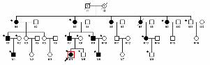
generations. Sixteen people in this big family (II:1, II:3, II:9, II:11, III:1, III:2, III:7-10, III:13,
IV:1, IV:4-6, IV:8) were willing to take part in the study (10 affected and 6 unaffected, Figure
1). The ethics committee of Beijing University approved the research and all participants
from the family provided their informed consent. The study protocol followed the principles of
the Declaration of Helsinki. All participants were determined by a medical history or
ophthalmologic examination, which included visual acuity, slit-lamp examination,
ultrasonography, intraocular pressure measurement, and fundus examination with dilated
pupils. Meanwhile, 100 unrelated ethnically-matched controls with no family history of
congenital cataracts were recruited.Five mL of venous blood was collected from
participating family members and controls in BD Vacutainers (BD, San Jose, CA) containing
EDTA. Genomic DNA was extracted using QIAamp DNA Blood Mini Kits (Qiagen Science,
Germantown, MD).
Mutation analysis
Twenty-one genes, including BFSP2, CRYAA, CRYAB, CRYBA1, CRYBB1, CRYBB2,

6
CRYGC, CRYGD, CRYGS, EPHA2, GJA3, GJA8, HSF4, LIM2, MAF, MIP, PITX3, VIM,
AGK, CHMP4B, and GALK1, were considered as candidate genes for hereditary cataracts
[2, 7, 15]. The coding regions of the candidate genes were amplified by polymerase chain
reaction (PCR) with previously published primer sequences [16-23] (Appendix 1) and
screened for mutations on both strands using bidirectional sequencing. Direct sequencing of
the100 ethnically-matched controls was used to screen any identified mutations in the genes
to confirm the mutations. PCR products were pooled, mixed with loading dye containing
internal size standards, denatured at 95°C for 5 minutes, and electrophoresed on 4%
denaturing polyacrylamide gels on a DNA sequencer (ABI-Prism 377; ABI, Foster City, CA).
The sequencing results were analyzed using Chromas 2.33 and compared to the reference
sequences in the NCBI database.
Bioinformatics analysis
The multiple-sequence alignment of the amino acid sequence in the mutated gene from
several different species was analyzed by the CLC Free Workbench 6.0 software (CLC bio,
Aarhus, Denmark). The three-dimensional (3D) structures of both wild-type and mutant
proteins were predicted andanalyzed by the online SWISS-MODEL tool
(http://swissmodel.expasy.org/) [24-26]. The possible impact of an amino acid substitution

7
on the structure and function of the protein was predicted by PolyPhen-2
(http://genetics.bwh.harvard.edu/pph2/) [27] and Mutation Taster
(http://www.mutationtaster.org) [28].
RESULTS
Clinical findings
We have identified a four-generation Chinese family (16 members) in which ten family
members have been diagnosed with bilateral autosomal dominant cortical cataracts (Figure
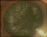
1). The proband (IV:4) was a 7-year-old girl who was diagnosed with bilateral congenital
cataracts. The rod-like opacities were primarily located in the lens cortex, and colorful dot
opacities were seen in nucleus (Figure 2).
According to the medical records, except for the proband IV:4, 8 affected individuals
(II:1, II:9, III:1, III:7, III:9, III:13, IV:1, IV:6) had a cataract extraction performed between the
ages of 7 and 25 years. The other affected patient had similar bilateral lens opacifications as
well as some age-related lens nucleus opacities. The clinical evaluation of the affected
individuals is provided in Table 1. Prior to surgery, the affected members had visual acuity
ranging from 0.05 to 0.6. After surgery, all patients achieved a best-corrected visual acuity of

8
0.6 to 1.0. There were no other ocular or other related systemic abnormalities in this family.
Mutation analysis
The 21 candidate genes were analyzed by sequence analysis of the coding regions.
Bidirectional sequence analysis of the HSF4 gene indicated a novel heterozygous c.69GT
variation in all ten affected individuals of the family (Figure 3). This heterozygous mutation
was not present in the unaffected family members or in 100 controls without congenital
cataracts. This c.69GT nucleotide alteration resulted in the substitution of a lysine with
anasparagine (p. K23N). We did not find any other mutations in this family, except for a few
non-pathogenic single nucleotide polymorphisms (SNPs) (Table 2).
Bioinformatics analysis
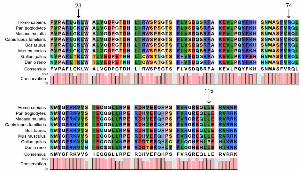
The p. K23N (c.69 G->T) mutation in the HSF4 gene detected in our present study
was located within the highly conserved HSF DNA binding region which is shared across
heat shock factors and between species, as shown by multiple-sequence alignment (Figure
4). The modeled residue range extended from amino acids13 to 124. As shown in Figure 5,
the predicted 3D structural model of the p.K23N mutated HSF4 protein was different from
that of the wild-type protein. Additionally, the p.K23N mutation was predicted to be “probably
damaging” by PolyPhen-2 analysis with a score of 1.000 and was predicted to be “disease

9
causing” by Mutation Taster analysis.
DISCUSSION
In the present study in an autosomal dominant congenital cataract (ADCC) family with
ten affected members in four generations, 21 known candidate genes were sequenced by
direct sequencing using polymerase chain reaction (PCR), but 20 of them were excluded as
pathogenic. The coding regions of these candidate genes were sequenced bidirectionally.
We observed anovelc.69 GT variation in the HSF4 gene in the individuals affected with
bilateral congenital cataracts. The heterozygous mutation resulted in the substitution of a
lysine with anasparagine (p. K23N). This mutation likely caused the cataracts since it
segregated with the phenotype and was not detected in either the unaffected family
members or the 100 ethnically matched controls.
HSF4 belongs to the family of heat shock transcription factors (HSFs) that regulate the
expression of heat shock proteins (HSPs) and mediate the inducible transcription response.
HSF4 regulates the expression of HSPs in response to different cellular stresses, such as
oxidants, heavy metals, elevated temperature, and bacterial or viral infections. HSPs play an
important role in the maintenance of the supramolecular organization of the lens protein [29],

10
which is essential for lens transparency. In human beings, HSF4 is widely expressed in the
body, especially in the heart, brain, skeletal muscle, lung, and pancreas [30]. The human
HSF4 gene has at least two alternatively spliced transcripts, HSF4a and HSF4b. Both these
forms of HSF4 protein have the same DNA-binding domain, which is important for the
function of HSF4.
Recently, the HSF4 gene has been reported to be responsible for both autosomal
dominant and autosomal recessive cataracts [31-33]. Congenital cataracts can partly be
distinguished by the location and severity of the mutations. So far, at least seven different
mutations in the HSF4 gene have been detected [32, 34-36]. Two of them can lead to
autosomal dominant cataracts, another three can cause autosomal recessive cataracts, and
the remaining two mutations were found in sporadic cases. So far, all known dominant
mutations in HSF4 ( p.R74H, p.L115P) are located in the α-helical DNA binding region,
which further highlights the importance of this domain [31-32]. In this study, the missense
mutation ( p.K23N ) also lies within this highly conserved functional domain. However, the
recessive mutations lie outside this highly conserved functional domain. Previously, Smaoui
et al reported a splice mutation in HSF4 associated with an autosomal recessive total
cataract in a Tunisian family [33].

11
Bioinformatics analyses were performed to elucidate a correlation between structural
disturbances and putative functional commitment, achieving a possible explanation for the
pathogenic mechanism of the novel p.K23N missense mutation of HSF4. The p.K23N
mutation is located within a highly conserved region across species, which suggests an
important role in the function and/or structure of HSF4. To evaluate the three-dimensional
impact of the mutation, we created a homology model to compute and compare mutant and
wild-type structures. The spatial structures of both wild-type and mutant proteins were
modeled by the online SWISS-MODEL tool (http://swissmodel.expasy.org/), base on temple:
2lduA (99.9 A). The 3D structural models also indicated the different structure between the
wild-type and p.K23N mutant proteins. Moreover, the p.K23N mutation was predicted to be
“probably damaging” by PolyPhen-2 analysis with a score of 1.000 and was predicted to be
“disease causing” by Mutation Taster analysis. Hence, the predicted change of the protein
structure could disrupt lens biochemistry and physiology early in development. The possible
mechanism of this mutation will require further investigation.
However, in this family, most patients, except the proband and II:2, had cataract
extraction performed between the ages of 7 and 25 years. The details of the phenotypes of
the other individuals could not be acquired. It is not certain whether the lens opacities of all

12
affected family members were similar. It is also unknown whether the opacity of the lenses
worsened with age. In spite of this, this novel mutation in the HSF4 gene could provide some
clues to the mechanism of developing congenital cataracts.
Moreover, Bagchi et al. [29] revealed that certain sequence changes in HSF4 resulted
in abnormal expression of HSPs and thereby influenced the function or level of HSPs. The
decrease of HSPs could be responsible for the loss of optimal protein organization and the
eventual appearance of age-related cataracts [37]. In addition, regarding the clinical
phenotype caused by mutations in this transcription factor, the reason that mutations in
HSF4 that are expressed in other tissues, including the heart, muscle, lung, and brain, cause
only nonsyndromic cataracts [33] is still unknown. More comprehensive studies will be
needed to answer this question.
In summary, we have shown a novel missense mutation in HSF4 that mapped to
16q21-22 and caused autosomal dominant cataracts in a large Chinese family. Sequencing
of the candidate genes showed a heterozygous c.69 GT variation in the HSF4 gene,
which resulted in the substitution of a lysine with anasparagine (p. K23N). This novel
mutation could enable proper genetic diagnostics and counseling in affected families and
could lead to a better understanding of the structure and function of HSF4 in health and

13
disease.
ACKNOWLEDGMENTS
The authors thank the family and controls for participating in this study. This research
was supported by the LinHu grand from Beijing University Third Hospital.
REFERENCES
1. Holmes, JM, Leske DA, Burke JP, Hodge DO. Birth prevalence of visually significant
infantile cataract in a defined U.S. population. Ophthalmic Epidemiol. 2003
Apr;10(2):67-74.
2. Reddy, MA, Francis PJ, Berry V, Bhattacharya SS, Moore AT. Molecular genetic basis
of inherited cataract and associated phenotypes. Surv Ophthalmol. 2004
May-Jun;49(3):300-15.
3. Rahi, JS, Dezateux C. Congenital and infantile cataract in the United Kingdom:
underlying or associated factors. British Congenital Cataract Interest Group. Invest
Ophthalmol Vis Sci. 2000 Jul;41(8):2108-14.
14
4. Francis, PJ, Berry V, Bhattacharya SS, Moore AT. The genetics of childhood cataract. J
Med Genet. 2000 Jul;37(7):481-8.
5. Santana, A, Waiswo M. The genetic and molecular basis of congenital cataract. Arq
Bras Oftalmol. 2011 Mar-Apr;74(2):136-42.
6. Vanita, Singh JR, Singh D. Genetic and segregation analysis of congenital cataract in
the Indian population. Clin Genet. 1999 Nov;56(5):389-93.
7. Hejtmancik, JF. Congenital cataracts and their molecular genetics. Semin Cell Dev Biol.
2008 Apr;19(2):134-49.
8. Zhao, R, Yang Y, He X, Liu Z, Wang P, Zhou L, Tang J, Xu W, Li L, Zhu Y. An autosomal
dominant cataract locus mapped to 19q13-qter in a Chinese family. Mol Vis.
2011;17:265-9.
9. Ouyang, S, Gao L, Zhang L, Zheng Y, Cao W, Feng G, He L, Liu P. A new locus in
chromosome 2q37-qter is associated with posterior polar cataract. Graefes Arch Clin
Exp Ophthalmol. 2012 Jun;250(6):907-13.
10. Shiels, A, Hejtmancik JF. Genetic origins of cataract. Arch Ophthalmol. 2007
Feb;125(2):165-73.
11. Devi, RR, Yao W, Vijayalakshmi P, Sergeev YV, Sundaresan P, Hejtmancik JF.
15
Crystallin gene mutations in Indian families with inherited pediatric cataract. Mol Vis.
2008;14:1157-70.
12. Reddy, MA, Bateman OA, Chakarova C, Ferris J, Berry V, Lomas E, Sarra R, Smith MA,
Moore AT, Bhattacharya SS, Slingsby C. Characterization of the G91del
CRYBA1/3-crystallin protein: a cause of human inherited cataract. Hum Mol Genet.
2004 May 1;13(9):945-53.
13. Gill, D, Klose R, Munier FL, McFadden M, Priston M, Billingsley G, Ducrey N,
Schorderet DF, Heon E. Genetic heterogeneity of the Coppock-like cataract: a mutation
in CRYBB2 on chromosome 22q11.2. Invest Ophthalmol Vis Sci. 2000
Jan;41(1):159-65.
14. Heon, E, Priston M, Schorderet DF, Billingsley GD, Girard PO, Lubsen N, Munier FL.
The gamma-crystallins and human cataracts: a puzzle made clearer. Am J Hum Genet.
1999 Nov;65(5):1261-7.
15. Wang, KJ, Li SS, Yun B, Ma WX, Jiang TG, Zhu SQ. A novel mutation in MIP associated
with congenital nuclear cataract in a Chinese family. Mol Vis. 2011;17:70-7.
16. Vanita, V, Singh JR, Hejtmancik JF, Nuernberg P, Hennies HC, Singh D, Sperling K. A
novel fan-shaped cataract-microcornea syndrome caused by a mutation of CRYAA in an

16
Indian family. Mol Vis. 2006;12:518-22.
17. Lu, S, Zhao C, Jiao H, Kere J, Tang X, Zhao F, Zhang X, Zhao K, Larsson C. Two
Chinese families with pulverulent congenital cataracts and deltaG91 CRYBA1 mutations.
Mol Vis. 2007;13:1154-60.
18. Litt, M, Carrero-Valenzuela R, LaMorticella DM, Schultz DW, Mitchell TN, Kramer P,
Maumenee IH. Autosomal dominant cerulean cataract is associated with a chain
termination mutation in the human beta-crystallin gene CRYBB2. Hum Mol Genet. 1997
May;6(5):665-8.
19. Zhang, LY, Yam GH, Fan DS, Tam PO, Lam DS, Pang CP. A novel deletion variant of
gammaD-crystallin responsible for congenital nuclear cataract. Mol Vis.
2007;13:2096-104.
20. Hansen, L, Yao W, Eiberg H, Funding M, Riise R, Kjaer KW, Hejtmancik JF, Rosenberg
T. The congenital "ant-egg" cataract phenotype is caused by a missense mutation in
connexin46. Mol Vis. 2006;12:1033-9.
21. Schmidt, W, Klopp N, Illig T, Graw J. A novel GJA8 mutation causing a recessive
triangular cataract. Mol Vis. 2008;14:851-6.
22. Bremond-Gignac, D, Bitoun P, Reis LM, Copin H, Murray JC, Semina EV. Identification

17
of dominant FOXE3 and PAX6 mutations in patients with congenital cataract and
aniridia. Mol Vis. 2010;16:1705-11.
23. Shiels, A, Bennett TM, Knopf HL, Yamada K, Yoshiura K, Niikawa N, Shim S, Hanson PI.
CHMP4B, a novel gene for autosomal dominant cataracts linked to chromosome 20q.
Am J Hum Genet. 2007 Sep;81(3):596-606.
24. Guex, N, Peitsch MC. SWISS-MODEL and the Swiss-PdbViewer: an environment for
comparative protein modeling. Electrophoresis. 1997 Dec;18(15):2714-23.
25. Schwede, T, Kopp J, Guex N, Peitsch MC. SWISS-MODEL: An automated protein
homology-modeling server. Nucleic Acids Res. 2003 Jul 1;31(13):3381-5.
26. Arnold, K, Bordoli L, Kopp J, Schwede T. The SWISS-MODEL workspace: a web-based
environment for protein structure homology modelling. Bioinformatics. 2006 Jan
15;22(2):195-201.
27. Adzhubei, IA, Schmidt S, Peshkin L, Ramensky VE, Gerasimova A, Bork P, Kondrashov
AS, Sunyaev SR. A method and server for predicting damaging missense mutations.
Nat Methods. 2010 Apr;7(4):248-9.
28. Schwarz, JM, Rodelsperger C, Schuelke M, Seelow D. MutationTaster evaluates
disease-causing potential of sequence alterations. Nat Methods. 2010 Aug;7(8):575-6.

18
29. Bagchi, M, Katar M, Maisel H. Heat shock proteins of adult and embryonic human ocular
lenses. J Cell Biochem. 2002;84(2):278-84.
30. Nakai, A, Tanabe M, Kawazoe Y, Inazawa J, Morimoto RI, Nagata K. HSF4, a new
member of the human heat shock factor family which lacks properties of a transcriptional
activator. Mol Cell Biol. 1997 Jan;17(1):469-81.
31. Bu, L, Jin Y, Shi Y, Chu R, Ban A, Eiberg H, Andres L, Jiang H, Zheng G, Qian M, Cui B,
Xia Y, Liu J, Hu L, Zhao G, Hayden MR, Kong X. Mutant DNA-binding domain of HSF4
is associated with autosomal dominant lamellar and Marner cataract. Nat Genet. 2002
Jul;31(3):276-8.
32. Ke, T, Wang QK, Ji B, Wang X, Liu P, Zhang X, Tang Z, Ren X, Liu M. Novel HSF4
mutation causes congenital total white cataract in a Chinese family. Am J Ophthalmol.
2006 Aug;142(2):298-303.
33. Smaoui, N, Beltaief O, BenHamed S, M'Rad R, Maazoul F, Ouertani A, Chaabouni H,
Hejtmancik JF. A homozygous splice mutation in the HSF4 gene is associated with an
autosomal recessive congenital cataract. Invest Ophthalmol Vis Sci. 2004
Aug;45(8):2716-21.
34. Forshew, T, Johnson CA, Khaliq S, Pasha S, Willis C, Abbasi R, Tee L, Smith U,

19
Trembath RC, Mehdi SQ, Moore AT, Maher ER. Locus heterogeneity in autosomal
recessive congenital cataracts: linkage to 9q and germline HSF4 mutations. Hum Genet.
2005 Sep;117(5):452-9.
35. Talamas, E, Jackson L, Koeberl M, Jackson T, McElwee JL, Hawes NL, Chang B,
Jablonski MM, Sidjanin DJ. Early transposable element insertion in intron 9 of the Hsf4
gene results in autosomal recessive cataracts in lop11 and ldis1 mice. Genomics. 2006
Jul;88(1):44-51.
36. Mellersh, CS, Pettitt L, Forman OP, Vaudin M, Barnett KC. Identification of mutations in
HSF4 in dogs of three different breeds with hereditary cataracts. Vet Ophthalmol. 2006
Sep-Oct;9(5):369-78.
37. Shi, Y, Shi X, Jin Y, Miao A, Bu L, He J, Jiang H, Lu Y, Kong X, Hu L. Mutation screening
of HSF4 in 150 age-related cataract patients. Mol Vis. 2008;14:1850-5.
FIGURE CAPTIONS:
Figure 1. Pedigree of a cataract family. Pedigree of a four-generation family with congenital
cataract. The proband is marked with an arrow. Squares and circles indicate males
20
and females, respectively. Black and white symbols represent affected and
unaffected individuals, respectively. The asterisks indicate family members who
attend this study.
Figure 2. Slit-lamp photograph of the proband. The photograph of the proband (IV:4) shows
rod-like opacities located in the lens cortex and colorful dots opacities in nucleus.
Figure 3. DNA sequence chromatograms of an affected and an unaffected individuals in the
ADCC Chinese family (Forward strand; individual III:1 and III:2, respectively). A single
transversion is observed at position 69(G->T) as a G/T double peak (indicated by an
black arrow).
Figure 4. A multiple-sequence alignment in HSF4 (16-120) from different species. The
alignment data indicates that the Phe at position 23 is highly conserved in different
species (indicated by an arrow). Both amino acids at positions 74 and 115, known as
causes of autosomal dominant cataracts, are also highly conserved in different
species (indicated by an arrow)
Figure 5. The three-dimensional structural models of the wild-type (A), the novel p.K23N
mutant (B) , the known p.R74H mutant (C) and p.L115P (D) HSF4 proteins. The
modelled residue range is from amino acids 13 to 124 for all proteins. The novel

21
p.K23N mutant (B) HSF4 protein and known p.L115P mutant (D) HSF4 protein
represent observed different modelled structures with the wide-type one (indicated by
an arrow).
Table 1. Clinical Features Of Affected Individuals
Affected Gender Age Surgery Phenotype
II1 Female 65 25 aphakia eye, after cataract surgery
II3 Female 60 - Rod-like cortical cataract with nuclear
II9 Female 51 20 aphakia eye, after cataract surgery
III1 Male 46 25 IOL, after cataract surgery
III7 Male 36 13 IOL, after cataract surgery
III9 Male 34 13 IOL, after cataract surgery
III13 Male 34 12 IOL, after cataract surgery
IV1 Male 17 11 IOL, after cataract surgery
IV4 Female 7 7 Rod-like cortical cataract with colorful dots opacities in nucleus
IV6 Male 11 8 IOL, after cataract surgery

22
Table 1. Clinical features of affected individuals. Besides the proband IV:4, 8 affected
individuals had a cataract extraction performed between the ages of 7 and 25 years. (IOL,
Intraocular Lens)
Table 2. All of single nucleotide polymorphisms (SNPS) have been found in all family
members.
Family
members Cataract
GJA3
p.L299M
CRYBB2
p R61T
CRYBB2
p.Q147R
CRYBB2
p.T150M
HSF4
p.K23N
II1 Yes + + + + +
II3 Yes + + + + +
II9 Yes + + + + +
II11 No + - - - -
III1 Yes + + + + +
III2 No + - - - -
III7 Yes + + + + +
III8 No + - - - -

23
III9 Yes + - - - +
III10 No + - - - -
III13 Yes + + + + +
IV1 Yes + + + + +
IV4 Yes + + + + +
IV5 No + + + + -
IV6 Yes + + + + +
IV8 No + + + + -
Table 2. SNPS in all family members. All of single nucleotide polymorphisms (SNPS) have
been found in this family are shown in table 2. While p.K23N is the only mutation which
co-segregated with all affected individuals and was not observed in unaffected family
members.