Embed Size (px)
Citation preview
© The Author 2014. Published by Oxford University Press on behalf of the UK Environmental Mutagen Society. All rights reserved. For permissions, please e-mail: [email protected].
doi:10.1093/mutage/geu019
A novel non-stop mutation in MSX1 causing autosomal dominant non-syndromic oligodontia
Sing-Wai Wong1,#, Hao-Chen Liu1,#, Dong Han1, Huai-Guang Chang1, Hong-Shan Zhao2,3, Yi-Xiang Wang4,* and Hai-Lan Feng1
1Department of Prosthodontics, School and Hospital of Stomatology, Peking University, Beijing 100081, China, 2Department of Medical Genetics and 3Peking University Center for Human Disease Genomics, Peking University Health Science Center, Beijing 100191, China and 4Central Laboratory, School and Hospital of Stomatology, Peking University, Beijing 100081, China.
#Authors contributing equally to this work.
*To whom correspondence should be addressed. Tel: +86 108 219 5537, Fax: +86 106 217 3402; Email: [email protected]
Received on January 22, 2014; revised on April 13, 2014; accepted on April 15, 2014
Oligodontia, which is the congenital absence of six or more permanent teeth, excluding the third molars, may contrib-ute to masticatory dysfunction, speech alteration, aesthetic problems and malocclusion. Msh homeobox 1 (MSX1) was the first gene identified as causing non-syndromic oligo-dontia. In this study, we identified a novel heterozygous non-stop mutation (c.910_911dupTA, p.*304Tyrext*48) in MSX1 in a Chinese family with autosomal dominant non-syndromic oligodontia. This novel mutation substitutes the stop codon with a tyrosine residue, potentially adding 48 amino acids to the C-terminus of MSX1. Further in vitro study found that mutant MSX1 could be expressed but had lost its ability to enter the nucleus. This is the first report indicating that a non-stop mutation in MSX1 is responsible for oligodontia. This study broadens the mutation spectrum for MSX1 and provides a new way to clarify the mechanism of MSX1 in tooth agenesis.
Introduction
Tooth agenesis is the most common dental developmental anomaly in humans, and it may contribute to masticatory dysfunction, speech alteration, aesthetic problems and malocclusion (1). The prevalence of dental agenesis of permanent teeth ranges from 2.2% to 10.1% in the general population, excluding the third molars (2). Tooth agenesis is classified into three categories: hypodontia, the absence of one to five permanent teeth, excluding the third molars; oligodontia, the absence of more than five permanent teeth, excluding the third molars; and anodontia, the absence of all teeth. Tooth agenesis may present as part of a syndrome; however, the non-syndromic form is more common. In the majority of cases, tooth agenesis has a genetic basis, although tooth agenesis may also be caused occasionally by environmental factors (e.g. by trauma, chemotherapy and radiation therapy and use of thalidomide during pregnancy) (1).
To the best of our knowledge, non-syndromic tooth agenesis has been associated with mutations or polymorphisms in EDA, AXIN2, MSX1, PAX9, WNT10A, EDAR and EDARADD (3–12). The first gene identified as causing non-syndromic tooth agen-esis was the transcription factor gene MSX1 (13). MSX1 belongs
to a family of transcription factors that are expressed in over-lapping patterns at multiple sites of tissue interactions during vertebrate development (14). MSX1 co-determines the position and shape of teeth, which involves the so-called homeobox code model, linking patterning of tooth types to spatially restricted expression of homeobox genes in the dental mesenchyme. Mice lacking the MSX1 protein function show a cleft palate, deficient mandibular and maxillary alveolar bones and failure of tooth development (15). Previous reports identified different MSX1 defects that could cause non-syndromic hypodontia or oligo-dontia (5,16–21). Most of these MSX1 mutations responsible for tooth agenesis are missense or nonsense mutations.
Here, we identified a novel non-stop MSX1 mutation (c.910_911dupTA, p.*304Tyrext*48) in a family with autoso-mal dominant non-syndromic oligodontia.
Materials and methods
Patient and control samplesThe 35-year-old Chinese woman was referred to the Department of Prosthodontics in Peking University Hospital of Stomatology to restore her congenital missing teeth. A panoramic radiograph was taken to verify the exact number of missing teeth. Her family members were subsequently recruited. Ninety-six normal volunteers were selected as control individuals. The Institutional Review Board of Peking University School and Hospital of Stomatology approved this study. Informed consent was obtained from all par-ticipants, including the patient’s family and the normal controls.
Mutation detectionGenomic DNA of the patient and her family members was isolated from peripheral blood lymphocytes as previously described (22). Four exons of PAX9, 2 exons of MSX1 and 10 exons of AXIN2, as well as their exon–intron boundaries, were ampli-fied by polymerase chain reaction according to the methods of Kim et al. (19), Lammi et al. (23) and Liang et al. (24) with modifications provided in supplementary file. Another pair of primers, MSX1x2bF (5'-AGCTGGAGAAGCTGAAGATG-3') and MSX1x2bR (5'-CATGGCCTCTAGCTCTGTTC-3'), was specifically designed to detect mutations. Once a mutation was detected, the PCR products harboring this mutation were cloned into pGEM-T Easy Vectors (Tiangen, Beijing, China) to determine the exact status of mutation.
Construction of expression plasmids and site-directed mutagenesisThe human MSX1 cDNA (accession number: NM_002448; Origene, Rockville, MD, USA) was cloned into pcDNA3.1 expression vector (Invitrogen, Grand Island, NY, USA) and pcDNA3.1-MSX1 was generated, incorporating a Myc epitope tag at the C-terminus of MSX1. In vitro site-directed mutagenesis using a QuikChange mutagenesis kit (Stratagene, La Jolla, CA, USA) was employed to generate the STOP304Tyrext*48 mutation using the following primers: forward—5'-CTACAGCATGTACCACCTGACATATAGAGGGTTCCAG-GTCGCCCACCTGTGG-3' and reverse—5'-CCACAGGTGGGCGACCTG-GAACCCTCTATATGTCAGGTGGTACATGCTGTAG-3', which inserted two nucleotides and mutated the SNP rs8670 site. The Myc epitope was also in frame with the amino terminus of mutant MSX1.
Cell culture, transient transfection and western blot analysisCOS7 cell were grown in Dulbecco’s modified Eagle’s medium (Invitrogen) supplemented with 10% fetal bovine serum and 2 mM l-glutamine in the pres-ence of 5% CO2. Transient transfection was performed using Lipofectamine 2000 (Invitrogen). Proteins were harvested 48 h after transfection, as previously described (25). The proteins subjected to 12% sodium dodecyl sulphate–poly-acrylimide gel electrophoresis, transferred to a polyvinylidenefluoride (PVDF) membrane, and incubated with anti-Myc-tag and anti-β-actin rabbit polyclonal
319
Mutagenesis vol. 29 no. 5 pp. 319–323Advance Access publication 9 June 2014
at Peking University on D
ecember 18, 2014
http://mutage.oxfordjournals.org/
Dow
nloaded from
S.-W. Wong et al.
antibodies (Cell Signaling, Danvers, MA, USA). The PVDF membrane was washed and then incubated with peroxidase-conjugate goat anti-rabbit second-ary antibodies (Origene, Rockville, MD, USA).
ImmunolocalisationCOS7 cells were transiently transfected with pcDNA3.1 expression plasmids containing Myc-tagged wild-type and mutant MSX1 cDNAs. At 48 h post-transfection, the cells were fixed with 95% ethanol and permeabilised with 0.1% Triton X-100. They were then stained with anti-Myc-tag antibody (Cell Signaling), followed by incubation with Alexa-647-labelled anti-rabbit IgG. The cells were mounted in mounting medium with diamidino-2-phenylin-dole and photographed with the confocal microscope LSM 510 Meta (Zeiss, Oberkochen, Germany) with a ×40/1.00 numerical aperture oil objective lens.
Secondary structural analysisPsiPred 3.3 (http://bioinf.cs.ucl.ac.uk/psipred) (26) was used to predict the secondary structure of wild-type MSX1 and elongated mutant MSX1 (p.*304Tyrext*48).
Results
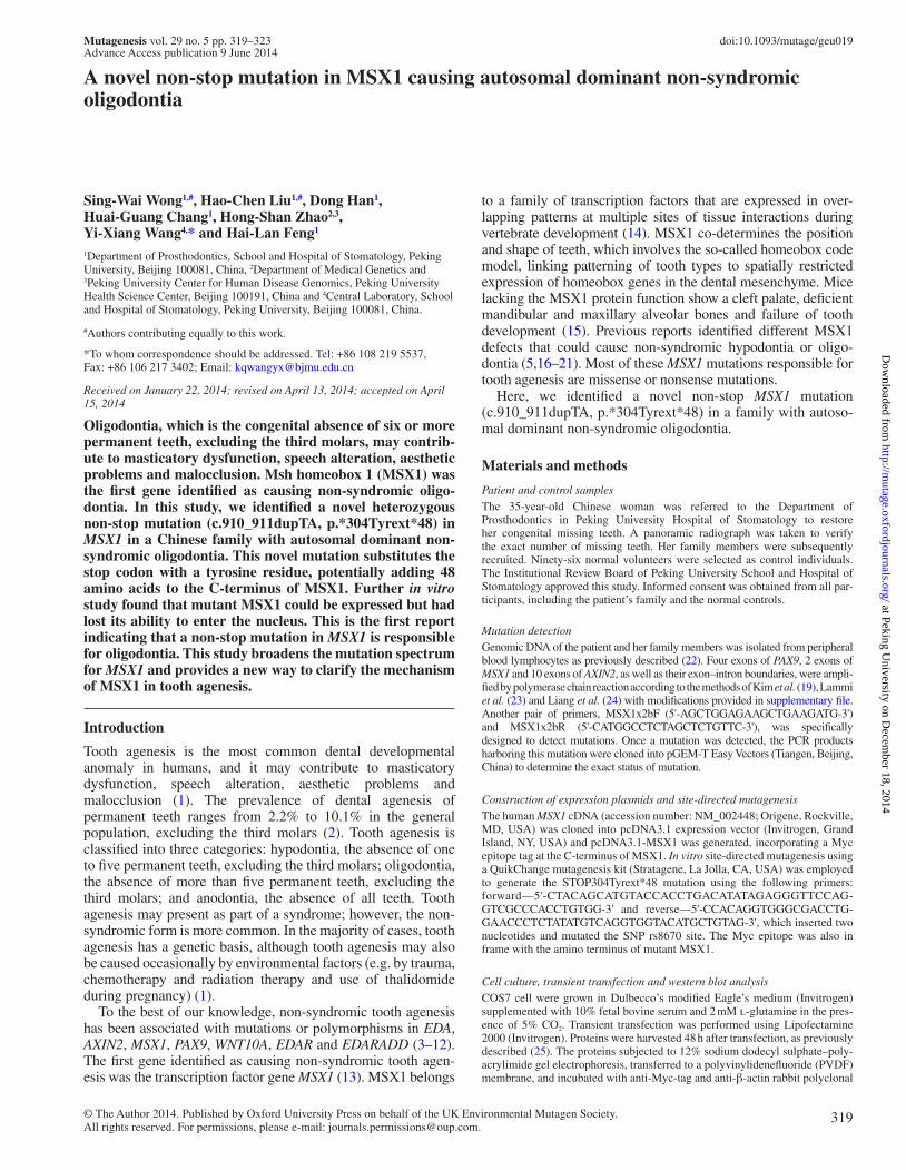
Clinical detailsProsthodontists at the Peking University hospital of Stomatology performed oral examinations of all participants. The proband was a 35-year-old female in good health. Clinical and radiographic examinations revealed that, in addition to retained deciduous teeth, the patient was missing 19 permanent teeth, including the third molar (Figure 1B and C). The mother
of the proband had normal dentition. The father of the proband was 72 years old and was in complete edentulous status. However, he stated that he and his mother had a history of tooth agenesis (pedigree shown in Figure 1A). All family members denied history of heat intolerance, and their facial features, hair, skin and nails appeared normal, which did not suggest the presence of any systemic disorder.
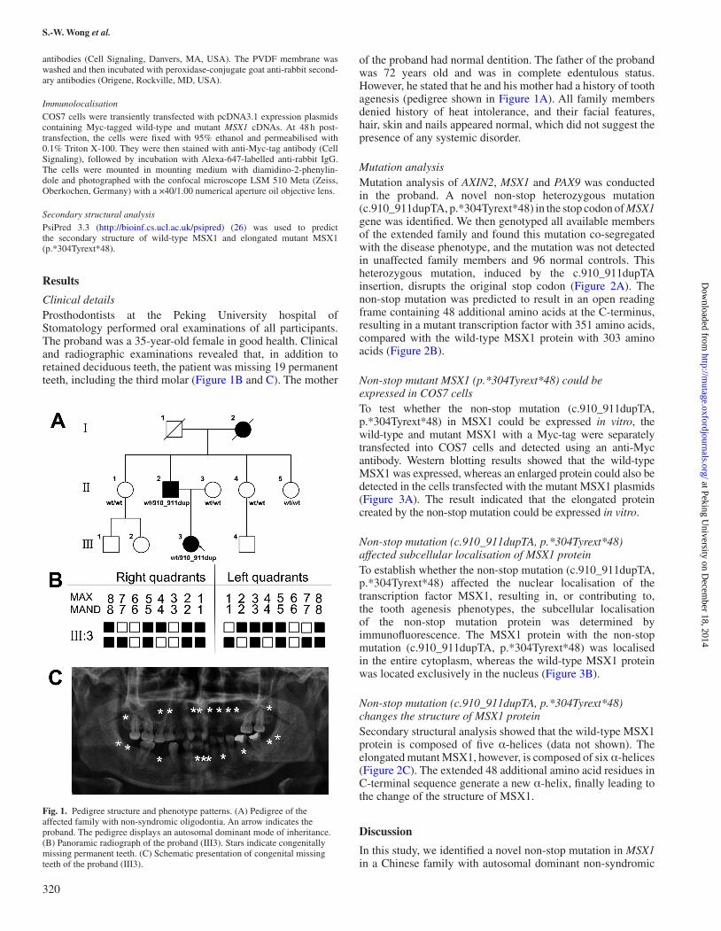
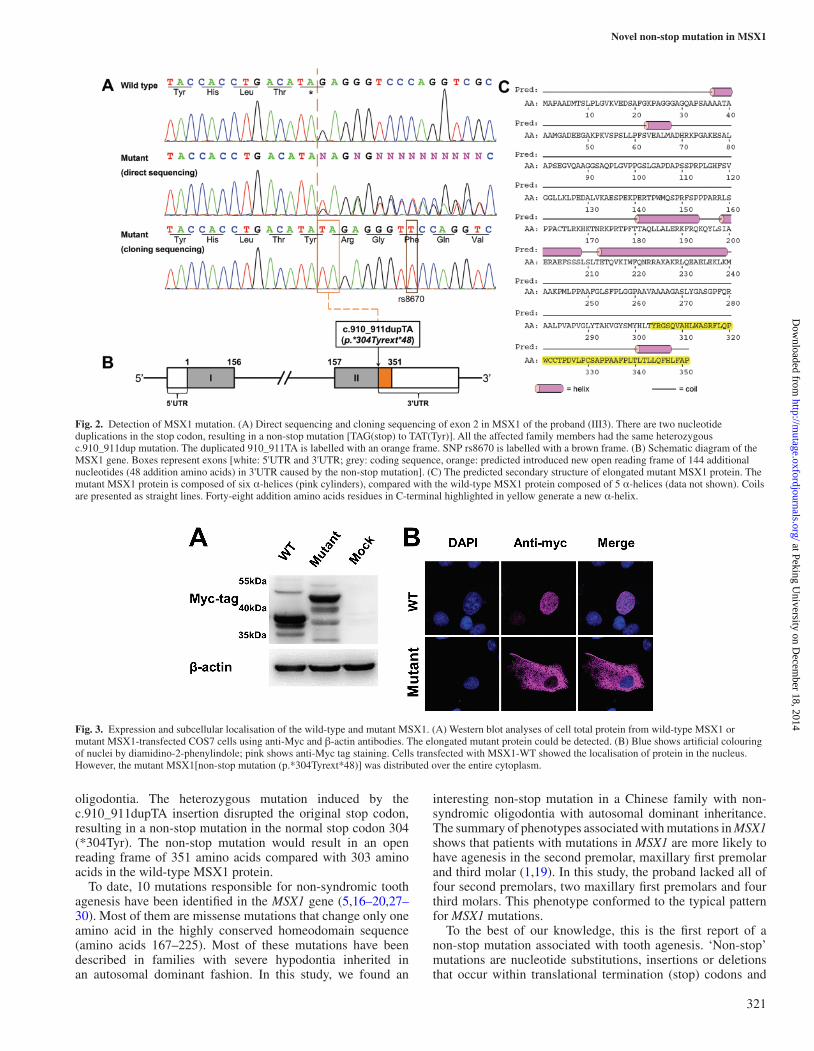
Mutation analysisMutation analysis of AXIN2, MSX1 and PAX9 was conducted in the proband. A novel non-stop heterozygous mutation (c.910_911dupTA, p.*304Tyrext*48) in the stop codon of MSX1 gene was identified. We then genotyped all available members of the extended family and found this mutation co-segregated with the disease phenotype, and the mutation was not detected in unaffected family members and 96 normal controls. This heterozygous mutation, induced by the c.910_911dupTA insertion, disrupts the original stop codon (Figure 2A). The non-stop mutation was predicted to result in an open reading frame containing 48 additional amino acids at the C-terminus, resulting in a mutant transcription factor with 351 amino acids, compared with the wild-type MSX1 protein with 303 amino acids (Figure 2B).
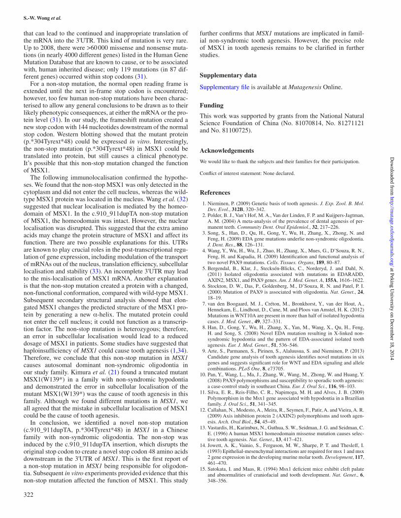
Non-stop mutant MSX1 (p.*304Tyrext*48) could be expressed in COS7 cellsTo test whether the non-stop mutation (c.910_911dupTA, p.*304Tyrext*48) in MSX1 could be expressed in vitro, the wild-type and mutant MSX1 with a Myc-tag were separately transfected into COS7 cells and detected using an anti-Myc antibody. Western blotting results showed that the wild-type MSX1 was expressed, whereas an enlarged protein could also be detected in the cells transfected with the mutant MSX1 plasmids (Figure 3A). The result indicated that the elongated protein created by the non-stop mutation could be expressed in vitro.
Non-stop mutation (c.910_911dupTA, p.*304Tyrext*48) affected subcellular localisation of MSX1 proteinTo establish whether the non-stop mutation (c.910_911dupTA, p.*304Tyrext*48) affected the nuclear localisation of the transcription factor MSX1, resulting in, or contributing to, the tooth agenesis phenotypes, the subcellular localisation of the non-stop mutation protein was determined by immunofluorescence. The MSX1 protein with the non-stop mutation (c.910_911dupTA, p.*304Tyrext*48) was localised in the entire cytoplasm, whereas the wild-type MSX1 protein was located exclusively in the nucleus (Figure 3B).
Non-stop mutation (c.910_911dupTA, p.*304Tyrext*48) changes the structure of MSX1 proteinSecondary structural analysis showed that the wild-type MSX1 protein is composed of five α-helices (data not shown). The elongated mutant MSX1, however, is composed of six α-helices (Figure 2C). The extended 48 additional amino acid residues in C-terminal sequence generate a new α-helix, finally leading to the change of the structure of MSX1.
Discussion
In this study, we identified a novel non-stop mutation in MSX1 in a Chinese family with autosomal dominant non-syndromic
Fig. 1. Pedigree structure and phenotype patterns. (A) Pedigree of the affected family with non-syndromic oligodontia. An arrow indicates the proband. The pedigree displays an autosomal dominant mode of inheritance. (B) Panoramic radiograph of the proband (III3). Stars indicate congenitally missing permanent teeth. (C) Schematic presentation of congenital missing teeth of the proband (III3).
320
at Peking University on D
ecember 18, 2014
http://mutage.oxfordjournals.org/
Dow
nloaded from
Novel non-stop mutation in MSX1
oligodontia. The heterozygous mutation induced by the c.910_911dupTA insertion disrupted the original stop codon, resulting in a non-stop mutation in the normal stop codon 304 (*304Tyr). The non-stop mutation would result in an open reading frame of 351 amino acids compared with 303 amino acids in the wild-type MSX1 protein.
To date, 10 mutations responsible for non-syndromic tooth agenesis have been identified in the MSX1 gene (5,16–20,27–30). Most of them are missense mutations that change only one amino acid in the highly conserved homeodomain sequence (amino acids 167–225). Most of these mutations have been described in families with severe hypodontia inherited in an autosomal dominant fashion. In this study, we found an
interesting non-stop mutation in a Chinese family with non-syndromic oligodontia with autosomal dominant inheritance. The summary of phenotypes associated with mutations in MSX1 shows that patients with mutations in MSX1 are more likely to have agenesis in the second premolar, maxillary first premolar and third molar (1,19). In this study, the proband lacked all of four second premolars, two maxillary first premolars and four third molars. This phenotype conformed to the typical pattern for MSX1 mutations.
To the best of our knowledge, this is the first report of a non-stop mutation associated with tooth agenesis. ‘Non-stop’ mutations are nucleotide substitutions, insertions or deletions that occur within translational termination (stop) codons and
Fig. 2. Detection of MSX1 mutation. (A) Direct sequencing and cloning sequencing of exon 2 in MSX1 of the proband (III3). There are two nucleotide duplications in the stop codon, resulting in a non-stop mutation [TAG(stop) to TAT(Tyr)]. All the affected family members had the same heterozygous c.910_911dup mutation. The duplicated 910_911TA is labelled with an orange frame. SNP rs8670 is labelled with a brown frame. (B) Schematic diagram of the MSX1 gene. Boxes represent exons [white: 5'UTR and 3'UTR; grey: coding sequence, orange: predicted introduced new open reading frame of 144 additional nucleotides (48 addition amino acids) in 3'UTR caused by the non-stop mutation]. (C) The predicted secondary structure of elongated mutant MSX1 protein. The mutant MSX1 protein is composed of six α-helices (pink cylinders), compared with the wild-type MSX1 protein composed of 5 α-helices (data not shown). Coils are presented as straight lines. Forty-eight addition amino acids residues in C-terminal highlighted in yellow generate a new α-helix.
Fig. 3. Expression and subcellular localisation of the wild-type and mutant MSX1. (A) Western blot analyses of cell total protein from wild-type MSX1 or mutant MSX1-transfected COS7 cells using anti-Myc and β-actin antibodies. The elongated mutant protein could be detected. (B) Blue shows artificial colouring of nuclei by diamidino-2-phenylindole; pink shows anti-Myc tag staining. Cells transfected with MSX1-WT showed the localisation of protein in the nucleus. However, the mutant MSX1[non-stop mutation (p.*304Tyrext*48)] was distributed over the entire cytoplasm.
321
at Peking University on D
ecember 18, 2014
http://mutage.oxfordjournals.org/
Dow
nloaded from
S.-W. Wong et al.
that can lead to the continued and inappropriate translation of the mRNA into the 3'UTR. This kind of mutation is very rare. Up to 2008, there were >60 000 missense and nonsense muta-tions (in nearly 4000 different genes) listed in the Human Gene Mutation Database that are known to cause, or to be associated with, human inherited disease; only 119 mutations (in 87 dif-ferent genes) occurred within stop codons (31).
For a non-stop mutation, the normal open reading frame is extended until the next in-frame stop codon is encountered; however, too few human non-stop mutations have been charac-terised to allow any general conclusions to be drawn as to their likely phenotypic consequences, at either the mRNA or the pro-tein level (31). In our study, the frameshift mutation created a new stop codon with 144 nucleotides downstream of the normal stop codon. Western blotting showed that the mutant protein (p.*304Tyrext*48) could be expressed in vitro. Interestingly, the non-stop mutation (p.*304Tyrext*48) in MSX1 could be translated into protein, but still causes a clinical phenotype. It’s possible that this non-stop mutation changed the function of MSX1.
The following immunolocalisation confirmed the hypothe-ses. We found that the non-stop MSX1 was only detected in the cytoplasm and did not enter the cell nucleus, whereas the wild-type MSX1 protein was located in the nucleus. Wang et al. (32) suggested that nuclear localisation is mediated by the homeo-domain of MSX1. In the c.910_911dupTA non-stop mutation of MSX1, the homeodomain was intact. However, the nuclear localisation was disrupted. This suggested that the extra amino acids may change the protein structure of MSX1 and affect its function. There are two possible explanations for this. UTRs are known to play crucial roles in the post-transcriptional regu-lation of gene expression, including modulation of the transport of mRNAs out of the nucleus, translation efficiency, subcellular localisation and stability (33). An incomplete 3'UTR may lead to the mis-localisation of MSX1 mRNA. Another explanation is that the non-stop mutation created a protein with a changed, non-functional conformation, compared with wild-type MSX1. Subsequent secondary structural analysis showed that elon-gated MSX1 changes the predicted structure of the MSX1 pro-tein by generating a new α-helix. The mutated protein could not enter the cell nucleus; it could not function as a transcrip-tion factor. The non-stop mutation is heterozygous; therefore, an error in subcellular localisation would lead to a reduced dosage of MSX1 in patients. Some studies have suggested that haploinsufficiency of MSX1 could cause tooth agenesis (1,34). Therefore, we conclude that this non-stop mutation in MSX1 causes autosomal dominant non-syndromic oligodontia in our study family. Kimura et al. (21) found a truncated mutant MSX1(W139*) in a family with non-syndromic hypodontia and demonstrated the error in subcellular localisation of the mutant MSX1(W139*) was the cause of tooth agenesis in this family. Although we found different mutations in MSX1, we all agreed that the mistake in subcellular localisation of MSX1 could be the cause of tooth agenesis.
In conclusion, we identified a novel non-stop mutation (c.910_911dupTA, p.*304Tyrext*48) in MSX1 in a Chinese family with non-syndromic oligodontia. The non-stop was induced by the c.910_911dupTA insertion, which disrupts the original stop codon to create a novel stop codon 48 amino acids downstream in the 3'UTR of MSX1. This is the first report of a non-stop mutation in MSX1 being responsible for oligodon-tia. Subsequent in vitro experiments provided evidence that this non-stop mutation affected the function of MSX1. This study
further confirms that MSX1 mutations are implicated in famil-ial non-syndromic tooth agenesis. However, the precise role of MSX1 in tooth agenesis remains to be clarified in further studies.
Supplementary data
Supplementary file is available at Mutagenesis Online.
Funding
This work was supported by grants from the National Natural Science Foundation of China (No. 81070814, No. 81271121 and No. 81100725).
Acknowledgements
We would like to thank the subjects and their families for their participation.
Conflict of interest statement: None declared.
References
1. Nieminen, P. (2009) Genetic basis of tooth agenesis. J. Exp. Zool. B. Mol. Dev. Evol., 312B, 320–342.
2. Polder, B. J., Van’t Hof, M. A., Van der Linden, F. P. and Kuijpers-Jagtman, A. M. (2004) A meta-analysis of the prevalence of dental agenesis of per-manent teeth. Community Dent. Oral Epidemiol., 32, 217–226.
3. Song, S., Han, D., Qu, H., Gong, Y., Wu, H., Zhang, X., Zhong, N. and Feng, H. (2009) EDA gene mutations underlie non-syndromic oligodontia. J. Dent. Res., 88, 126–131.
4. Wang, Y., Wu, H., Wu, J., Zhao, H., Zhang, X., Mues, G., D’Souza, R. N., Feng, H. and Kapadia, H. (2009) Identification and functional analysis of two novel PAX9 mutations. Cells. Tissues. Organs, 189, 80–87.
5. Bergendal, B., Klar, J., Stecksén-Blicks, C., Norderyd, J. and Dahl, N. (2011) Isolated oligodontia associated with mutations in EDARADD, AXIN2, MSX1, and PAX9 genes. Am. J. Med. Genet. A, 155A, 1616–1622.
6. Stockton, D. W., Das, P., Goldenberg, M., D’Souza, R. N. and Patel, P. I. (2000) Mutation of PAX9 is associated with oligodontia. Nat. Genet., 24, 18–19.
7. van den Boogaard, M. J., Créton, M., Bronkhorst, Y., van der Hout, A., Hennekam, E., Lindhout, D., Cune, M. and Ploos van Amstel, H. K. (2012) Mutations in WNT10A are present in more than half of isolated hypodontia cases. J. Med. Genet., 49, 327–331.
8. Han, D., Gong, Y., Wu, H., Zhang, X., Yan, M., Wang, X., Qu, H., Feng, H. and Song, S. (2008) Novel EDA mutation resulting in X-linked non-syndromic hypodontia and the pattern of EDA-associated isolated tooth agenesis. Eur. J. Med. Genet., 51, 536–546.
9. Arte, S., Parmanen, S., Pirinen, S., Alaluusua, S. and Nieminen, P. (2013) Candidate gene analysis of tooth agenesis identifies novel mutations in six genes and suggests significant role for WNT and EDA signaling and allele combinations. PLoS One, 8, e73705.
10. Pan, Y., Wang, L., Ma, J., Zhang, W., Wang, M., Zhong, W. and Huang, Y. (2008) PAX9 polymorphisms and susceptibility to sporadic tooth agenesis: a case-control study in southeast China. Eur. J. Oral Sci., 116, 98–103.
11. Silva, E. R., Reis-Filho, C. R., Napimoga, M. H. and Alves, J. B. (2009) Polymorphism in the Msx1 gene associated with hypodontia in a Brazilian family. J. Oral Sci., 51, 341–345.
12. Callahan, N., Modesto, A., Meira, R., Seymen, F., Patir, A. and Vieira, A. R. (2009) Axis inhibition protein 2 (AXIN2) polymorphisms and tooth agen-esis. Arch. Oral Biol., 54, 45–49.
13. Vastardis, H., Karimbux, N., Guthua, S. W., Seidman, J. G. and Seidman, C. E. (1996) A human MSX1 homeodomain missense mutation causes selec-tive tooth agenesis. Nat. Genet., 13, 417–421.
14. Jowett, A. K., Vainio, S., Ferguson, M. W., Sharpe, P. T. and Thesleff, I. (1993) Epithelial-mesenchymal interactions are required for msx 1 and msx 2 gene expression in the developing murine molar tooth. Development, 117, 461–470.
15. Satokata, I. and Maas, R. (1994) Msx1 deficient mice exhibit cleft palate and abnormalities of craniofacial and tooth development. Nat. Genet., 6, 348–356.
322
at Peking University on D
ecember 18, 2014
http://mutage.oxfordjournals.org/
Dow
nloaded from
Novel non-stop mutation in MSX1
16. De Muynck, S., Schollen, E., Matthijs, G., Verdonck, A., Devriendt, K. and Carels, C. (2004) A novel MSX1 mutation in hypodontia. Am. J. Med. Genet. A, 128A, 401–403.
17. Kamamoto, M., Machida, J., Yamaguchi, S., et al. (2011) Clinical and func-tional data implicate the Arg(151)Ser variant of MSX1 in familial hypodon-tia. Eur. J. Hum. Genet., 19, 844–850.
18. Xuan, K., Jin, F., Liu, Y. L., Yuan, L. T., Wen, L. Y., Yang, F. S., Wang, X. J., Wang, G. H. and Jin, Y. (2008) Identification of a novel missense mutation of MSX1 gene in Chinese family with autosomal-dominant oligodontia. Arch. Oral Biol., 53, 773–779.
19. Kim, J. W., Simmer, J. P., Lin, B. P. and Hu, J. C. (2006) Novel MSX1 frameshift causes autosomal-dominant oligodontia. J. Dent. Res., 85, 267–271.
20. Mostowska, A., Biedziak, B. and Jagodzinski, P. P. (2012) Novel MSX1 mutation in a family with autosomal-dominant hypodontia of second pre-molars and third molars. Arch. Oral Biol., 57, 790–795.
21. Kimura, M., Machida, J., Yamaguchi, S., et al. (2014) Novel nonsense mutation in MSX1 in familial nonsyndromic oligodontia: subcellu-lar localization and role of homeodomain/MH4. Eur. J. Oral Sci., 122, 15–20.
22. Wong, S., Liu, H., Bai, B., Chang, H., Zhao, H., Wang, Y., Han, D. and Feng, H. (2014) Novel missense mutations in the AXIN2 gene associated with non-syndromic oligodontia. Arch. Oral Biol., 59, 349–353.
23. Lammi, L., Arte, S., Somer, M., Jarvinen, H., Lahermo, P., Thesleff, I., Pirinen, S. and Nieminen, P. (2004) Mutations in AXIN2 cause familial tooth agenesis and predispose to colorectal cancer. Am. J. Hum. Genet., 74, 1043–1050.
24. Liang, J., Song, G., Li, Q. and Bian, Z. (2012) Novel missense mutations in PAX9 causing oligodontia. Arch. Oral Biol., 57, 784–789.
25. Liu, H., Han, D., Wong, S., Nan, X., Zhao, H. and Feng, H. (2013) rs929387 of GLI3 is involved in tooth agenesis in Chinese Han population. PLoS One, 8, e80860.
26. Buchan, D. W., Ward, S. M., Lobley, A. E., Nugent, T. C., Bryson, K. and Jones, D. T. (2010) Protein annotation and modelling servers at University College London. Nucleic Acids Res., 38, W563–W568.
27. Vastardis, H., Karimbux, N., Guthua, S. W., Seidman, J. G. and Seidman, C. E. (1996) A human MSX1 homeodomain missense mutation causes selec-tive tooth agenesis. Nat. Genet., 13, 417–421.
28. Lidral, A. C. and Reising, B. C. (2002) The role of MSX1 in human tooth agenesis. J. Dent. Res., 81, 274–278.
29. Chishti, M. S., Muhammad, D., Haider, M. and Ahmad, W. (2006) A novel missense mutation in MSX1 underlies autosomal recessive oligodontia with associated dental anomalies in Pakistani families. J. Hum. Genet., 51, 872–878.
30. Mostowska, A., Biedziak, B. and Trzeciak, W. H. (2006) A novel c.581C>T transition localized in a highly conserved homeobox sequence of MSX1: is it responsible for oligodontia? J. Appl. Genet., 47, 159–164.
31. Hamby, S. E., Thomas, N. S., Cooper, D. N. and Chuzhanova, N. (2011) A meta-analysis of single base-pair substitutions in translational termination codons (‘nonstop’ mutations) that cause human inherited disease. Hum. Genomics, 5, 241–264.
32. Wang, Y., Kong, H., Mues, G. and D’Souza, R. (2011) Msx1 mutations: how do they cause tooth agenesis? J. Dent. Res., 90, 311–316.
33. Mignone, F., Gissi, C., Liuni, S. and Pesole, G. (2002) Untranslated regions of mRNAs. Genome Biol., 3, REVIEWS0004.
34. Hu, G., Vastardis, H., Bendall, A. J., et al. (1998) Haploinsufficiency of MSX1: a mechanism for selective tooth agenesis. Mol. Cell. Biol., 18, 6044–6051.
323
at Peking University on D
ecember 18, 2014
http://mutage.oxfordjournals.org/
Dow
nloaded from

![Autosomal recessive ichthyosis with limb reduction defect ... · including autosomal dominant, autosomal recessive and X-linked inheritance [1,2]. Associated cutaneous and extracutaneous](https://img.pdfslide.net/doc/110x75/5ec8c9b91adfdf12ab3e663c/autosomal-recessive-ichthyosis-with-limb-reduction-defect-including-autosomal.jpg)