Embed Size (px)
Citation preview

A Novel Pharmacophore Model to Identify Leadsfor Simultaneous Inhibition of Anti-coagulationand Anti-inflammatory Activities of Snake VenomPhospholipase A2
Abdul Wadood1,3, Syed Abid Ali2,Rabia Sattar1, Muhammad Arif Lodhi2
and Zaheer Ul-Haq1,*
1Dr Panjwani Center for Molecular Medicine and Drug Research,International Center for Chemical and Biological Sciences, Universityof Karachi, Karachi 75270, Pakistan2HEJ Research Institute of Chemistry, International Center forChemical and Biological Sciences, University of Karachi, Karachi75270, Pakistan3Department of Biochemistry, Shankar Campus, Abdul Wali KhanUniversity, Mardan, Pakistan*Corresponding author: Zaheer Ul-Haq, [email protected]
In addition to catalytic action, snake venomphospholipase A2 induces several pharmacologicaleffects including neurotoxicity, cardiotoxicity aswell as anti-coagulant and anti-platelet aggrega-tion effects. Therefore, strategy to identify dualinhibitor for this enzyme will be of much impor-tance in medical research. In this paper, structure-based pharmacophore mapping, moleculardocking, protein–ligand interaction fingerprints,binding energy calculations, and binding affinitypredictions were employed in a virtual screeningstrategy to identify new hits for dual inhibition ofanti-coagulation and inflammation of phospholi-pase A2. A structure-based pharmacophore mapwas modeled which comprised of important inter-actions as observed in co-crystal of phospholipaseA2 and its dual inhibitor indomethacin. Thegenerated model was used to retrieve moleculesfrom ChemBridge, a free database of commerciallyavailable compounds. A total of 381 moleculesmapped on the developed pharmacophore modelfrom ChemBridge database. The hits retrievedwere further screened by molecular docking, pro-tein–ligand interaction fingerprints, binding energycalculations, and binding affinity predictions usingGenetic Optimization for Ligand Docking and MOE.Based on these results, 32 chemo-types moleculeswere predicted as potential lead scaffolds fordeveloping novel, potent and structurally diversedual inhibitor of phospholipase A2.
Key words: docking, dual inhibitor, pharmacophore, phospholipaseA2, snake, venom, virtual screening
Received 14 March 2011, revised 30 September 2011 and accepted forpublication 25 November 2011
Phospholipase A2 (PLA2) is an important family of different enzymesthat exhibits diverse substrate specificities, subcellular localization,requirements of cofactor, and cellular function (1). The PLA2 class ofenzymes especially catalyzes the hydrolysis of the 2-acyl ester bondof phospholipids to yield arachidonic acid that is used as a sub-strate by cyclooxygenase (COX) and lipoxygenase enzymes, as aresult eicosanoids is formed (2,3). Snake venom PLA2, in addition tocatalytic action, also mediates several pharmacological disorders,for example, myotoxicity, neurotoxicity, cardiotoxicity as well asanti-coagulant, edema inducing, hemolytic and platelets modulatingeffects (4). The primary consideration is that the catalytic propertiesand pharmacological actions were related, and all the activitieswere based on the specific and critical phospholipids hydrolysis (5).Alternatively, it has been reported that the pharmacological proper-ties of snake venom PLA2 are not necessarily dependent on theircatalytic activity, and it might be due to the protein–protein interac-tions involving a specific site on the surface of protein (6,7). So far,these pharmacological sites are neither clearly defined nor theirbinding modes are established. Consequently, the effective chemicalentities have not been designed to prevent the interactions of thesesites to overcome the harmful pharmacological effects, whereas thesubstrate binding region and the residues involved in the catalyticactivities of PLA2 enzymes are well established. A number of three-dimensional (3D) structures of different PLA2 isoforms from a varietyof sources have been recently resolved (8–18). To understand themechanism of catalytic actions of PLA2, a number of inhibitors havebeen designed for the inhibition of their enzymatic activity. Severalcomplex structures of PLA2 with natural compounds, syntheticallydesigned inhibitors as well as substrate analogues, have also beenpublished (9,19–26). It has also been reported that the anti-inflam-matory activity showed by non-steroidal anti-inflammatory drugs hasbeen attributed to their binding to PLA2 and COX enzymes (27–33).
The anti-coagulant activity in snake venom PLA2 was also reportedmany years ago by Buffa et al. (34,35). It has been reported thatthe anti-coagulant activity in various snake venom PLA2 is greatlyvaried. Based on their activities, the PLA2 was further classifiedinto strong, weak, and non-coagulant compounds (4,36). It has alsobeen suggested that the anti-coagulant site exists between resi-dues 54–77. Although the exact nature of the structure of this
431
Chem Biol Drug Des 2012; 79: 431–441
Research Article
ª 2011 John Wiley & Sons A/S
doi: 10.1111/j.1747-0285.2011.01281.x
anti-coagulant loop is not clearly understood so far, it has beenshown that the strong anti-coagulant activity of PLA2 attributed tothe presence of basic residues at precise positions in the segment54–77. For example, isoform of PLA2 from Daboia russelli pulchellapossesses five basic residues in the loop 54–77 demonstrating itspotential to be powerful anti-coagulant. However, the ligand-bindingmode for inhibiting the anti-coagulant activity of PLA2 is not fullyunderstood, and hence, the ligand could not be designed to inhibitthe anti-coagulant action of PLA2. Indomethacin has been reportedlong time ago as a non-selective COX-2 inhibitor (37). In recentstudies, it has also been reported that indomethacin inhibits groupII PLA2, but its unusual kinetic properties could not be explained(38,39). These studies demonstrate that the binding mode of indo-methacin is very different as compare to other molecules that bindto the substrate binding site of PLA2 enzyme. It was also indicatedthat indomethacin blocks the anti-thrombotic action of PLA2, thussuggesting that indomethacin might also be involved in inhibitingits anti-coagulant binding site (40). Very recently, the co-crystallizedstructure of PLA2 with indomethacin has been resolved, to under-stand its binding mode with PLA2 and the mechanisms of itsactions against both catalytic and anti-coagulant actions. A remark-able new ligand-binding site has been identified, which demon-strates that indomethacin binds to the amino acid residues that aresignificant for the catalytic action of the enzyme as well as for itsanti-coagulant activity.
In recent years, high-throughput virtual screening has been emerg-ing as a complementary to high-throughput screening in an attemptto discover novel potential lead compounds in the process of drugdiscovery (41). Thus, to identify new and potent compounds thatinhibit both the catalytic and anti-coagulant activities of snakevenom PLA2 like indomethacin, structure-based pharmacophoremodeling and virtual screening may consider as an effectiveapproach. This paper describes the structure-based pharmacophoremodeling to identify the pharmacophoric features required for simul-taneous inhibition of anti-coagulant and inflammatory effects byvirtual screening, molecular docking, protein–ligand interactionfingerprints (PLIFs), binding energy calculations, and binding affinitypredictions.
Materials and Methods
Generation of structure-based pharmacophoremodelIn the present study, the only crystal structure of PLA2 in complexwith a dual inhibitor indomethacin (3H1X.pdb) was used as startingstructure for the generation of structure-based pharmacophore mod-els (42). LIGANDSCOUT (LS) is a tool that allows the automatic con-struction and visualization of 3D pharmacophore from structuraldata of macromolecules ⁄ ligand complexes (43). For the LS algorithm,chemical features include hydrogen bond donors and acceptors asdirected vectors, and positive and negative ionizable regions as wellas lipophilic areas are represented by spheres. Moreover, toincrease the selectivity, the LS model includes spatial informationregarding areas inaccessible to any potential ligand, thus reflectingpossible steric restrictions. In particular, excluded volume spheresplaced in positions that are sterically forbidden are automatically
added to the generated pharmacophore model. The software LS
was applied to the detection and interpretation of crucial inter-action patterns between PLA2 and ligand. LS extracts and interpretsligands and their macromolecular environment from PDB files andautomatically generates and visualizes advanced 3D pharmacophoremodels.
LIGANDSCOUT may also be used to construct pharmacophore of vary-ing degrees of sophistication, suitable for export to different pro-grams. In the present study, MOE-compatible 3D-pharmacophoremodel was first developed by LS using default parameters, and then,it was exported and converted into a Molecular Operating Environ-ment, pharmacophore query for virtual screening (http://www.chemcomp.com). Prior to screening, it was necessary to make a numberof adjustments, because feature interpretation differs slightlybetween the two programs. Those aromatic rings that LS classifiedsimply as hydrophobic groups were classified as either aromatic orhydrophobic in MOE, using the PPCH_All scheme (which incorporatesdirectionality of hydrogen bond donors and acceptors, and orienta-tion of aromatic rings). As in LS pharmacophore, the aromatic ring isnot directly classified as such (because of lack of detection of p–pstacking or cation–p interactions) but rather as a set of hydrophobicatoms, can be interpreted in MOE in a manner that is useful in theprediction of right compounds in virtual screening.
Pharmacophore-based virtual screeningThe ChemBridge database (http://www.chembridge.com), whichallows the user to download compounds structures from a varietyof vendors as SDF files, was used in this preliminary screen. UsingMOE, the database was washed, and the 3D structure of each com-pound was built using the MMFF94x force field. Then for eachcompound, the low-energy conformers were generated using Con-formation Import methodology implemented in MOE software. Afterassessing the pharmacophore query, virtual screening was carriedout using the software MOE against ChemBridge database. Becausesome changes may occur when the pharmacophore is exported fromLS to MOE environment, therefore, the pharmacophore queries werevalidated before using it for virtual screening. To reduce the data ofidentified hits, they were docked into the recently identified bindingpocket of PLA2, and the PLIFs were developed using MOE. Bindingenergies and binding affinities were calculated using LIGX (ChemicalComputing Group, Montreal, Quebec, Canada) implemented in MOE
to prioritize the final hits.
Molecular dockingMolecular docking studies were performed using Genetic Optimiza-tion for Ligand Docking (GOLD) from Cambridge Crystallographic DataCenter, UK (44). GOLD uses genetic algorithm for docking flexiblesmall molecules (ligands) into binding site of macromolecules (targetproteins) to investigate the full range conformational flexibility ofligand with partial flexibility of the receptor. The ligand-bindingenergy with receptor was predicted via GOLD score implemented inGOLD. The total GOLD score, which is represented as 'Fitness', was cal-culated from the contribution of hydrogen bonds and van der Waalsinteractions between receptor and ligand as well as the contributionof intra-molecular hydrogen bonds and intra-molecular strain in the
Wadood et al.
432 Chem Biol Drug Des 2012; 79: 431–441
ligand. Receptor co-ordinates of the crystal structure of PLA2-inhibi-tor complex were used to define the binding site for moleculardocking studies. All the solvent molecules except involved in ligandbinding (i.e. W158, W261, and W284) in the crystal structure wereremoved, and hydrogen atoms were added to the whole protein. Theligand-binding site for docking was defined as a collection of aminoacids enclosed within a sphere of 10 � radius around theco-ordinates of indomethacin, which is the inhibitor molecule presentin the binding site of PLA2-inhibitor complex. Top 10 docked poseswere allowed to be saved with the early termination option of quit-ting the genetic optimization calculation for a ligand if the RMSDbetween any five conformations of the particular ligand is <1.5 �.All other parameters were kept at their default values. Molecularinteractions were observed using PLIF and LIGX software imple-mented in MOE.
Computation of generalized Born interactionenergies and binding affinityTo predict the most promising compounds, binding affinities of thehits-PLA2 complexes were calculated with generalized Born ⁄ volumeintegral (GB ⁄ VI) implicit solvent method using LIGX tool in MOE (45).a
Generalized Born interaction energy is the non-bonded interactionenergy between the macromolecule and the ligand, which comprisesvan der Waals, Coulomb electrostatic interaction, and implicitsolvent interaction energies; however, ligand and receptor macro-molecule strain energies are not taken into account. LIGX is thename for a collection of procedures and capabilities in MOE for con-ducting interactive ligand modification and energy minimization inthe binding pocket of a flexible receptor. In LIGX calculations, thereceptor atoms far from the ligand are held fixed (constrained notto move) while receptor atoms in the vicinity of the ligand (in theactive site) are allowed to move but are subject to tether restraintsthat discourage gross movement. The ligand atoms can be config-ured as either free to be move or subject to similar tethers as thereceptor. Prior to calculation of binding energy, binding pocket ofPLA2 and the ligand in the complex were subjected to energy mini-mization. During the minimization, the positions of residues of PLA2that are located 10 � away from ligand were constrained. The cal-
culated binding affinities of each compound are reported in theunits of pki.
Results and Discussion
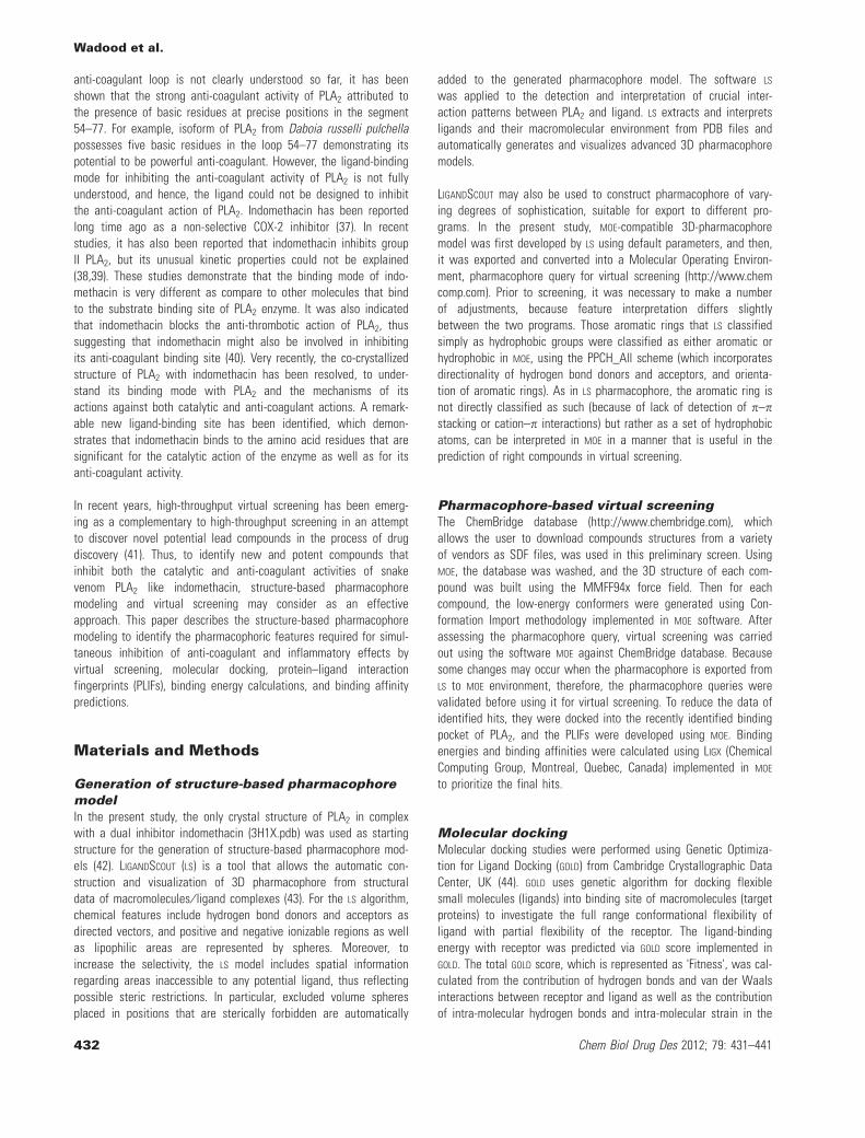
Generation of structure-based pharmacophoremodelAs shown in Figures 1 and 2B, the pharmacophore model automati-cally generated by the LS program includes four features: three hydro-gen bond acceptors (HBA) and one hydrophobic group. Besides, theprogram automatically generated several excluded volumes in themodel. The HBA features points toward the carboxylic and methoxyoxygen atoms of the ligand from the Lys69 and water moleculesW158, 261, 284, and 358, respectively. The hydrophobic groups arelocated on the methyl group adjacent to the indole ring of the ligand.The developed pharmacophore model was exported into MOE. Prior toscreening, it was necessary to make a number of adjustments, asfeature interpretation varies slightly between the two programs. Asin LS pharmacophore, the aromatic ring of the compound in the com-plex was not classified as aromatic or hydrophobic features; thus,these were interpreted in MOE, using the PPCH_All scheme. Two mod-ifications were made on this model to obtain appropriate model forvirtual screening. The first modification is about the chlorobenzyl ring.It is clear that it is an aromatic group, but the LS could not interpretthis ring as an aromatic group automatically. In MOE, additional fea-tures were developed using the MOE pharmacophore query editor.First, an aromatic feature was developed on the chlorobenzyl ring,and a hydrophobic feature was developed on the phenyl ring ofindole moiety of the ligand. This modified pharmacophore model wasthen validated by screening the test database. In the test database,we kept the compound (i.e. indomethacin) present in complex struc-ture. First, the indomethacin was extracted, and then, hydrogenatoms were added and energy minimized by using MOE. The mini-mized structure of indomethacin was added to the test database.After screening, the test compound was correctly mapped by themodified pharmacophore model as shown in Figure 2A. The resultverified the validity of our modified pharmacophore model that canbe used for the screening of large databases.
Pharmacophore-based virtual screeningThe modified validated pharmacophore model was then used asin silico filter to screen the ChemBridge database (http://www.chembridge.com) of commercially available compounds. TheChemBridge database compounds in SDF format were loaded intoMOE environment where the 3D structure of each compound wasmodeled using MMFF94x force field. The Conformation Importmethodology was applied to generate low-energy conformationsfor each compound. All these compounds and their respective con-formations were saved in MOE database. The conformers of eachcompound were then filtered by the pharmacophore model. To beconsidered as hit, the compound has to fit all the features of thepharmacophore. From the pharmacophore-based virtual screening,381 hits were identified that mapped on the developed pharmaco-phore model (i.e. having the specified requirements). These ini-tially identified hits were selected for further evaluation usingdocking studies.
Figure 1: Two-dimensional pharmacophore model generated byLIGANDSCOUT from the complex structure of phospholipase A2 andindomethacin. The dotted arrows indicated the hydrogen bondacceptor features, and the yellow sphere represented the hydropho-bic feature in the ligand.
Pharmacophore Model to Identify Leads for Simultaneous Inhibition of Snake Venom PLA2
Chem Biol Drug Des 2012; 79: 431–441 433
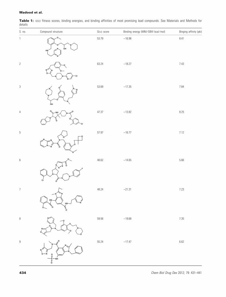
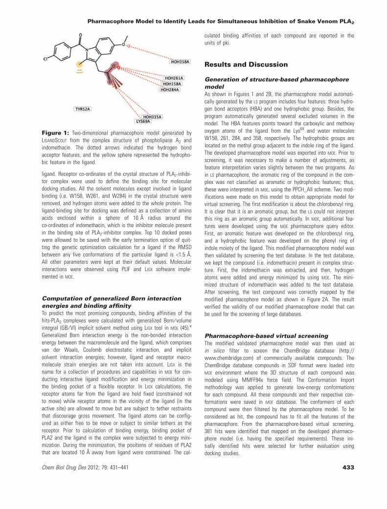
Table 1: GOLD fitness scores, binding energies, and binding affinities of most promising lead compounds. See Materials and Methods fordetails
S. no. Compound structure GOLD score Binding energy (MM ⁄ GBVI kcal ⁄ mol) Binging affinity (pki)
1 O
HN
N
N
O N
OH
53.79 )16.98 8.41
2
NO
N
N NN
Cl
O
O
63.24 )18.27 7.43
3
N
N
ON
NNO
OH
53.69 )17.35 7.84
4
N
NHO
O
NO
O
Cl
47.37 )13.82 8.25
5
N
N
N
N
O
NO
O
O
O
57.97 )16.77 7.12
6
F
NN
O
N
NN
N
O
O
Cl
48.62 )14.65 5.66
7
O
HN
O
HN
O
N
NN
48.24 )21.31 7.23
8
N
O
NO
NO
O
O
N
O
59.56 )19.68 7.35
9
N
O N
NO
NHS
O
ON
N55.24 )17.47 6.62
Wadood et al.
434 Chem Biol Drug Des 2012; 79: 431–441

Table 1: (Continued)
S. no. Compound structure GOLD score Binding energy (MM ⁄ GBVI kcal ⁄ mol) Binging affinity (pki)
10 Cl
HN
O
NO
O O
HN
59.82 )19.46 7.40
11S
N
N
N
O O
NH O
O
66.57 )17.69 9.47
12
N
HN
ON
O N 47.54 )14.35 8.22
13
NH
O
OO
N
ONO
46.77 )16.45 6.49
14O
O
NN
HO
S
O
O67.76 )22.50 9.12
15
O
N
OO
SO
O
ON
50.60 )21.31 7.60
16 O O
O
NH
N
NN
SO
O
63.90 )23.21 6.76
17 SO
N
O
O
O
58.16 )21.38 7.71
18N
O
O NO
O
48.16 )21.10 7.07
19
NN
HO
O
HNN
52.53 )24.66 8.46
Pharmacophore Model to Identify Leads for Simultaneous Inhibition of Snake Venom PLA2
Chem Biol Drug Des 2012; 79: 431–441 435

Table 1: (Continued)
S. no. Compound structure GOLD score Binding energy (MM ⁄ GBVI kcal ⁄ mol) Binging affinity (pki)
20
O
O
HN
N HN N
N
59.18 )17.48 8.18
21
NH
HO
HO
O
HO
N
55.22 )16.20 8.16
22
O
O
HO
NO
NN
47.22 )13.90 6.58
23
NN O
N NN
S
O
O
60.46 )20.41 8.49
24O
O
NS
N
OO
49.78 )13.88 7.23
25 O
HNN
O
OH
NO
52.78 )15.53 7.93
26NH
O
N
O
N
O
O
51.20 )18.91 6.39
27 O
N
N N
SCl
O HN
S
O
O
N
65.73 )17.79 9.15
28 O
O NN
NHO
53.54 )17.78 8.30
29 O
NH
O
O N
N
N
58.25 )20.39 8.59
Wadood et al.
436 Chem Biol Drug Des 2012; 79: 431–441
Molecular dockingTo deduce the promising compounds for dual inhibitors of PLA2, allthe initial hits were docked into the recently identified binding pocketof PLA2 using the automated GOLD docking program. Prior to dock theinitial hits, the ligand (i.e. indomethacin) from the complex structure
was extracted and re-docked into the binding pocket to validate thedocking protocol. The docked conformation corresponding to the highGOLD fitness score was selected as the most promising bindingconformation. The root mean square deviation (RMSD) between thedocked and experimentally determined conformation was calculated
Table 1: (Continued)
S. no. Compound structure GOLD score Binding energy (MM ⁄ GBVI kcal ⁄ mol) Binging affinity (pki)
30
O
O
N
N
O
NN
O
52.22 )20.51 7.37
31
O
ON
O
O
N
O
55.99 )13.38 7.25
32N
N
OH
O
HN S
O
57.92 )25.17 7.15
Indomethacine
N
O
OH
O
O 58.00 )13.28 6.98
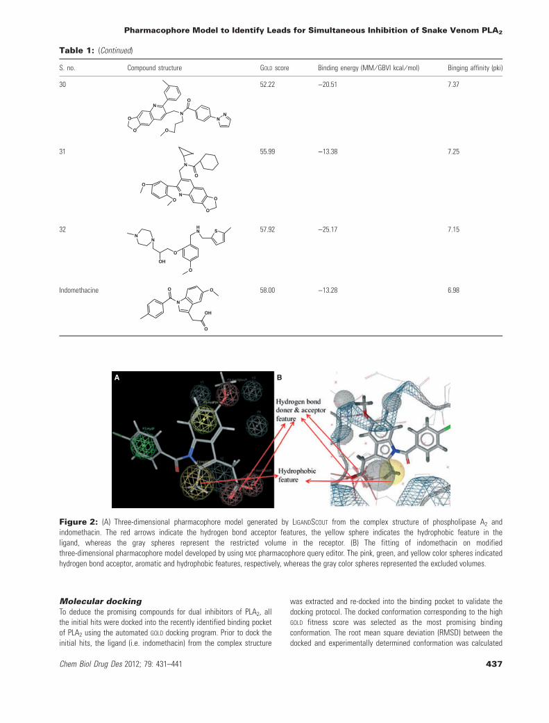
A B
Figure 2: (A) Three-dimensional pharmacophore model generated by LIGANDSCOUT from the complex structure of phospholipase A2 andindomethacin. The red arrows indicate the hydrogen bond acceptor features, the yellow sphere indicates the hydrophobic feature in theligand, whereas the gray spheres represent the restricted volume in the receptor. (B) The fitting of indomethacin on modifiedthree-dimensional pharmacophore model developed by using MOE pharmacophore query editor. The pink, green, and yellow color spheres indicatedhydrogen bond acceptor, aromatic and hydrophobic features, respectively, whereas the gray color spheres represented the excluded volumes.
Pharmacophore Model to Identify Leads for Simultaneous Inhibition of Snake Venom PLA2
Chem Biol Drug Des 2012; 79: 431–441 437
using SVL script of MOE and found to be equal to 1.33 �, suggestingthat a high docking reliability of GOLD in reproducing theexperimentally determined binding mode for PLA2. The GOLD dockingsoftware and the parameters set could be extended to search thePLA2 binding conformations for other compounds accordingly. Usingthe same docking protocol, all the initial hits were docked into thebinding pocket of PLA2. Furthermore, on the basis of GOLD fitnessscore, the top-ranked 100 compounds were selected for further detailinvestigations. For the top-ranked pose of all the selected com-pounds, PLIFs were generated by MOE, and those compounds thatrevealed significant interactions with important residues of recentlyidentified binding pocket (i.e. Asp49, His48, Lys69) and water mole-cules in the catalytic and anti-coagulant active site regions of PLA2
were picked as promising hits. Protein–ligand interaction fingerprintsimplemented on MOE classify the interactions between a ligand andthe residues on the binding site into various types of weak andstrong interactions. Among the top-ranked 100 compounds, 53 com-pounds showed the crucial interaction with the enzyme via PLIFs.
Computation of binding affinity and expertvisual inspectionFinally, binding energy and binding affinity for all the 53 com-pounds including indomethacin were calculated using LIGX imple-mented in MOE to prioritize identified promising hits. The criteria forthe selection of the most promising hits were compounds havingbinding energy and binding affinity equal to or good as compare tothe binding energy and binding affinity calculated for indomethacin,visualization of each compound in the binding pocket and theselection of only those compounds exhibiting interactions with bothcatalytic and anti-coagulation region of PLA2, and diversity of thecompounds. Applying the above-mentioned criteria, 32 compoundsof 53 fulfill the specific requirements summarized in Table 1. Thepharmacophore mapping, PLIFs, binding mode, binding energy, andbinding affinity prediction showed that these predicted lead hitsmight act as good lead compounds for the development of novel,potent, and structurally diverse dual inhibitors of snake venom PLA2
enzyme.
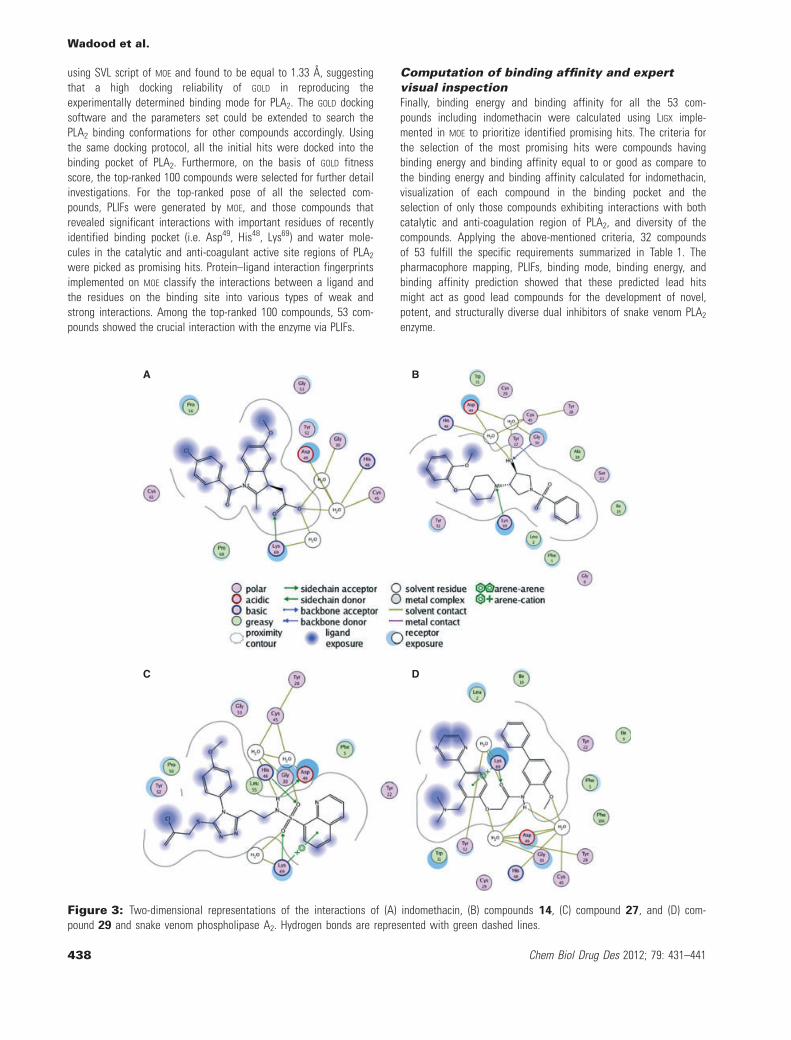
A B
C D
Figure 3: Two-dimensional representations of the interactions of (A) indomethacin, (B) compounds 14, (C) compound 27, and (D) com-pound 29 and snake venom phospholipase A2. Hydrogen bonds are represented with green dashed lines.
Wadood et al.
438 Chem Biol Drug Des 2012; 79: 431–441
Binding interactions of finally selectedcompoundsThe docking studies revealed that almost all finally selected lead hitsshowed similar binding interactions as that of indomethacin to bothcatalytic and anti-coagulant regions of PLA2 enzyme. For example,compound 14 for which the strong binding affinity (09.1 pki), lowerbinding energy ()22.5), and high GOLD fitness score (67.76) wereobserved showed the binding interaction similar to that of indometh-acin (Table 1). From the docked conformation, it was also predictedthat the oxygen atom of hydroxyl group attached to the pyrrolidinering and nitrogen atom in pyridine ring of the compound 14 estab-lished interaction with the amino acid residues Asp49, Gly30, His40,and Lys69 in a similar manner of indomethacin to the same residues(Figure 3A,B). Similarly, it was also predicted that the oxygen atomsattached to the sulfur atom in compound 27 interact in the samefashion to the important residues of the enzyme (Figure 3C). Like-wise, in the binding mode of compound 29, oxygen atom of methoxygroup attached to the phenyl ring of the compound interacts with thecatalytic region, whereas the carbonyl oxygen interacts with anti-coagulant region of PLA2 in a similar fashion as indomethacin(Figure 3D). From the docking poses of the selected compounds, itwas observed that there are some specific functional groups thatinteract with the catalytic and anti-coagulation regions and fit wellin the newly identified binding pocket of snake venom PLA2 enzyme.
Conclusion
The aim of this study was to generate a pharmacophore model toidentify structurally diverse lead hits. The identified hits might beused for developing novel and potent inhibitors for simultaneousinhibition of anti-coagulant and inflammatory affects of snakevenom enzyme PLA2. A structure-based pharmacophore was devel-oped based on the complex structure of PLA2 and indomethacin.The developed pharmacophore model was used for the screening ofChemBridge database. The identified hits were further evaluated bymolecular docking, PLIFs development, binding energy calculation,and binding affinity prediction. As a result, 32 lead hits werereported that fulfilled all the criteria for the design of compoundsthat might act as good leads for development of novel, potent, andstructurally diverse compounds for dual inhibition. From the bindingmode, predicted by docking, it was observed that there are somespecific groups that mimic the binding mode of indomethacin andfit well to both of the catalytic and ant-coagulation region of PLA2
enzyme. Further studies toward the synthesis and structure–activityrelationship of the above-mentioned lead compounds with differentsnake venom PLA2 are in progress and will be reported elsewhere.
Acknowledgments
Financial support from ASEA-UNINET under the Austrian-PakistanCooperation Project in Computational Chemistry (APCPC) ishighly acknowledged. We are extremely grateful to Dr Reaz uddin(Dr Panjwani Center for Molecular Medicine and Drug Research,University of Karachi, Pakistan) for his useful suggestions andmeaningful discussions.
References
1. Dennis E.A. (1997) The growing phospholipase A2 superfamily ofsignal transduction enzymes. Trends Biochem Sci;22:1–2.
2. Smith W.L., De Witt D.L., Garavito R.M. (2000) Cyclooxygenase:structural, cellular, and molecular biology. Annu Rev Bio-chem;69:145–182.
3. Dennis E.A. (1983) Phospholipases. In: Boyer P., editor. TheEnzymes. New York: Academic Press; p. 307–353.
4. Kini R.M., Evans H.J. (1987) Structure-function relationships ofphospholipases. The anticoagulant region of phospholipases A2.J Biol Chem;262:14402–14407.
5. Rudrammaji L.M., Machiah K.D., Kantha T.P., Gowda T.V. (2001)Role of catalytic function in the antiplatelet activity of phospho-lipase A2 cobra (Naja naja naja) venom. Mol Cell Bio-chem;219:39–44.
6. Kerns R.T., Kini R.M., Stefansson S., Evans H.J. (1999) Targetingof venom phospholipases: the strongly anticoagulant phospholi-pase A2 from Naja nigricollis venom binds to coagulation factorXa to inhibit the prothrombinase complex. Arch Biochem Bio-phys;369:107–113.
7. Kini R.M. (2005) Structure-function relationships and mechanismof anticoagulant phospholipase A2 enzymes from snake venoms.Toxicon;45:1147–1161.
8. Thunnissen M.G.M., Ab E., Kalk K.H., Drenth J., Dijkstra B.W.,Kuipers O.P., Dijkman R., de Haas G.H., Verheij H.M. (1990) X-raystructure of phospholipase A2 complexed with a substrate-derived inhibitor. Nature;347:689–691.
9. Scott D.L., White S.P., Browning J.L., Rosa J.J., Gelb M.H., SiglerP.B. (1991) Structures of free and inhibited human secretory phos-pholipase A2 from inflammatory exudate. Science;254:1007–1010.
10. Chandra V., Kaur P., Jasti J., Betzel C., Singh T.P. (2001) Regula-tion of catalytic function by molecular association: structure ofphospholipase A2 from Daboia russelli pulchella (DPLA2) at 1.9 �resolution. Acta Crystallogr D Biol Crystallogr;D57:1793–1798.
11. Singh G., Gourinath S., Sharma S., Paramasivam M., SrinivasanA., Singh T.P. (2001) Sequence and crystal structure determina-tion of a basic phospholipase A2 from common krait (Bungaruscaeruleus) at 2.4 � resolution: identification and characterizationof its pharmacological sites. J Mol Biol;307:1049–1059.
12. Banumathi S., Rajashankar K.R., Notzel C., Aleksiev B., SinghT.P., Genov N., Betzel C. (2001) Structure of the neurotoxic com-plex vipoxin at 1.4 � resolution. Acta Crystallogr D Biol Crystal-logr;D57:1552–1559.
13. Gu L., Zhang H., Song S., Zhou Y., Lin Z. (2002) Structure of anacidic phospholipase A2 from the venom of Deinagkistrodonacutus. Acta Crystallogr D Biol Crystallogr;D58:104–110.
14. Matoba Y., Katsube Y., Sugiyama M. (2002) The crystal structureof prokaryotic phospholipase A2. J Biol Chem;277:20059–20069.
15. Xu S., Gu L., Jiang T., Zhou Y., Lin Z. (2003) Structures ofcadmium-binding acidic phospholipase A2 from the venom ofAgkistrodon halys Pallas at 1.9 � resolution. Biochem BiophysRes Commun;300:271–277.
16. Xu S., Gu L., Wang Q., Shu Y., Song S., Lin Z. (2003) Structureof a King cobra phospholipase A2 determined from a hemihed-rally twinned crystal. Acta Crystallogr D Biol Crystal-logr;D59:1574–1581.
Pharmacophore Model to Identify Leads for Simultaneous Inhibition of Snake Venom PLA2
Chem Biol Drug Des 2012; 79: 431–441 439
17. Jasti J., Paramasivam M., Srinivasan A., Singh T.P. (2004)Structure of an acidic phospholipase A2 from Indian saw-scaledviper (Echis carinatus) at 2.6 � resolution reveals a novel inter-molecular interaction. Acta Crystallogr D Biol Crystallogr;D60:66–72.
18. Lok S.M., Gao R., Rouault M., Lambeau G., GopalakrishnakoneP., Swaminathan K. (2005) Structure and function comparison ofMicropechis ikaheka snake venom phospholipase A2 isoenzymes.FEBS J;272:1211–1220.
19. Chandra V., Jasti J., Kaur P., Betzel C., Singh T.P. (2002) Struc-tural basis of phospholipase A2 inhibition for the synthesis ofprostaglandins by the plant alkaloid aristolochic acid from a1.7 � crystal structure. Biochemistry;41:10914–10919.
20. Chandra V., Jasti J., Kaur P., Srinivasan A., Betzel C., Singh T.P.(2002) First structural evidence of a specific inhibition of phos-pholipase A2 by alpha-tocopherol (vitamin E) and its implicationsin inflammation: crystal structure of the complex formedbetween phospholipase A2 and alpha-tocopherol at 1.8 � resolu-tion. J Mol Biol;320:215–222.
21. Singh N., Jabeen T., Pal A., Sharma S., Perbandt M., Betzel C.,Singh T.P. (2006) Crystal structures of the complexes of a groupIIA phospholipase A2 with two natural anti-inflammatory agents,anisic acid, and atropine reveal a similar mode of binding. Pro-teins;64:89–100.
22. Schevitz R.W., Bach N.J., Carlson D.G., Chirgadze N.Y., ClawsonD.K., Dillard R.D., Draheim S.E., Hartley L.W., Jones N.D., Mihe-lich E.D., Olkowski J.L., Snyder D.W., Sommers C., Wery J.P.(1995) Structure-based design of the first potent and selectiveinhibitor of human non-pancreatic secretory phospholipase A2.Nat Struct Biol;2:458–465.
23. Chandra V., Jasti J., Kaur P., Dey S., Perbandt M., Srinivasan A.,Betzel C., Singh T.P. (2002) Design of specific peptide inhibitorsof phospholipase A2: structure of a complex formed betweenRussell's viper phospholipase A2 and a designed peptide Leu-Ala-Ile-Tyr-Ser (LAIYS). Acta Crystallogr D Biol Crystal-logr;D58:1813–1819.
24. Chandra V., Jasti J., Kaur P., Dey S., Perbandt M., Srinivasan A.,Betzel C., Singh T.P. (2002c) Crystal structure of a complexformed between a snake venom phospholipase A2 and a potentpeptide inhibitor Phe-Leu-Ser-Tyr-Lys at 1.8 � resolution. J BiolChem;277:41079–41085.
25. Singh R.K., Vikram P., Makker J., Jabeen T., Sharma S., Dey S.,Kaur P., Srinivasan A., Singh T.P. (2003) Design of specific pep-tide inhibitors for group I phospholipase A2: structure of a com-plex formed between phospholipase A2 from Naja najasagittifera (Group I) and a designed peptide inhibitor Val-Ala-Phe-Arg-Ser (VAFRS) at 1.9 � resolution reveals unique features.Biochemistry;42:11701–11706.
26. Scott D.L., Otwinowski Z., Gelb M.H., Sigler P.B. (1990) Crystalstructure of bee-venom phospholipase A2 in a complex with atransition-state analogue. Science;250:1563–1566.
27. Singh N., Jabeen T., Sharma S., Somvanshi R.K., Dey S., SinghT.P. (2004) Phospholipase A2 as a target protein for non-steroidalanti-inflammatory drugs (NSAIDs): crystal structure of the com-plex formed between phospholipase A2 and oxyphenbutazone at1.6 � resolution. Biochemistry;43:14577–14583.
28. Singh R.K., Ethayathulla A.S., Jabeen T., Sharma S., Kaur P.,Singh T.P. (2005) Aspirin induces its anti-inflammatory effects
through its specific binding to phospholipase A2: crystal structureof the complex formed between phospholipase A2 and aspirin at1.9 � resolution. J Drug Target;2:113–119.
29. Singh N., Jabeen T., Sharma S., Somvanshi R.K., Dey S., Sriniva-san A., Singh T.P. (2006) Specific binding of non-steroidal anti-inflammatory drugs (NSAIDs) to phospholipase A2: structure of thecomplex formed between phospholipase A2 and diclofenac at2.7 � resolution. Acta Crystallogr D Biol Crystallogr;D62:410–416.
30. Jabeen T., Singh N., Singh R.K., Sharma S., Somvanshi R.K., DeyS., Singh T.P. (2005) Non-steroidal anti-inflammatory drugs aspotent inhibitors of phospholipase A2: structure of the complexof phospholipase A2 with niflumi acid at 2.5 � resolution. ActaCrystallogr D Biol Crystallogr;D61:1579–1586.
31. Gierse J.K., Koboldt C.M., Walker M.C., Seibert K., Isakson P.C.(1999) Kinetic basis for selective inhibition of cyclo-oxygenases.Biochem J;339:607–614.
32. Vane J.R., Botting R.M. (1998) Mechanism of action of nonste-roidal anti-inflammatory drugs. Am J Med;104:2S–8S. discussion21S–22S.
33. Vane J.R., Bakhle Y.S., Botting R.M. (1998) Cyclooxygenases 1and 2. Annu Rev Pharmacol Toxicol;38:97–120.
34. Boffa G.A., Boffa M.C., Winchenne J.J. (1976) A phospholipaseA2 with anticoagulant activity. I. Isolation from Vipera berusvenom and properties. Biochim Biophys Acta;429:828–838.
35. Boffa M.C., Rothen C., Verheij H.M., Verger R., DeHaas G.H.(1980) Correlation of enzymatic activity and anticoagulant proper-ties of phospholipase A2. In: Eaker D., Walstrom T., editors. Nat-ural Toxins. Oxford: Pergamon Press; p. 131–138.
36. Verheij H.M., Boffa M.C., Rothen C., Bryckaert M.C., Verger R.,de Haas G.H. (1980) Correlation of enzymatic activity and anti-coagulant properties of phospholipase A2. Eur J Bio-chem;112:25–32.
37. Callan O.H., So O.Y., Swinney D.C. (1996) The kinetic factors thatdetermine the affinity and selectivity for slow binding inhibitionof human prostaglandin H synthase 1 and 2 by indomethacinand flurbiprofen. J Biol Chem;271:3548–3554.
38. Kaplan L., Weiss J., Elsbach P. (1978) Low concentrations ofindomethacin inhibit phospholipase A2 of rabbit polymorpho-nuclear leukocytes. Proc Natl Acad Sci USA;75:2955–2958.
39. Lobo I.B., Hoult J.R. (1994) Groups I, II and III extracellular phos-pholipases A2: selective inhibition of group II enzymes by indo-methacin but not other NSAIDs. Agents Actions;41:111–113.
40. Nygard G., Hudson M., Mazure G., Anthony A., Dhillon A.P.,Pounder R.E., Wakefield A.J. (1995) Procoagulant andprothrombotic responses of human endothelium to indomethacinand endotoxin in vitro. Relevance to non-steroidal anti-inflamma-tory drug enteropathy. Scand J Gastroenterol;30:25–32.
41. Lyne P.D. (2002) Structure-based virtual screening: an overview.Drug Discov Today;7:1047–1055.
42. Sing N., Kumar R.P., Kumar S., Sharma S., Mir R., Kaur P., Srini-vasan A., Singh T.P. (2009) Simultaneous inhibition of anti-coagu-lation and inflammation: crystal structure of phospholipase A2
complexed with indomethacin at 1.4 � resolution reveals thepresence of the new common ligand-binding site. J Mol Recog-nit;22:437–445.
43. Wolber G., Langer T. (2005) LigandScout: 3-D pharmacophoresderived from protein-bound ligands and their use as virtualscreening filters. J Chem Inf Model;45:160–169.
Wadood et al.
440 Chem Biol Drug Des 2012; 79: 431–441
44. Jones G., Willett P., Glen R.C., Leach A.R., Taylor R. (1997)Development and validation of genetic a algorithm for flexibledocking. J Mol Biol;267:727–748.
45. Labute P. (2008) The Generalized Born ⁄ Volume Integral (GB ⁄ VI)implicit solvent model: estimation of the free energy of hydrationusing London dispersion instead of atomic surface area. J CompChem;29:1693–1698.
Note
aMOE, 2009.10. Montreal: Chemical Computing Group, Inc, available
at: http://www.chemcomp.com/.
Pharmacophore Model to Identify Leads for Simultaneous Inhibition of Snake Venom PLA2
Chem Biol Drug Des 2012; 79: 431–441 441