Embed Size (px)
Citation preview

American Journal of Biochemistry and Biotechnology 4 (2): 146-166, 2008 ISSN 1553-3468 © 2008 Science Publications
*Corresponding Author: Derrick F. MacFabe, Director: The Kilee Patchell-Evans Autism Research Group, Departments of Psychology and Psychiatry, (Division of Developmental Disabilities), University of Western Ontario, Social Science Centre, Room 7252, London, Canada, N6A 5C2 Tel: 1-519-661-2111 x 84703, Fax: 1-519-661-3961
146
A Novel Rodent Model of Autism: Intraventricular Infusions of Propionic
Acid Increase Locomotor Activity and Induce Neuroinflammation and Oxidative Stress in Discrete Regions of Adult Rat Brain
Derrick F. MacFabe, Karina Rodríguez-Capote, Jennifer E. Hoffman, Andrew E. Franklin,
Yalda Mohammad-Asef, A. Roy Taylor, Francis Boon, Donald P. Cain, Martin Kavaliers, Fred Possmayer and Klaus-Peter Ossenkopp
The Kilee Patchell-Evans Autism Research Group, The University of Western Ontario, London, Ontario, Canada N6A 5C2
Abstract: Innate neuroinflammatory changes, increased oxidative stress and disorders of glutathione metabolism may be involved in the pathophysiology of autism spectrum disorders (ASD). Propionic acid (PPA) is a dietary and gut bacterial short chain fatty acid which can produce brain and behavioral changes reminiscent of ASD following intraventricular infusion in rats. Adult Long-Evans rats were given intraventricular infusions of either PPA (.26M, 4µl animal−1) or phosphate buffered saline (PBS) vehicle, twice daily for 7 days. Immediately following the second daily infusion, the locomotor activity of each rat was assessed in an automated open field (Versamax) for 30 min. PPA-treated rats showed significant increases in locomotor activity compared to PBS vehicle controls. Following the last treatment day, specific brain regions were assessed for neuroinflammatory or oxidative stress markers. Immunohistochemical analyses revealed reactive astrogliosis (GFAP), activated microglia (CD68, Iba1) without apoptotic cell loss (Caspase 3’ and NeuN) in hippocampus and white matter (external capsule) of PPA treated rats. Biomarkers of protein and lipid peroxidation, total glutathione (GSH) as well as the activity of the antioxidant enzymes superoxide dismutase (SOD), catalase, glutathione peroxidase (GPx), glutathione reductase (GR) and glutathione S-transferase (GST) were examined in brain homogenates. Some brain regions of PPA treated animals (neocortex, hippocampus, thalamus, striatum) showed increased lipid and protein oxidation accompanied by decreased total GSH in neocortex. Catalase activity was decreased in most brain regions of PPA treated animals suggestive of reduced antioxidant enzymatic activity. GPx and GR activity was relatively unaffected by PPA treatment while GST was increased, perhaps indicating involvement of GSH in the removal of PPA or related catabolites. Impairments in GSH and catalase levels may render CNS cells more susceptible to oxidative stress from a variety of toxic insults. Overall, these findings are consistent with those found in ASD patients and further support intraventricular PPA administration as an animal model of ASD. Key words: Animal model, glutathione, clostridia, movement disorder, short chain fatty acids,
metabolic disorder, organic acidemia, oxidative stress
INTRODUCTION Autism spectrum disorder (ASD) comprises a family of neurodevelopmental disorders characterized by language impairment, restricted interests, stereotypic motor behaviors, hyperactivity, sensory disturbances and self injury[1,2]. ASD is also associated with seizure disorder[3]. Imaging and neuropathological studies of ASD patients have noted increased brain size, white
matter abnormalities as well as increased neuronal density in neocortical, limbic and cerebellar areas[4-7]. These findings suggest altered neurodevelopmental processes in some brain areas in ASD patients. A recent neuropathological study of autopsy material from ASD patients of varying ages found evidence of a diffuse innate neuroinflammatory response of unknown etiology. This response was characterized by reactive astrogliosis, activated
Am. J. Biochem. & Biotech., 4 (2): 146-166, 2008
147
microglia and cytokine abnormalities[8]. These findings suggest that an immune-mediated response may persist throughout the life of ASD patients[9]. ASD has a strong genetic basis as the disorder shows high heritability with a concordance rate of 82% amongst monozygotic twins and close to 0% for dizygotic twins[10,11]. ASD is also co-morbid with a number of other inherited conditions including fragile X syndrome, tuberous sclerosis, Smith-Lemli Opitz syndrome and Mobius syndrome[12-15]. A panel of genes involved in brain development[16] and serotonin[17], glutamate, or GABA metabolism, have also been associated with a sub-set of ASD patients[18]. The lack of complete concordance as well as variance in the severity of autism in monozygotic twins[19] suggests that environmental factors are also likely important in ASD etiology. Environmental factors may account for some observed geographical clustering of cases[20] and may be involved in the recent postulated increase in the incidence of ASD cases in North America (approximately 1 in 166)[21]. There has been growing interest in a group of environmental toxins and infectious agents which could act at critical periods of pre- and post-natal development to putatively trigger or exacerbate this disorder in genetically vulnerable individuals. An increased risk of ASD is associated with pre-natal exposure to ethanol[22], valproic acid[23], terbutaline[24], some metals and viral infections[25,26]. Many of these effects have been observed in animal models using these toxic or infectious agents. One common mechanism by which diverse environmental and/or genetic factors could induce the diffuse CNS effects seen in ASD is increased oxidative stress[27]. CNS effects may be produced by increased production of reactive intermediates, impairments in the detoxification of these agents, or a combination of these factors[28,29]. Studies of plasma from ASD patients with diverse genetic backgrounds provide evidence of increased oxidative stress involving reduced capacity for methylation and sulfuration as measured by methionine transmethylation and transulfuration pathways and lipid peroxidation. Additional observations have included impaired xenobiotic detoxification and clearance at a systemic level[29-31]. Findings from these studies may reflect some innate neuroinflammatory processes in the brains of ASD patients[8]. Of particular interest is the evidence of an impairment in glutathione (GSH) associated pathways in the plasma of ASD patients[27,30]. GSH participates in both antioxidant defense and xenobiotic detoxification over a wide range of environmental organic compounds
and metals[32]. The GSH redox system consists of primary and secondary antioxidants, including glutathione peroxidase (GPx), glutathione reductase (GR), glutathione S-transferase (GST), γ -glutamylcysteine synthetase ( γ -GCS) and glucose 6-phosphate dehydrogenase (G6PD). Alterations in the activities of these enzymes are suggestive of reduced cellular defense and are surrogate markers of increased oxidative stress[33]. Furthermore, genetic alterations in glutathione metabolism have been noted in some ASD populations suggesting a plausible inherited mechanism for environmental sensitivity[34,35]. Many environmental factors have been proposed to contribute to ASD pathogenesis. There is growing interest in the role that gastrointestinal factors may play in the behavioral, neuropathological and neuroinflammatory sequelae observed in some ASD cases. Anecdotal reports have suggested that ASD symptomology may be induced or exacerbated following acute gastrointestinal abnormalities[36], routine and antibiotic resistant paediatric infections[37], or ingestion of wheat or dairy containing foods[38]. Gastroenterological studies have revealed evidence of ileal lymphoid nodular hyperplasia[39] and have isolated unique clostridial species[40], in some ASD patients exhibiting regressive symptoms after apparently attaining normal postnatal developmental milestones. The linkage between brain and gastrointestinal function is interesting in the light of the recent discovery of reduced display of MET gene expression, a factor involved in neocortical and cerebellar growth, immune function and gastrointestinal repair[41] in some subpopulations of ASD. These findings raise the possibility that some gut-derived factor may contribute to the pathogenesis and behavioral abnormalities of ASD. Our laboratory has investigated the role of propionic acid (PPA) which is a metabolic intermediate of fatty acid metabolism[42]. PPA is also an end-product of enteric bacterial fermentation, particularly from those bacteria implicated in antibiotic associated diarrhea[43,44]. Moreover, PPA is commonly used as a food preservative in wheat and dairy food products[45]. A number of effects have been shown following PPA treatment, including catecholamine and proenkephalin gene induction[46], cytoskeletal phosphorylation[47], histone modulation[48], second messenger metabolism[49], impaired mitochondrial respiratory transport chain function[50,51], modulation of gap junctions[52] and immune system activation[53]. There is also evidence of variable PPA metabolism in a number of metabolic disorders such as propionic and methylmalonic acidemia[54], disorders of biotin[55]
Am. J. Biochem. & Biotech., 4 (2): 146-166, 2008
148
and B12 metabolism[56], as well as following valproate[57] and ethanol exposure[58]. These disorders are associated with developmental delay, seizure and episodes of paroxysmal metabolic dysfunction resulting in increased oxidative stress and are to some degree reminiscent of autism[59,60]. We have hypothesized that PPA may be a candidate environmental factor putatively involved in the diverse behavioral, neuropathological and gastrointestinal aspects of ASD[61]. In our initial studies we found that repeated micro-infusions of PPA into the lateral cerebral ventricles of adult rats produced behavioral, electrophysiological and neuropathological effects consistent with ASD[61]. PPA treated animals exhibited increased locomotor activity and repetitive behaviors such as dystonic limb movements, retropulsion and axial hyperextension. Simultaneous electrophysiological examination of cortical, hippocampal and striatal EEG evidenced electrographic changes reminiscent of human complex partial seizure and movement disorder. These effects were apparent within minutes of microinfusion. Furthermore, repeated exposures resulted in increased behavioral and electrographic effects suggestive of a kindling process. Collectively these findings indicated that PPA may have permanent effects on brain function. Neuropathological analysis of hippocampus and external capsule white matter tissue revealed PPA-related increases in reactive astrocytes and activated microglia in the absence of gross neuronal loss and apoptotic effects. Additional analyses of whole brain homogenates produced evidence of increased oxidative stress and impaired glutathione metabolism in PPA treated animals. These findings are consistent with the neuropathological changes found in ASD autopsy brains[8] as well as biochemical studies in ASD patients[27,31]. The present study extended the examination of the effects of intraventricular PPA infusion on behavior, neuropathology and oxidative stress levels in the adult rodent ASD model. In particular, we characterized PPA induced increases in locomotor activity with additional automated measures of horizontal and vertical activity. We also examined the neuropathological responses following the twice-daily treatment schedule, utilizing additional indices of microglial activation and neurotoxicity. Lastly, we examined pro- and anti-oxidative stress pathways in brain regions which have been previously implicated in ASD pathophysiology.
MATERIALS AND METHODS Animals and housing facilities: A total of 30 adult male Long-Evans (Charles River Laboratories, Quebec)
rats were used. Rats weighed approximately 200-225 g (approximately 47 to 49 days old) at the time of delivery from the supplier. Animals were individually housed at a controlled temperature (21±1°C) with ad libitum access to food (Prolab rat chow) and tap water. All behavioral testing occurred during the light phase of a 12:12 hr light:dark cycle (lights on 0700h to 1900h) under normal lighting conditions. Rats were naive to all experimental procedures prior to surgery. All procedures were in accordance with guidelines of the Canadian Council on Animal Care (CCAC) and approved by the University of Western Ontario Animal Use Committee. Surgical procedures for cannula implantation: Surgical procedures were completed under aseptic conditions. All rats were implanted with a 23 ga guide cannula using standard stereotaxic techniques. Prior to surgery each rat was pretreated with atropine methyl nitrate (0.1ml SC). Rats were anaesthetized using inhaled Isoflurane and 3% oxygen and were placed in a stereotaxic apparatus. During surgery body temperature was maintained at normothermia using a heating pad. The tip of the guide cannula was placed immediately below the border of the corpus callosum into the lateral ventricle (AP 1.3 mm, ML 1.8 mm)[62]. The tip of the 30 ga injection cannula protruded 0.5 mm beyond the tip of the guide cannula to allow infusion into the lateral ventricle. Each cannula was sealed with an obtruator, which was removed prior to each intracerebroventricular (ICV) infusion. Small screws were placed in the top of the skull and the cannula was affixed to the skull with dental acrylic. Testing procedures began approximately 14 days after surgery. Treatment groups and intracerebroventricular infusion procedure: Rats were randomly assigned to two groups: PPA (4.0 µl of a 0.26 M solution, n = 12); and PBS control (4.0µl of .1 M PBS, n = 12). PPA was dissolved in PBS vehicle and buffered to pH 7.5 using concentrated HCl or NaOH. Each group received intracerebroventricular (ICV) infusions twice daily for 7 consecutive days. Compounds were infused using a 30 ga injection cannula attached to a Sage syringe pump with sterile PE10 tubing. Infusions took place over a 60 sec period. The infusion cannula was allowed to remain in place for an additional 60 sec before being removed. Locomotor activity assessments were made each day (D1 to D7) following the second infusion. On day 8, brain tissue was extracted and subsequently assayed for biochemical markers of oxidative stress and neuropathological markers (see procedures below). A random subset of 6 rats from each
Am. J. Biochem. & Biotech., 4 (2): 146-166, 2008
149
treatment group was used for the neuropathological and biochemical analyses. An additional set of 6 rats were cannulated and treated with PPA twice daily; however, locomotor activity was not assessed. Brain tissue from this second group of PPA animals was used in the biochemical assays. Unless otherwise specified each of the biochemical markers were completed with 6 PBS and 12 PPA treated rats. Locomotor activity assessments: Locomotor activity was quantified with eight VersaMax Animal Activity Monitors (Model NVMA16TT/W, Accuscan Instruments Inc., Columbus, OH). Each monitor consisted of a 40×40×30.5 cm Plexiglas open field covered by a Plexiglas lid with air holes. Sets of infrared beams for horizontal activity measurement surrounded the perimeter of each open field. Each beam was located every 2.54 cm for a total of 16 beams on each of four sides. Each set of infrared beams, used to measure horizontal activity, was located 4.5 cm above the floor of the open-field[63]. Vertical activity was measured by two additional sets of beams that were located 15cm above the floor of each open-field. Light levels at the floor level of each open-field were approximately 900lux. A VersaMax Analyser (Accuscan Model VSA-16, Columbus, OH) processed and relayed data from each automated open-field to a computer located in a room adjacent to the testing room. We previously reported that rats treated with PPA traveled significantly greater distances in the VersaMax automated open field compared to PBS-treated controls[61] . The locomotor activity analyses reported in the present study are from the same group of animals previously studied but include additional measures of locomotor activity. The multiple activity variables[64] analyzed in the present study included the following: Number of movements (NM): Total number of horizontal movements separated by 1s of stop time. Movement time (MT): Total amount of time (s) engaged in horizontal movement. Vertical movements (VM): Total number of vertical movements separated by 1s of stop time. Vertical time (VT): Total amount of time (s) spent in a vertical position. Rats were habituated to the apparatus for two 30 min sessions prior to the treatment sessions. Locomotor activity was recorded during a third baseline session to establish activity levels for the untreated rats. Habituation and baseline activity recordings were completed at times that corresponded with future treatment sessions.
Rats received ICV infusions for 7 days twice daily at 9 A.M. and 1 P.M. during the light period. Locomotor activity was recorded for 30 min on each treatment day (D1 to D7) immediately following the second infusion at 1300h. The half life of PPA is estimated to be between 18 and 57 min when administered to rats which supports the use of the 30 min test time[65]. Assessments of neuropathology Tissue preparation: On day 8 animals were deeply anaesthetized with sodium pentobarbital (270 mg mL−1 IP) and transcardially perfused with ice cold PBS (0.1M) followed by 4% paraformaldehyde in PBS. Brains were removed and placed in 4% paraformaldehyde solution and stored at 4°C for 24 h. Following the fixation period, brains were placed in an 18% sucrose solution prior to paraffin embedding. Selected coronal blocks from various brain regions (frontal cortex/striatum, dorsal hippocampus, midbrain and cerebellum/brainstem), were dehydrated and defatted by increasing concentrations of ethanol/xylenes and embedded in paraffin wax for permanent storage. Immunohistochemistry procedures: Using a Leica microtome (model RM2125) serial 4 µm sections were obtained through the following regions: 1) right lateral ventricle (cannula site) and 2) ipsilateral dorsal hippocampus, including adjacent white matter of the external capsule. These anatomical regions were chosen for preliminary analyses based on: 1) their close proximity to the intraventricular administration site of PPA or PBS vehicle, 2) the well-known cytoarchitectonics of the hippocampus which allowed for reliable quantification of possible PPA-induced neuroplastic changes, 3) the known role of the hippocampus in experimental kindling, human seizure disorder and autism and 4) for analysis of oligodendroglial changes in white matter of the external capsule, also implicated in autism. The following antibodies were used: 1) anti-glial fibrillary acidic protein (GFAP) (1:500, rabbit polyclonal, DakoCytomation, Glostrup, Denmark) a marker for reactive astrogliosis, found elevated in human autism neuropathology studies[66] and in cerebrospinal fluid from autism patients[67], 2) anti-rat CD68 antigen (1: 200, monoclonal, Serotec, Oxford, UK), 3) anti-IbA1 ( 1:10, 000, rabbit polyclonal, Wako, Richmond, VA) both markers for activated microglia and found elevated in human autism brain[66] and epilepsy[68], 4) anti-cleaved caspase 3 (1: 100, rabbit polyclonal, Cell Signaling Technology, Danvers, MA),
Am. J. Biochem. & Biotech., 4 (2): 146-166, 2008
150
a marker for apoptotic cytotoxicity[69,70] and; 5) anti-NeuN (1:1000, monoclonal, Chemicon USA ), a marker for neurons[71]. Tissue sections were mounted on glass slides (SurgiPath, Canada) and dried overnight at 37°C. Sections were deparaffinized and rehydrated using standard immunohistochemical procedures for antigen recovery[72]. Endogenous peroxidase activity was blocked using a 3% hydrogen peroxide in distilled water solution for 5min. For antigen recovery, sections were immersed in boiling 0.21% citric acid buffer (pH 6.0) for 30 min in a 1250W microwave oven. Slides were counterstained with Gill haematoxylin (EMD Biosciences) and rinsed with PBS for 5 min. A 10% normal horse serum in PBS solution was applied for 5 min followed by the primary antibodies for 1hr at room temperature. Following the incubation period, sections were washed with PBS and secondary antibodies, either biotinylated anti-mouse IgG (Vector Laboratories, Burlingame, CA-BA2000) or biotinylated anti-rabbit (Vector Laboratories, Burlingame CA-BA1000) for 30 min. Tissues were again washed with PBS and stained using the avidin-biotin complex (Vectastain Elite ABC, Vector Laboratories, Burlingame, CA-PK6100) for 30 min at room temperature. Following incubation, slides were washed with PBS and 3, 3-diaminobenzidine DAB chromagen (Sigma-D8001) was applied for 5 min. After final rinsing, slides were dehydrated, cleared and coverslipped. Immunohistochemistry quantification: Using a standard light microscope, 8 non-overlapping digital photomicrographs (area = 160,000 µm2) spanning the pyramidal cell layer of the hippocampus (CA1 to CA2 and CA3 to hilus of the dentate gyrus) as well as the stratum oriens to stratum radiatum were captured at 250×magnification. From the same section of tissue, an additional 7 digital images (area = 160,000 µm2) of the white matter of the external capsule, dorsally adjacent to the hippocampus, were also captured sequentially starting at the corpus callosum and ending at the lateral ventricle. A total of 15 digital photomicrographs were taken for each animal in each treatment group (PPA, n = 6, PBS, n = 6). Photos were captured under fixed microscope illumination settings and exposure times to ensure consistent image quality across all pictures. To quantify immunoreactivity, a standard set of color recognition criteria were created for each antibody to ensure that only DAB labelled immunopositive cells were recognized by the software. Standard color criteria were created for each individual antibody to counter the effects of variance in the intensity DAB labelling. Data from images were
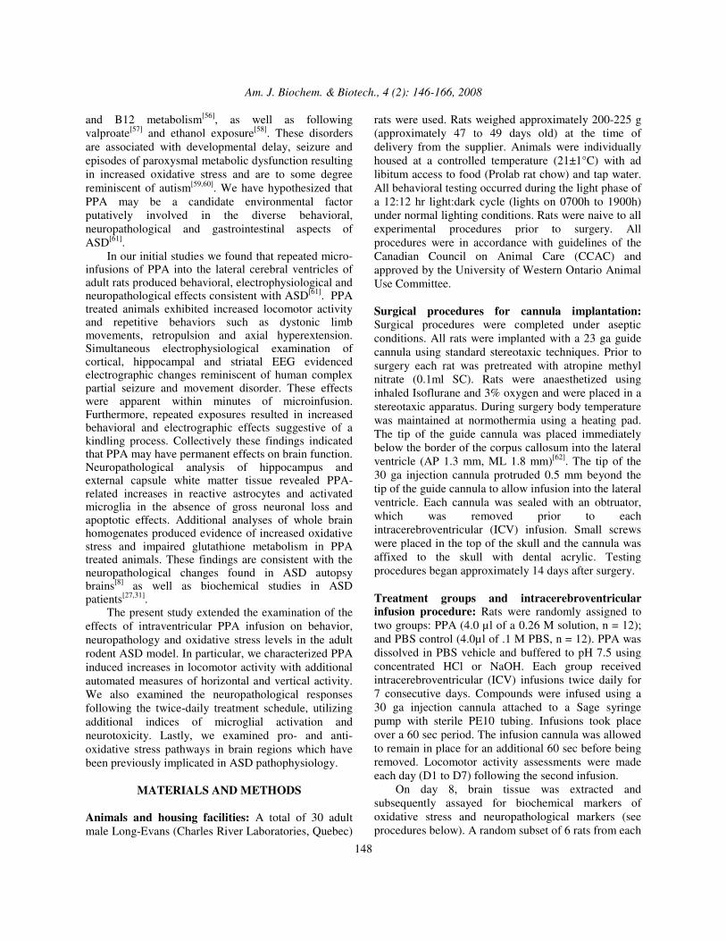
summed on a per-region basis to yield totals for both the hippocampus (i.e., 8 images summed) and white matter (i.e., 7 images summed)[64]. Two analysis methods were used for quantification of immunoreactivity. Due the diffuse nature of GFAP, IbA1 and NeuN staining, analyses were completed by using the ‘area stained’ function within ImagePro Plus which sums the immunopositive area within a digital image to provide a total immunopositive area per picture (µm2). CD68 and caspase 3’ staining was restricted within the cell membranes hence these antibodies were quantified using the ‘cell count’ function which counted immunopositive cells only. Finally, the cytotoxicity of PPA was also examined via direct visual cell counts of NeuN stained neuron cell bodies (hippocampal pyramidal cells) to determine potential cell losses in the hippocampus only. Cell bodies were counted by a trained scorer who was blind to the treatment. Cell bodies were counted only if they displayed an intact cell membrane and possessed a visible nucleus. Assessment of oxidative stress: Biomarkers for lipid and protein oxidation, glutathione (GSH) and the activity of enzymes involved in glutathione metabolism (glutathione peroxidase (GPx), glutathione reductase (GR) and glutathione S-transferase (GST)) were studied in whole brain homogenates. Assays were conducted at room temperature unless otherwise indicated. Tissue preparation: At approximately 9 A.M. on day 8, rats were decapitated without anaesthesia. Brain tissue was rapidly excised and dissected, washed with ice cold PBS, weighed and kept on ice until homogenized. The following brain areas were grossly dissected on ice: 1) neocortex ipsilateral to drug infusion cannula, encompassing all tissue lateral to the external capsule; 2) bilateral hippocampal formations; 2) basal ganglia, encompassing caudate, putamen, nucleus accumbens and septum; 3) thalamus to caudal division between superior and inferior colliculi; 4) brain stem, encompassing inferior colliculi to medulla and 5) cerebellum, dissected from cerebellar peduncles (Fig. 1). Each brain was homogenized in 10 volumes of PBS (1:10, w/v; pH 7.4) and centrifuged at 800×g for 10 min at 4°C to discard nuclei and cell debris. The supernatant was separated and stored at -80°C until needed for biochemical analyses and the pellet was discarded.
Am. J. Biochem. & Biotech., 4 (2): 146-166, 2008
151
Fig. 1: Schematic representation of gross anatomical
brain areas of rat brain homogenized for analysis of biochemical oxidative stress markers
Oxidative stress marker assays: Lipid peroxidation was determined by measuring the amounts of the secondary products of malondialdehyde (MDA) and 4-hydroxyalkenals (HAE), using a commercial kit (LPO-586; Calbiochem, La Jolla, CA), according to the manufacturer’s instructions. Protein carbonyl concentration was determined using the Protein Carbonyl Assay kit from Cayman Chemical ®, based on the reaction between protein carbonyls and 2,4-dinitrophenylhydrazine (DNPH) to form the corresponding hydrazone[73]. Superoxide dismutase (SOD) activity was assessed in the brain homogenates by measuring the dismutation of superoxide radicals generated by xanthine oxidase and hypoxanthine in a 96 well format using the Cayman Chemical® SOD Assay kit (Cayman Chemical Cat. No. 706002). Catalase activity was measured by using the Cayman Chemical® Catalase Assay kit (Cayman Cat. No. 707002) which is based on the reaction of the enzyme with methanol in the presence of an optimal concentration of H2O2. Glutathione (GSH) system assays: Glutathione system assays were done using available commercial kits from Cayman Chemical®. All absorbance measurements were performed in a Multiskan ® Spectrum microplate spectrophotometer from Thermo Labsystems. Glutathione peroxidase (GPx) activity was assayed based on the procedure described by Paglia et al.[74] using cumene hydroperoxide as substrate. Glutathione reductase (GR) activity was determined according to Carlberg and Mannervik[75]. Glutathione S-transferase (GST) was assessed by the method of Habig et al.[76] which measures the conjugation of 1-chloro-2,4-dinitrobenzene (CDNB) with reduced GSH.
Total GSH concentrations were detected by the GSH disulphide reductase 5,5´-dithiobis (2-nitrobenzoic acid) recycling method using the procedure described in the GSH assay kit from Cayman Chemicals ®. Statistical Analyses: Locomotor activity data were analyzed using a mixed design analysis of variance (ANOVA) to assess group differences (i.e., PPA and PBS) in activity levels during the baseline (BL) session as well as across each of the 7 test days (i.e., D1 to D7). Means of the PPA and PBS treated animals were compared by t-tests for the neuropathological and biochemical data. Hypothesis tests were completed using α = 0.05 as the criterion for significant effects. All statistical tests were calculated using SPSS 13.0 for Windows.
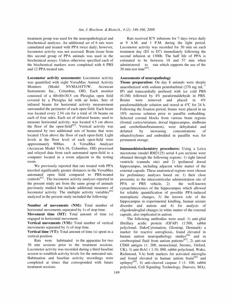
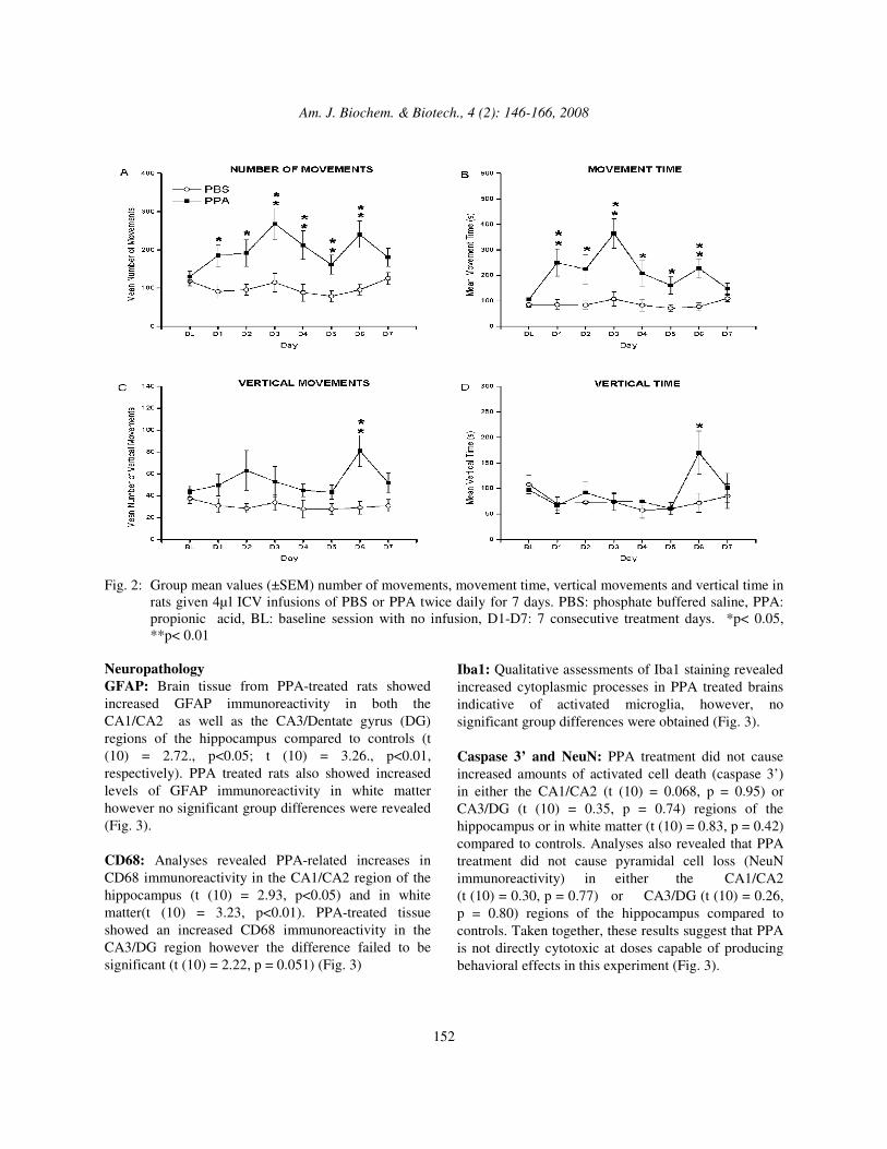
RESULTS AND DISCUSSION Baseline locomotor activity: Analyses of baseline activity levels of untreated rats revealed no significant differences between animals assigned to the PPA and PBS groups. This finding suggests that these samples of rats showed no inherent differences in locomotor activity prior to ICV infusions and that locomotor activity differences exhibited over treatment days were attributable to PPA treatment. Effects of PPA treatment on locomotor activity: The analyses revealed a significant day×treatment interaction only for MT (F (7, 154) = 3.02, p = 0.019) suggesting that the activity levels differed between treatment groups across the treatment days. A significant main effect of treatment was also obtained for NM and MT (p<0.001, for each) and VM (p <0.01) Horizontal activity variables: PPA-treated rats showed increased locomotor activity that peaked on D3. Simple effects tests were performed for each variable across baseline and treatment days. PPA-treated rats exhibited a significantly greater number of movements (NM) compared to the PBS controls on each of treatment days 1-6 (p <0.05 or better). PPA-treated rats also spent more time moving compared to PBS controls on treatment days 1-6 (p <0.05) (Fig. 2). Vertical activity variables: Analyses revealed a significant main effect for treatment on vertical activity measures. PPA-treated rats produced more vertical movements (VM) and spent more time in a vertical position (VT) compared to PBS-treated animals on treatment day 6 (p< 0.05 or better) (Fig. 2.)
Am. J. Biochem. & Biotech., 4 (2): 146-166, 2008
152
Fig. 2: Group mean values (±SEM) number of movements, movement time, vertical movements and vertical time in
rats given 4µl ICV infusions of PBS or PPA twice daily for 7 days. PBS: phosphate buffered saline, PPA: propionic acid, BL: baseline session with no infusion, D1-D7: 7 consecutive treatment days. *p< 0.05, **p< 0.01
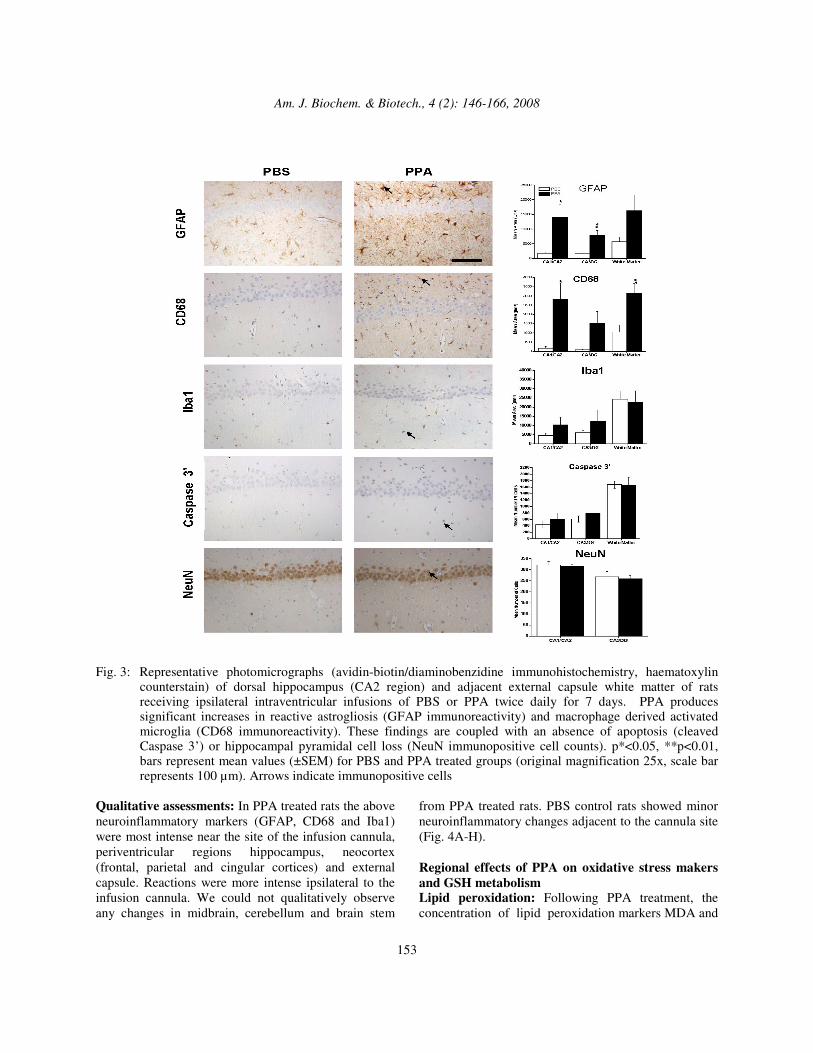
Neuropathology GFAP: Brain tissue from PPA-treated rats showed increased GFAP immunoreactivity in both the CA1/CA2 as well as the CA3/Dentate gyrus (DG) regions of the hippocampus compared to controls (t (10) = 2.72., p<0.05; t (10) = 3.26., p<0.01, respectively). PPA treated rats also showed increased levels of GFAP immunoreactivity in white matter however no significant group differences were revealed (Fig. 3). CD68: Analyses revealed PPA-related increases in CD68 immunoreactivity in the CA1/CA2 region of the hippocampus (t (10) = 2.93, p<0.05) and in white matter(t (10) = 3.23, p<0.01). PPA-treated tissue showed an increased CD68 immunoreactivity in the CA3/DG region however the difference failed to be significant (t (10) = 2.22, p = 0.051) (Fig. 3)
Iba1: Qualitative assessments of Iba1 staining revealed increased cytoplasmic processes in PPA treated brains indicative of activated microglia, however, no significant group differences were obtained (Fig. 3). Caspase 3’ and NeuN: PPA treatment did not cause increased amounts of activated cell death (caspase 3’) in either the CA1/CA2 (t (10) = 0.068, p = 0.95) or CA3/DG (t (10) = 0.35, p = 0.74) regions of the hippocampus or in white matter (t (10) = 0.83, p = 0.42) compared to controls. Analyses also revealed that PPA treatment did not cause pyramidal cell loss (NeuN immunoreactivity) in either the CA1/CA2 (t (10) = 0.30, p = 0.77) or CA3/DG (t (10) = 0.26, p = 0.80) regions of the hippocampus compared to controls. Taken together, these results suggest that PPA is not directly cytotoxic at doses capable of producing behavioral effects in this experiment (Fig. 3).
Am. J. Biochem. & Biotech., 4 (2): 146-166, 2008
153
Fig. 3: Representative photomicrographs (avidin-biotin/diaminobenzidine immunohistochemistry, haematoxylin
counterstain) of dorsal hippocampus (CA2 region) and adjacent external capsule white matter of rats receiving ipsilateral intraventricular infusions of PBS or PPA twice daily for 7 days. PPA produces significant increases in reactive astrogliosis (GFAP immunoreactivity) and macrophage derived activated microglia (CD68 immunoreactivity). These findings are coupled with an absence of apoptosis (cleaved Caspase 3’) or hippocampal pyramidal cell loss (NeuN immunopositive cell counts). p*<0.05, **p<0.01, bars represent mean values (±SEM) for PBS and PPA treated groups (original magnification 25x, scale bar represents 100 µm). Arrows indicate immunopositive cells
Qualitative assessments: In PPA treated rats the above neuroinflammatory markers (GFAP, CD68 and Iba1) were most intense near the site of the infusion cannula, periventricular regions hippocampus, neocortex (frontal, parietal and cingular cortices) and external capsule. Reactions were more intense ipsilateral to the infusion cannula. We could not qualitatively observe any changes in midbrain, cerebellum and brain stem
from PPA treated rats. PBS control rats showed minor neuroinflammatory changes adjacent to the cannula site (Fig. 4A-H). Regional effects of PPA on oxidative stress makers and GSH metabolism Lipid peroxidation: Following PPA treatment, the concentration of lipid peroxidation markers MDA and
Am. J. Biochem. & Biotech., 4 (2): 146-166, 2008
154
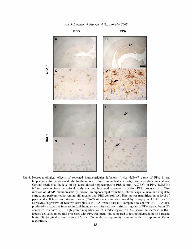
Fig. 4: Neuropathological effects of repeated intraventricular infusions (twice daily×7 days) of PPA in rat
hippocampal formation (avidin-biotin/diaminobenzidine immunohistochemistry, haematoxylin counterstain): Coronal sections at the level of ispilateral dorsal hippocampus of PBS control (A,C,E,G) or PPA (B,D,F,H) infused rodents from behavioral study eliciting increased locomotor activity. PPA produced a diffuse increase of GFAP immunoreactivity (arrows) in hippocampal formation, internal capsule, neo- and cingulate cortex, and periventricular regions (B) greater than PBS controls (A). High power magnification at level of pyramidal cell layer and stratum oriens (CA-2) of same animals showed hypertrophy of GFAP labeled astrocytes suggestive of reactive astrogliosis in PPA treated rats (D) compared to controls (C). PPA also produced a qualitative increase in Iba1 immunoreactivity (arrow) in similar regions of PPA treated brain (F) compared to control (E). High power magnification of similar region in CA-2 shows an increase in Iba1 labeled activated microglial processes with PPA treatment (H), compared to resting microglia in PBS treated brain (G) (original magnifications 1.6x and 63x, scale bar represents 1mm and scale bar represents 50µm, respectively)
Am. J. Biochem. & Biotech., 4 (2): 146-166, 2008
155
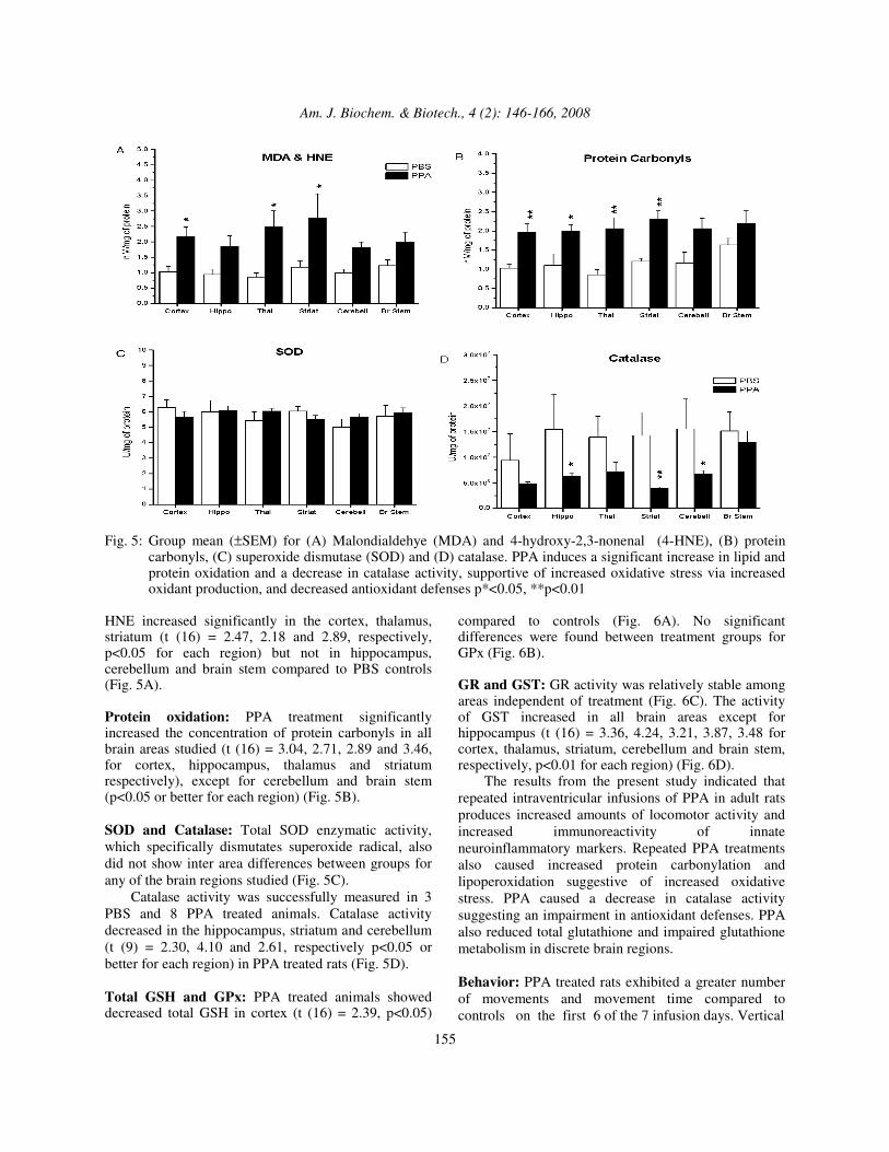
Fig. 5: Group mean (±SEM) for (A) Malondialdehye (MDA) and 4-hydroxy-2,3-nonenal (4-HNE), (B) protein
carbonyls, (C) superoxide dismutase (SOD) and (D) catalase. PPA induces a significant increase in lipid and protein oxidation and a decrease in catalase activity, supportive of increased oxidative stress via increased oxidant production, and decreased antioxidant defenses p*<0.05, **p<0.01
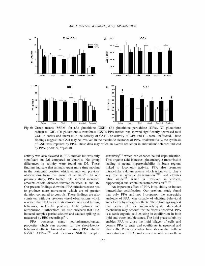
HNE increased significantly in the cortex, thalamus, striatum (t (16) = 2.47, 2.18 and 2.89, respectively, p<0.05 for each region) but not in hippocampus, cerebellum and brain stem compared to PBS controls (Fig. 5A). Protein oxidation: PPA treatment significantly increased the concentration of protein carbonyls in all brain areas studied (t (16) = 3.04, 2.71, 2.89 and 3.46, for cortex, hippocampus, thalamus and striatum respectively), except for cerebellum and brain stem (p<0.05 or better for each region) (Fig. 5B). SOD and Catalase: Total SOD enzymatic activity, which specifically dismutates superoxide radical, also did not show inter area differences between groups for any of the brain regions studied (Fig. 5C). Catalase activity was successfully measured in 3 PBS and 8 PPA treated animals. Catalase activity decreased in the hippocampus, striatum and cerebellum (t (9) = 2.30, 4.10 and 2.61, respectively p<0.05 or better for each region) in PPA treated rats (Fig. 5D). Total GSH and GPx: PPA treated animals showed decreased total GSH in cortex (t (16) = 2.39, p<0.05)
compared to controls (Fig. 6A). No significant differences were found between treatment groups for GPx (Fig. 6B). GR and GST: GR activity was relatively stable among areas independent of treatment (Fig. 6C). The activity of GST increased in all brain areas except for hippocampus (t (16) = 3.36, 4.24, 3.21, 3.87, 3.48 for cortex, thalamus, striatum, cerebellum and brain stem, respectively, p<0.01 for each region) (Fig. 6D). The results from the present study indicated that repeated intraventricular infusions of PPA in adult rats produces increased amounts of locomotor activity and increased immunoreactivity of innate neuroinflammatory markers. Repeated PPA treatments also caused increased protein carbonylation and lipoperoxidation suggestive of increased oxidative stress. PPA caused a decrease in catalase activity suggesting an impairment in antioxidant defenses. PPA also reduced total glutathione and impaired glutathione metabolism in discrete brain regions. Behavior: PPA treated rats exhibited a greater number of movements and movement time compared to controls on the first 6 of the 7 infusion days. Vertical
Am. J. Biochem. & Biotech., 4 (2): 146-166, 2008
156
Fig. 6: Group means (±SEM) for (A) glutathione (GSH), (B) glutathione peroxidase (GPx), (C) glutathione
reductase (GR), (D) glutathione s-transferase (GST). PPA treated rats showed significantly decreased total GSH in cortex and increase in the activity of GST. The activity of GPx and GR were unaffected. These findings suggest that GSH may be involved in the metabolic clearance of PPA, or alternatively, the synthesis of GSH was impaired by PPA. These data may reflex an overall reduction in antioxidant defenses induced by PPA. p*<0.05, **p<0.01
activity was also elevated in PPA animals but was only significant on D6 compared to controls. No group differences in activity were found on D7. These findings indicate that animals spent more time moving in the horizontal position which extends our previous observations from this group of animals[61]. In our previous study, PPA treated rats showed increased amounts of total distance traveled between D1 and D6. Our present findings show that PPA infusions cause rats to produce more movements which are of greater duration compared to controls. These findings are also consistent with our previous visual observations which revealed that PPA treated rats showed increased turning behaviors, snake-like postures, limb dystonia and retropulsion. Furthermore, we also observed that PPA induced complex partial seizures and caudate spiking as measured by EEG recordings[61]. PPA possesses many neuropharmacological properties which can account for the short-latency behavioral effects observed in this study. PPA inhibits Na+/K+ ATPase[78] and increases NMDA receptor
sensitivity[47] which can enhance neural depolarization. This organic acid increases glutamatergic transmission leading to neural hyperexcitability in brain regions linked to locomotor activity. PPA also promotes intracellular calcium release which is known to play a key role in synaptic transmission[79,80] and elevates nitric oxide[60] which is involved in cortical, hippocampal and striatal neurotransmission[81-85]. An important effect of PPA is its ability to induce intracellular acidification. Our previous study found that only PPA and not 1-propanol, the non-acidic analogue of PPA, was capable of eliciting behavioral and electrophysiological effects. These findings suggest that some pH or monocarboxylate dependent mechanism may account for the effects observed. PPA is a weak organic acid existing in equilibrium in both lipid and water soluble states. The lipid phase solubility enables PPA to cross the lipid bilayer of cells and permits PPA to enter and equilibrate in neuronal and glial cells. Previous studies have shown that cellular concentration of PPA produces a reversible intracellular
Am. J. Biochem. & Biotech., 4 (2): 146-166, 2008
157
acidosis and this effect is potentiated by minor reductions in extracellular pH[52,86-89]. In addition to passive accumulation in CNS cells, there is also evidence that PPA and other short chain fatty acids are specifically taken up by monocarboxylate receptors in cerebrovascular endothelium, neurons and glia[90-92]. PPA is metabolized by glial cells which involves the propionyl CoA carboxlyase enzyme[93]. PPA increases the levels of the cytotoxin propionyl CoA and depletes cytosolic carnitine stores leading to a reduction in fatty acid metabolism by mitochondria which can cause further reductions in intracellular pH[50,51]. Intracellular neuronal acidification produces widespread effects on neurotransmitter release including glutamate, dopamine, norepinepherine and serotonin, each of which can influence the production of locomotor activity[94-98]. Furthermore, alkylating analogues of PPA including 3’3’-iminodipropionitrile and 3-nitroprioprionic acid have been used to produce rodent models for movement disorders such as Huntington’s disease[99,100]. Therefore intracellular pH reduction by PPA provides a possible explanation for the PPA-induced increases in locomotor activity observed in the present study. One interesting pH dependent effect of PPA is its ability to rapidly and reversibly reduce intercellular coupling by the closure of gap junctions[52,101]. Gap junctions are composed of a family of membrane proteins known as the connexins. They are know to play an important role in electrotonic neurotransmission in the basal ganglia, deep cerebellar nuclei, prefrontal cortex, nucleus accumbens and the hippocampal formation which are involved in the production of locomotor activity[102,103]. Closure of neural gap junctions creates functional groups of neuronal clusters while closure of glial gap junctions causes neural hyperexcitability from elevations of extracellular potassium. This is thought to occur through the development of discrete gap junction linked clusters of neurons[88,104]. Furthermore closure of glial gap junctions by PPA could lead to neuronal hyperexcitablilty by impaired glial spatial buffering of cytosolic potassium or glutamate[105]. Moreover, intrastriatal infusions of gap junction blockers produce movement disorder in rodents[106]. Thus, the altered neural excitablility of neocortical, hippocampal and striatal neuronal groups in PPA treated rats would be consistent with the closure of neural or glial gap junctions by PPA[101]. PPA infusions did not induce significant increases in locomotor activity on D7 suggesting that the animals either became tolerant to PPA treatment or a
compensatory mechanism was engaged. Pathways underlying a compensatory mechanism include increased synthesis of PPA metabolizing enzymes such as propionyl CoA decarboxylase, induction of carbonic anhydrase in response to acidosis[107] or induction of pro- inflammatory (IL1-� and TNF-�) or anti-inflammatory cytokines such as TGF-� or interferon-β by the observed proliferation of microglia[108]. In summary, the observation of increased locomotor activity in PPA-treated is consistent with the expectations of an animal model for autism. These findings resemble the idiosyncratic bouts of repetitive behaviors noted in autism. Autism has been grouped with the movement disorders[109] and the contribution of seizures to these behaviors may be underappreciated[3]. Neuropathology: PPA treated rats showed increased amounts of GFAP and CD68 immunoreactivity in the hippocampus and as well as a significant increase in CD68 in white matter. The most significant findings occurred in the CA1/CA2 region of the hippocampus for each marker. Qualitatively, this pathological response was concentrated around the cerebrovascular endothelium. These findings collectively suggest an innate neuroinflammatory response characterized by reactive astrogliosis and activated microglia. These findings are similar to, but not as robust as, the GFAP and CD68 immunoreactivity seen in rats treated with PPA for 13 consecutive days[61]. Reactive astrogliosis is a common indicator for direct neurotoxic[110] and neuroplastic effects[111]. The more pronounced immunoreactivity of CD68, a marker of microglia and macrophages[112] coupled with the lack of significant Iba1 staining may suggest a PPA-induced recruitment of peripheral macrophages to the CNS. This is conceivable as short chain fatty acid receptors are known to exist on immune cells and have chemotactic and proliferative effects[53]. Interestingly, neither group showed gross neuronal loss in either the hippocampus or white matter as evidenced by the lack of group differences in either caspase 3’ or NeuN immunoreactivity (direct cell counts and total area stained). This suggests that PPA is not grossly cytotoxic, at least in the brain areas directly studied. This finding is in direct contrast to a number of other neuropathological conditions that produce neuroinflammation coupled with neuronal loss such as Parkinson[113] and Alzheimer disease[114], epilepsy[68], and AIDS dementia complex[115]. These observations have been substantiated by a number of complementary rodent models such as kainic acid induced seizures[116], experimental autoimmune
Am. J. Biochem. & Biotech., 4 (2): 146-166, 2008
158
encephalomyelitis[117] and bacterial lipopoly-saccharide[113,118]. The above neuropathological findings in the PPA rat model are similar to findings from brain tissue of autistic patients. Similarities include activated microglia and reactive astrocytes in hippocampus and neocortex, changes in white matter, coupled with comparatively minor effects in neuronal cytoarchitecture[6]. Reactive astrocytes and activated microglia release cytokines including tumor necrosis factor and macrophage chemoattractant protein, which contribute to the neuroinflammatory response and are elevated in autism[66]. Microglia produce inducible nitric oxide synthetase, leading to the production of nitric oxide, reactive oxygen species (ROS) and increased oxidative stress[119]. This neuroinflammatory process is localized near the cerebral endovasculature suggesting that altered permeability of the blood brain barrier and impaired cerebral blood flow may be components of autism[120;121]. Furthermore, specific G-protein coupled receptors (e.g., GPR41 and GPR43) for short chain fatty acids have been identified on a number of immune cells including neutrophils[122] suggesting that PPA may be involved in the activation of the immune response. It is unclear whether this neuroinflammatory response is responsible for, or restorative to the behavioral and pathophysiological effects of autism. One could propose the latter, since the PPA induced behavioral effects occurred within minutes of infusion, while activation of astrocytes and microglia, require hours to occur[123]. Further studies to examine the temporal distribution of these and other relevant neuroinflammatory markers will need to be performed. Biochemistry: PPA infusions caused an increase in oxidative stress markers, a decrease in antioxidant production and an impairment in glutathione metabolism in most brain regions. Interestingly, these biochemical effects were more widespread in the CNS and occurred in areas distal from the infusion site (e.g. cerebellum) compared to pathological findings. These observations are similar to the findings reported in our recent paper[61] which analyzed homogenates from whole brain and another recent report that examined the effects of direct intrastriatal infusion of PPA in rodents[124]. Malondialdehyde (MDA) and 4-hydroxy-2, 3-nonenal (4-HNE) are both used as key indicators of lipid peroxidation, while many of the reactions mediated by ROS conclude in the introduction of carbonyl groups into proteins[73]. Although increased trends existed in all brain regions, PPA infusions produced significantly increased amounts of
MDA/HNA and protein carbonyls in the cortex, thalamus and striatum and increased carbonylation in hippocampus. These findings suggest that ICV infusions of PPA produce widespread induction of lipoperoxidation and protein carbonylation, presumably by the production of ROS. The activity of the antioxidant enzyme SOD was not altered by PPA treatment. However, the activity of the antioxidant enzyme catalase was decreased by PPA in many brain regions with significant group differences occurring in the hippocampus, striatum and cerebellum. These findings are similar to studies which found a reduction in catalase activity in erythrocytes from autistic patients[28]. Increased oxidative damage, putatively via neuroinflammation, mitochondrial dysfunction, carnitine reduction and impaired GSH metabolism, may be an important aspect of autism[29,30,125]. Lipoperoxidation of CNS membranes are thought to lead to alterations in membrane lipid composition and fluidity, which has been proposed as playing a causative role in the disorder[27]. Furthermore, biochemical processes common to those observed in oxidative stress have been proposed as mechanisms of synaptic plasticity[126]. PPA treated animals showed decreased total GSH in cerebral cortex. GPx and GR activity were unchanged. PPA treatment produced increased GST levels in PPA treated animals compared to controls suggesting that perhaps GSH was being used for the removal of PPA or related catabolites or, alternatively, that the production of GSH was impaired. These findings are interesting since GSH plays a major role in cellular antioxidant defense, methylation pathways and in the integrity of the blood brain barrier[32]. GSH also is a major detoxifier of a broad range of xenobiotics and certain metals, some of which have been suggested to be relevant as risk factors for autism[30,33]. Genetic variations in GSH metabolism may exist in some populations at increased risk for autism[31]. Interestingly, oxidation of glutathione has been shown to result in the closure of gap junctions and thus may be a mechanism for reduction of connexin-dependent intercellular communication during acidosis or increased oxidative stress[127]. Reductions in brain GSH have been found in other pathological conditions such as experimental methylmalonic acidemia[128], 2-chloropropionic acid administration[129], experimental hyperphenylalaninemia[130] and picrotoxin induced seizures[131]. Possible mechanisms mediating the GSH decrease in neocortex include increased oxidation, release from the mitochondria and/or decreased import from the

Am. J. Biochem. & Biotech., 4 (2): 146-166, 2008
159
cytosol. Since our analysis system recycles GSSG to GSH, decreases observed are due to loss of GSH, possibly by conjugation in xenobiotics metabolism. Furthermore, GST was also increased suggesting that the decreased concentration of total GSH induced by PPA is likely due to GSH consumption by its conjugation with xenobiotics. This decline in GSH could render cells more susceptible to oxidative stress, which might account for the increased protein and lipid oxidation detected in the brain of PPA treated rats[132]. However, the decreased concentration of total GSH may also be related to a reduction in ATP- dependent synthesis. The reductions in GSH could be due to the impaired synthesis of GSH by PPA. However, because this decline was associated with increased GST activity these results are more suggestive of GSH being involved in the metabolic clearance of PPA or related metabolites in most brain areas. Whether the observed reductions in GSH metabolism in our study were the result of a general impairment in GSH synthesis or due to possible sequestration of GSH by PPA or related metabolites is unclear. Whether increased oxidative stress observed in our study leads to mitochondrial failure, or alternatively, mitochondrial failure induced by PPA leads to oxidative stress is a subject of further study. Nonetheless, the observed decline in GSH concentration may render CNS cells more susceptible to oxidative stress[33], which could account for the increased protein and lipid oxidation detected in the brain of PPA treated rats and possibly human autism. It is interesting to note that the oxidative stress effect of PPA administration was far more widespread in the brain than the innate neuroinflammatory effects, which were primarily located in ipsilateral hippocampus, neocortex and external capsule white matter. Other studies have also implicated subcortical areas such as basal ganglia[133] and thalamus[134] in autism. As well, behavioral effects of PPA occurred shortly after the first infusion suggesting that at least some of the behavioral effects of PPA may be independent of astro- and microglial activation. Indeed, astrogliosis may be protective and play a key role in repair in the CNS following injury[111]. As we used gross dissection regions for these initial oxidative stress studies, we cannot rule out PPA induced biochemical and neuropathological changes in discrete cell populations (i.e., brain stem).
SUMMARY These behavioral, neuropathological and biochemical findings in our rodent PPA model provide further support for the hypothesis that autism may be a
systemic metabolic encephalopathic process affecting widespread brain regions rather than a static primary brain disorder involving discrete CNS areas[21]. This work, coupled with our initial study[61], shows that PPA can produce behavioral changes that resemble those found in ASD. Recently we have also found evidence of reversible impairments in social behavior following PPA exposure[135]. The similarities in innate neuroinflammatory and oxidative stress changes between the animal model and human ASD cases could represent similar metabolic or immune mediated processes[9] directly or indirectly associated with PPA. Of particular interest are our observations of broad impairments in the GSH and catalase metabolism which could provide a common mechanism for increased oxidative stress and increased environmental sensitivity to a variety of environmental compounds[27]. Despite the promise of the PPA rodent model fulfilling some of the behavioral, neuropathological and biochemical criteria for autism[136], some caution needs to be used in suggesting PPA as a possible risk factor for the human condition. Firstly, it should be noted that there are no studies directly measuring PPA in ASD patients. However, there is indirect evidence linking PPA to alterations in fatty acid metabolism[137], deficiencies in carnitine[125] and alterations in biotin[138] in autism. There are similar metabolic trends noted following exposure to valproate, a teratogenic risk factor in autism[57]. Furthermore PPA producing gut clostridial species have been found in a subset of patients with regressive ASD [40]. Secondly, our initial studies have used mature rodents. Given the known effects of PPA on neurodevelopmental processes[65] (i.e., cell signalling[139], gene expression[140,141], apoptosis[142] and modulation of gap junctions[52]) future studies need to examine the effects of PPA at pre- and peri-natal developmental stages. However, intraventricular administration of PPA does show promise as an environmental trigger that links the disparate genetic, neuropathological and systemic findings[31] associated with ASD.
ACKNOWLEDGEMENTS The authors thank Jue Fan, Yudith Ramos and Lisa Tichenoff for their expert technical assistance. We also thank Carlos Pardo (Johns Hopkins University) for advice on Iba1 staining. This research was supported by contributions from GoodLife Children’s Foundation and Round for a Reason Charities to DFM. We also
Am. J. Biochem. & Biotech., 4 (2): 146-166, 2008
160
extend our gratitude to David Patchell-Evans, Tamara Rogerson and Kilee Patchell-Evans. Supported in part by grants from The Natural Sciences and Engineering Research Council of Canada to DPC and KPO.
REFERENCES 1. Andres, C., 2002. Molecular genetics and animal
models in autistic disorder. Brain Res. Bull., 57: 109-119.
2. Zwaigenbaum, L., S. Bryson, T. Rogers, W. Roberts, J. Brian and P. Szatmari, 2005. Behavioral manifestations of autism in the first year of life. Int. J. Dev. Neurosci., 23: 143-152.
3. Besag, F.M., 2004. Behavioral aspects of pediatric epilepsy syndromes. Epilepsy Behav., 5 (Suppl 1): S3-S13.
4. Cody, H., K. Pelphrey and J. Piven, 2002. Structural and functional magnetic resonance imaging of autism. Int. J. Dev. Neurosci., 20: 421-438.
5. Courchesne, E. and K. Pierce, 2005. Brain overgrowth in autism during a critical time in development: implications for frontal pyramidal neuron and interneuron development and connectivity. Int. J. Dev. Neurosci., 23: 153-170.
6. Bauman, M.L. and T.L. Kemper, 2005. Neuroanatomic observations of the brain in autism: a review and future directions. Int. J. Dev. Neurosci., 23: 183-187.
7. Herbert, M.R., D.A. Ziegler, C.K. Deutsch, L.M. O'Brien, D.N. Kennedy, P.A. Filipek, A.I. Bakardjiev, J. Hodgson, M. Takeoka, N. Makris and V.S. Caviness, Jr., 2005. Brain asymmetries in autism and developmental language disorder: a nested whole-brain analysis. Brain 128: 213-226.
8. Vargas, D.L., C. Nascimbene, C. Krishnan, A.W. Zimmerman and C.A. Pardo. 2005. Neuroglial activation and neuroinflammation in the brain of patients with autism. Ann. Neurol., 57: 67-81.
9. Ashwood, P., S. Wills and W.J. Van de, 2006. The immune response in autism: a new frontier for autism research. J. Leukoc. Biol., 80: 1-15.
10. Dawson, G., S. Webb, G.D. Schellenberg, S. Dager, S. Friedman, E. Aylward and T. Richards. 2002. Defining the broader phenotype of autism: genetic, brain and behavioral perspectives. Dev. Psychopathol., 14: 581-611.
11. Folstein, S.E. and B. Rosen-Sheidley, 2001. Genetics of autism: complex aetiology for a heterogeneous disorder. Nat. Rev. Genet., 2: 943-955.
12. Trottier, G., L. Srivastava and C.D. Walker, 1999. Etiology of infantile autism: a review of recent advances in genetic and neurobiological research. J. Psychiatry Neurosci., 24: 103-115.
13. Waltereit, R., H. Welzl, J. Dichgans, H.P. Lipp, W.J. Schmidt and M. Weller, 2006. Enhanced episodic-like memory and kindling epilepsy in a rat model of tuberous sclerosis. J. Neurochem. 96: 407-413.
14. Tierney, E., N.A. Nwokoro, F.D. Porter, L.S. Freund, J.K. Ghuman and R.I. Kelley, 2001. Behavior phenotype in the RSH/Smith-Lemli-Opitz syndrome. Am. J. Med. Genet., 98: 191-200.
15. Bandim, J.M., L.O. Ventura, M.T. Miller, H.C. Almeida and A.E. Costa, 2003. Autism and Mobius sequence: an exploratory study of children in northeastern Brazil. Arq Neuropsiquiatr., 61: 181-185.
16. Persico, A.M. and T. Bourgeron, 2006. Searching for ways out of the autism maze: genetic, epigenetic and environmental clues. Trends Neurosci., 29: 349-358.
17. Bartlett, C.W., N. Gharani, J.H. Millonig and L.M. Brzustowicz, 2005. Three autism candidate genes: a synthesis of human genetic analysis with other disciplines. Int. J. Dev. Neurosci., 23: 221-234.
18. Scherer, S.W., J. Cheung, J.R. MacDonald, L.R. Osborne, K. Nakabayashi, J.A. Herbrick, A.R. Carson, L. Parker-Katiraee, J. Skaug, R. Khaja, J. Zhang, A.K. Hudek, M. Li, M. Haddad, G.E. Duggan, B.A. Fernandez, E. Kanematsu, S. Gentles, C.C. Christopoulos, S. Choufani, D. Kwasnicka, X.H. Zheng, Z. Lai, D. Nusskern, Q. Zhang, Z. Gu, F. Lu, S. Zeesman, M.J. Nowaczyk, I. Teshima, D. Chitayat, C. Shuman, R. Weksberg, E.H. Zackai, T.A. Grebe, S.R. Cox, S.J. Kirkpatrick, N. Rahman, J.M. Friedman, H.H. Heng, P.G. Pelicci, F. Lo-Coco, E. Belloni, L.G. Shaffer, B. Pober, C.C. Morton, J.F. Gusella, G.A. Bruns, B.R. Korf, B.J. Quade, A.H. Ligon, H. Ferguson, A.W. Higgins, N.T. Leach, S.R. Herrick, E. Lemyre, C.G. Farra, H.G. Kim, A.M. Summers, K.W. Gripp, W. Roberts, P. Szatmari, E.J. Winsor, K.H. Grzeschik, A. Teebi, B.A. Minassian, J. Kere, L. Armengol, M.A. Pujana, X. Estivill, M.D. Wilson, B.F. Koop, S. Tosi, G.E. Moore, A.P. Boright, E. Zlotorynski, B. Kerem, P.M. Kroisel, E. Petek, D.G. Oscier, S.J. Mould, H. Dohner, K. Dohner, J.M. Rommens, J.B. Vincent, J.C. Venter, P.W. Li, R.J. Mural, M.D. Adams and L.C. Tsui, 2003. Human chromosome 7: DNA sequence and Biology. Sci., 300: 767-772.
Am. J. Biochem. & Biotech., 4 (2): 146-166, 2008
161
19. Hu, V.W., B.C. Frank, S. Heine, N.H. Lee and J. Quackenbush, 2006. Gene expression profiling of lymphoblastoid cell lines from monozygotic twins discordant in severity of autism reveals differential regulation of neurologically relevant genes. BMC.Genomics, 7: 118.
20. Bertrand, J., A. Mars, C. Boyle, F. Bove, M. Yeargin-Allsopp and P. Decoufle, 2001. Prevalence of autism in a United States population: the Brick Township, New Jersey, investigation. Pediatrics, 108: 1155-1161.
21. Herbert, M.R., J.P. Russo, S. Yang, J. Roohi, M. Blaxill, S.G. Kahler, L. Cremer and E. Hatchwell, 2006. Autism and Environmental Genomics. Neurotoxicology.
22. Arndt, T.L., C.J. Stodgell and P.M. Rodier, 2005. The teratology of autism. Int. J. Dev. Neurosci., 23: 189-199.
23. Ingram, J.L., S.M. Peckham, B. Tisdale and P.M. Rodier, 2000. Prenatal exposure of rats to valproic acid reproduces the cerebellar anomalies associated with autism. Neurotoxicol. Teratol, 22: 319-324.
24. Zerrate, M.C., M. Pletnikov, S.L. Connors, D.L. Vargas, F.J. Seidler, A.W. Zimmerman, T.A. Slotkin and C.A. Pardo, 2007. Neuroinflammation and Behavioral Abnormalities after Neonatal Terbutaline Treatment in Rats: Implications for Autism. J. Pharmacol. Exp. Ther.
25. Fatemi, S.H., A.E. Cuadra, E.E. El Fakahany, R.W. Sidwell and P. Thuras, 2000. Prenatal viral infection causes alterations in nNOS expression in developing mouse brains. Neuroreport 11: 1493-1496.
26. Lancaster, K., D.M. Dietz, T.H. Moran and M.V. Pletnikov, 2006. Abnormal social behaviors in young and adult rats neonatally infected with Borna disease virus. Behav. Brain Res.
27. Chauhan, A. and V. Chauhan, 2006. Oxidative stress in autism. Pathophysiology, 13: 171-181.
28. Zoroglu, S.S., F. Armutcu, S. Ozen, A. Gurel, E. Sivasli, O. Yetkin and I. Meram, 2004. Increased oxidative stress and altered activities of erythrocyte free radical scavenging enzymes in autism. Eur. Arch. Psychiatry Clin. Neurosci., 254: 143-147.
29. Chauhan, A., V. Chauhan, W.T. Brown and I. Cohen, 2004. Oxidative stress in autism: increased lipid peroxidation and reduced serum levels of ceruloplasmin and transferrin--the antioxidant proteins. Life Sci., 75: 2539-2549.
30. James, S.J., P. Cutler, S. Melnyk, S. Jernigan, L. Janak, D.W. Gaylor and J.A. Neubrander, 2004. Metabolic biomarkers of increased oxidative stress and impaired methylation capacity in children with autism. Am. J. Clin. Nutr., 80: 1611-1617.
31. James, S.J., S. Melnyk, S. Jernigan, M.A. Cleves, C.H. Halsted, D.H. Wong, P. Cutler, K. Bock, M. Boris, J.J. Bradstreet, S.M. Baker and D.W. Gaylor, 2006. Metabolic endophenotype and related genotypes are associated with oxidative stress in children with autism. Am. J. Med. Genet. B Neuropsychiatr. Genet.
32. Dringen, R., J.M. Gutterer and J. Hirrlinger, 2000. Glutathione metabolism in brain metabolic interaction between astrocytes and neurons in the defense against reactive oxygen species. Eur. J. Biochem., 267: 4912-4916.
33. Monks, T.J., J.F. Ghersi-Egea, M. Philbert, A.J. Cooper and E.A. Lock, 1999. Symposium overview: the role of glutathione in neuroprotection and neurotoxicity. Toxicol. Sci., 51: 161-177.
34. Buyske, S., T.A. Williams, A.E. Mars, E.S. Stenroos, S.X. Ming, R. Wang, M. Sreenath, M.F. Factura, C. Reddy, G.H. Lambert and W.G. Johnson, 2006. Analysis of case-parent trios at a locus with a deletion allele: association of GSTM1 with autism. BMC. Genet., 7: 8.
35. Williams, T.A., A.E. Mars, S.G. Buyske, E.S. Stenroos, R. Wang, M.F. Factura-Santiago, G.H. Lambert and W.G. Johnson, 2007. Risk of autistic disorder in affected offspring of mothers with a glutathione S-transferase P1 haplotype. Arch. Pediatr. Adolesc. Med., 161: 356-361.
36. Horvath, K., J.C. Papadimitriou, A. Rabsztyn, C. Drachenberg and J.T. Tildon, 1999. Gastrointestinal abnormalities in children with autistic disorder. J. Pediatr., 135: 559-563.
37. Fallon, J., 2005. Could one of the most widely prescribed antibiotics amoxicillin/clavulanate augmentin be a risk factor for autism? Med Hypotheses, 64: 312-315.
38. Jyonouchi, H., S. Sun and N. Itokazu, 2002. Innate immunity associated with inflammatory responses and cytokine production against common dietary proteins in patients with autism spectrum disorder. Neuropsychobiology, 46: 76-84.
39. Wakefield, A.J., S.H. Murch, A. Anthony, J. Linnell, D.M. Casson, M. Malik, M. Berelowitz, A.P. Dhillon, M.A. Thomson, P. Harvey, A. Valentine, S.E. Davies and J.A. Walker-Smith, 1998. Ileal-lymphoid-nodular hyperplasia, non-specific colitis and pervasive developmental disorder in children. Lancet, 351: 637-641.
Am. J. Biochem. & Biotech., 4 (2): 146-166, 2008
162
40. Finegold, S.M., D. Molitoris, Y. Song, C. Liu, M.L. Vaisanen, E. Bolte, M. McTeague, R. Sandler, H. Wexler, E.M. Marlowe, M.D. Collins, P.A. Lawson, P. Summanen, M. Baysallar, T.J. Tomzynski, E. Read, E. Johnson, R. Rolfe, P. Nasir, H. Shah, D.A. Haake, P. Manning and A. Kaul, 2002. Gastrointestinal microflora studies in late-onset autism. Clin. Infect. Dis., 35: S6-S16.
41. Campbell, D.B., J.S. Sutcliffe, P.J. Ebert, R. Militerni, C. Bravaccio, S. Trillo, M. Elia, C. Schneider, R. Melmed, R. Sacco, A.M. Persico and P. Levitt, 2006. A genetic variant that disrupts MET transcription is associated with autism. Proc. Natl. Acad. Sci. USA, 103: 16834-16839.
42. Thompson, G.N., J.H. Walter, J L. Bresson, G.C. Ford, S.L. Lyonnet, R.A. Chalmers, J.M. Saudubray, J.V. Leonard and D. Halliday, 1990. Sources of propionate in inborn errors of propionate metabolism. Metabolism, 39: 1133-1137.
43. Kuijper, E.J., B. Coignard and P. Tull, 2006. Emergence of Clostridium difficile-associated disease in North America and Europe. Clin. Microbiol. Infect., 12 (Suppl 6): 2-18.
44. Jan, G., A.S. Belzacq, D. Haouzi, A. Rouault, D. Metivier, G. Kroemer and C. Brenner, 2002. Propionibacteria induce apoptosis of colorectal carcinoma cells via short-chain fatty acids acting on mitochondria. Cell Death Differ, 9: 179-188.
45. Zarate, G., S. Gonzalez and A.P. Chaia, 2004. Assessing survival of dairy propionibacteria in gastrointestinal conditions and adherence to intestinal epithelia. Methods Mol. Biol., 268: 423-432.
46. Mally, P., R. Mishra, S. Gandhi, M.H. Decastro, B.B. Nankova and E.F. LaGamma, 2004. Stereospecific regulation of tyrosine hydroxylase and proenkephalin genes by short-chain fatty acids in rat PC12 cells. Pediatr. Res., 55: 847-854.
47. De Mattos-Dutra, A., R. Meirelles, d.R. Bevilaqua, T. Kommers, S.T. Wofchuk, M. Wajner and R. Pessoa-Pureur, 2000. Methylmalonic and propionic acids increase the in vitro incorporation of 32P into cytoskeletal proteins from cerebral cortex of young rats through NMDA glutamate receptors. Brain Res., 856: 111-118.
48. Hinnebusch, B.F., S. Meng, J.T. Wu, S.Y. Archer and R.A. Hodin, 2002. The effects of short-chain fatty acids on human colon cancer cell phenotype are associated with histone hyperacetylation. J. Nutr., 132: 1012-1017.
49. Nicot, A., T. Otto, P. Brabet and E.M. Dicicco-Bloom, 2004. Altered social behavior in pituitary adenylate cyclase-activating polypeptide type I receptor-deficient mice. J. Neurosci., 24: 8786-8795.
50. Brass, E.P. and R.A. Beyerinck, 1987. Interactions of propionate and carnitine metabolism in isolated rat hepatocytes. Metabolism, 36: 781-787.
51. Brass, E.P. and R.A. Beyerinck, 1988. Effects of propionate and carnitine ton the hepatic oxidation of short- and medium-chain-length fatty acids. J. Biochem., 250: 819-825.
52. Rorig, B., G. Klausa and B. Sutor, 1996. Intracellular acidification reduced gap junction coupling between immature rat neocortical pyramidal neurones. J. Physiol., 490 (pt-1): 31-49.
53. Cavaglieri, C.R., A. Nishiyama, L.C. Fernandes, R. Curi, E.A. Miles and P.C. Calder, 2003. Differential effects of short-chain fatty acids on proliferation and production of pro- and anti-inflammatory cytokines by cultured lymphocytes. Life Sci., 73: 1683-1690.
54. De Baulny, H.O., J.F. Benoist, O. Rigal, G. Touati, D. Rabier and J.M. Saudubray, 2005. Methylmalonic and propionic acidaemias: management and outcome. J. Inherit. Metab. Dis., 28: 415-423.
55. Ozand, P.T., G.G. Gascon, M. Al Essa, S. Joshi, E. Al Jishi, S. Bakheet, J. Al Watban, M.Z. Al Kawi and O. Dabbagh, 1998. Biotin-responsive basal ganglia disease: a novel entity. Brain, 121 (pt7): 1267-1279.
56. Brass, E.P. and L.J. Ruff, 1989. Effect of carnitine on propionate metabolism in the vitamin B-12-deficient rat. J. Nutr., 119: 1196-1202.
57. Schulpis, K.H., G.A. Karikas, J. Tjamouranis, S. Regoutas and S. Tsakiris, 2001. Low serum biotinidase activity in children with valproic acid monotherapy. Epilepsia, 42: 1359-1362.
58. Calabrese, V. and V. Rizza, 1999. Formation of propionate after short-term ethanol treatment and its interaction with the carnitine pool in rat. Alcohol, 19: 169-176.
59. Nyhan, W.L., C. Bay, E.W. Beyer and M. Mazi, 1999. Neurologic nonmetabolic presentation of propionic acidemia. Arch. Neurol., 56: 1143-1147.
60. Wajner, M., A. Latini, A.T. Wyse and C.S. Dutra-Filho. 2004. The role of oxidative damage in the neuropathology of organic acidurias: insights from animal studies. J. Inherit. Metab. Dis., 27: 427-448.
Am. J. Biochem. & Biotech., 4 (2): 146-166, 2008
163
61. MacFabe, D.F., D.P. Cain, K. Rodriguez-Capote, A.E. Franklin, J.E. Hoffman, F. Boon, A.R. Taylor, M. Kavaliers and K.P. Ossenkopp, 2007. Neurobiological effects of intraventricular propionic acid in rats: possible role of short chain fatty acids on the pathogenesis and characteristics of autism spectrum disorders. Behav. Brain Res., 176: 149-169.
62. Paxinos, G. and C. Watson, 1986. The Rat Brain in Stereotaxic Coordinates, 2nd Edn. Ed. Academic Press, Sydney, Montreal.
63. Ossenkopp, K.P. and M. Kavaliers, 1996. Measuring spontaneous locomotor activity in small mammals. In K.P. Ossenkopp, M. Kavaliers and P.R. Sanberg, editors Measuring Movement and Locomotion: From Invertabrates to Humans R.G. Landes Company, Austin, Texas., 33-59.
64. Ossenkopp, K.P. and D.S. Mazmanian, 1985. The measurement and integration of behavioral variables: aggregation and complexity as important issues. Neurobehav. Toxicol. Teratol., 7: 95-100.
65. Brusque, A.M., C.F. Mello, D.N. Buchanan, S.T. Terracciano, M.P. Rocha, C.R. Vargas, C.M. Wannmacher and M. Wajner, 1999. Effect of chemically induced propionic acidemia on neurobehavioral development of rats. Pharmacol. Biochem. Behav., 64: 529-534.
66. Vargas, D.L., C. Nascimbene, C. Krishnan, A.W. Zimmerman and C.A. Pardo, 2004. Neuroglial activation and neuroinflammation in the brain of patients with autism. Ann. Neurol., 57: 67-81.
67. Rosengren, L.E., G. Ahlsen, M. Belfrage, C. Gillberg, K.G. Haglid and A. Hamberger, 1992. A sensitive ELISA for glial fibrillary acidic protein: application in CSF of children. J. Neurosci. Methods, 44: 113-119.
68. Beach, T.G., W.B. Woodhurst, D.B. MacDonald and M.W. Jones, 1995. Reactive microglia in hippocampal sclerosis associated with human temporal lobe epilepsy. Neurosci. Lett., 191: 27-30.
69. Kondratyev, A. and K. Gale, 2000. Intracerebral injection of caspase-3 inhibitor prevents neuronal apoptosis after kainic acid-evoked status epilepticus. Brain Res. Mol. Brain Res., 75: 216-224.
70. Noyan-Ashraf, M.H., F. Brandizzi and B.H. Juurlink. 2005. Constitutive nuclear localization of activated caspase 3 in subpopulations of the astroglial family of cells. GLIA, 49: 588-593.
71. Casella, G.T., M.B. Bunge and P.M. Wood, 2004. Improved immunocytochemical identification of neural, endothelial and inflammatory cell types in paraffin-embedded injured adult rat spinal cord. J. Neurosci. Methods, 139: 1-11.
72. Shi, S.R., R.J. Cote and C.R. Taylor, 2001. Antigen retrieval techniques: current perspectives. J. Histochem. Cytochem., 49: 931-937.
73. Levine, R.L., J.A. Williams, E.R. Stadtman and E. Shacter, 1994. Carbonyl assays for determination of oxidatively modified proteins. Methods Enzymol., 233: 346-357.
74. Paglia, D.E. and W.N. Valentine, 1967. Studies on the quantitative and qualitative characterization of erythrocyte glutathione peroxidase. J. Lab Clin. Med., 70: 158-169.
75. Carlberg, I. and B. Mannervik, 1985. Glutathione reductase. Methods Enzymol., 113: 484-490.
76. Habig, W. H., M.J. Pabst and W.B. Jakoby, 1974. Glutathione S-transferases. The first enzymatic step in mercapturic acid formation. J. Biol. Chem., 249: 7130-7139.
77. Lowry, O.H., N.J. Rosenbrough, A.L. FARR and R.J. Randall, 1951. Protein measurement with the Folin phenol reagent. J. Biol. Chem., 193: 265-275.
78. Wyse, A.T., A.M. Brusque, C.G. Silva, E.L. Streck, M. Wajner and C.M. Wannmacher, 1998. Inhibition of Na+,K+-ATPase from rat brain cortex by propionic acid. Neuroreport, 9: 1719-1721.
79. Bronstein, J.M., D.B. Farber and C.G. Wasterlain, 1993. Regulation of type-II calmodulin kinase: functional implications. Brain Res. Rev., 18: 135-147.
80. Nakao, S., A. Fujii and R. Niederman, 1992. Alteration of cytoplasmic Ca2+ in resting and stimulated human neutrophils by short-chain carboxylic acids at neutral pH. Infect. Immun., 60: 5307-5311.
81. Dawson, V.L. and T.M. Dawson, 1996. Nitric oxide neurotoxicity. J. Chem. Neuroanat., 10: 179-190.
82. Centonze, D., P. Gubellini, A. Pisani, G. Bernardi and P. Calabresi, 2003. Dopamine, acetylcholine and nitric oxide systems interact to induce corticostriatal synaptic plasticity. Rev. Neurosci., 14: 207-216.
83. Shih, Y.H., A.W. Lee, Y.H. Huang, M.H. Ko and Y.S. Fu, 2004. GABAergic neuron death in the striatum following kainate-induced damage of hippocampal neurons: evidence for the role of NO in locomotion. Int. J. Neurosci., 114: 1119-1132.
Am. J. Biochem. & Biotech., 4 (2): 146-166, 2008
164
84. Akyol, O., S.S. Zoroglu, F. Armutcu, S. Sahin and A. Gurel, 2004. Nitric oxide as a physiopathological factor in neuropsychiatric disorders. In vivo., 18: 377-390.
85. Virmani, A., F. Gaetani, S. Imam, Z. Binienda and S. Ali, 2003. Possible mechanism for the neuroprotective effects of L-carnitine on methamphetamine-evoked neurotoxicity. Ann. N Y. Acad. Sci., 993: 197-207.
86. Karuri, A.R., E. Dobrowsky and I.F. Tannock, 1993. Selective cellular acidification and toxicity of weak organic acids in an acidic microenvironment. Br. J. Cancer, 68: 1080-1087.
87. Spray, D.C., J. Nerbonne, d.C. Campos, A.L. Harris and M.V. Bennett, 1984. Substituted benzyl acetates: a new class of compounds that reduce gap junctional conductance by cytoplasmic acidification. J. Cell Biol., 99: 174-179.
88. Perez-Velazquez, J.L., T.A. Valiante, Carlen and PL., 1994. Modulation of gap junctional mechanisms during calcium-free induced field burst activity: a possible role for electrotonic coupling in epileptogenesis. J. Neurosci., 14: 4308-4317.
89. Bonnet, U., D. Bingmann and M. Wiemann, 2000. Intracellular pH modulates spontaneous and epileptiform bioelectric activity of hippocampal CA3-neurones. Eur. Neuropsychopharmacol., 10: 97-103.
90. Maurer, M.H., M. Canis, W. Kuschinsky and R. Duelli, 2004. Correlation between local monocarboxylate transporter 1 (MCT1) and glucose transporter 1 (GLUT1) densities in the adult rat brain. Neurosci. Lett., 355: 105-108.
91. Bergersen, L., A. Rafiki and O.P. Ottersen, 2002. Immunogold cytochemistry identifies specialized membrane domains for monocarboxylate transport in the central nervous system. Neurochem. Res., 27: 89-96.
92. Nehlig, A., 2004. Brain uptake and metabolism of ketone bodies in animal models. Prostaglandins Leukot. Essent. Fatty Acids, 70:265-275.
93. Nguyen, N.H., C. Morland, S.V. Gonzalez, F. Rise, J. Storm-Mathisen, V. Gundersen and B. Hassel, 2007. Propionate increases neuronal histone acetylation, but is metabolized oxidatively by glia. Relevance for propionic acidemia. J. Neurochem., 101: 806-814.
94. Remblier, C., R. Pontcharraud, C. Tallineau, A. Piriou and F. Huguet, 1999. Lactic acid-induced increase of extracellular dopamine measured by microdialysis in rat striatum: evidence for glutamatergic and oxidative mechanisms. Brain Res., 837: 22-28.
95. Cannizzaro, C., R. Monastero, M. Vacca and M. Martire, 2003. [3H]-DA release evoked by low pH medium and internal H+ accumulation in rat hypothalamic synaptosomes: involvement of calcium ions. Neurochem. Int., 43: 9-17.
96. Smith, I.D., M.A. Sutton and R.J. Beninger, 1997. Rotational bias in intact rats following intrastriatal injections of dopaminergic drugs. Pharmacol. Biochem. Behav., 58: 431-441.
97. Filosa, J.A., J.B. Dean and R.W. Putnam, 2002. Role of intracellular and extracellular pH in the chemosensitive response of rat locus coeruleus neurones. J. Physiol., 541: 493-509.
98. Severson, C.A., W. Wang, V.A. Pieribone, C.I. Dohle and G.B. Richerson, 2003. Midbrain serotonergic neurons are central pH chemoreceptors. Nat. Neurosci., 6: 1139-1140.
99. Moser, V.C. and W.K. Boyes, 1993. Prolonged neurobehavioral and visual effects of short-term exposure to 3,3'-iminodipropionitrile (IDPN) in rats. Fundam. Appl. Toxicol., 21: 277-290.
100. Borlongan, C.V., T.K. Koutouzis and P.R. Sanberg, 1997. 3-Nitropropionic acid animal model and Huntington's disease. Neurosci. Biobehav. Rev., 21: 289-293.
101. Fujita, K., K. Nakanishi, K. Sobue, T. Ueki, K. Asai and T. Kato, 1998. Astrocytic gap junction blockage and neuronal Ca2+ oscillation in neuron-astrocyte cocultures in vitro. Neurochem. Int., 33: 41-49.
102. O'Donnell, P. and A.A. Grace, 1997. Cortical afferents modulate striatal gap junction permeability via nitric oxide. Neurosci., 76: 1-5.
103. O'Donnell, P., J. Greene, N. Pabello, B.L. Lewis and A.A. Grace, 1999. Modulation of cell firing in the nucleus accumben. Ann. N. Y. Acad. Sci., 877: 157-175.
104. LeBeau, F.E., R.D. Traub, H. Monyer, M.A. Whittington and E.H. Buhl, 2003. The role of electrical signaling via gap junctions in the generation of fast network oscillations. Brain Res. Bull., 62: 3-13.
105. Anders, J.J., 1988. Lactic acid inhibition of gap junctional intercellular communication in in vitro astrocytes as measured by fluorescence recovery after laser photobleaching. GLIA, 1: 371-379.
106. Moore, H. and A.A. Grace, 2002. A role for electrotonic coupling in the striatum in the expression of dopamine receptor-mediated stereotypies. Neuropsychopharmacol., 27: 980-992.
107. Nogradi, A., F. Domoki, R. Degi, S. Borda, M. Pakaski, A. Szabo and F. Bari, 2003. Up-regulation of cerebral carbonic anhydrase by anoxic stress in piglets. J. Neurochem., 85: 843-850.
Am. J. Biochem. & Biotech., 4 (2): 146-166, 2008
165
108. Hinkerohe, D., D. Smikalla, A. Haghikia, K. Heupel, C.G. Haase, R. Dermietzel and P.M. Faustmann, 2005. Effects of cytokines on microglial phenotypes and astroglial coupling in an inflammatory coculture model. GLIA, 52: 85-97.
109. Palomo, T., R.J. Beninger, R.M. Kostrzewa and T. Archer, 2003. Brain sites of movement disorder: genetic and environmental agents in neurodevelopmental perturbations. Neurotox. Res., 5: 1-26.
110. O'Callaghan, J.P., K.F. Jensen and D.B. Miller, 1995. Quantitative aspects of drug and toxicant-induced astrogliosis. Neurochem. Int., 26: 115-124.
111. Briones, T.L., J. Woods, M. Wadowska, M. Rogozinska and M. Nguyen, 2006. Astrocytic changes in the hippocampus and functional recovery after cerebral ischemia are facilitated by rehabilitation training. Behav. Brain Res., 171: 17-25.
112. Mittelbronn, M., K. Dietz, H. J. Schluesener and R. Meyermann, 2001. Local distribution of microglia in the normal adult human central nervous system differs by up to one order of magnitude. Acta Neuropathol. (Berl), 101: 249-255.
113. Iravani, M.M., K. Kashefi, P. Mander, S. Rose and P. Jenner, 2002. Involvement of inducible nitric oxide synthase in inflammation-induced dopaminergic neurodegeneration. Neurosci., 110: 49-58.
114. Sheng, J.G., R.E. Mrak and W.S. Griffin, 1997. Glial-neuronal interactions in Alzheimer disease: progressive association of IL-1alpha+ microglia and S100beta+ astrocytes with neurofibrillary tangle stages. J. Neuropathol. Exp. Neurol., 56: 285-290.
115. Seilhean, D., K. Kobayashi, Y. He, T. Uchihara, O. Rosenblum, C. Katlama, F. Bricaire, C. Duyckaerts and J.J. Hauw, 1997. Tumor necrosis factor-alpha, microglia and astrocytes in AIDS dementia complex. Acta Neuropathol. (Berl), 93: 508-517.
116. Sola, C., C. Casal, J.M. Tusell and J. Serratosa, 2002. Astrocytes enhance lipopolysaccharide-induced nitric oxide production by microglial cells. Eur. J. Neurosci., 16: 1275-1283.
117. Ponomarev, E.D., L.P. Shriver, K. Maresz and B.N. Dittel, 2005. Microglial cell activation and proliferation precedes the onset of CNS autoimmunity. J. Neurosci. Res., 81: 374-389.
118. Sugaya, K., S. Chou, S.J. Xu and M. McKinney, 1998. Indicators of glial activation and brain oxidative stress after intraventricular infusion of endotoxin. Mol. Brain Res., 58: 1-9.
119. Dringen, R., 2005. Oxidative and antioxidative potential of brain microglial cells. Antioxid. Redox. Signal., 7: 1223-1233.
120. Yao, Y., W.J. Walsh, W.R. McGinnis and D. Pratico, 2006. Altered vascular phenotype in autism: correlation with oxidative stress. Arch. Neurol., 63: 1161-1164.
121. Stolp, H.B., K.M. Dziegielewska, C.J. Ek, A.M. Potter and N.R. Saunders, 2005. Long-term changes in blood-brain barrier permeability and white matter following prolonged systemic inflammation in early development in the rat. Eur. J. Neurosci., 22: 2805-2816.
122. Le Poul, E., C. Loison, S. Struyf, J.Y. Springael, V. Lannoy, M.E. Decobecq, S. Brezillon, V. Dupriez, G. Vassart, J. Van Damme, M. Parmentier and M. Detheux, 2003. Functional characterization of human receptors for short chain fatty acids and their role in polymorphonuclear cell activation. J. Biol. Chem., 278: 25481-25489.
123. Benjelloun, N., S. Renolleau, A. Represa, Y. Ben Ari and C. Charriaut-Marlangue, 1999. Inflammatory responses in the cerebral cortex after ischemia in the P7 neonatal rat. Stroke, 30: 1916-1923.
124. Rigo, F.K., L. Pasquetti, C.R. Malfatti, M.R. Fighera, R.C. Coelho, C.Z. Petri and C.F. Mello, 2006. Propionic acid induces convulsions and protein carbonylation in rats. Neurosci. Lett., 408: 151-154.
125. Filipek, P.A., J. Juranek, M.T. Nguyen, C. Cummings and J.J. Gargus, 2004. Relative carnitine deficiency in autism. J. Autism Dev. Disord., 34: 615-623.
126. Mattson, M.P. and D. Liu, 2002. Energetics and oxidative stress in synaptic plasticity and neurodegenerative disorders. Neuromolecular. Med., 2: 215-231.
127. Caruso, R.L., B.L. Upham, C. Harris and J.E. Trosko, 2005. Biphasic lindane-induced oxidation of glutathione and inhibition of gap junctions in myometrial cells. Toxicol. Sci., 86: 417-426.
128. Schuck, P.F., R.B. Rosa, L.F. Pettenuzzo, A. Sitta, C.M. Wannmacher, A.T. Wyse and M. Wajner, 2004. Inhibition of mitochondrial creatine kinase activity from rat cerebral cortex by methylmalonic acid. Neurochem. Int., 45: 661-667.
129. Wyatt, I., A. Gyte, G. Mainwaring, P.S. Widdowson and E.A. Lock, 1996. Glutathione depletion in the liver and brain produced by 2-chloropropionic acid: relevance to cerebellar granule cell necrosis. Arch. Toxicol., 70: 380-389.
Am. J. Biochem. & Biotech., 4 (2): 146-166, 2008
166
130. Kienzle Hagen, M.E., C.D. Pederzolli, A.M. Sgaravatti, R. Bridi, M. Wajner, C.M. Wannmacher, A.T. Wyse and C.S. Dutra-Filho, 2002. Experimental hyperphenylalaninemia provokes oxidative stress in rat brain. Biochim. Biophys. Acta, 1586: 344-352.
131. Rajasekaran, K., 2005. Seizure-induced oxidative stress in rat brain regions: blockade by nNOS inhibition. Pharmacol. Biochem. Behav., 80: 263-272.
132. Brusque, A.M., S.T. Terracciano, F.U. Fontella, C. Vargas, C.G. da Silva, A.T. Wyse, V.M. Trindade, C.M. Wannmacher and M. Wajner, 1998. Chronic administration of propionic acid reduces ganglioside N-acetylneuraminic acid concentration in cerebellum of young rats. J. Neurol. Sci., 158: 121-124.
133. Singer, H.S., C.M. Morris, P.N. Williams, D.Y. Yoon, J.J. Hong and A.W. Zimmerman, 2006. Antibrain antibodies in children with autism and their unaffected siblings. J. Neuroimmunol., 178: 149-155.
134. Tsatsanis, K.D., B.P. Rourke, A. Klin, F.R. Volkmar, D. Cicchetti and R.T. Schultz, 2003. Reduced thalamic volume in high-functioning individuals with autism. Biol. Psychiatry, 53: 121-129.
135. Shultz, S.R., Whelan, J. Cain, D.P., Boon, F., Ossenkopp, K.P. and MacFabe, D.F., 2006. Intraventricular infusions of propionic acid reversibly impair social behavior in Long--Evans rats. Neuroscience Meeting Planner.Atlanta, GA: Society for Neuroscience.
136. Crawley, J.N., 2004. Designing mouse behavioral tasks relevant to autistic-like behaviors. Ment. Retard. Dev. Disabil. Res. Rev., 10: 248-258.
137. Wu, K., C. Wasterlain, L. Sachs and P. Siekevitz, 1990. Effect of septal kindling on glutamate binding and calcium/calmodulin-dependent phosphorylation in a postsynaptic density fraction isolated from rat cerebral cortex. Proc. Natl. Acad. Sci. USA, 87: 5298-5302.
138. Zaffanello, M., G. Zamboni, E. Fontana, L. Zoccante and L. Tato, 2003. A case of partial biotinidase deficiency associated with autism. Child Neuropsychol., 9: 184-188.
139. Shah, P., B.B. Nankova, S. Parab and E.F. La Gamma, 2006. Short chain fatty acids induce TH gene expression via ERK-dependent phosphorylation of CREB protein. Brain Res., 1107: 13-23.
140. DeCastro, M., B.B. Nankova, P. Shah, P. Patel, P.V. Mally, R. Mishra and E.F. La Gamma, 2005. Short chain fatty acids regulate tyrosine hydroxylase gene expression through a cAMP-dependent signaling pathway. Brain Res. Mol. Brain Res., 142: 28-38.
141. Menegola, E., F. Di Renzo, M.L. Broccia, M. Prudenziati, S. Minucci, V. Massa and E. Giavini, 2005. Inhibition of histone deacetylase activity on specific embryonic tissues as a new mechanism for teratogenicity. Birth Defects Res. B Dev. Reprod. Toxicol., 74: 392-398.
142. Frantseva, M.V., L. Kokarovtseva, C.G. Naus, P.L. Carlen, D. MacFabe and J.L. Perez Velazquez, 2002. Specific gap junctions enhance the neuronal vulnerability to brain traumatic injury. J. Neurosci., 22: 644-653.