Embed Size (px)
Citation preview
CASE REPORT
146 Acta Medica Indonesiana - The Indonesian Journal of Internal Medicine
A Patient With Plaque Type Morphea Mimicking Systemic Lupus Erythematosus
Wardhana1, EA Datau2
1 Department of Internal Medicine, Siloam International Hospitals. Karawaci, Indonesia.2 Department of Internal Medicine, Prof. Dr. RD Kandou General Hospital & Sitti Maryam Islamic Hospital, Manado, North Sulawesi, Indonesia.
Correspondence mail:Siloam Hospitals Group’s CEO Office, Siloam Hospital Lippo Village. 5th floor. Jl. Siloam No.6, Karawaci, Indonesia. email: [email protected]
ABSTRAKMorfea merupakan penyakit jaringan penyambung yang jarang dengan gambaran utama berupa penebalan
dermis tanpa disertai keterlibatan organ dalam. Penyakit ini juga dikenal sebagai bagian dari skleroderma terlokalisir. Berdasarkan gambaran klinis dan kedalaman jaringan yang terlibat, morfea dikelompokkan ke dalam beberapa bentuk dan sekitar dua pertiga orang dewasa dengan morfea mempunyai tipe plak. Produksi kolagen yang berlebihan oleh fibroblast merupakan penyebab kelainan pada morfea dan mekanisme terjadinya aktivitas fibroblast yang berlebihan ini masih belum diketahui, meskipun beberapa mekanisme pernah diajukan. Morfe tipe plak biasanya bersifat ringan dan dapat sembuh dengan sendirinya. Morfea tipe plak yang penampilan klinisnya menyerupai lupus eritematosus sistemik, misalnya meliputi alopesia dan ulkus mukosa di mulut, jarang dijumpai.
Sebuah kasus morfea tipe plak pada wanita berusia 20 tahun dibahas. Pasien ini diobati dengan imunosupresan dan antioksidan local maupun sistemik. Kondisi paisen membaik tanpa disertai efek samping yang berarti.
Kata kunci: morfea, tipe plak.
ABSTRACTMorphea is an uncommon connective tissue disease with the most prominent feature being thickening or
fibrosis of the dermal without internal organ involvement. It is also known as a part of localized scleroderma. Based on clinical presentation and depth of tissue involvement, morphea is classified into several forms, and about two thirds of adults with morphea have plaque type. Overproduction of collagen production by fibroblast is the cause of abnormality in morphea, and the hyperactivity mechanism of fibroblast is still unknown, although there are several mechanisms already proposed. Plaque type morphea is actually a benign and self limited. Plaque type morphea that mimicking systemic lupus erythematosus in clinical appearance, such as alopecia and oral mucosal ulcers, is uncommon.
A case of plaque type morphea mimicking systemic lupus erythematosus in a 20 year old woman was discussed. The patient was treated with local and systemic immunosuppressant and antioxydant. The patient’s condition is improved without any significant side effects.
Key words: morphea, plaque type.
147
Vol 47 • Number 2 • April 2015 A patient with plaque type morphea mimicking SLE
INTRODUCTIONMorphea is an uncommon persistent
condition in which there are areas of thickened skin and cutaneus tissue from excessive collagen deposition and one of its type is plaque type. It is also known as localized scleroderma, and it may affect adults and children. Morphea includes specific condition raging from very small plaques only involving the skin to widespread disease, and it is discriminated from systemic sclerosis by its supposed no internal organ involvement.1,2
Plaque type morphea is mainly involved women. Adequate studies on the incidence and prevalence have not been performed. Plaque type morphea also may be under-reported as a physician may be unaware of this disorder and small morphea plaques may be less often referred to a Dermatologist or Rheumatologist.2,3
Overproduction of collagen by fibroblast in affected tissues is common in plaque type morphea, although the mechanism by which these fibroblasts are activated is unknown. Possible causes of morphea are radiation, infection and vaccination, trauma, genetic factor, endothelial cell injury, immunologic (autoimmunity) and inflammatory activation, and dysregulation of collagen production. In histologic finding, there is a thickening process and homogenous collagen bundles of the skin, and since the autoimmunity is the most possible cause of plaque type morphea, there are many autoantibodies may be found in individual affected.1,2 Plaque type morphea is usually a benign and self-limiting disease.1
Reported below is a case of a 20-year old woman with plaque type morphea, presenting with difficulty in swallowing, ulcers on oral mucosal and tongue, face redness, loss of hair, localized upper and lower arms skin tightness and redness, and also pain on chest, and abdomen.
CASE ILLUSTRATIONA 20-year old woman from Ternate, North
Mollucas, came to our clinic with chief complaint of difficulty of swallowing since 1 month ago, accompanied by painless oral mucosal ulcers. She had a routine evaluation by an internist at Ternate and regularly took Celecoxib, Esomeprazol, and Chloroquin since the doctor
said that she had lupus. Before she felt this complaint, she already lost her hair slowly since 2 months ago, face redness about 1 month ago, and fever because of the influenza about 2 weeks ago followed by the appearance of redness and localized, painful, and hard circular plaques on the skin, each on her right upper and lower arms.
Since 9 years ago, the patient had 3 episodes of pain on her joints especially on both of her
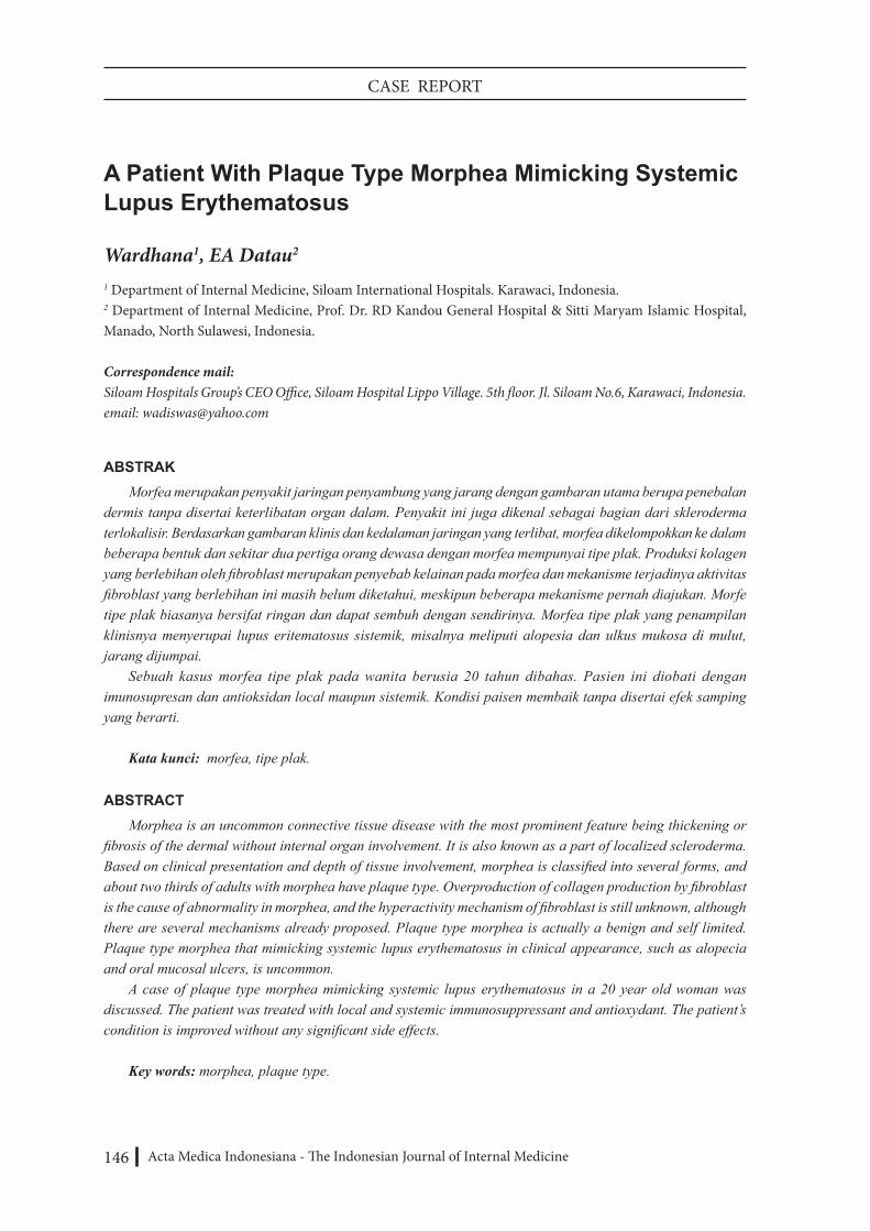
Figure 1. The patient’s alopecia
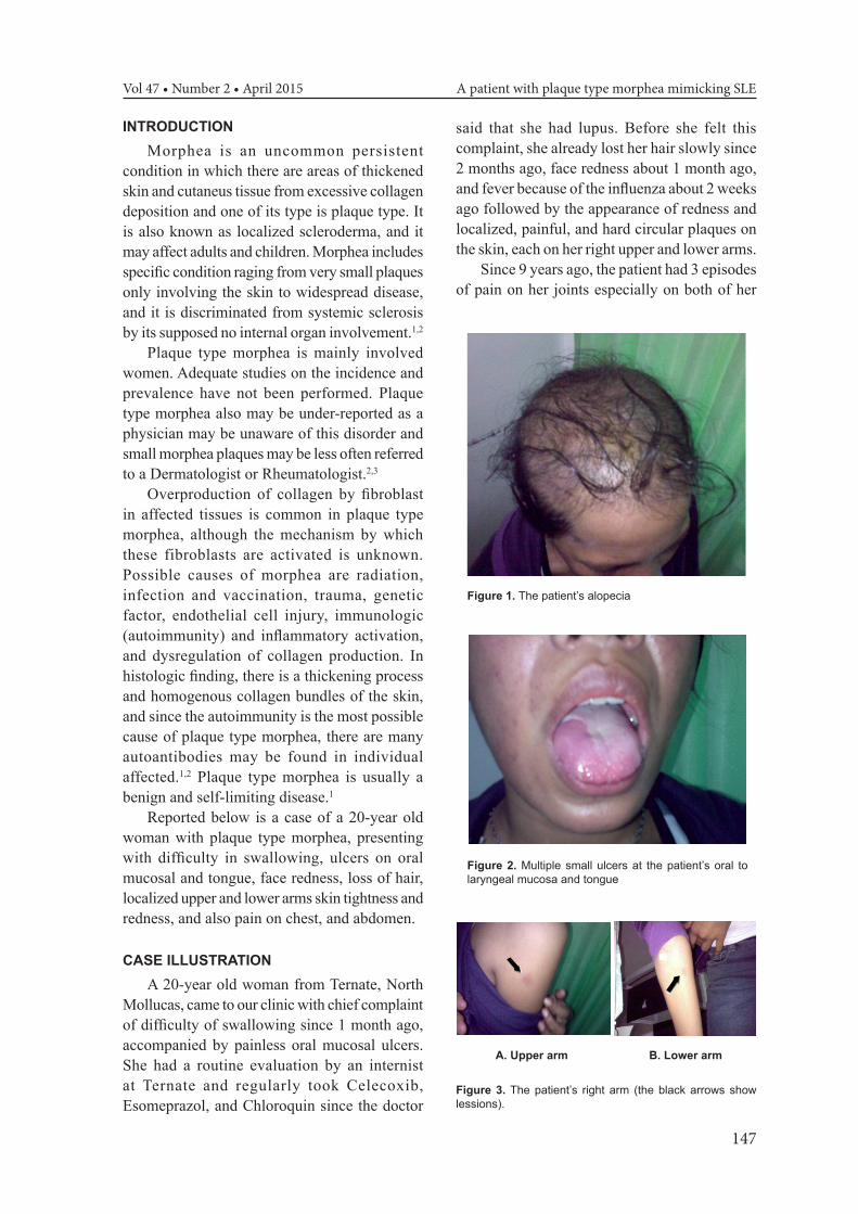
Figure 2. Multiple small ulcers at the patient’s oral to laryngeal mucosa and tongue
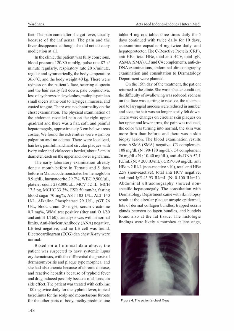
A. Upper arm B. Lower arm
Figure 3. The patient’s right arm (the black arrows show lessions).
148
Wardhana Acta Med Indones-Indones J Intern Med
feet. The pain came after she got fever, usually because of the influenza. The pain and the fever disappeared although she did not take any medication at all.
In the clinic, the patient was fully conscious, blood pressure 120/80 mmHg, pulse rate 87 x/minute regularly, respiratory rate 20 x/minute, regular and symmetrically, the body temperature 36.6°C, and the body weight 40 kg. There were redness on the patient’s face, scarring alopecia and the hair easily felt down, pale conjunctiva, loss of eyebrows and eyelashes, multiple painless small ulcers at the oral to laryngeal mucosa, and coated tongue. There was no abnormality on the chest examination. The physical examination of the abdomen revealed pain on the right upper quadrant and there was a flat, soft, and painful hepatomegaly, approximately 3 cm below arcus costae. We found the extremities were warm on palpation and no edema. There were localized, hairless, painfull, and hard circular plaques with ivory color and violaceous border, about 3 cm in diameter, each on the upper and lower right arms.
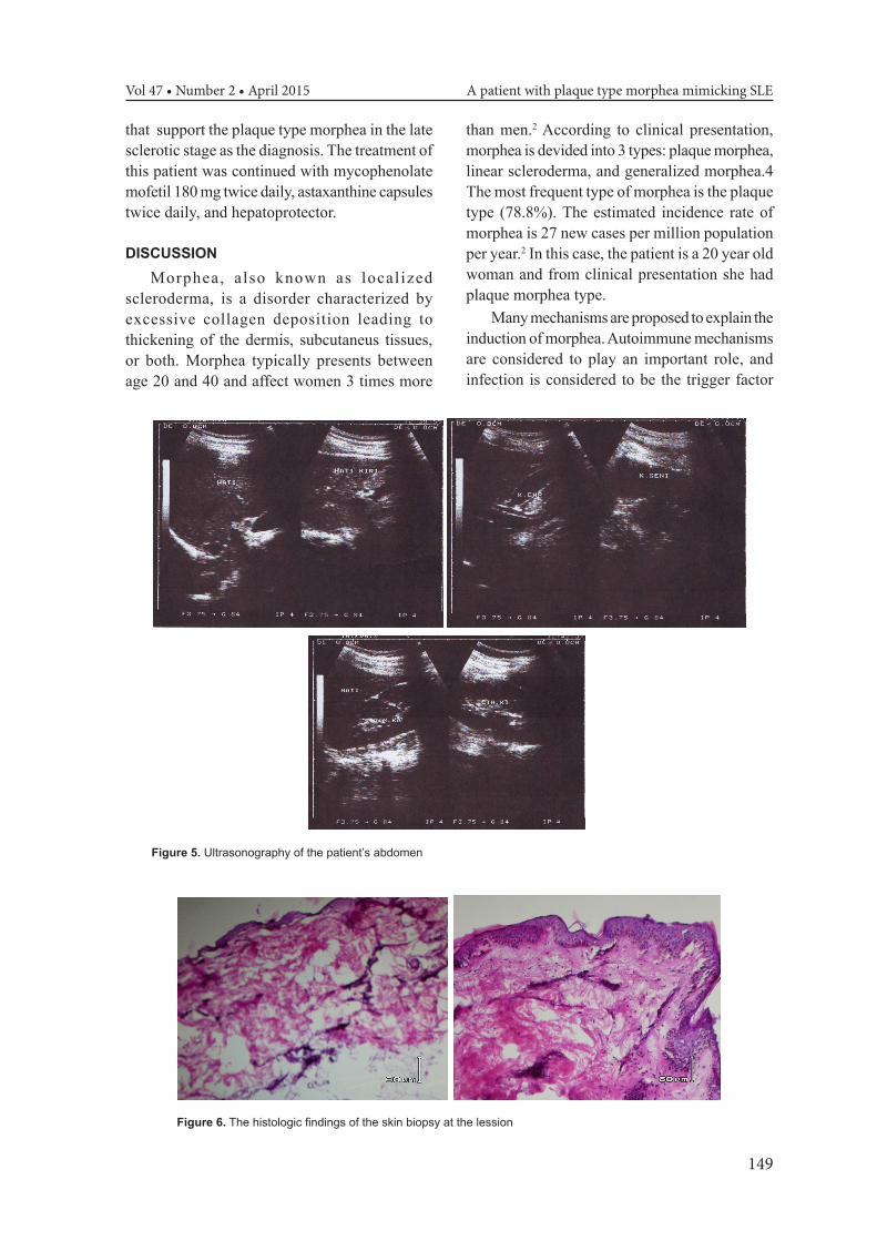
The early laboratory examination already done a month before in Ternate and 5 days before in Manado, demonstrated her hemoglobin 9.9 g/dL, haematocrite 29.7%, WBC 9,900/µL, platelet count 238,000/µL, MCV 52 fL, MCH 17.3 pg, MCHC 33.3%, ESR 50 mm/hr, fasting blood sugar 70 mg%, AST 103 U/L, ALT 140 U/L, Alkaline Phosphatase 79 U/L, γGT 76 U/L, blood ureum 20 mg%, serum creatinine 0.7 mg%, Widal test positive (titer anti O 1/80 and anti H 1/160), urinalysis was with in normal limits, Anti-Nuclear Antibody (ANA) negative, LE test negative, and no LE cell was found. Electrocardiogram (ECG) dan chest X-ray were normal.
Based on all clinical data above, the patient was suspected to have systemic lupus erythematosus, with the differential diagnosis of dermatomyositis and plaque type morphea, and she had also anemia because of chronic disease, and reactive hepatitis because of typhoid fever and drug induced possibly because of chloroquin side effect. The patient was treated with cefixime 100 mg twice daily for the typhoid fever, topical tacrolimus for the scalp and mometasone furoate for the other parts of body, methylprednisolone
tablet 4 mg one tablet three times daily for 5 days continued with twice daily for 10 days, astaxanthine capsules 4 mg twice daily, and hepatoprotector. The C-Reactive Protein (CRP), anti HBs, total HBc, total anti HCV, total IgE, ASMA (SMA), C3 and C4 complements, anti-ds-DNA examinations, abdominal ultrasonography examination and consultation to Dermatology Department were planned.
On the 15th day of the treatment, the patient returned to the clinic. She was in better condition, the difficulty of swallowing was reduced, redness on the face was starting to resolve, the ulcers at oral to laryngeal mucosa were reduced in number and size, the hair was no longer easily felt down. There were changes on circular skin plaques on her upper and lower arms, the pain was reduced, the color was turning into normal, the skin was more firm than before, and there was a skin biopsy lesion. The blood examination results were ASMA (SMA) negative, C3 complement 108 mg/dL (N : 90-180 mg/dL), C4 complement 26 mg/dL (N : 10-40 mg/dL), anti-ds-DNA 52.1 IU/mL (N: ≤ 200 IU/mL), CRP 0.39 mg/dL, anti HBs < 2 IU/L (non-reactive <10), total anti HBc 2.58 (non-reactive), total anti HCV negative, and total IgE 43.93 IU/mL (N: 0-100 IU/mL). Abdominal ultrasonography showed non-specific hepatomegaly. The consultation with Dermatology Department came with skin biopsy result at the circular plaque: atropic epidermal, lots of dermal collagen bundles, trapped eccrin glands between collagen bundles, and bundels found also at the fat tissue. The histologic findings were likely a morphea at late stage,
Figure 4. The patient’s chest X-ray
149
Vol 47 • Number 2 • April 2015 A patient with plaque type morphea mimicking SLE
that support the plaque type morphea in the late sclerotic stage as the diagnosis. The treatment of this patient was continued with mycophenolate mofetil 180 mg twice daily, astaxanthine capsules twice daily, and hepatoprotector.
DISCUSSIONMorphea, also known as local ized
scleroderma, is a disorder characterized by excessive collagen deposition leading to thickening of the dermis, subcutaneus tissues, or both. Morphea typically presents between age 20 and 40 and affect women 3 times more
than men.2 According to clinical presentation, morphea is devided into 3 types: plaque morphea, linear scleroderma, and generalized morphea.4 The most frequent type of morphea is the plaque type (78.8%). The estimated incidence rate of morphea is 27 new cases per million population per year.2 In this case, the patient is a 20 year old woman and from clinical presentation she had plaque morphea type.
Many mechanisms are proposed to explain the induction of morphea. Autoimmune mechanisms are considered to play an important role, and infection is considered to be the trigger factor
Figure 5. Ultrasonography of the patient’s abdomen
Figure 6. The histologic findings of the skin biopsy at the lession
150
Wardhana Acta Med Indones-Indones J Intern Med
of morphea.1 The immune system responses to autoantigens are induced by cryptic self-epitops that are generated by modification of the self-antigens during apoptosis.5
The clinical presentation of patients with plaque type morphea varies depending on the level of tissue involvement and extend of the lesion. In general, we may find scalp alopecia, loss of eyebrows and eyelashes, nail dystrophy, restricted respiration if there is extensive truncal morphea, restricted mobility, contractures, and deformity, muscle weakness if there is peripheral nerves involvement, ptosis, extra-ocular muscle dysfunction, anterior uveitis, episcleritis, glaucoma, xerophthalmia, keratitis, altered dentition, malocclusion, and asymmetry of the tongue.1 The Plaque type of morphea is the most common and the lessions are relatively superficial, primarily involving the dermis.1 Plaque-type morphea lesions are characterized as circular, indurated palques that range from 1 cm to more than 20 cm in diameter. They often begins as erythematous to violaceous patches or slightly edematous plaques. With the disease progression, sclerosis develops centrally as the lesions undergo peripheral expansion. The surface become smooth and shiny over time, with loss of hair follicles and sweat glands. The margins are often surrounded by a zone of violaceous color or telengiectasias. Over a period of months to years, the skin softens and the dermis becomes atrophic.1,2,6 In this patient, we found the patient had scalp alopecia, loss of eyebrows and eyelashes, and 2 plaque type morphea lesions, about 3 cm in diameter, each on the upper and lower right arms.
On the additional examination in the patients with plaque type morphea, there are several blood parameters and also the histology examination of skin biopsy and other additional examinations. To make sure that there is no internal organs involvement, beside the routine blood parameters, the liver and kiney function should be measured and the ECG and chest X-ray should be done.1,2 Several studies have shown increased levels of anti nuclear antibody, rheumatoid factor, anti-single-stranded DNA antibodies, anticentromer antibody, antibodies to Th/To ribonucleoprotein, antihistone antibody,
anti-topoisomerase II antibody, Circulating Intercellular Adhesion Mollecule-1 (ICAM-1), Soluble Vascular Cell Adhesion Mollecule-1 (sVCAM-1), E-selectin, soluble CD4, CD8, CD23, and fibrogenic T-helper 2 cytokines such as interleukin (IL)-4, IL-6, and Transforming Growth Factor-Beta (TGF-β) in the patients with plaque type morphea, and anti-Cu/Zn-superoxide dismutase antibodies.7-13 These cytokines recruit eosinophils and other inflammatory cells, induce fibroblast to synthesize excessive collagen and connective-tissue growth factor which enchances and perpetuates the fibrotic effect of TGF-β.14 Among those serum autoantibodies, antitopoisomerase 2-alpha is one of the major autoantibodies in localized scleroderma.4
Histologic findings of the skin biopsy from the patients with plaque type morphea in the early inflammatory stage, the epidermis is flattened and athrophic with loss of the rete ridges. There is dermal edema and the collagen fibrils become eosinophylic. There is a perivascular infiltrate of lymphocytes, plasma cells or macrophages.1 In the late sclerotic stage, the inflammatory infiltrate typically disappears. Collagen bundles in the reticular dermis and subcutis become thick, closely packed, and deeply eosinophylic. Atrophic eccrine glands appear to be trapped within the middle of the thickened dermis as subcutaneus fat is replaced by collagen. A paucity of blood vessels is seen and the dermal appendages are lost.1,2,6
The patient in this case gave a clinical presentation mimicking systemic lupus erythematosus but did not give any positive results in autoantibody measurements. The histologic findings supported the diagnosis as late sclerotic stage of plaque type morphea. The possibility of the autoantibody measurements did not give any positive results was the morphea was in the late sclerotic stage. We did not measure all the autoantibodies mentioned above because not all of them were available. Anemia was possibly caused from chronic disease, and the increase of liver function was possibly from reactive hepatitis because of typhoid fever and the side effects of chloroquine. Chloroquine can form higly reactive radicals, the hydroperoxides, which can cause hepatotoxicity.15 Reactive hepatitis
151
Vol 47 • Number 2 • April 2015 A patient with plaque type morphea mimicking SLE
because of typhoid fever may happen in people with malnutrition and weakness of the immune system.16
The treament of morphea is difficult and only a few controlled clinical trials have been published. There are 2 kinds of therapy, the topical and systemic therapies.16 The topical therapies are the ultra-potent topical steroids cream in superficial active lessions to reduce inflammation, tacrolimus to inhibit T cells activation, vitamin D derivatives inhibit proliferation of fibroblast lead to softening and repigmentation of morphea lession, phototherapy using PUVA and UVA-1 will induce matrix metallo proteinase that reduces the procollagen in the skin.17-19 The systemic therapies are immunosuppression using the combination of methotrexate and corticosteroids, penicillin and penicillamine, oral vitamin D derivatives, mycophenolate mofetil which inhibits inosine mohophosphate dehydrogenase, an enzyme for controlling the novo purine synthesis used by proliferating lymphocytes, and vitamin A derivatives which inhibits TGF-β.20-22 This patient got cefixime 100 mg twice daily for the typhoid fever, mometasone furoate, methylprednisolone tablet 4 mg one tablet three times daily for 5 days continued with twice daily for 10 days then changed with mycofenolate mofetil 180 mg twice daily for the next 15 days, astaxanthine capsules 4 mg twice daily as antioxydant, and hepatoprotector.
The prognosis of the plaque type morphea improved spontaneously within 3-5 years although it may last as long as 25 years, but atropy, induration and pigment changes may persist.6,23 The patient in this case had a good prognosis because of the significant clinical improvements to the therapies given.
CONCLUSIONA case of plaque type morphea in a 20 year-
old woman has been discussed. The patient was diagnosed with plaque type morphea, anemia because of chronic disease, and reactive hepatitis hepatitis because of typhoid fever and drug induced possibly because of chloroquin side effect. The management of this patient were cefixime 100 mg twice daily for her typhoid
fever, topical tacrolimus for the scalp and mometasone furoate for other parts of the body, methylprednisolone tablet 4 mg one tablet three times daily for 5 days continued with twice daily for 10 days then changed with mycophenolate mofetil 180 mg twice daily, astaxanthine capsules mg twice daily as antioxydant, and hepatoprotector. Significant improvements were observed. The prognosis of this patient is good.
REFERENCES1. Rocken M, Ghoreschi K. Morphea and lichen
sclerosus. In: Bolognia JL, Jorizzo JL, Rapini RP, eds. Dermatology. 1st ed. Philadelphia: Mosby; 2003. p. 1503-17.
2. James DW, Berger TG, Elston DM. Connective tissue diseases. Philadelphia: Saunders Elsevier; 2006. p. 171-6.
3. Toledano C, Rabhi S, Kettaneh A, et al. Localized scleroderma: a series of 52 patients. Eur J Intern Med. 2000;20(3):331-6.
4. Takehara K, Sato S. Localized scleroderma is an autoimmune disorder. Rheumatol. 2005;44:274-9.
5. Hayakawa I, Hasegawa M, Takehara K, Sato S. Anti-DNA topoisomerase IIα autoantibodies in localized scleroderma. Arthritis Rheum. 2004;50(1):227-32.
6. Docrat ME. Morphea (localized scleroderma). Curr Allergy Clin Immunol. 2006;19(4):192-4.
7. Ruffatti A, Peserica A, Glorioso S, et al. Anticentromere antibody in localized scleroderma. J Am Acad Dermatol. 1986;15(4 Pt 1):637-42.
8. Yamane K, Ihn H, Kubo M, et al. Antibodies to Th/To ribonucleoprotein in patients with localized scleroderma. Rheumatol (Oxford). 2001;40(6):683-6.
9. Yamane K, Ihn H, Kubo M, er al. Increased serum levels of soluble vascular cell adhesion molecule 1 and E-selectin in patients with localized scleroderma. J Am Acad Dermatol. 2000;42(1 Pt 1):64-9.
10. Ihn H, Fujimoto M, Sato S, et al. Increased levels of circulating intercellular adhesion molecule-1 in patients with localized scleroderma. J Am Acad Dermatol. 1994;31(4):591-5.
11. Sato S, Fujimoto M, Kikuchi K, et al. Soluble CD4 and CS8 in serum from patients with localized scleroderma. Arch Dermatol Res. 1996;288(7):358-62.
12. Sato S, Fujimoto M, Kikuchi K, et al. Elevated soluble CD23 levels in the sera from patients with localized scleroderma. Arch Dermatol Res. 1996;288(2):74-8.
13. Leask A, Denton CP, Abraham DJ. Insights into moleculer mechanism of chronic fibrosis:the role of connective tissue groeth factor in scleroderma. J Invest Dermatol. 2004;122:1-6.
14. Nagai M, Hasegawa M, Takehara K, Sato S. Novel Autoantibody to Cu/Zn superoxide dismutase in patients with localized scleroderma. J Invest Dermatol.
152
Wardhana Acta Med Indones-Indones J Intern Med
2004;122:594-601.15. Pari L, Amali DR. Protective role of tetrahydrocurcumin
(THC) an active principle of turmeric on chloroquin induced hepatotoxicity in rats. J Pharm Pharmaceut Sci. 2005;8(1):115-23.
16. Widodo D. Demam tifoid. In: Sudoyo AW, Setiyohadi B, Alwi I, Kolopaking MS, Setiati S, eds. Buku ajar ilmu penyakit dalam. Jakarta: Pusat Penerbitan Departemen Ilmu Penyakit Dalam FKUI; 2006. p. 1174-9.
17. Kieffer MA. Topical vitamin D analogs. Dermatol Nurs. 2004;16(1):89-90.
18. Kreuter A, Gambichler T, Avermaete A, et al. Combined treatment with calcipotriol ointment and low-dose ultraviolet A1 phototherapy in childhood morphea. Pediatr Dermatol. 2001;18(3):241-5.
19. Kreuter A, Hyun J, Stuker M, et al. A randomized controlled study of low-dose UVA1, medium-dose UVA1, and narrowed band UVB phototherapy in the treatment of localized scleroderma. J Am Acad Dermatol. 2006;54(3):440-7.
20. Hulshof MM, Bouwes Bavinck JN, Bergman W, et al. Double-blind, placebo-controlled study of oral calcitriol for the treatment of localized and systemic scleroderma. J Am Acad Dermatol. 2000;43(6):1017-23.
21. Falanga V, Medsger TA Jr. D-Penicillamine in the treatment of localized scleroderma. Arch Dermatol. 1990;126(5):609-12.
22. Fitch PG, Rettig P, Burnham JM, et al. Treatment of pediatric localized scleroderma with methotrexate. J Rheumatol. 2006;33(3):609-14.
23. Mayes MD. Classification and epidemiology of scleroderma. Semin Cutan Med Surg. 1998;17(1):22-6.