Embed Size (px)
Citation preview
Vol. 144, No. 1, 1987
April 14, 1987
BIOCHEMICAL AND BIOPHYSICAL RESEARCH COMMUNICATIONS Pages 123-131
A PHOSPHOLIPASE A 2 ISOENZYME PROVOKES LIPOXIN B FORMATION FROM ENDOGENOUS SOURCES OF ARACHIDONIC ACID IN PORCINE LEUKOCYTES
Bing K. Lam, Charles N. Serhan +*, Bengt Samuelsson*, and Patrick Y-K Wong ++
Department of Pharmacology, New York Medical College, Valhalla, New York 10595
and
*Department of Physiological Chemistry, Karolinska Institute, Stockholm, Sweden
Received February 16, 1987
SUMMARY: Porcine leukocytes incubated with an isoenzyme of phospholipase A 2 (PLA2) (isolated from snake venom) produced several trihydroxytetraene- containing compounds ,~hich were derived from endogenous sources of arachidonic acid. The formation of these compounds was dose-dependent with an EC50 of approximately 1.25 x IO-8M. At this concentration of the isoenzyme and time of explosure the cells remained viable as determined by the exclusion of trypan blue. The compounds were purified by HPLC and their identities were determined by physical criteria which included U.V. spectrometry, GC/MS and by comparison with both synthetic and authentic materials. The biologically derived compounds proved to be lipoxin B (5S,14R,15S-trihydroxy-6,10,12-trans-8-cis- eicosatetraenoic acid) and its two structural isomers (8-trans-LXB and 14S-8-trans-LXB). Of interest, only small amounts of lipoxin A and its isomers were found in these incubations. Results of the present study indicate that porcine leukocytes can generate lipoxin B and its isomers from endogenous sources of arachidonic acid. Moreover, they suggest that certain PLA 2 isoenzymes may initiate the formation of lipoxins and related compounds. ® 1987 Academic Press, Inc.
The lipoxins, a new series of trihydroxytetraenes (1,2) and trihydroxypentaenes (3)
derivatives of arachidonic acid (AA) and eicosapentaenoic acid (EPA), were initially
isolated following incubation of either 15-HPETE or 15-HPEPE with leukocytes,
respectively. These lipoxygenase products display several biological activities. For
example, at submicromolar concentrations, lipoxin A stimulates superoxide anion
+ Present address: Hematology Division, Brigham and Womens Hospital, Harvard Medical School, 75 Francis St., Boston, MA.
++ To whom all correspondence should be addressed.
Abbreviations Used: 15-HPETE, 15-hydroperoxyeicosatetraenoic acid; 15-HPEPE, 15-hydroperoxyeicosapentaenoic acid; AA, Arachidonic acid; EPA, Eicosapentaenoic acid; RP-HPLC, Reverse phase-High Performance Liquid Chromatography; GC/MS, Gas Chromatography/Mass Spectrometry; LXA, lipoxin A: (SS,6R,15S)-5,6,15-trihydroxy-7,9,13- -trans-ll-cis-eieosatetraenoic acid); LXB, lipoxin B: (5S,14R,15S)-5,14,15-trihydroxy- 6,10,12-trans-8-cis-eicosatetraenoic acid; 8-trans-LXB, (5S,14R,15S)-5,14,15-trihydroxy- 6,8,10,12-trans-eicosatetraenoic acid; 14S-8-trans LXB, (5S,14S,15S)-5,14,15-trihydroxy- 6,8,10.12-trans-eicosatetraenoic acid; PLA2, Phospholipase A2; PBS, phosphate buffered saline.
123
0006-291X/87 $1.50 Copyright © 1987 by Academic Press, Inc.
All rights of reproduction in any form reserved.
Vol. 144, No. 1, 1987 BIOCHEMICAL AND BIOPHYSICAL RESEARCH COMMUNICATIONS
generation and some degranulation in human neutrophils without provoking substantial
aggregatory responses (2,6). At a similar dose range both lipoxin A and lipoxin B
inhibit natural killer cell activity (7). In addition, lipoxin A induces contraction of
lung parenchymal strips and stimulates microvascular changes (8). More recently, lipoxin
A (LXA) has been found to activate isolated preparations of human placental-derived
protein kinase C (in vitro) (9) and proved to be 30 times more potent than
diacylglycerol, a proposed intracellular signal for the activation of protein kinase C
(i0). Like lipoxin A, lipoxin A 5 derived from 15-HPEPE also induces superoxide anion
generation with similar potency to that of LXA without causing aggregation in canine
neutrophils (ii).
Although both series of eicosanoids have distinct biological activities different
than those of either LT or PGs in several systems (12,13,14), their importance as
mediators or regulators has been questioned since these compounds had not yet been
isolated from endogenous sources (4,15). Recently, we have demonstrated that lipoxins of
the 4 series (AA derived products) and 5 series (EPA derived products) can be isolated
from porcine leukocytes exposed to either AA or EPA, respectively (ii). The results of
these studies (ii) taken together with the stereospecific nature and spectrum of
biological activities observed with synthetic and authentic lipoxins suggest that
lipoxins may play a role in inflammation. Recently, Yamamoto et al. (16,17) had
demonstrated that multiple oxygenation of AA by 5-, 12- and 15-1ipoxygenases resulted i~
the formation of lipoxins. However, the mechanism involved in the generation of lipoxins
from endogenous sources remained obscure. Using an isoenzyme of PLA 2 purified from
snake venom (Vipera Russelli), we present evidence indicating that this PLA 2 provokes
the formation of LXB from endogenous sources of AA in porcine leukocytes.
MATERIALS AND METHODS
Phospholipase A2 isoenzvme
Phospholipase A 2 isoenzymes were prepared from Vipera Russelli venom (Sigma Chemical, St. Louis, MO), according to the method of Salach et al. (18), by isoelectric focusing and further purified by HPLC using a Protein-PAl< DEAE 5 PW column and 20 mM Tris-acetate Buffer, pH 7.8 with 5% glycerol as mobile phase (Waters Assoc., Milford, MA). The isoenzyme with an isoelectric point of 8.8 to 9.0 was used in this study (Molecular weight of this isoenzyme was estimated to be 15,000 Dalton as determined By SDS-gel electrophoresis).
124
Vol. 144, No. 1, 1987 BIOCHEMICAL AND BIOPHYSICAL RESEARCH COMMUNICATIONS
Cell preparation, incubation condition and separation
Leukoeytes were prepared as previously described (3) and was suspended in PBS. After preincubation of the leukocytes (10-20 ml) for 5 min at 37°C in a shaking water bath, phospholipase A 2 suspended in deionized distilled water was added and the incubation
continued for i0 min with continuous shaking. The incubations were terminated by addition of ice cold ethanol followed by immediate eentrifugation at 300 g for 20 min. The ethanolic solution was evaporated under vacuum and the residues was redissolved in 3 ml of distilled water. After being acidified with IN HCI to a pH range of 3.5 to 4.0, the residue was extracted with 9 volumes of ethylacetate. The ethylaeetate extracts were dried under nitrogen. The residue was dissolved in 50 ~i methanol and then separated by RP-HPLC on a Waterfs Associates Dual Pump System equipped with an RP-HPLC ultrasphere ODS column (CI8-ODS , 5#, 4.6 mm x 25 cm, Beckman, Palo Alto, CA), a U6-K injector and a 481 max variable wavelength detector. The products were eluted on a linear gradient of methanol-water/acetic acid (50:50:0.05, v/v) (solvent A) to methanol (solvent B) for 15 min at a flow rate of i ml/min. Column effluents were monitored with a Water's Associates ~ 481 max variable wavelength detector set at 301 nm. PGB 2 (2 ~g) was added to the incubation after termination as internal standard. Following treatment of the tetraene-containing fraction with diazomethane (CH3N2), samples were repurified by a second RP-HPLC using a solvent system as described (5). Viability of ~orcine leukocytes was also tested by Trypan blue exclusion method after treatment with varlous concentrations of PLA 2 isoenzyme (10 .9 to 3 x 10⁻⁷M).
Gas Chromatography-Mass Spectrometry
The methylesters of the tetraene containing materials (Fig IA and IB) were converted to trimethylsilyl ethers by addition of 25 ~i of pyridine followed by 50 ~i of trimethylchlorosilane and 50 #I of hexamethyldisilazine (Supelco). The mixtures were kept at room temperature for 20 min and dried under N 2. The samples were dissolved in 5 ~i hexane and injected into the gas chromatograph-mass spectrometer. GC/mass spectrometry was performed with a Dani 3800 gas chromatograph HR PRV-2CH equipped with a fused silica capillary column (20m x 0.32, Orion) SE-30 and 7070E VG analytical mass spectrometer. The electron energy was set at 22.5 eV, with an oven temperature of 230°C.
RESULTS AND DISCUSSION
Preliminary studies with crude PLA 2 from snake venom incubated with leukocytes
indicated that several novel products were generated from leukoeytes. Therefore, the
PLA 2 isoenzymes responsible for this activity were isolated (18). Porcine leukocytes
generated several tetraene containing materials following exposure to the isolated PLA 2
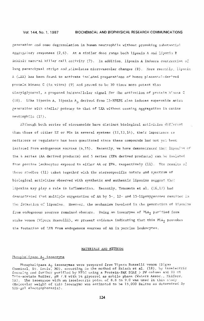
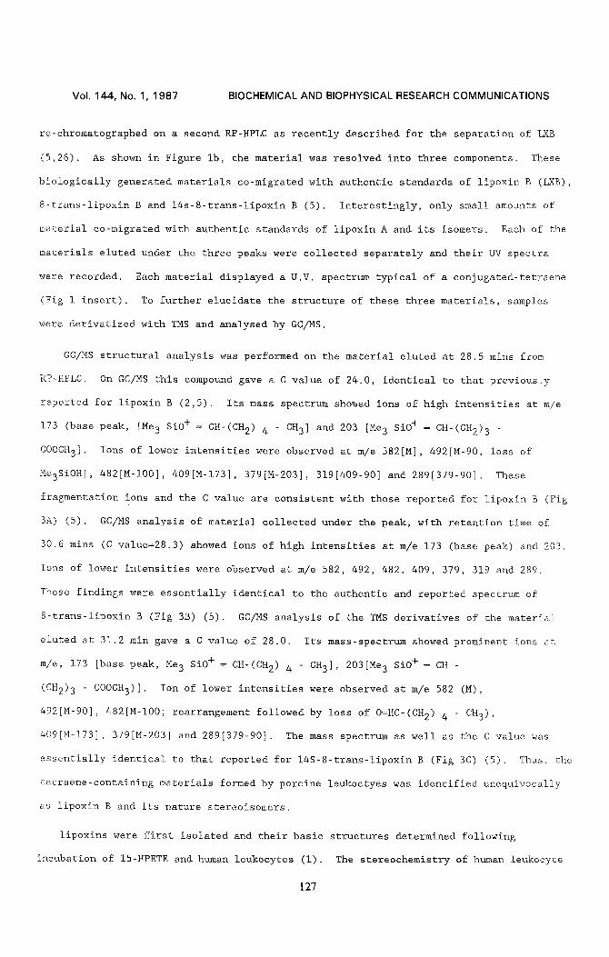
(Fig. IA). The formation of these materials by PLA 2 was dose dependent as indicated in
Fig 2A. At the highest dose of PLA 2 (3 x 10 .7 M), the viability of leukocytes after
I0 min of incubation was between 65 and 729 (Fig 2B) (n=3). Following i0 min exposure to
10-8M of PLA 2 the viability of the leukocytes was greater than 909, and the formation
of these materials from these cells was more than half of the maximum response
(EC50:I.25 x 10-SM) (Fig 2B). This result suggests that the generation of these
tetraene containing compounds is not likely a result of cell injury or cellular toxicity
caused by PLA 2.
125
Vol. 144, No. 1, 1987 BIOCHEMICAL AND BIOPHYSICAL RESEARCH COMMUNICATIONS
g co v
z _o m n- O ¢0 m
Fig. i.
1
FRACTION-A i
o co
LU o
O3
, , , , ,
240 280 3 2 0 3 6 0 W A V E L E N G T H (nm)
i I I 5 i0 15
TIME (min)
B-trans-LXB
14s- B - t r a n s - LXB / .¢
LXB o C
i I 1~0 I 30 20 0
TIME (rain)
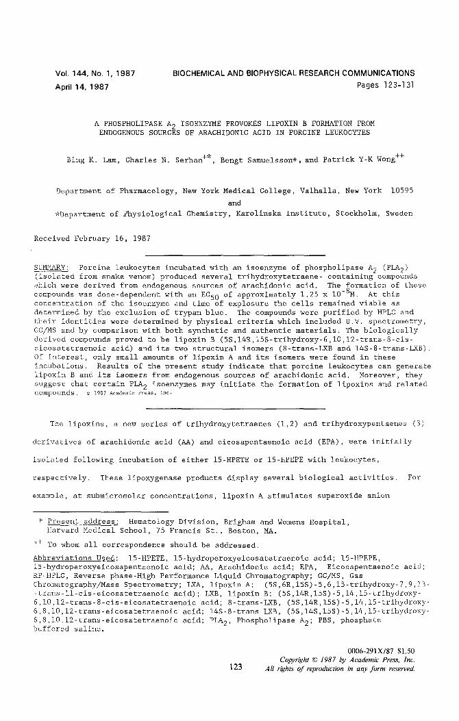
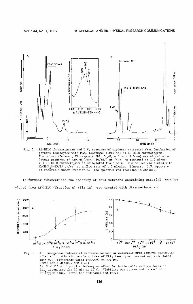
RP-HPLC chromatograms and U.V. spectrum of products extracted from incubation of porcine leukocytes with PLA 2 isoenzyme (ixl0-7M) A) RP-HPLC chromatogram. The column (Beckman, Ultrasphere ODS, 5 #H, 4.6 mm x 2.5 cm) was eluted on a linear gradient of MeOH/H20/HAC , 50/50/0.05 (V/V) to methanol at 1.0 ml/min; (B) RP-HPLC chromatogram of methylated fraction A. The column was eluted with MeOH/H20/65/35 (v/v), at a flow rate of 1.0 ml/min. (insert) U.V. spectrum of materials under Fraction A. The spectrum was recorded in ethanol.
To further substantiate the identity of this tetraene-containing material, samples
eluted from RP-HPLC fraction A) (Fig IA) were treated with diazomethane and
A = 5 0 0 - o
4 0 0 -
3 o o - ==
v z 200- x O o_ 100" .J
. . . . 0 , •
10"9M 3x10⁻⁹M 10-8M 3x10 -8M10-TM 3x10⁻⁷M
PLA 2 CONC.
Fig. 2.
100,
80.
v 6 0 . >-
"J 40- m
> 20'
, , , , , ,
10 °9 3x100 9 10 -8 3x100 8 100 7 3 x 1 0 -7
PLA 2 (M)
A) Endogenous release of tetraene-containing materials from porcine leukocytes after stimulatin with various doses of PLA 2 isoenzyme. Amount was calculated from U.V. absorbance using E=50,000 at 302 rim. Error bar indicates SEM (n=3) B) Viability of porcine leukocytes after incubation with various doses of PLA 2 isoenzymes for I0 min at 37°C. Viability was determined by exclusion of Trypan blue. Error bar indicates SEM (n=3).
126
Vol. 144, No. 1, 1987 BIOCHEMICAL AND BIOPHYSICAL RESEARCH COMMUNICATIONS
re-chromatographed on a second RP-HPLC as recently described for the separation of LXB
(5,26). As shown in Figure Ib, the material was resolved into three components. These
biologically generated materials co-migrated with authentic standards of lipoxin B (LXB),
8-trans-lipoxin B and 14s-8-trans-lipoxin B (5). Interestingly, only small amounts of
material co-migrated with authentic standards of lipoxin A and its isomers. Each of the
materials eluted under the three peaks were collected separately and their UV spectra
were recorded. Each material displayed a U.V. spectrum typical of a eonjugated-tetraene
(Fig i insert). To further elucidate the structure of these three materials, samples
were derivatized with TMS and analysed by GC/MS.
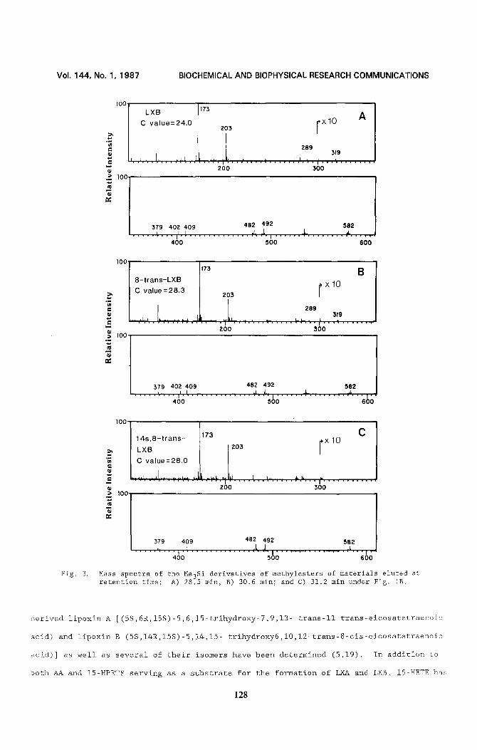
GC/MS structural analysis was performed on the material eluted at 28.5 mins from
RP-HPLC. On GC/MS this compound gave a C value of 24.0, identical to that previously
reported for lipoxin B (2,5). Its mass spectrum showed ions of high intensities at m/e
173 (base peak, [Me3 SiO+ = CH'(CH2) 4 - CH3] and 203 [Me 3 SiO + = CH-(CH2) 3 -
COOCH3]. Ions of lower intensities were observed at m/e 582[M], 492[M-90, loss of
Me3SiOH], 482[M-I00], 409[M-173], 379[M-203], 3191409-90] and 289[379-90]. These
fragmentation ions and the C value are consistent with those reported for lipoxin B (Fig
3A) (5). GC/MS analysis of material collected under the peak, with retention time of
30.6 mins (C value=28.3) showed ions of high intensities at m/e 173 (base peak) and 203.
lons of lower intensities were observed at m/e 582, 492, 482, 409, 379, 319 and 289.
These findings were essentially identical to the authentic and reported spectrum of
8-trans-lipoxin B (Fig 3B) (5). GC/MS analysis of the TMS derivatives of the material
eluted at 31.2 min gave a C value of 28.0. Its mass-spectrum showed prominent ions at
m/e, 173 [base peak, Me 3 SiO + = CH-(CH2) 4 " CH3], 203[Me3 SiO+ = CH -
(CH2) 3 - COOCH3) ]. Ion of lower intensities were observed at m/e 582 (M),
492[M-90], 482[M-I00; rearrangement followed by loss of O=HC-(CH2) 4 - CH3)'
409[M-173], 379[M-203] and 289[379-90]. The mass spectrum as well as the C value was
essentially identical to that reported for 14S-8-trans-lipoxin B (Fig 3C) (5). Thus, the
tetraene-containing materials formed by porcine leukoctyes was identified unequivocally
as lipoxin B and its nature stereoisomers.
lipoxins were first isolated and their basic structures determined following
incubation of 15-HPETE and human leukocytes (i). The stereochemistry of human leukocyte
127
Vol. 14.4, No. 1, 1 9 8 7 B I O C H E M I C A L A N D B I O P H Y S I C A L R E S E A R C H C O M M U N I C A T I O N S
Fig. 3.
l°O I LXB I, C value=24,0 173 Z0:3 r x l 0 A
L "~ 289 319
N
;~ 100
'1 200 300
[ 379 402 409 482 492 582 J I
. . . . . , . . . . , . ~ ' . . . . . . . . . . . . . t l , , ~ . , . . . . . . . . . . . . . ~, . . . . . p 400 500 600
100 173 B
8-t rans-LXB I x 10 I C value = 28'3 J 203 I
. . . . . . " ; i . I ' , " ; . . i " ' i ~ I I i ' ~ , ' 1 i i ~ . . . . . . - - ' ~ - - i ' i , ~ I I . ~ . . . . . . .
-- Z00 300 .~ 100
379 402 409 482 492 582 . . . . . ~ . . . . iJ.I . . . . . . . . . . . . . . ~ 1 , ~ , . = . . . . . . ~ . . . . . . . . ; 'h . . l "
400 500 600
1 0 0
;~ I00
14;1~8_ t r ans- 17:3 I 203 iX 10 C
. . . . . . . . . . . . . . . . . . 2~o . . . . . . . . . . . . . . . . . ~oo . . . . . . . . . . .
379 409 482 492 582 I I
. . . . . ,Ij r . . i . l . . . . . . . . . . . . . ~ll~,~l I . . . . . . . . . . . . . . . A , . . i . 400 500 600
Mass spectra of the Me3Si derivatives of methylesters of materials eluted at retention time: A) 28.5 min, B) 30.6 min; and C) 31.2 min under Fig. IB.
derived lipoxin A [(5S,6R,15S)-5,6,15-trihydroxy-7,9,13- trans-ll trans-eicosatetraenoic
acid) and lipoxin B (5S,14R,15S)-5,14,15- trihydroxy6,10,1i-trans-8-cis-eicosatetraenoic
acid)] as well as several of their isomers have been determined (5,19). In addition to
both AA and 15-HPETE serving as a substrate for the formation of LXA and LXB, 15-HETE has
128
Vol. 144, No. 1, 1987 BIOCHEMICAL AND BIOPHYSICAL RESEARCH COMMUNICATIONS
been found to be transformed to LXA and LXB in activated human leukocytes (5,11,20). The
results of these studies suggest that lipoxin can be formed in part by transeellular
metabolism of 15-HETE (5,19). It is obvious that several biosynthetic routes could yield
tetraene compounds. Thus it remained to be determined whether lipoxins can be formed
from endogenous AA released from cellular pools. Recently, we demonstrated that porcine
leukocytes incubated with either AA or EPA generated lipoxins (0.05% for AA and 0.1% for
EPA) which is about 15-30% of the amount of leukotriene B 4 produced. These results
were based on RP-HPLC, U.V. and GC/MS data (Wong et al., unpublished data). We therefore
hypothesized that lipoxins may also be formed from endogenous AA if appropriate stimuli
are applied as in the case with bradykinin stimulation of human platelets to release
15-1ipoxygenase products from endogenous sources (21). In this study we have found that
Ca ++ ionophore A23187 (I-i0 ~M) was relatively weak stimulus for lipoxin production
by porcine leukocytes (less than 50±10 ng of lipoxin B and its isomers were isolated from
each I00 x 106 cells incubation, n=8). Bradykinin was not active with the doses tested
(up to I0 #M). In contrast, the PLA 2 isoenzyme (pi=8.9) provokes the formation of
lipoxin B and its isomers (approximately 30 ng/ml; i00 x 106 cells) from porcine
leukocytes at doses as low as 10-9M.
The use of purified PLA 2 isoenzyme in the present study is based on the following:
(I) granule associated PLA 2 of inflammatory cells may be released to the extracellular
environment upon cell activation (20,22-27); (2) that similar PLA 2 isoenzyme has
previously been demonstrated to induce leukotriene release in isolated perfused guinea
pig lung (28); (3) we have found that other PLA 2 isoenzymes (other than pI of 8.9)
isolated from crude Russelli Vipera's venom do not induce the release of lipoxins from
porcine leukocytes; finally, 4) we have also found that this isoenzyme provokes the
formation of these compounds by certain cell types, i.e., porcine and human leukocytes,
rat PMN and not by guinea pig PMNs, guinea macrophage and rat macrophages (Wong et al,
unpublished data). The responses of porcine leukocytes to exogenously added PLA 2 as
shown in this study may mimick the in situ pathophysiologic response of leukocytes
exposed to extracellular PLA 2 which may be released by macrophages or leukocytes during
cell activation and phagocytosis or in antigen induced chronic inflammation (24,25).
Furthermore, PLA 2 have been reported to be released by rat platelets during platelet
129
Vol. 144, No. 1, 1987 BIOCHEMICAL AND BIOPHYSICAL RESEARCH COMMUNICATIONS
aggregation (26) and has been found at site(s) of inflammation and in lymph draining
nodes with tuberculin reactions in rabbits (27). Thus, PLA 2 and its isoenzymes
released from macrophages, platelets or isolated from snake venom may be useful tools in
studying the formation of eicosanoids from endogenous sources during cell activation.
In the present study, the identities of these materials were established by U.V.,
HPLC, and by GC/MS and comparison with synthetic materials. The formation of lipoxin B
and its isomers provoked by PLA 2 was dose dependent (Fig. 2A). Maximal recovery of LXB
was achieved at about ixl0-7M PLA 2. At this dose, approximately 70-80~ of leukocytes
were viable after I0 min of incubation at 37°C as determined by the Trypan blue
exclusion technique (Fig. 2b). Similar results were also obtained from lactate
dehydrogenase release assay (data not shown). The selective formation of large amounts
of LXB but not LXA, may reflect the involvement of a specific biosynthetic pathway in
porcine leukoeytes. Previous studis in human leukocytes using 15-HETE suggested that
lipoxins can be formed via 5,6-epoxide tetraene intermediates (5,19). The high activity
of 12-1ipoxygenase in porcine leukocytes (29) may accelerate formation of lipoxins, since
12-1ipoxygenase can also metabolize 15-HPETE to form 14,15 DHETE (29) and may represent
another biosynthetic pathway for LXB formation. It is of interest to note that purified
12-1ipoxygenase from porcine leukocytes has recently been reported to generate LXB from
5,15-DHPETE (17). From the results of the presenty study, however, it is not possible to
determine the precise route and intermediates involved in the of formation of LXB and its
isomers by porcine leukoeytes. Lipoxins have been demonstrated to affect the functions
of inflammatory cells, natural killer cells, protein kinase C, and microvaseular
circulation (2,6,9). Thus, demonstration that lipoxins can be formed from endogenously
derived AA (Fig. IA and 2A) in addition to transcellular metabolism of 15-HETE (5,19)
provides further evidence to suggest that lipoxins may serve as lipid mediators or
intracellular regulators in various inflammatory diseases and immune responses.
Moreover, the result of the present study provide further evidence to support the
proposal that the release of PLA 2 activity by inflammatory cells may result in the
formation of agents which are active in chronic inflammation.
ACKNOWLEDGEMENTS
This work was supported by Swedish Medical R.C. (03X-217 B.S.) and National Institute
130
Vol. 144, No. 1, 1987 BIOCHEMICAL AND BIOPHYSICAL RESEARCH COMMUNICATIONS
of Health Grants HL-25316 and HL-00811. We thank Carl Ehmer Farm and personnel of
Poughkeepsie, N.Y. for the collaboration and the generous supply of porcine blood used in
this study. The assistance of Gail Price in the preparation of this manuscript is
gratefully acknowledged. Dr. C.N. Serhan is a fellow of the Arthritis Foundation.
REFERENCES
i. Serhan, C.N., Hamberg, M., and Samuelsson, B. (1984) Biochim. Biophys. Res. Commun. 118, 943-949.
2. Serhan, C.N., Hamberg, M., and Samuelsson, B. (1984) Proc. Natl. Acad. Sei. U.S.A. 81, 5335-5339.
3. Wong, P.Y-K., Hughes, R.A., and Lam, B. (1985) Biochim. Biophys. Res. Commun. 126, 763-772.
4. Fitzsimmons, B.J., Adams, J., Evans, J.F., Leblanc, Y., and Rokach, J., (1985) J. Biol. Chem. 260, 13008-13012.
5, Serhan, C.N., Hamberg, M., Samuelsson, B., Morris, J., and Wishka, D.G. (1986) Proc. Natl. Acad. Sci. U.S.A. 83, 1983-1987.
6. Samuelsson, B. and Serhan, C.N. (1986) International Conference on PG and Cancer, Rome, July 1986, Raven Press, N.Y., in press.
7. Ramstedt, U., Ng, J., Wigzell, H., Serhan, C.N., and Samuelsson, B. (1985) J. Immunol. 135, 3434-3436.
8. Dahlen, S.E., Serhan, C.N., and Samuelsson, B. (1987) Acta. Scand. Physiol. in press.
9. Hansson, A., Serhan, C.N., Haeggstrom, J., Ingelman-Sundberg, M., and Samuelsson, B. (1986) Biochem. Biophys. Res. Commun. 134, 1215-1222.
i0. Nishizuka, Y. (1984) Nature (Lond.) 225, 1365-1370. ii. Wong, P.Y-K., Spur, B., Hirai, A., Yoshida, S., Tamura, Y., and Lam, B. (1986)
Federation Proceedings 45, 927 (Abstract) 12. Von Euler, U.S. (1936) J. Physiol. 88, 213-234. 13. Hamberg, M., Svensson, J. and Samuelsson, B. (1975) Proc. Natl. Acad. Sci. USA 72,
2994-2998. lo
14. Samuelsson, B. and Hammarstrom, S. (1982) Vit. and Hormones 39, 1-30. 15. Rainsford, K.D. (1985) Trends in Pharmacology June, 230-231. 16. Yamamoto, S., Neda, N., Yakoyama, C., Fitzsimmons, B.J., Rokach, J., Oates, J. and
Brash, A.R. (1987) IN Symposium of "Lipoxin: Biosynthesis and Pharmacology", Fed. Proc. (in press).
17. Yokoyama, C., Yoshimoto, T., Yamamoto, S., Oates, J.A. and Brash, A.R. (1986) Proc. 5th International Conference on PGs and Related Compounds. June 2, 1986, Florence, Italy.
18. Salach, J.I., Turini, P., Seng, R., Hauber, J., and Singer T.P. (1971) J. Biol. Chem. 246, 331-339.
19. Serhan, C.N., Nicolaou, K.C., Webber, S.E., Veale, C.A., Dahlen, S.E., Puustinen, T.J. and Samuelsson, B. (1986) J. Biol. Chem. 261, 16340-16345.
20. Movat, H.Z. (1984) in: Inflammation, Immunity and Hypersensitivity pp. 1-161, Harper and Row Publishers, N.Y.
21. Wong, P.Y-K., Westlund, P., Hamberg, M., Granstrom, E., Chao, P.H-W, and Samuelsson, B. (1985) J. Biol. Chem. 260, 9162-9165.
22. Weissmann, G., Smolen, J.E. and Korchak, H.M. (1980) N. Engl. J. Med. 303:27-34. 23. Victor, M., Weiss, J., Klempner, M.S. and Elsbach (1981) FEBS LETTERS 136:298-300. 24. Vadas, P., Wasi,' S., Movat, H.Z. and Hay, J.B. (1981) Nature 293, 583-585. 25. Vadas, P. and Hay, J.B. (1980) Int. Arch. Allergy Appl. Immunol. 62, 142-151. 26. Vadas, P. and Hay, J.B. (1980) Life Sciences 26, 1721-1729. 27. Vadas, P. and Hay, J.B. (1982) Am. J. Pathol. 107, 285-291. 28. Huang, H-C (1984) Toxicon. 22, 359-372. 29. Maas, R.L. and Brash, A.R. (1983) Proc. Natl. Acad. Sei. U.S.A. 80, 2884-2888.
131


![Мінрегіонdfrr.minregion.gov.ua/foto/projt_addition/2016/02/... · 150 aBTOM06iJ1iB ga n06 ] [10;aaBaTH a60 Ha lipoxin 3MiHi KiJ1bKOCTi Flpox0ÄiB HOPMH 2-4-33 [MPH BHKOHaHHi](https://img.pdfslide.net/doc/110x75/60d1547388e155243e230173/oedfrr-150-abtom06ij1ib-ga-n06-10aabath-a60-ha-lipoxin-3mihi.jpg)