Embed Size (px)
Citation preview
Plant Physiol. (1997) 114: 715-722
A Plant Chloroplast Glutamyl Proteinase’
William A. Laing* and John T. Christeller
HortResearch, Private Bag 921 69, Auckland, New Zealand (W.A.L.); and HortResearch, Private Bag 11 030, Palmerston North, New Zealand (J.T.C.)
A glutamyl proteinase was partially purified from Percoll gradient- purified spinach (Spinacia oleracea) chloroplast preparations and appeared to be predominantly localized in the chloroplast stroma. The enzyme degraded casein, but of the 11 synthetic endopeptidase substrates tested, only benzyloxycarbonyl-leucine-leucine-glutamic acid-p-napthylamide was hydrolyzed at measurable rates. In addi- tion, the enzyme cleaved the oxidized /3-chain of insulin after a glutamic acid residue. There was no evidence that native ribulose- 1,5-bisphosphate carboxylase/oxygenase was cleaved by this pro- teinase. The apparent K,,, for benzyloxycarbonyl-leucine-leucine- glutamic acid-PNA at the pH optimum of 8.0 was about 1 mM. CI- ions were required for both activity and stability. Of the proteinase inhibitors covering all four classes of the endopeptidases, only 442- aminoethy1)-benzenesulfonyl-fluoride HCI and ~-1-chloro-3-[4- tosylamido]-4-phenyl-2-butanone significantly inhibited the protein- ase. The partially purified enzyme had a molecular weight of about 350,000 to 380,000, based on size-exclusion chromatography. The enzyme has both similar and distinctive properties to those of the bacterial glutamyl proteinases. To our knowledge, this ís the first
description of a plant glutamyl proteinase found predominantly or exclusively in the chloroplast.
Proteinases are important during protein breakdown in senescence-regulated protein turnover during normal me- tabolism, protein remobilization during development, and protein degradation during stress responses (Ryan and Walker-Simmons, 1981; Vierstra, 1993; Callis, 1995). In ad- dition, proteinases are involved in the processing of pro- teins during their synthesis and transport within the cell (Lord and Robinson, 1986).
The chloroplast is a . complex organelle containing its own partia1 genome (Taylor, 1989; Sugiura, 1992) and pro- tein synthesis machinery (Gillham et al., 1994), as well as the capacity to degrade proteins (Dalling and Nettleton, 1986). The chloroplast has proven to be a source of several different proteinases, some of which have been isolated and partially characterized. Chloroplast proteinases in- clude both soluble stromal and membrane-located en- zymes. Some of the thylakoid-located proteinases de- scribed to date include enzymes that process protein presequences as well as others that degrade specific tar- gets. Examples of the latter include three thylakoid-bound enzymes: a prolyl endoproteinase from spinach (Spinacia
‘This project was funded by the Foundation for Research, Sci-
* Corresponding author; e-mail [email protected]; fax 64-9- ence and Technology, New Zealand.
815-4201.
oleracea) that degrades the 18-kD protein from PSII (Ku- wabara, 1992); another that degrades the Chl a J b-binding protein (Hoober and Hughes, 1992); and the membrane- bound NADPH-protochlorophyllide oxidoreductase pro- teinase of barley (Hordeum vulgare; Hauser et al., 1984). Examples of processing proteinases include one that pro- cesses the thylakoid D1 protein (Bowyer et al., 1992) and another that processes plastocyanin (Hageman et al., 1986). Other thylakoid proteinases have been characterized, but have yet to be ascribed a specific function (e.g. Kuwabara and Hashimoto, 1990). In addition, several soluble chloro- plast proteinases have been described, including a stromal metalloproteinase that degrades Rubisco (Bushnell et al., 1993), metallo- and Ser-proteinases (Musgrove et al., 1989), a Cys proteinase (Liu and Jagendorf, 1986), and a soluble, metallo precursor-processing proteinase (Robinson and El- lis, 1984). A range of aminopeptidases have also been de- scribed from chloroplasts (Waters et al., 1982; Liu and Jagendorf, 1986; Thayer et al., 1988).
There have been reports of ATP-dependent proteolysis in chloroplasts (Liu and Jagendorf, 1984; Malek et al., 1984). Subsequently, a gene for the ATP-regulated proteinase ho- mologous to the Ti proteinase (the Escherichia coli ClpP gene product) has been shown to be encoded by the chlo- roplast genome (Gray et al., 1990; Weglohner et al., 1992; Moore and Keegstra, 1993; Clarke et al., 1994). There has also been a recent report of the presence of the Ti protein in the chloroplast (Shankin et al., 1995).
Prokaryotic glutamyl proteinases (e.g. the Staphylococcus aureus V8 proteinase widely used in protein sequencing studies; Birktoft and Breddam, 1994) cleave a peptide bond that usually follows a Glu and, to a much lesser extent, an Asp. They are Ser proteinases, belonging to family S2, and have only been reported in eubacteria (Rawlings and Bar- rett, 1994). They are resistant to inhibition by most protein- ase inhibitors, with a pH optimum in the range of 7.0 to 9.0 (Birktoft and Breddam, 1994). To our knowledge, no such proteinase with a specificity toward glutamyl residues has been described from plants to date, except for the Z-LLE- PNA-hydrolyzing activity associated with the proteasome (Ozaki et al., 1992; W.A. Laing, unpublished observations). However, there have been reports of proteinase inhibitors that appear to target putative glutamyl proteinases in plants (eg. Margossian et al., 1988; Fujita et al., 1993;
Abbreviations: AEBSF, 4-(2-aminoethyl)-benzenesulfonyl- fluoride HC1; AMC, 7-amido-4-methylcoumarin; PNA, p- napthylamide Boc, t-butyloxycarbonyl; Chl, chlorophyll; Succ, succinyl; Z-LLE-PNA, benzyloxycarbonyl-Leu-Leu-Glu-PNA.
71 5 www.plantphysiol.orgon December 13, 2018 - Published by Downloaded from Copyright © 1997 American Society of Plant Biologists. All rights reserved.
71 6 Laing and Christeller Plant Physiol. Vol. 114, 1997
Linthorst et al., 1993), although their in vivo function is unknown.
This paper describes a high-molecular-weight chloro- plast glutamyl proteinase with a mildly alkaline pH opti- mum and a dependency on CI- ions for activity. This enzyme shares many of the characteristics of the bacterial glutamyl proteinase, although in other features it differs markedly.
MATERIALS A N D METHODS
Materiais
Spinach (Spinacia oleracea L.; hybrid number 7R, Yates New Zealand Ltd., Auckland, New Zealand) plants were grown hydroponically under fluorescent lights (Roughan, 1994). Synthetic peptide substrates were obtained from Bachem AG (Bubendorf, Switzerland) or Sigma, and fluo- rescent (Bodipy-labeled) casein was obtained from Molec- ular Probes (San Diego, CA). Other biochemicals were obtained from Sigma. Chromatography media were ob- tained from Pharmacia Biotech (Uppsala, Sweden) or, in the case of hydroxyapatite, from Bio-Rad, whereas the Miniprotean I1 and Prepcell electrophoresis systems from Bio-Rad were used for gel electrophoresis. Fluorescence was measured on a microwell-plate-reading fluorimeter (Fluroskan I1 96, Labsystems, Hampshire, UK). Filtration equipment (Amicon, Beverly, MA) and 10,000 molecular weight cutoff membrane filters (Amicon) were used to concentrate samples.
Partia1 Purification of the Proteinase
A11 procedures were carried out at 4°C or lower. Intact chloroplasts were isolated and purified using the method described by Roughan (1994). This included centrifugation on a Perco11 gradient, which results in highly purified chloroplasts. The chloroplasts were immediately frozen in liquid nitrogen and stored at -80°C until used. The frozen chloroplasts were thawed and diluted 3-fold into 25 mM potassium phosphate buffer, pH 7.5, with 20% (v /v) glyc- erol and 0.1% (v/v) 2-mercaptoethanol. A11 other buffers, unless specified, contained these concentrations of glycerol and 2-mercaptoethanol. The lysed chloroplast extract was centrifuged at 30,OOOg for 10 min, and the clear supernatant fluid was applied to a 10 X 2.5-cm hydroxyapatite column equilibrated with phosphate buffer. The column was washed with this buffer and enzyme activity eluted at 1 mL min-l using a 25 to 250 mM linear phosphate gradient.
The two peaks of Z-LLE-PNA-hydrolyzing activity were collected and concentrated to about 1.5 mL using 10,000 molecular weight cutoff membrane, and the concentrated samples were applied separately to a G75 Sephadex col- umn (50 X 1.25 cm) equilibrated with 50 mM Tris-HCI buffer, pH 8.0. The column was eluted at 1 mL min-' with the same buffer, and void-volume fractions able to hydro- lyze Z-LLE-PNA were combined and applied to a 1-mL anion-exchange column (HiTrap Q, Pharmacie Biotech, Uppsala, Sweden). The column was washed with the Tris- HCl buffer and the proteins were eluted with a O to 0.25 M
NaCl linear gradient at 1 mL min-' in this buffer. Active
fractions were concentrated as above, and applied to a 50 X 1.25 cm high-resolution gel-filtration column (Sephacryl 5-300, Pharmacie Biotech) equilibrated with Tris buffer. This column was calibrated using a range of protein stan- dards to allow calculation of the M, of the Z-LLE-PNA- hydrolyzing activity. Active fractions were concentrated, frozen in liquid nitrogen, and stored at -80°C. Most of the work characterizing the proteinase was performed using this leve1 of purity. However, to further purify the enzyme, it was subjected to preparative native PAGE at pH 8.8 (Hames and Rickwood, 1983) at 4°C using a Prepcell with 20% (v /v) glycerol present in the 5% polyacrylamide gel.
Assays
The Z-LLE-PNA (dissolved in DMSO as a 2.5 mM stock solution) and fluorescent-casein-hydrolyzing activities were measured using a microwell-format fluorometer with interference filter peak excitation and emission wave- lengths at 340 and 405 nm, respectively, for Z-LLE-PNA, and 485 and 538 nm, respectively, for caseinase activity. Assays of blocked peptide substrates with AMC deriva- tives (also dissolved in DMSO) had excitation and emission wavelengths of 355 and 460 nm, respectively. Standard assays (e.g. assays of column fractions) contained 50 mM Tris-HC1, pH 8.0, with 0.1% (v/v) 2-mercaptoethanol, 20% (v/v) glycerol, 0.25 mM Z-LLE-PNA substrate, and 10% (v/v) DMSO in a final concentration of 100 FL. These assays automatically contained about 30 mM C1-, since the Tris base was adjusted to pH 8.0 with HC1. Assays were initiated by adding the substrate. In inhibition studies the inhibitor and enzyme were incubated together for at least 10 min before the substrate was added to ensure that equilibrium between the proteinase and the inhibitor was established. At least six time points were read in any assay with the plate incubated at 30°C between readings, and the results were collected by a computer and analyzed using programs similar to those described by Christeller et al. (1990). Only those points that showed a linear time course were used to calculate the reaction rate. Except for the caseinase reaction, reaction rates were a linear function of the amount of enzyme. Fluorescence calibration curves were carried out using AMC and PNA standards under a range of experimental conditions, and rates of reaction in appropriate units were calculated.
The effect of assay pH was determined as above, except that the effect of buffer composition was checked in a preliminary experiment. Since a11 buffers used except borate gave a continuous set of curves, subsequent pH curves were determined using 1,3-bis (Tris[hydroxymethyl] -methylamino)propane. Fluorescence yield was measured in PNA standard solutions as a function of pH, and was found to be almost independent of pH. Background assays at each pH value without added enzyme showed that the substrate was stable at all pH values. Substrate response curves were obtained by adding different amounts of sub- strate dissolved in a constant added volume of DMSO to each reaction. Background activities were measured in the absence of enzyme.
www.plantphysiol.orgon December 13, 2018 - Published by Downloaded from Copyright © 1997 American Society of Plant Biologists. All rights reserved.
A Plant Chloroplast Glutamyl Proteinase 717
Activities were measured in crude leaf (the centrifugedextract made immediately after the initial leaf homogeni-zation during chloroplast preparation) and chloroplaststromal extracts by freezing the extracts in liquid nitrogen,and slowly thawing them twice before centrifugation at12,000g for 10 min to remove membrane-bound pigmentsthat quench fluorescence during assay. Assays were carriedout as above over a range of extract amounts, and resultswere calculated where assays responded linearly with theamount of extract assayed. Rubisco (Laing and Christeller,1976) and NADH-malic dehydrogenase (Ting, 1968) werealso used as markers for chloroplast purity. Protein wasmeasured using BSA as a standard (Bradford, 1976) andChl was measured as described by Roughan (1994).
Substrate cleavage specificity was measured using theoxidized /3-chain of insulin. Enzyme was incubated withinsulin for up to 48 h in the standard reaction mixture at25°C, and at intervals 200-ju.L aliquots were removed into800 p,L of 0.1% (v/v) trifluoroacetic acid in water andimmediately frozen at — 20°C. Cleaved fragments wereseparated from the intact insulin chain by HPLC using a250 X 4.6 mm reverse-phase column (C8, Vydac, Hesperia,CA) and a 0 to 100% (v/v) acetonitrile gradient containing0.1% (v/v) trifluoroacetic acid at 1 mL min"1. Representa-tive samples of the newly generated fragments were se-quenced on a Precise sequencer (Applied Biosystems) forfour cycles. Cleavage of Rubisco was measured by incubat-ing the Prepcell-purified proteinase (0.26 nmol min"1 ac-tivity for up to 40 h) in the presence of excess Rubisco(obtained from the Prepcell native gel purification step)both in the absence and presence of 5 HIM MgCl2 and ATP.The reaction was stopped by adding SDS sample buffer,and accumulated samples were analyzed by SDS-PAGEand Coomassie blue staining of the gel. Controls wereperformed without added glutamyl proteinase.
Polyacrylamide Electrophoresis
Tricine-SDS-PAGE was carried out as described bySchagger and Von Jagow (1987), and native gels (Hamesand Rickwood, 1983) were electrophoresed at pH 8.8 and4°C with the addition of 20% (v/v) glycerol to the gel. Gelswere stained using a silver-staining procedure, and thenative gels were stained using an activity assay (Van DerValk et al., 1989). This consisted of a 20-min wash of the gel(with shaking) after electrophoresis in the Tris-HCl buffercontaining 10 mM MgCl2- Then, 10 mL of fresh buffercontaining 0.125 mM Z-LLE-/3NA was added and the gelwas incubated at 30°C for 15 to 30 min. The gel was thenbriefly washed with distilled water to remove excess sub-strate and 20 mL of 0.1% (w/v) fast garnet dye was added.The development of dark-red-stained bands on an orangebackground was followed and, when staining appearedcomplete, the gel was washed several times with water,photographed, and dried under a vacuum at 80°C.
Data Analysis
Data were fitted to untransformed theoretical equationsusing the SAS (SAS Institute, 1985) statistical package.
Calculated parameters are presented as the value ± theasymptotic SE. Experiments were repeated two or threetimes, and either representative experiments are shown orthe means of several experiments are presented as noted inthe figures and tables.
RESULTS
Purity of Chloroplasts and Location of the Proteinase
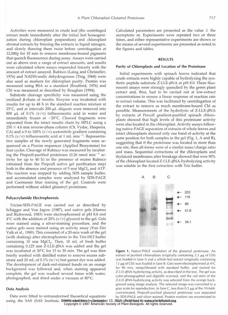
Initial experiments with spinach leaves indicated thatcrude extracts were highly capable of hydrolyzing the syn-thetic peptide substrate Z-LLE-j8NA at pH 8.0. These fluo-rescent assays were strongly quenched by the green plantextract and, thus, had to be carried out at low-extractconcentrations to ensure a linear response of reaction rateto extract volume. This was facilitated by centrifugation ofthe extract to remove as much membrane-bound Chl aspossible. Measurements of the hydrolysis of Z-LLE-/3NAby extracts of Percoll gradient-purified spinach chloro-plasts showed that high levels of this proteinase activitywere also located in the chloroplast. Activity assays follow-ing native-PAGE separation of extracts of whole leaves andintact chloroplasts showed only one band of activity at thesame position for both samples in the gel (Fig. 1, A and B),suggesting that if the proteinase was located in more thanone site, then all forms were of a similar mass/charge ratioand mass. Sequential extractions of the chloroplasts andthylakoid membranes after breakage showed that over 90%of the chloroplast-located Z-LLE-j3NA-hydrolyzing activitywas soluble in the first extraction with Tris buffer.
A B
203118
86
51.6
34.1
Figure 1. Native-PAGE resolution of the glutamyl proteinase. Anextract of purified chloroplasts (originally containing 3.3 /xg of Chl)was loaded in lane A and a whole-leaf extract (originally containing13 jig of Chl) was loaded in lane B. Gels were electrophoresed at 4°Cfor 90 min, reequilibrated with standard buffer, and stained forZ-LLE-^NA-hydrolyzing activity, as described in the text. The gel wascolor-photographed and digitally scanned, and the red stain of theZ-LLE-(3NA-hydrolyzing activity was selected from the orange back-ground using image analysis. The selected image was converted to agray scale for reproduction. In lane C, less than 0.5 /ig of the 10-fold-concentrated, Prepcell-purified glutamyl proteinase was separatedby SDS-PAGE and silver-stained. Protein markers are enumerated tothe right of lane C (values in kilodaltons). www.plantphysiol.orgon December 13, 2018 - Published by Downloaded from
Copyright © 1997 American Society of Plant Biologists. All rights reserved.
71 8 Laing and Christeller Plant Physiol. Vol. 114. 1997
To determine what proportion of the proteinase was located in the chloroplast, we assayed whole-leaf extracts and the purified chloroplasts made from this extract, and expressed the results on a Chl basis. If activity was pre- dominantly located in the chloroplast, then we would ex- pect a similar activity per unit of Chl in whole-leaf and chloroplast extracts. This was indeed the case, with the ratio of Z-LLE-PNA-hydrolyzing activity per milligram of Chl of chloroplast extract to whole-leaf extract being 0.80, implying four times as much Z-LLE-PNA-hydrolyzing ac- tivity in the chloroplast as in the rest of the cell. Two marker enzymes were also measured, NADH-malic dehy- drogenase (cytoplasm, peroxisome, and mitochondrion [Ting, 19681) and Rubisco (chloroplast). The purified chlo- roplasts had about 1% of the total leaf malic dehydrogenase activity on a Chl basis. On the other hand, the levels of Rubisco activity were similar in the two extracts on a Chl basis (1.2-1.0). These results clearly show that the isolated chloroplasts were highly pure, and that at least two-thirds and possibly a11 of the Z-LLE-PNA-hydrolyzing activity is located in the chloroplast.
Enzyme Purification
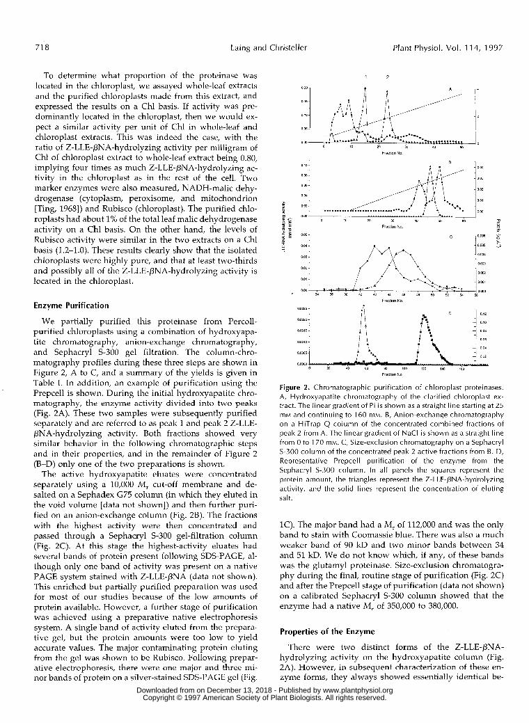
We partially purified this proteinase from Percoll- purified chloroplasts using a combination of hydroxyapa- tite chromatography, anion-exchange chromatography, and Sephacryl S-300 gel filtration. The column-chro- matography profiles during these three steps are shown in Figure 2, A to C, and a summary of the yields is given in Table I. In addition, an example of purification using the Prepcell is shown. During the initial hydroxyapatite chro- matography, the enzyme activity divided into two peaks (Fig. 2A). These two samples were subsequently purified separately and are referred to as peak 1 and peak 2 Z-LLE- PNA-hydrolyzing activity. Both fractions showed very similar behavior in the following chromatographic steps and in their properties, and in the remainder of Figure 2 (B-D) only one of the two preparations is shown.
The active hydroxyapatite eluates were concentrated separately using a 10,000 M , cut-off membrane and de- salted on a Sephadex G75 column (in which they eluted in the void volume [data not shown]) and then further puri- fied on an anion-exchange column (Fig. 2B). The fractions with the highest activity were then concentrated and passed through a Sephacryl S-300 gel-filtration column (Fig. 2C). At this stage the highest-activity eluates had severa1 bands of protein present following SDS-PAGE, al- though only one band of activity was present on a native PAGE system stained with Z-LLE-PNA (data not shown). This enriched but partially purified preparation was used for most of our studies because of the low amounts of protein available. However, a further stage of purification was achieved using a preparative native electrophoresis system. A single band of activity eluted from the prepara- tive gel, but the protein amounts were too low to yield accurate values. The major contaminating protein eluting from the gel was shown to be Rubisco. Following prepar- ative electrophoresis, there were one major and three mi- nor bands of protein on a silver-stained SDS-PAGE gel (Fig.
1 2
Fraction Na
0 o 10
o O6
: 0 0 2 - o o4 j ...................... I 1 -.
TI
J B r
Fractlon No
Figure 2. Chromatographic purification of chloroplast proteinases. A, Hydroxyapatite chromatography of the clarified chloroplast ex- tract. The linear gradient of Pi is shown as a straight l ine starting at 25 mM and continuing to 160 mM. B, Anion-exchange chromatography on a HiTrap Q column of the concentrated combined fractions of peak 2 from A. The linear gradient of NaCl is shown as a straight line from O to 170 mM. C, Size-exclusion chromatography o n a Sephacryl S-300 column of the concentrated peak 2 active fractions from B. D, Representative Prepcell purification of the enzyme from the Sephacryl S-300 column. In all panels the squares represent the protein amount , t he triangles represent the Z-LLE-PNA-hydrolyzing activity, and the solid lines represent the concentration of eluting salt.
1C). The major band had a M, of 112,000 and was the only band to stain with Coomassie blue. There was also a much weaker band of 90 kD and two minor bands between 34 and 51 kD. We do not know which, if any, of these bands was the glutamyl proteinase. Size-exclusion chromatogra- phy during the final, routine stage of purification (Fig. 2C) and after the Prepcell stage of purification (data not shown) on a calibrated Sephacryl S-300 column showed that the enzyme had a native M , of 350,000 to 380,000.
Properties of the Enzyme
There were two distinct forms of the Z-LLE-PNA- hydrolyzing activity on the hydroxyapatite column (Fig. 2A). However, in subsequent characterization of these en- zyme forms, they always showed essentially identical be-
www.plantphysiol.orgon December 13, 2018 - Published by Downloaded from Copyright © 1997 American Society of Plant Biologists. All rights reserved.
A Plant Chloroplast Glutamyl Proteinase 71 9
Table 1. Purification o f Z-LLE-PNA-hydrolyzing activity from purif ied spinach chloroplasts
All assays were carried out in 50 mM Tris-HCI buffer at p H 8.0 with no added CI- ions. Chloroplasts contain about 40% of the soluble protein in the cell. Consequently, the specific activity of the 2-LLE-PNA-hydrolyzing activity on a whole-leaf basis would be about 0.12 nmol min-' mg-' (crude protein). The data in this table are representative of two major preparations, and similar fractionation results were obtained in a preparation made from leaves. -
- Fraction Protein Activitv
Chloroplasts Hydroxyapatite
Peak 1 Peak 2
HiTrap Q Peak 1 Peak 2
Sephacryl/
Peak 1 Peak 2
S-300
mg
a7
5.4 4.4
0.49 0.1 5
0.1 2 0.03
nmol min-'
22.7
11.9 12.8
4.8 3.7
2.8 3.7
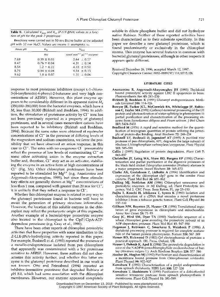
havior and properties. Consequently, for clarity, in some cases only one form is shown in the figures. The rate of Z-LLE-PNA hydrolysis depended upon the salt concentra- tion in the assay. Time courses of Z-LLE-PNA hydrolysis were linear for at least the first 50 min at a high salt concentration (above 20 mM NaCI), whereas the rate de- clined with time at low salt concentrations (Fig. 3A). Con- sequently, the response of Z-LLE-pNA hydrolysis to salt concentration depended on the extent of the time course, showing a much greater response to salt when rates were calculated later in the time course (Fig. 3B). We have not yet established the specific ion(s) responsible for this salt effect, although NaCl and MgCI, are almost equally effec- tive in stimulating the enzyme when measured at the same C1- concentration (Fig. 3B).
We tested the partially purified Z-LLE-PNA-hydrolyzing activity at pH 8.0 against a range of potential substrates. Rates of reaction with a11 substrates were measured by flu- orescence assays, which are about 100 times more sensitive than the corresponding spectrophotometric or radiometric assays. The substrates tested included (at 0.25 mM): Succ- Ala-Ala-Phe-AMC (chymotrypsin-like), Succ-Phe-Leu-Phe- 4-methoxy-PNA (chymotrypsin-like and a substrate of the Escherichia coli proteinase La), Boc-Phe-Ser-Arg-AMC (tryp- sin-like), Z-Gly-Gly-Arg-AMC (trypsin-like), Succ-Gly-Pro- Leu-Gly-Pro-AMC (prolyl endopeptidase substrate), Boc- Gly-Arg-Arg-AMC (trypsin-like), Succ-Leu-Leu-Val-Tyr- AMC (chymotrypsin-like), Succ-Leu-Tyr-AMC (a substrate for the E. coli proteinase Ti), Succ-Tyr-Val-Ala-Asp-AMC (another minor substrate of the V8 glutamyl proteinase), and casein (general proteinase). None of these artificial sub- strates was hydrolyzed at rates faster thán 0.1% of the rate of Z-LLE-PNA (the limit of sensitivity of the assay; data not shown). In addition, separate assays established that there were no benzoyl-Arg-4-nitroanilide (trypsin4ike)- or benzoyl- Tyr-4-nitroanilide (chymotrypsin-1ike)-hydrolyzing activity present in spinach chloroplasts.
When the partially purified enzyme (after the Prepcell step) was incubated with insulin, two new peptide peaks
Purification Saecific Activitv
nmol min- 'mg- ' protein
0.26
2.2 2.9
9.8 24.7
23.3 123
-fold
1
a
38
11
95
90 473
on the HPLC trace were produced. The size of these peaks increased over 44 h of incubation, with 30% hydrolysis of the insulin occurring after 16 h and 68% hydrolysis after 44 h. Complete recovery of the sum of the amount of insulin and the two cleavage fragments was maintained throughout the time course. One had the sequence FVNQ, the initial N-terminal sequence of the 30-amino acid pro- tein, and the other had the sequence ALYL, corresponding to residue numbers 14 to 17, immediately after a glutamyl residue. No other new peaks were observed. The enzyme did not cleave oxidized RNase A and the glutamyl protein- ase did not cleave native spinach Rubisco in the presence or absence of ATP and MgCI,, as shown by the fact that neither the large nor small subunits were altered in size or amount, as shown by SDS-PAGE (data not shown).
The response to the concentration of the substrate Z-LLE- PNA was measured at pH 8.5. Activity increased with substrate concentration to 1.0 mM and decreased slightly thereafter (Fig. 4). The apparent K , (Z-LLE-PNA) values for peaks 1 and 2 (as identified from the hydroxyapatite column) were 1.04 (?0.04) and 1.17 (?0.22) mM, respec- tively. There appeared to be substrate inhibition at the highest concentrations, as determined from the deviation of the data points from the fitted hyperbola, which may reflect fluorescence quenching at high substrate concentra- tions. All other assays were done at 0.25 mM Z-LLE-PNA (or as noted), which is close to the linear part of the substrate response curve.
The pH response curve for the Z-LLE-PNA-hydrolyzing activity in 1,3-bis(Tris[hydroxymethyl]-methylamino)pro- pane buffer adjusted between pH 6.5 and 10.5 was deter- mined. In preliminary experiments it was established that the response to ionic strength was unaffected by pH (data not shown) and, thus, all assays were done at a constant saturating CI- concentration of 33 mM. The pH optimum under the constant substrate concentration used in this assay (0.14 m M ) was dose to pH 8.0 (data not shown). In a second experiment the effect of pH near the pH opti- mum on the apparent K,(Z-LLE-PNA) and V,,, was mea-
www.plantphysiol.orgon December 13, 2018 - Published by Downloaded from Copyright © 1997 American Society of Plant Biologists. All rights reserved.

720 Laing and Christeller Plant Physiol. Vol. 114, 1997
2501 200
O 20 40 60 80 100 120 140
Time (Min)
2.0 7
O
0.0: O 20 40 60 80 100
Cr ion concentration (mM)
Figure 3. The response of Z-LLE-PNA-hydrolyzing activity to CI- ions. A, Time courses of activity at CI- concentrations of O (h), 5 (O), 20 (A), 30 (V), and 50 ( + ) mM. B, Enzyme activity at the calculated initial rate (0-8 min [H]) and the final rate (47-133 min [O]), as affected by NaCl concentration. The open symbols (O and O) rep- resent the corresponding rates carried out using MgCI,. Peak 2 enzyme after the Sephacryl S-300 column was desalted using a HiTrap desalting column equilibrated with 50 mM Bicine, pH 8.5, and was essentially CI- free. Assays were carried out in the same buffer, with the indicated added [CI-I, a ta Z-LLE-PNA concentration of 0.25 mM.
sured (Table 11). with increasing pH, the K , rose to a maximum of about 1 mM Z-LLE-PNA and the V,,, in- creased to a maximum at pH 8.75 at saturating substrate concentrations and then declined.
A range of proteinase inhibitors was evaluated against the Z-LLE-PNA-hydrolyzing activity to classify this proteinase. A range of inhibitors specific to Cys proteinases (E64 and cystatin), Ser proteinases (bovine pancreatic trypsin inhibi- tor, potato inhibitor I and 11, soybean trypsin inhibitor, and AEBSF), both Cys and Ser proteinases (PMSF, leupeptin, chymostatin, and ~-l-chloro-3-[4-tosylamido]-7-amino-2- heptanone), metalloproteinases (1, 10-phenanthroline-2, 2'- bipyrridyl, EDTA, and EGTA), and Asp proteinases (pep- statin), as well as a nonspecific inhibitor a-2 macroglobulin had little or no effect on the Z-LLE-PNA-hydrolyzing activ- ity (data not shown). ~-l-chloro-3-[4-tosylamido]-4-phenyl- 2-butanone, a Cys and Ser proteinase inhibitor, was the most effective (e.g. 90% inhibition at 2.6 mM), whereas at very high concentrations the Ser proteinase inhibitor AEBSF was also an effective inhibitor of the Z-LLE-PNA-hydrolyzing activity, with one-half-maximal inhibitory activity at about 8 mM AEBSF (data not shown).
DI SCUSSION
To our knowledge, this is the first report of a proteinase showing strong specificity toward glutamyl residues in plants. Proteinases with a similar preference for cleaving after Glu residues have been reported from a range of bacteria, including Staphylococcus aureus, Actinomyces spe- cies, and various Streptomyces species, but they have not been previously reported in eukaryotes. The bacterial groups of glutamyl proteinases are characterized as Ser proteinases, with a preference for cleaving the peptide bond following a Glu or, to a much lesser extent, an Asp residue, and are generally not sensitive to inhibition by a range of proteinase inhibitors (Birktoft and Breddam, 1994). The proteinase reported in this paper showed no tendency to cleave after a range of hydrophobic or basic residues, or after Pro. Similarly, it was resistant to inhibi- tion by a wide range of proteinase inhibitors. Although glutamyl proteinases have not been reported from plants, genes for glutamyl proteinase inhibitors have been shown to be expressed in ripening fruit (Margossian et al., 1988), in tobacco cell tumors (Fujita et al., 1993), and under stress (Linthorst et al., 1993). However, the possible function of these proteinase inhibitors, such as to inhibit endogenous proteinases or to protect the fruit from microbial disease, is unknown.
This proteinase from spinach-leaf chloroplasts is similar to the archetypal glutamyl-cleaving V8 proteinase from S. auyeus in severa1 of its properties. The chloroplast enzyme cleaves the substrate Z-LLE-PNA, which is diagnostic of this type of proteinase, but does not cleave significantly after an aspartic residue. The V8 proteinase also has about 1/1,000 the activity after an Asp residue compared with a Glu residue (Birktoft and Breddam, 1994). This stromal proteinase definitely has no activity for a range of other typical Ser proteinase substrates. In addition, it cleaved insulin after one of two residues present in the p-chain, again confirming its substrate specificity. The pH optimum of around 8.0 is typical of V8 proteinases, as is the lack of
q , , , , , , , , , , O0 0 5 1 0 1 5 20
LLE-BNA (mM)
Figure 4. Substrate response curves of peaks 1 and 2 proteinases after the Sephacryl S-300 column step. Assays were carried out in 50 mM Bicine buffer at pH 8.5 and 32 mM CI-. The hyperbolic response curve was fitted to all points except the two highest Z-LLE-PNA concentrations. O, Peak 1 enzyme; H, peak 2 enzyme.
www.plantphysiol.orgon December 13, 2018 - Published by Downloaded from Copyright © 1997 American Society of Plant Biologists. All rights reserved.
A Plant Chloroplast Clutamyl Proteinase 72 1
Table II. Calculated V,,, and K,, (Z-LLE-PNA) values as a func- tion of pH for the peak 2 proteinase
pH with 32 mM NaCI. Values are means ? asymptotic SE. Reactions were carried out in 50 mM Bicine buffer at the adjusted
mM nmol min- pL- enzyme
7.69 0.39 ? 0.05 2.64 ? 0.17 8.47 0.76 2 0.04 4.31 2 0.14 8.54 1.2 1 0 . 2 2 4.33 ? 0.51 8.75 0.94 -C_ 0.04 4.54 2 0.13 9.62 1 .O 2 0.07 1.31 ? 0.06
soluble in dilute phosphate buffer and d id not hydrolyze native Rubisco. Neither of these reported activities have been characterized a s to their substrate specificity. In this paper we describe a new glutamyl proteinase, which is found predominantly or exclusively i n the chloroplast stroma. This enzyme has several features i n common with bacterial glutamyl proteinases, although in other respects it appears quite different.
Received December 26, 1996; accepted March 12, 1997. Copyright Clearance Center: 0032-0889/ 97/ 114/0715/08
response to most proteinase inhibitors (except L-l-chloro- 3-[4-tosylamido]-4-phenyl-2-butanone and very high con- centrations of AEBSF). However, this plant enzyme ap- pears to be considerably different i n its apparent native M , (350,000-380,000) from the bacterial enzymes, which have a M , less than 30,000 (Birktoft and Breddam, 1994). In addi- tion, the stimulation of proteinase activity by C1- ions has not been previously reported as a property of glutamyl proteinases, a n d i n several cases monovalent cations have been reported to be inhibitory (Birktoft a n d Breddam, 1994). Because the same rates were obtained a t equimolar concentrations of C1- i n the presence of differing levels of the magnesium and sodium counterions, we favor the pos- sibility that we have observed an anion response, in this case to CI-. The rates with no exogenous C1- presumably reflect the presence of low levels of contaminating C1- or some other activating anion i n the enzyme extraction buffer and, therefore, CI- may act as a n activator, stabiliz- ing the enzyme i n a n active form a t high concentrations of CI- (Fig. 3A). Other chloroplast proteinases have been reported to be stimulated by Mg2+ (e.g. Anastassiou a n d Argyroudi-Akoyunoglou, 1995), but since these stimula- tory effects are generally shown a t Mg2' concentrations of less than 1 mM, compared with greater than 20 mM for CI-, it is unlikely that they reflect a response to C1-.
Whether this chloroplast enzyme is related i n any way t o the glutamyl proteinases found i n bacteria will have to await the generation of primary structure information. However, the location of this soluble enzyme i n the chlo- roplast may reflect the prokaryotic origin of this organelle. Another example of a bacterial-type proteolytic enzyme also located i n the chloroplast is the ClpP/ ClpA-ATP- dependent proteinase ( e g . Gray et al., 1990).
There have been other reports of chloroplast proteolytic activities that have properties with some similarities to the Z-LLE-PNA-hydrolyzing activity reported i n this paper. For example, Bushnell e t al. (1993) reported the presence of a metallo-endoproteinase isolated from pea chloroplasts that apparently was contaminated with a Ser proteinase sensitive to AEBSF. However, these authors d id not char- acterize this activity further, and whether this latter en- zyme is the glutamyl proteinase described i n our work is not known. Otto and Feierabend (1994) reported an inhibitor-insensitive proteinase that degraded Rubisco a t pH 8.0, which had some association with the chloroplast membranes. However, our enzyme appeared completely
LITERATURE ClTED
Anastassiou R, Argyroudi-Akoyunoglou JH (1995) Thylakoid bound proteolytic activity against LHC I1 apoprotein in bean. Photosynthesis Res 43: 241-250
Birktoft JJ, Breddam K (1994) Glutamyl endoproteinases. Meth- ods Enzymol244: 114-126
Bowyer JR, Packer JCL, McCormick BA, Whitelegge JP, Robin- son C, Taylor MA (1992) Carboxyl-terminal processing of the D1 protein and photoactivation of water-splitting in photosystem 11: partial purification and characterization of the processing en- zyme from Scenedesmus obliquus and Pisum sativum. J Biol chem 267 5424-5433
Bradford M (1976) A rapid and sensitive method for the quanti- fication of microgram quantities of protein utilizing the princi- ple of protein-dye binding. Ana1 Biochem 72: 248-254
Bushnell TP, Bushnell D, Jagendorf AT (1993) A purified zinc protease of pea chloroplasts, EP1, degrades the large subunit of ribulose-1,5-bisphosphate carboxylase / oxygenase. Plant Physiol 103: 585-591
Callis J (1995) Regulation of protein degradation. Plant Cell 7
Christeller JT, Laing WA, Shaw BD, Burgess EP (1990) Charac- terization and partial purification of the digestive proteases of the black field cricket Teleogryllus commodus (Walker): elastase is a major component. Insect Biochem 20: 165-172
Clarke AK, Gustafsson P, Lidholm A (1994) Identification and expression of the chloroplast clpP gene in the conifer Pinus contorta. Plant Mo1 Biol 26: 851-862
Dalling MJ, Nettleton AM (1986) Chloroplast senescence and proteolytic enzymes. Zn MJ Dalling, ed, Plant Proteolytic En- zymes, Vol 2. CRC Press, Boca Raton, FL, pp 25-153
Fujita T, Kouchi H, Ichikawa T, Syono K (1993) Isolation and characterization of a cDNA that encodes a nove1 proteinase inhibitor I from a tobacco genetic tumor, Plant Cell Physiol 34: 137-142
Gillham NW, Boynton JE, Hauser CR (1994) Translational regu- lation of gene expression in chloroplasts and mitochondria. Annu Rev Genet 28: 71-93
Gray JC, Hird SM, Dyer TA (1990) Nucleotide sequence of a wheat chloroplast gene encoding the proteolytic subunit of an ATP-dependent protease. Plant Mo1 Biol 15: 947-950
Hageman J, Robinson C, Smeekens S, Weisbeek P (1986) A thylakoid processing protease is required for complete matura- tion of the lumen protein plastocyanin. Nature 324: 567-569
Hames BD, Rickwood D (1983) Gel electrophoresis of proteins: a practical approach. IRL Press, Oxford, UK
Hauser I, Dehesh K, Apel K (1984) The proteolytic degradation in vitro of the NADPH-protochlorophyllide oxidoreductase of bar- ley (Hordeum vulgare L). Arch Biochem Biophys 228: 577-586
Hoober JK, Hughes MJ (1992) Purification and characterization of a membrane bound protease from Chlamydomonas reinhardtii. Plant Physiol 99: 932-937
Kuwabara T (1992) Characterization of a prolyl endoproteinase from spinach thylakoids. FEBS Lett 300: 127-130
Kuwabara T, Hashimoto Y (1990) I'urification of a dithiothreitol sensitive tetrameric protease from spinach photosynthesis 11 membranes. Plant Cell Physiol 31: 581-589
845-857
www.plantphysiol.orgon December 13, 2018 - Published by Downloaded from Copyright © 1997 American Society of Plant Biologists. All rights reserved.
722 Laing and Christeller Plant Physiol. Vol. 11 4, 1997
Laing WA, Christeller JT (1976) A model for the kinetics of activation and catalysis of ribulose-1,5-bisphosphate carboxy- lase. Biochem J 159: 563-570
Linthorst HJM, Brederode FT, van der Does C, Bol JF (1993) Tobacco proteinase inhibitor I genes are locally but not system- atically induced by stress. Plant Mo1 Biol 21: 985-992
Liu XQ, Jagendorf AT (1984) ATP-dependent proteolysis in pea chloroplasts. FEBS Lett 166: 248-252
Liu XQ, Jagendorf AT (1986) Neutra1 peptidases in the stroma of pea chloroplasts. Plant Physiol 81: 603-608
Lord JM, Robinson C (1986) Role of proteolytic enzymes in the post-translational modification of proteins. In MJ Dalling, ed, Plant Proteolytic Enzymes, Vol2. CRC Press, Boca Raton, FL, pp
Malek L, Bogorad L, Ayers AR, Goldberg AL (1984) Newly synthesized proteins are degraded by an ATP stimulated pro- teolytic process in isolated pea chloroplasts. FEBS Lett 166:
Margossian LJ, Federman AD, Giovannoni JJ, Fischer RL (1988) Ethylene-regulated expression of a tomato fruit ripening gene encoding a proteinase inhibitor I with a glutamic residue at the reactive site. Proc Natl Acad Sci USA 85: 8012-8016
Moore T, Keegstra K (1993) Characterization of a cDNA clone encoding a chloroplast-targeted Clp homologue. Plant Mo1 Biol 21: 525-537
Musgrove JE, Elderfield PD, Robinson C (1989) Endopeptidases in the stroma and thylakoids of pea chloroplasts. Plant Physiol 90: 1616-1621
Otto S , Feierabend J (1994) Assay and comparative characteriza- tion of the proteolytic degradation of isolated small subunit and holoenzyme of ribulose-1,5-bisphosphate carboxylase/ oxygen- ase in chloroplasts from rye leaves. J Plant Physiol 144: 26-33
Ozaki M, Fujinami K, Tanaka K, Amemiya Y, Sato T, Ogura N, Nakagawa H (1992) Purification and initial characterization of the proteasome from the higher plant Spinacia oleracea. J Biol Chem 267: 21678-21684
Rawlings ND, Barrett A J (1994) Families of serine peptidases. Methods Enzymol 244: 19-61
Robinson C, Ellis RJ (1984) Transport of proteins into chloro- plasts: partia1 purification of a chloroplast protease involved in
69-80
253-257
the processing of imported precursor polypeptides. Eur J Bio- chem 142: 337-342
Roughan G (1994) A semi-preparative enzymic synthesis of malonyl-COA from [I4C]acetate and 14C0,: labeling in the 1, 2 or 3 position. Biochem J 300 355-358
Ryan CA, Walker-Simmons M (1981) Plant proteinases. In A Marcus, ed, The Biochemistry of Plants, Vol 6. Academic Press, New York, pp 321-350
SAS Institute (1985) SAS Users Guide. SAS Institute, Cary, NC Schagger H, Von Jagow G (1987) Tricine-sodium dodecyl sulfate-
polyacrylamide gel electrophoresis for the separation of proteins in the range 1 to 100 kDa. Ana1 Biochem 166: 368-379
Shankin J, DeWitt ND, Flanagan JM (1995) The stroma of higher plant plastids contains ClpP and ClpC functional homologues of Escherichia coli ClpP and CplA: an archetypal two-component ATP dependent protease. Plant Cell 7: 1713-1722
Sugiura M (1992) The chloroplast genome. Plant Mo1 Biol 19:
Taylor WC (1989) Regulatory interactions between nuclear and plastid genomes. Annu Rev Plant Physiol Plant Mo1 Biol 40:
Thayer S S , Chou HT, Raussere S , Huffaker RC (1988) Character- ization and subcellular localization of aminopeptidases in se- nescing barley leaves. Plant Physiol 87: 894-897
Ting IP (1968) Malic dehydrogenase in corn root tips. Arch Bio- chem Biophys 126: 1-7
Van Der Valk HCPM, Van Bentum MIA, Van Loon LC (1989) Proteolytic enzymes in developing leaves of oats (Avena sativa L.). I1 Aminoacyl-2-naphthylamidases. J Plant Physiol 135: 489-494
Vierstra RD (1993) Protein degradation in plants. Annu Rev Plant Physiol Plant Mo1 Biol 44: 385410
Waters SP, Noble ER, Dalling MJ (1982) Intracellular localization of peptide hydrolases in Wheat (Triticum aestivum L) leaves. Plant Physiol 69: 575-579
Weglohner W, Subramanian AR (1992) Nucleotide sequence of a region of maize chloroplast DNA containing the 3’ end of ClpP exon 1 of rpsl2 and rp120 and their cotranscription. Plant Mo1 Biol 18: 415-418
149-168
211-233
www.plantphysiol.orgon December 13, 2018 - Published by Downloaded from Copyright © 1997 American Society of Plant Biologists. All rights reserved.