Embed Size (px)
Citation preview

Acta Biomaterialia 73 (2018) 388–399
Contents lists available at ScienceDirect
Acta Biomaterialia
journal homepage: www.elsevier .com/locate /ac tabiomat
Full length article
A polypeptide based podophyllotoxin conjugate for the treatment ofmulti drug resistant breast cancer with enhanced efficiency and minimaltoxicity
https://doi.org/10.1016/j.actbio.2018.04.0161742-7061/� 2018 Acta Materialia Inc. Published by Elsevier Ltd. All rights reserved.
⇑ Corresponding authors.E-mail addresses: [email protected] (X. Zhang), [email protected] (Z. Tang).
Huicong Zhou a,b, Shixian Lv b, Dawei Zhang b, Mingxiao Deng c, Xuefei Zhang a,⇑, Zhaohui Tang b,⇑,Xuesi Chen b
aKey Laboratory of Environmentally Friendly Chemistry and Applications of Ministry of Education and Key Laboratory of Polymeric Materials & Application Technology of HunanProvince, Xiangtan University, Xiangtan 411105, PR ChinabKey Laboratory of Polymer Ecomaterials, Changchun Institute of Applied Chemistry, Chinese Academy of Sciences, Changchun 130022, PR ChinacCollege of Chemistry, Northeast Normal University, Changchun 130024, PR China
a r t i c l e i n f o
Article history:Received 23 December 2017Received in revised form 3 April 2018Accepted 5 April 2018Available online 22 April 2018
Keywords:P-glycoproteinPodophyllotoxinPolypeptideConjugateDrug delivery
a b s t r a c t
Podophyllotoxin (PPT) is a chemotherapeutic agent which has shown significant activity againstP-glycoprotein (P-gp) mediated multi drug resistant cancer cells. However, because of the poor aqueoussolubility and high toxicity, PPT cannot be used in clinical cancer therapy. In order to enhance the effi-ciency and reduce side effect of PPT, a polypeptide based PPT conjugate PLG-g-mPEG-PPT was developedand used for the treatment of multi drug resistant breast cancer. The PLG-g-mPEG-PPT was prepared byconjugating PPT to poly(L-glutamic acid)-g-methoxy poly(ethylene glycol) (PLG-g-mPEG) via ester bonds.The PPT conjugates self-assembled into nanoparticles with average sizes about 100 nm in aqueous solu-tion. Western blotting assay showed that the PLG-g-mPEG-PPT could effectively inhibit the expression ofP-gp in the multiple drug resistant MCF-7/ADR cells. In vitro cytotoxicity assay indicated that the resis-tance index (RI) values of PLG-g-mPEG-PPT on different drug-resistant cancer cell lines exhibited 57–270folds reduction than of traditional microtubule inhibitor chemotherapeutic drug PTX or DTX. Hemolysisassay demonstrated that the conjugation greatly decreased the hemolytic activity of free PPT. Maximumtolerated dose (MTD) of PLG-g-mPEG-PPT increased greatly (13.3 folds) as compared to that of free PPT. Invivo study showed that the PLG-g-mPEG-PPT conjugate remarkably enhanced the antitumor efficacyagainst MCF-7/ADR xenograft tumors with a tumor suppression rate (TSR) of 82.5%, displayed signifi-cantly improved anticancer efficacy as compared to free PPT (TSR = 37.1%) with minimal toxicity whenboth of the two formulations were used in MTD.
Statement of Significance
The development of multiple drug resistance (MDR) of cancer cells is the main cause of chemotherapyfailure. The over-expression of P-glycoprotein (P-gp) has been recognized to be the most important causeof MDR in cancer. Podophyllotoxin (PPT) is a chemotherapeutic agent which has shown strong activityagainst P-gp mediated multidrug resistant cancer cells by simultaneously inhibiting the over-expression of P-gp and the growth of cancer cells. However, PPT can not be used in clinical cancer treat-ment due to its poor aqueous solubility and high toxicity. Herein, we developed a polypeptide based PPTconjugate PLG-g-mPEG-PPT by conjugating PPT to poly(L-glutamic acid)-g-methoxy poly(ethylene gly-col). The PLG-g-mPEG-PPT shows significantly decreased hemolytic activity, greatly improved maximumtolerated dose and remarkably enhanced antitumor efficacy against MCF-7/ADR xenograft tumors ascompared to free PPT.
� 2018 Acta Materialia Inc. Published by Elsevier Ltd. All rights reserved.
1. Introduction
Cancer is the leading disease that threatens human health.Chemotherapy is still the basic method of clinical malignant tumor

H. Zhou et al. / Acta Biomaterialia 73 (2018) 388–399 389
therapy [1–4]. To minimize the side effects and improve the ther-apeutic efficacy of chemotherapy, nanomedicines based on variousbiomaterials have been devoted in the past decade [5–9]. Amongthese biomaterials, synthetic polypeptides are one of the mostimportant and widely studied biomaterials [10–12]. The chemicaldiversity of the side chains of synthetic polypeptides enables theirbroad applications in the field of gene delivery, bio-imaging anddrug delivery [13]. These polypeptide polymers have shownimproved aqueous solubility, enhanced in vivo stability, and pro-longed blood circulation time, which contributed to better perfor-mances of drugs in the biological environment [7,14].
In the cancer chemotherapy, the development of multi drugresistant (MDR) of cancer cells during treatment is the main causeof chemotherapy failure and accounts for as more than nearly 90%death of tumor patients [15–18]. So far, the over-expression of P-glycoprotein (P-gp) has been recognized to be the most importantcause of MDR in cancer [19–22]. In order to overcome the MDR ofcancer cells, various strategies have been developed which largelydepend on the combination of MDR inhibitors and chemothera-peutic drugs [23,24]. However, these combination strategies stillface challenges that limit their efficacy in overcoming MDR: (1)the toxicity of inhibitors at effective P-gp inhibiting doses, (2) dif-ficulties of regulation and control of the combination dosage accu-rately [25,26], (3) complexity of the formulation. Thus, specificchemotherapeutics which can simultaneously inhibit the growthof tumor cells and the over-expression of P-gp will be advanta-geous to treat the drug resistant tumor cells without complicateddesign.
Podophyllotoxin (PPT) is a kind of lignan anti-tubulin agentextracted from natural plants. After the successful use of PPT totreat venereal warts in 1942, there has been a growing interestin the application of PPT against tumors [27]. PPT can effectivelyinhibit the assembly of microtubule of tumor cells, therefore, PPTalone has shown potent antitumor activity [28,29]. Later,Podophyllotoxin was found to exhibit significant activity againstP-glycoprotein mediated MDR tumor cell lines [30,31]. However,due to the severe side effect caused by nonspecific cytotoxicityand poor water solubility, PPT has not been well applied in cancertreatment [28]. Recently, several PPT-nanoparticle systems havebeen developed [32–34], however, to the best of my knowledge,polypeptide-based PPT delivery system has never been reported[35–37].
In order to overcome the limitations of free PPT, we describehere the construction of PPT conjugated polymeric nanomedicinefor the treatment of multi drug resistant breast cancer. In thisdesign, PPT was conjugated to poly(L-glutamic acid)-g-methoxypoly(ethylene glycol) (PLG-g-mPEG) copolymer via ester bonds toobtain the polymer-drug conjugate PLG-g-mPEG-PPT. The PLG-g-mPEG-PPT was synthesized, characterized and evaluated in vitroand in vivo in detail.
2. Materials and methods
2.1. Materials.
PLG-g-mPEG was prepared according to our team’s previouswork [14]. The PLG-g-mPEG has an average of 160 L-glutamic acidrepeating units and an average of 8.3 mPEG5k chains. PPT was pur-chased from Dalian Meilun Biological Technology Co., Ltd., China.N,N-Dimethylformamide (DMF) was stored over CaH2 for 3 daysand distilled under vacuum prior to use. Diisopropylcarbodiimide(DIC), 4-dimethylaminopyridine (DMAP), fluoresceinamine isomerII (FI), 3-(4,5-dimethyl-thiazol-2-yl)-2,5-diphenyl tetrazolium bro-
mide (MTT) and 40,6-diamidino-2-phenylindole dihydrochloride(DAPI) were all supplied by Sigma-Aldrich Co., LLC. Benzotriazol-1-yl-oxytripyrrolidino-phosphonium hexafluorophosphate(PyBOP) and trypsin were purchased from Aladdin Industrial Cor-poration, Shanghai, China. For all the in vitro and in vivo studies,free drugs were first dissolved in an ethanol/castor oil mixture(1:1, v/v) to 6 mg mL�1 and then diluted with culture medium orPBS. All other reagents and solvents were purchased from Sino-pharm Chemical Reagent Co., Ltd., China and used as received.
2.2. Cells cultures and animals
MCF-7 cells (Human breast cancer cell line), A549 cells (Humannon-small cell lung cancer cell line) were obtained from the CellBank of the Chinese Academy of Sciences (Shanghai, China).MCF-7/ADR cells (Human breast cancer drug-resistant cell line)were originated from the Cancer Center of the Second AffiliatedHospital of Zhejiang University School of Medicine. A549/PTX cells(Human non-small cell lung cancer paclitaxel-resistant cell line)were obtained from Shanghai Bogu Biotechnology Co., Ltd., China.MCF-7 and A549 cells were cultured at 37 �C in a 5% CO2 atmo-sphere in Dulbecco’s modified Eagle’s medium (DMEM, Gibco) sup-plemented with 10% fetal bovine serum (FBS), penicillin (50 UmL�1) and streptomycin (50 Um L�1). MCF-7/ADR and A549/PTXcells were maintained in RPMI medium 1640 (Gibco) supple-mented with 10% FBS in a humidified atmosphere containing 5%CO2 at 37 �C.
Female Balb/C nude mice (6–8 weeks old) were purchased fromBeijing Huafukang Biological Technology Co. Ltd. (HFK Bioscience,Beijing). Kunming mice (6–8 weeks old, male) were purchasedfrom Laboratory Animal Center, Jilin University (Changchun,China). All experimental animals received well care and approvedby the Animal Care and Use Committee of Jilin University.
2.3. Characterizations
1H NMR spectra were carried on a Bruker AV 400 NMRspectrometer in chloroform-d (CDCl3) or trifluoroacetic acid-d(CF3COOD). Dynamic laser scattering (DLS) measurement wasperformed as our previous study [38,39]. Unconjugated PPT inthe PLG-g-mPEG-PPT conjugate was determined using a Waters1525 Binary HPLC pump with the detector set at 220 nm using ace-tonitrile and water (4:1, v/v) as a mobile phase. Gel permeationchromatography (GPC) measurement of PLG-g-mPEG was con-ducted on a water GPC system (Waters Ultrahydrogel Linear col-umn, 1515 HPLC pump with 2414 Refractive Index detector)using phosphate buffer (0.2 M, pH 7.4) as eluent (flow rate: 1 mLmin�1 at 25 �C, and polyethylene glycol as standards). GPC mea-surement of the PLG-g-mPEG-PPT was carried out on a GPC systemequipped with a Waters 1515 HPLC pump, a series of linear TskgelSuper columns (AW3000 and AW5000), and a OPTILAB DSP inter-ferometric refractometer. The eluent was DMF containing 0.05 Mlithium bromide (LiBr) at a flow rate of 1 mL min�1 at 50 �C. Poly(methyl methacrylate) (PMMA) standards with different molecularweights were used to generate the calibration curve. The zeta-potential of PLG-g-mPEG-PPT was measured by a Zeta Potential/BI-90Plus Particle Size Analyzer (Brookhaven, USA). Critical micelleconcentration (CMC) was measured according to the previousmethod [40]. Confocal laser scanning microscopy (CLSM) observa-tions were performed on a Carl Zeiss LSM 780 confocal laser micro-scope. Circular dichroism (CD) spectroscopy was performed onCircular dichroism (Applied Photophysics Ltd., UK.). FT-IR spectrawere recorded on a Bio-Rad Win-IR instrument using the potas-sium bromide method.

390 H. Zhou et al. / Acta Biomaterialia 73 (2018) 388–399
2.4. Synthesis of PLG-g-mPEG-PPT.
PLG-g-mPEG-PPT was prepared through the condensation reac-tion of PLG-g-mPEG and PPT using DIC as condensing agent andDMAP as catalytic agent. In brief, PLG-g-mPEG (2.0 g, 0.032 mmol),PPT (534.6 mg, 1.290 mmol), and DMAP (78.7 mg, 0.645 mmol)were added to a flame-dried flask and dissolved in dry DMF (20mL), then heated gently and stirred for about 0.5 h until the solu-tion was completely clear. Subsequently, DIC (162.5 mg, 1.290mmol) in 5 mL DMF was added via a syringe under the ice bath.The reaction was maintained in the dark at 25 �C for 24 h. The solu-tion was precipitated with excess amount of cold diethyl ether toremove unreacted PPT and other small molecules. The precipita-tion was repeated twice before pumping vacuum and PLG-g-mPEG-PPT crude product was obtained. Then the crude productwas dissolved in DMF and dialyzed against distilled water for 3days. The purified product was obtained as a white solid afterfreeze-drying. 1H NMR of PLG-g-mPEG-PPT was measured usingCF3COOD as a solvent. FT-IR spectra of non salinized PLG-g-mPEG, non salinized PLG-g-mPEG-PPT and the freeze-drying pro-duct of sodium salinized PLG-g-mPEG (pH 9.0) and sodium salin-ized PLG-g-mPEG-PPT (pH 9.0) were also recorded.
2.5. Synthesis of FI-labeled PLG-g-mPEG-PPT.
Briefly, PLG-g-mPEG-PPT (200.0 mg), FI (10.0 mg) and PyBOP(22.5 mg) were dissolved in DMF (5 mL) and the reaction solutionwas stirred for 24 h at room temperature in the dark. Then themixture was dialyzed against distilled water. The FI-labeled PLG-g-mPEG-PPT yellow powder was obtained after lyophilizationand stored at dark place.
2.6. Determination of DLC and DLE of PLG-g-mPEG-PPT.
Drug loading content (DLC, wt%) and drug loading efficiency(DLE, wt%) of the PLG-g-mPEG-PPT were determined by UV–Visspectrophotometry at 292 nm. DLC and DLE were calculatedaccording to the following formulas:
DLC ðwt%Þ ¼ ðweight of loadedPPT=weight of conjugatesÞ � 100%
DLCðwt%Þ¼ ðweightof loadedPPT=weightof feedingPPTÞ�100%
2.7. In vitro drug release study.
The in vitro drug release was investigated in PBS buffer (pH 7.4and pH 5.0) with or without trypsin (0.40 mg mL�1). Typically,weighted PLG-g-mPEG-PPT powder was dissolved in 10 mL ofrelease medium and placed into a dialysis bag (MWCO 3500 Da).Afterwards, the dialysis bag was transferred into 40 mL of releasemedium. The release study was performed in a thermotank undergently shaking at 100 rpm at 37 �C. At desired time intervals, 4 mLof release solution was withdrawn and replaced with equalamount of fresh release medium. The PPT release amount wasdetermined by UV–Vis spectrometer at 292 nm.
2.8. Cellular uptake
The cellular uptake behaviors of FI-labeled PLG-g-mPEG-PPTwere investigated by CLSM toward MCF-7/ADR cells. Cells wereseeded on the coverslips in 6-well plates with a density of 1.0 �105 cells per well in 2 mL of RMPI 1640 medium and incubatedfor 24 h, then the cells were treated with FI-labeled PLG-g-mPEG-PPT. The culture media were removed after 1 or 3 h incubation at37 �C. The cells were washed with fresh PBS and fixed with
formaldehyde (4% in PBS) for 10 min at 37 �C. Then the cell nucleiwere stained with 0.1% DAPI for 10 min in the dark and washedwith PBS three times. The treated cells were visualized with a CarlZeiss LSM 780 confocal laser microscope.
2.9. In vitro cytotoxicity assays.
The in vitro cytotoxicity of free PPT, PTX, DTX and PLG-g-mPEG-PPT were evaluated by MTT assay on four tumor cell lines (MCF-7,MCF-7/ADR; A549, A549/PTX). Cells were seeded in 96-well platesat 6.0 � 103 cells per well in 100 lL DMEM or RMPI 1640 mediumfor 24 h. Subsequently, the original culture medium was removed,fresh culture medium containing free drug (PPT, PTX or DTX) orPLG-g-mPEG-PPT was added in different concentrations. Afterother 72 h incubation, cell viability was analyzed using MTT assaywith a Bio-Rad 680 microplate reader at a wavelength of 490 nm.The relative cell viability was determined by comparing the absor-bance at 490 nm with control wells containing only cell culturemedium. Data are presented as mean ± STD (n = 3).
2.10. Apoptotic activity
The apoptotic activities of free PPT and PLG-g-mPEG-PPT onMCF-7/ADR cells were determined by fluorescent-activated cellsorting (FACS) using propidium iodide (PI) and annexin V staining.Briefly, MCF-7/ADR cells were seeded in 6-well plates at 3.0 � 105
cells per well and incubated for 24 h, and then treated with freePPT or PLG-g-mPEG-PPT (10 or 20 lM on the PPT basis) for 48 h.The cells were treated with an Annexin V-FITC apoptosis detectionkit (KeyGEN Biotech, China) following the manufacturer’s instruc-tion. The apoptotic activities of free PPT and PLG-g-mPEG-PPT wereperformed using flow cytometry in the same manner.
2.11. Western blot analysis
MCF-7/ADR cells were exposed to various concentrations of freePPT and PLG-g-mPEG-PPT for 48 h. Untreated MCF-7 and MCF-7/ADR cells were used as control groups. Then, all the cells were col-lected and lysed to extract the whole protein. The lysate wascleared by centrifugation, quantified, and boiled at 100 �C for 10min in SDS loading buffer. Then, the cell extracts were equallyloaded onto 8% SDS–PAGE and electrophoretically transferred toPVDF membrane (Bio-Rad, New Orleans, Louisiana, USA). Themembrane was blocked with Tris-buffered saline plus 0.1% Tween20 plus 5.0% skimmilk (BSA). Then PVDF membrane was incubatedovernight with primary antibody against Mdr-1 (Santa Cruz) at 4�C. The membrane was then washed with TBST and incubated for1 h with a secondary antibody before being visualized. Image Jwas used to analysis the gray-scale values of the straps.
2.12. Hemolysis assay
Hemolytic activities of free PPT and PLG-g-mPEG-PPT wereevaluated according to the previous protocol [41]. In brief, freshrabbit blood obtained from the Experimental Animal Center of JilinUniversity was diluted by physiological saline and EDTA was addedas anticoagulant. After centrifugation, red blood cells (RBCs) wereobtained. After carefully washing and diluting, 2% RBC suspensionwas prepared. Then, free PPT or PLG-g-mPEG-PPT micelle solutionat systematically varied concentrations were added and mixed byvortex and incubated at 37 �C for 2 h. PBS and double distilledwater were used as negative and positive controls, respectively.After that, RBCs were centrifuged at 3000 rpm for 10 min and100 lL of supernatant of each sample was transferred to a 96-well plate. Free hemoglobin in the supernatant was measured with

Scheme 1. (A) Preparation of PLG-g-mPEG-PPT and (B) self-assemble behavior of PLG-g-mPEG-PPT micelles.
Fig. 1. (A) 1H NMR spectrums of PPT in CDCl3 and PLG-g-mPEG-PPT in CF3COOD, (B) HPLC curves of free PPT and PLG-g-mPEG-PPT (acetonitrile and water (4:1, v/v) at 220nm), (C) UV spectrograms of free PPT, PLG-g-mPEG and PLG-g-mPEG-PPT in DMF. (D) GPC curves of PLG-g-mPEG and PLG-g-mPEG-PPT.
H. Zhou et al. / Acta Biomaterialia 73 (2018) 388–399 391

392 H. Zhou et al. / Acta Biomaterialia 73 (2018) 388–399
a Bio-Rad 680 microplate reader at 540 nm. The hemolysis ratio(HR) of RBCs was calculated using the following formula:
Hemolysis (%) = (A sample � A negative control)/(A positive control �A negative control) � 100%, where A sample, A negative control, and A positive
control were denoted as the absorbencies of samples, negative andpositive controls, respectively. All hemolysis experiments werecarried out in triplicates.
2.13. Maximum tolerated dose (MTD)
Kunming mice were divided into 10 groups (n = 10) and admin-istered intravenously with the free PPT (5, 10, 15, 20, 30 mg kg�1)or PLG-g-mPEG-PPT (20, 50, 100, 150, 200, 250, 300 mg kg�1 in PPTequivalent). Changes in body weight and survival of mice weremeasured daily for 2 weeks. The MTD was identified as the maxi-mum dose of a drug that does not induce animal death or >20%body weight loss or other remarkable changes in the generalappearance within the entire period of the experiments.
2.14. In vivo anti-tumor efficacy study
MDR human breast tumor xenograft model was established bysubcutaneous inoculation of 1.0 � 107 MCF-7/ADR cells in 100 lLserum-free RPMI 1640 media into the hind flank of each mouse.When the tumor volume reached about 50–60 mm3, the mice weredivided into 3 groups (n = 4) and then treated with PBS, free PPT(15 mg kg�1), PLG-g-mPEG-PPT (200 mg kg�1 in PPT equivalent)by tail intravenous injection only one time at day 0. Tumor sizeand body weight were measured every two days to evaluate theantitumor activity and systemic toxicity. Tumor volume was mea-sured using a Vernier caliper. Tumor volume (V) = a � b2/2, wherea is the length and b is the width of each tumor. Similarly, tumorsuppression rate (TSR) was calculated according to the previouscalculation method [42]. TSR (%) = [(Vc � Vx)/Vc] � 100%, where crepresents the control group and x represents the treatment group.
Fig. 3. PPT release profiles of PLG-g-mPEG-PPT in PBS or PBS with enzyme (trypsinat 0.4 mg/mL) at pH 7.4 or 5.0 at 37 �C. Each point was an average of threemeasurements.
2.15. Statistical analysis
Data are expressed as the mean ± STD. Statistical significancewas determined using the Student’s t-test. p < 0.05 was consideredstatistically significant, and p < 0.01 was considered highlysignificant.
Fig. 2. (A) Hydrodynamic radius, and (B) critical mi
3. Results and discussion
3.1. Synthesis and characterization of PLG-g-mPEG-PPT conjugate
As shown in Scheme 1A, PLG-g-mPEG-PPT was simply synthe-sized by condensation of the hydroxyl group of PPT with the car-boxyl groups of PLG-g-mPEG. Selection PLG-g-mPEG as thevehicle material was because of its excellent biocompatibility,good biodegradability and long blood circulation time [42–46].The structures of PPT and PLG-g-mPEG-PPT were confirmed by1H NMR in Fig. 1A. The signals at d 5.89–7.07 ppm were assignedto the protons of phenyl groups of PPT. The appearance of signalsof phenyl groups of PPT in the 1H NMR spectrum of PLG-g-mPEG-PPT suggested the existence of PPT in the obtained PLG-g-mPEG-PPT. HPLC curves of PPT and PLG-g-mPEG-PPT were shownin Fig. 1B. The peak of PPT at 2.96 min disappeared in the spectrumof the PLG-g-mPEG-PPT, indicating the absence of free PPT in theconjugate. The UV spectra of free PPT, PLG-g-mPEG and PLG-g-mPEG-PPT were shown in Fig. 1C. PLG-g-mPEG had no UV absorp-
celle concentration (CMC) of PLG-g-mPEG-PPT.

H. Zhou et al. / Acta Biomaterialia 73 (2018) 388–399 393
tion in the range of 275–400 nm, both free PPT and PLG-g-mPEG-PPT presented the maximum absorption at 292 nm. GPC analyses(Fig. 1D) revealed that the PLG-g-mPEG and PLG-g-mPEG-PPThad a narrow, unimodal molecular weight distribution(PDI = 1.17 and 1.37, respectively). The FT-IR of non salinized
Fig. 4. Confocal laser scanning microscopy observation of MCF-7/ADR cells a
Fig. 5. Effect of P-gp expression in MCF-7/ADR and MCF-7 cells evaluated by western buntreated MCF-7/ADR control group. Lanes 2–7 represent 0.01, 0.02, 0.05, 0.1, 0.5 and 2.5g-mPEG-PPT (PPT equivalent) and (D) Lane 1 represents the untreated MCF-7/ADR cequivalent), respectively. (n = 3).
PLG-g-mPEG, non salinized PLG-g-mPEG-PPT and the freeze-drying product of sodium salinized PLG-g-mPEG (pH 9.0) andsodium salinized PLG-g-mPEG-PPT (pH 9.0) were shown inFig. S2. The peaks at 1735 cm�1 (tC(O)–O) are attributed to the car-boxyl acid groups (–COOH) of poly(L-glutamic acid) and carboxylic
fter incubation with FI-labeled PLG-g-mPEG-PPT for 1 and 3 h at 37 �C.
lotting using b-actin as an internal control. (A) Free PPT; (B) Lane 1 represents thelM PPT, respectively. Lane 8 represents the untreated MCF-7 control group. (C) PLG-ontrol group. Lanes 2–5 represent 0.5, 2.5, 12.5, 62.5 lM PLG-g-mPEG-PPT (PPT

394 H. Zhou et al. / Acta Biomaterialia 73 (2018) 388–399
ester of PLG-g-mPEG-PPT [47]. For non salinized PLG-g-mPEG andPLG-g-mPEG-PPT, the characteristic peaks (–COOH) at 1735 cm�1
were strong. After sodium salinization of the carboxyl groups, thispeak disappeared for the PLG-g-mPEG because the carboxyl acidgroups (–COOH) had changed to carboxylate ions groups (–COO�).In contrast, because of the formation of PLG-PPT ester linkages inthe PLG-g-mPEG-PPT, the peak at 1735 cm�1 still existed for thesodium salinized PLG-g-mPEG-PPT. These further confirmed theexistence of PPT moiety in the obtained PLG-g-mPEG-PPT. Stan-dard curve of free PPT was obtained and DLC of the PLG-g-mPEG-PPT was determined by UV–Vis spectrophotometry. The DLC andDLE of PPT conjugate were 21.0 wt% and 74.8%, respectively. Fur-thermore, the conformation of PLG-g-mPEG and PLG-g-mPEG-PPTin PB at pH 7.4 was measured by circular dichroism spectroscopy.PLG-g-mPEG predominantly adopted a random-coil conformationindicated by a positive maximum at 217 nm and a minimum at201 nm in Fig. S1. The CD curve exhibited a positive maximum at220 nm and a minimum at 209 nm, indicating that the PLG-g-mPEG-PPT also adopted a random-coil conformation. These shouldbe attributed to the fact that most of the carboxyl groups on the
Fig. 6. In vitro cytotoxicity studies of PPT, PTX, DTX and PLG-g-
Table 1The IC50 values and resistance index (RI) of PPT, PTX, DTX and PLG-g-mPEG-PPT on differ
MCF-7/ADR MCF-7
PPT 0.12 0.12PTX 4.89 0.04DTX 3.74 0.01PLG-g-mPEG-PPT 12.30 5.80
side chain remained at an ionized status at pH 7.4. According toa report of Cheng’s group [48], poly glutamic acid transform tothe sodium salt form under physiological pH and the repulsionbetween charges is very strong. Because of the strong inter-charge force, both PLG-g-mPEG and PLG-g-mPEG-PPT kept arandom-coil conformation.
3.2. Self-assembly of PLG-g-mPEG-PPT conjugate
After bonding of hydrophobic PPT, amphiphilic PLG-g-mPEG-PPT can self-assemble to micelles in aqueous solution. Because ofthe hydrophilicity of PEG segments, PLG-g-mPEG-PPT can bedirectly dissolved in water to obtain the micelles (Scheme 1B).The hydrodynamic sizes of the micelles were determined by DLSmeasurements. As shown in Fig. 2A, the hydrodynamic radius(Rh) of the PLG-g-mPEG-PPT micelles was 50.0 ± 15.1 nm, suggest-ing that PLG-g-mPEG-PPT could self-assemble into nanoscaleparticle in aqueous phase to maintain the balance between thehydrophilic moieties and hydrophobic ones. The size of the PLG-g-mPEG-PPT micelles will also bring advantages for solid tumor
mPEG-PPT on different cells for 72 h by MTT assay. (n = 3).
ent cell lines for 72 h (unit of IC50 value: lM).
RI A549/PTX A549 RI
1.00 0.31 0.03 10.33122.30 1.23 0.01 123.00374.00 1.19 0.004 297.502.12 6.72 6.11 1.10

H. Zhou et al. / Acta Biomaterialia 73 (2018) 388–399 395
targeting delivery via EPR effect because they are large enough toavoid filtration by the kidney (Rh > 10 nm) and small enough todecrease the capture of reticulo-endothelial system (RES) [49].After PLG-g-mPEG-PPT self-assembled into micellar-type nanopar-ticles in the aqueous phase, the self-assembly stability and thesurface charge of the micelles were determined. Pyrene was usedas the fluorescence probe. The CMC value of PLG-g-mPEG-PPTwas determined to be 2.8 � 10�2 mg mL�1 (Fig. 2B). The zetapotential of the micelles was measured. Owing to the pendant car-boxylic acid groups of the glutamic acid units, PLG-g-mPEG-PPTwas negative charged at the physiological environment and thezeta potential was �10.5 ± 0.3 mV in water. For drug delivery sys-tems by intravenous administration, low or neutral charge is moresuitable for in vivo applications. Due to the strong interactions withnegatively charged serum proteins, positively charged nanoparti-cles are not stable during blood circulation [50,51].
3.3. In vitro drug release
The PPT release profiles of PLG-g-mPEG-PPT were studied in PBSat various pH values (7.4 and 5.0) with and without trypsin at 37�C. As shown in Fig. 3, PPT kept a sustained and relatively slowrelease rate in PBS at both pH 7.4 and 5.0 without enzyme, <10%of PPT released even when the time extended to 72 h, demonstrat-ing the stability of the covalent ester linker between the PPT andPLG-g-mPEG. On the contrary, when incubated with trypsin, themicelles showed a significant increase release trend of PPT within5 h. After that, the PPT conjugate exhibited a stable PPT releasebehavior for a long period of time. At the selected 72 h time point,about 74% PPT were released from the nanoparticles at pH 5.0 and
Fig. 7. Apoptotic cell populations determined by flow cytometric analysis with Annexin Vwith (A) free PPT 10 lM; (B) free PPT 20 lM; (C) PLG-g-mPEG-PPT 10 lM (based on PPTlower-left and upper-left quadrants in each panel indicate the populations of normal cellsin each panel indicate the populations of early and late apoptotic cells, respectively.
about 59% PPT were released at pH 7.4. Once the PLG-g-mPEGnanoparticles penetrating into the tumor tissues by EPR effect,PPT could be easily released from the nanoparticles through theenzyme hydrolysis effect. In addition, considering the excellentlong circulating capability and tumor accumulation of PLG-g-mPEG, the sustained drug release would lead to extended tumorgrowth inhibition in vivo.
3.4. Cell uptake
To investigate the cellular internalization, FI-labeled PLG-g-mPEG-PPT was incubated with MCF-7/ADR cells for 1 h or 3 h at37 �C. The cell uptake was then observed by CLSM (Fig. 4), thenuclei were stained with DAPI (blue) for subcellular observation,and the green fluorescence from FI was carried out to visualizethe location of PPT conjugates after internalized by MCF-7/ADRcells. For the samples, a time dependent cellular accumulationwas observed as much higher fluorescent intensity was seen at 3h than those at 1 h. After 1 h incubation at 37 �C with FI-labeledPLG-g-mPEG-PPT, the green fluorescence was started to appear inthe cytosol and surround the nuclei. With time extended to 3 h,enhancement green fluorescence could be distinguished. Thisimplied that more nanoparticles entered into the cells as theculture time prolonged.
3.5. Western blot analysis
In order to examine the effects of PPT and PLG-g-mPEG-PPT ontumor cell P-gp expression, varied concentrations of free PPT andPLG-g-mPEG-PPT were incubated with MCF-7/ADR cells for 48 h.
-FITC and propidium iodide (PI) staining after incubating MCF-7/ADR cells for 48 h,concentration); and (D) PLG-g-mPEG-PPT 20 lM (based on PPT concentration). Theand necrotic cells, respectively, whereas the lower-right and upper-right quadrants

396 H. Zhou et al. / Acta Biomaterialia 73 (2018) 388–399
Total cell lysates were analysed by western blot analysis. As shownin Fig. 5A, MCF-7/ADR cells had a high P-gp expression level but italmost couldn’t be detected in MCF-7 cells. Low concentration ofPPT didn’t affect the P-gp expression but when the concentrationincreased to 0.1 lM, the P-gp expression decreased obviouslyand exhibited dose dependence. Free PPT induced 39.4% P-gpexpression down-regulation at the concentration of 2.5 lM(Fig. 5B). Similarly, PLG-g-mPEG-PPT induced 30.1% and 61.3% P-gp expression down-regulation at the concentration of 12.5 lMand 62.5 lM on PPT equivalent, respectively (Fig. 5D). These indi-cated that PLG-g-mPEG-PPT had as effective capacity of inhibitingP-gp expression as free PPT.
3.6. In vitro cytotoxicity and apoptotic studies
To further compare the ability of PPT and PLG-g-mPEG-PPT inkilling drug-resistant cells with common cancer cells, the in vivocytotoxicity of free PPT and PLG-g-mPEG-PPT were evaluatedagainst two types of normal tumor cell lines and correspondingresistant cell lines for 72 h by MTT assay. Meanwhile, PTX andDTX, two widely used chemotherapeutic drugs, were used as the
Fig. 8. Hemolytic activities of (A) PLG-g-mPEG-PPT, (B) Free PPT on rabbit red blood cellsand free PPT. The red hemoglobin in the supernatant indicates the damage to RBCs. Drespectively. (For interpretation of the references to colour in this figure legend, the rea
control. The resistance index (RI) was denoted as IC50 of resistantcell/IC50 of sensitive cell [36]. The MTT results were shown inFig. 6 and Table 1. For the RI values of different cell lines showedin Table 1, the RI (72 h) of PTX and DTX against MCF-7/ADR(MCF-7) cells were about 122.3 and 374.0, respectively. The RI(72 h) of PTX and DTX against A549/PTX (A549) cells were about123.0 and 297.5 respectively, demonstrating the disability of com-mon chemotherapeutic drugs in the treatment of MDR cancer cells.In contrast, the RIs of free PPT against these two cell lines were 1.0(MCF-7 and MCF-7/ADR couple) and 10.3 (A549 and A549/PTXcouple), respectively. The RIs of PLG-g-mPEG-PPT against the twocell lines were 2.12 (equaled to 1/57.7 of PTX and 1/176.4 of DTXon MCF-7 and MCF-7/ADR couple) and 1.10 (equaled to 1/111.8of PTX and 1/270.5 of DTX on A549 and A549/PTX couple), respec-tively. These were much lower than those of PTX and DTX, suggest-ing that free PPT and PLG-g-mPEG-PPT had enormous advantagesin inhibiting the P-gp overexpressed MDR cancer cell lines in vitro.
Further, the apoptosis of MCF-7/ADR cells treated with free PPTand PLG-g-mPEG-PPT at different concentration was evaluated byflow cytometry. Cells were double stained for viability (negativefor propidium iodide) and apoptosis (positive for Annexin V-
and (C) Photographs of hemolysis of RBCs after the treatment with PLG-g-mPEG-PPTouble distilled water and PBS are used as positive (+) and negative (�) controls,der is referred to the web version of this article.)

H. Zhou et al. / Acta Biomaterialia 73 (2018) 388–399 397
FITC). Incubated with the cells at a concentration of 10 lM PPT-equivalent for 48 h, free PPT and the PLG-g-mPEG-PPT resulted in15.4% and 9.5% early apoptotic cells, respectively (Fig. 7). A higherratio of early apoptotic cells (26.4% and 13.9%) were observed forboth the free PPT and PLG-g-mPEG-PPT at 20 lM PPT equivalent.Similarly to the MTT assay results, the decreased apoptotic activityof PLG-g-mPEG-PPT compared with free PPT is probably due to therelatively low level of cell uptake through endocytosis during theendocytosis process.
3.7. Safety evaluation of the drug conjugate
Stabile blood compatibility of the drug-loaded micelle is crucial,because it will be finally injected intravenously into blood vessels.A hemolysis assay was carried out based on the previous report[52,53]. As shown in Fig. 8A, PLG-g-mPEG-PPT showed slighthemolysis toxicity (<10%) to RBCs at the concentration of 1.0 mgmL�1 but for free PPT it was about 50% hemolysis (Fig. 8B), demon-strating the excellent blood compatibility of PPT conjugate. Thephotographs of the RBC samples also showed that PLG-g-mPEG-PPT could significantly decrease the hemolysis of the RBCs com-pared to free PPT (Fig. 8C). The low hemolytic activity should beoriginated from the PEG shell serving as a protective layer, andthe negatively charged surface of the PLG-g-mPEG-PPT micelle[54].
The MTD was assessed in tumor-free Kunming mice. The micewere administered intravenously with different doses of free PPT
Fig. 9. MTD studies for free PPT and PLG-g-mPEG-PPT (PPT conjugate) on survival rate aweight change for free PPT; (C, D) survival rate and body weight change for PLG-g-mPE
or PPT conjugates, followed by daily body weight measurementand observation of toxic death. As shown in Fig. 9A, the MTD of freePPT was 15 mg kg�1, basically consistent with the previous litera-ture report [36]. In contrast, the MTD of PLG-g-mPEG-PPT was200 mg kg�1 (PPT-equivalent), increasing about 13.3 folds thanthat of free PPT. Except for the death group, no weight loss wasobserved in all groups (Fig. 9B, D). As far as we know, once smallmolecule PPT was administrated, it could exert toxicity to the bodyimmediately and induce death of mice soon after 1 or 2 days. Butfor PLG-g-mPEG-PPT, it possessed a sustained PPT release progressand the drug was persistent to be effective, so it needed a long per-iod of time to produce side effect. In summary, the high MTD forPPT conjugate may be attributed to the slow release kinetics ofPPT under physiological conditions (Fig. 3) and the remarkable bio-compatibility and safety of the PLG-g-mPEG vehicle.
3.8. In vivo antitumor efficacy
To evaluate the antitumor activity of PLG-g-mPEG-PPT, efficacystudies were performed in mice bearing MCF-7/ADR xenografts.After the tumor volume reached about 60 mm3, mice were treatedwith free PPT or PLG-g-mPEG-PPT for only one time at 15 and 200mg kg�1 (PPT equivalents at the MTD respectively) via the tail vein.PBS was used as a control. The tumor volumes and the bodyweights were measured. As shown in Fig. 10A, the tumor volumesin the control group (PBS) increased progressively rapidly to over830.0 mm3 in 30 days. For the free PPT group, the tumors were
nd body weight change in tumor-free Kunming mice: (A, B) survival rate and bodyG-PPT.
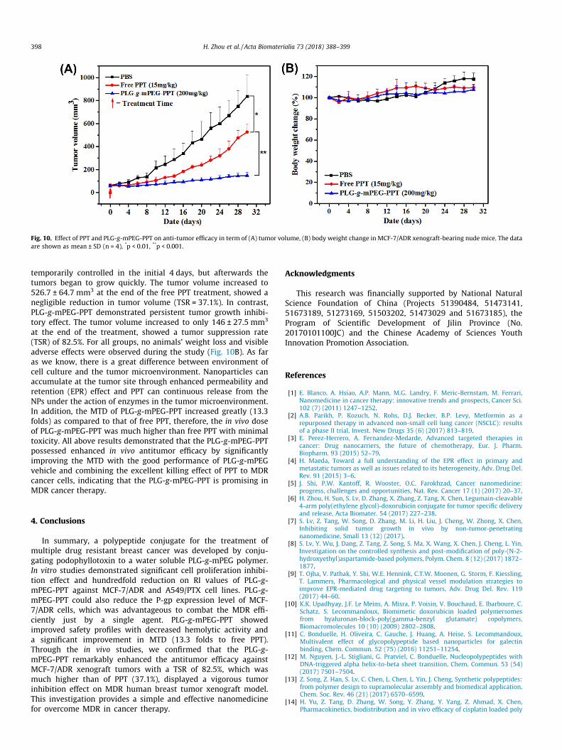
Fig. 10. Effect of PPT and PLG-g-mPEG-PPT on anti-tumor efficacy in term of (A) tumor volume, (B) body weight change in MCF-7/ADR xenograft-bearing nude mice. The dataare shown as mean ± SD (n = 4), *p < 0.01, **p < 0.001.
398 H. Zhou et al. / Acta Biomaterialia 73 (2018) 388–399
temporarily controlled in the initial 4 days, but afterwards thetumors began to grow quickly. The tumor volume increased to526.7 ± 64.7 mm3 at the end of the free PPT treatment, showed anegligible reduction in tumor volume (TSR = 37.1%). In contrast,PLG-g-mPEG-PPT demonstrated persistent tumor growth inhibi-tory effect. The tumor volume increased to only 146 ± 27.5 mm3
at the end of the treatment, showed a tumor suppression rate(TSR) of 82.5%. For all groups, no animals’ weight loss and visibleadverse effects were observed during the study (Fig. 10B). As faras we know, there is a great difference between environment ofcell culture and the tumor microenvironment. Nanoparticles canaccumulate at the tumor site through enhanced permeability andretention (EPR) effect and PPT can continuous release from theNPs under the action of enzymes in the tumor microenvironment.In addition, the MTD of PLG-g-mPEG-PPT increased greatly (13.3folds) as compared to that of free PPT, therefore, the in vivo doseof PLG-g-mPEG-PPT was much higher than free PPT with minimaltoxicity. All above results demonstrated that the PLG-g-mPEG-PPTpossessed enhanced in vivo antitumor efficacy by significantlyimproving the MTD with the good performance of PLG-g-mPEGvehicle and combining the excellent killing effect of PPT to MDRcancer cells, indicating that the PLG-g-mPEG-PPT is promising inMDR cancer therapy.
4. Conclusions
In summary, a polypeptide conjugate for the treatment ofmultiple drug resistant breast cancer was developed by conju-gating podophyllotoxin to a water soluble PLG-g-mPEG polymer.In vitro studies demonstrated significant cell proliferation inhibi-tion effect and hundredfold reduction on RI values of PLG-g-mPEG-PPT against MCF-7/ADR and A549/PTX cell lines. PLG-g-mPEG-PPT could also reduce the P-gp expression level of MCF-7/ADR cells, which was advantageous to combat the MDR effi-ciently just by a single agent. PLG-g-mPEG-PPT showedimproved safety profiles with decreased hemolytic activity anda significant improvement in MTD (13.3 folds to free PPT).Through the in vivo studies, we confirmed that the PLG-g-mPEG-PPT remarkably enhanced the antitumor efficacy againstMCF-7/ADR xenograft tumors with a TSR of 82.5%, which wasmuch higher than of PPT (37.1%), displayed a vigorous tumorinhibition effect on MDR human breast tumor xenograft model.This investigation provides a simple and effective nanomedicinefor overcome MDR in cancer therapy.
Acknowledgments
This research was financially supported by National NaturalScience Foundation of China (Projects 51390484, 51473141,51673189, 51273169, 51503202, 51473029 and 51673185), theProgram of Scientific Development of Jilin Province (No.20170101100JC) and the Chinese Academy of Sciences YouthInnovation Promotion Association.
References
[1] E. Blanco, A. Hsiao, A.P. Mann, M.G. Landry, F. Meric-Bernstam, M. Ferrari,Nanomedicine in cancer therapy: innovative trends and prospects, Cancer Sci.102 (7) (2011) 1247–1252.
[2] A.B. Parikh, P. Kozuch, N. Rohs, D.J. Becker, B.P. Levy, Metformin as arepurposed therapy in advanced non-small cell lung cancer (NSCLC): resultsof a phase II trial, Invest. New Drugs 35 (6) (2017) 813–819.
[3] E. Perez-Herrero, A. Fernandez-Medarde, Advanced targeted therapies incancer: Drug nanocarriers, the future of chemotherapy, Eur. J. Pharm.Biopharm. 93 (2015) 52–79.
[4] H. Maeda, Toward a full understanding of the EPR effect in primary andmetastatic tumors as well as issues related to its heterogeneity, Adv. Drug Del.Rev. 91 (2015) 3–6.
[5] J. Shi, P.W. Kantoff, R. Wooster, O.C. Farokhzad, Cancer nanomedicine:progress, challenges and opportunities, Nat. Rev. Cancer 17 (1) (2017) 20–37.
[6] H. Zhou, H. Sun, S. Lv, D. Zhang, X. Zhang, Z. Tang, X. Chen, Legumain-cleavable4-arm poly(ethylene glycol)-doxorubicin conjugate for tumor specific deliveryand release, Acta Biomater. 54 (2017) 227–238.
[7] S. Lv, Z. Tang, W. Song, D. Zhang, M. Li, H. Liu, J. Cheng, W. Zhong, X. Chen,Inhibiting solid tumor growth in vivo by non-tumor-penetratingnanomedicine, Small 13 (12) (2017).
[8] S. Lv, Y. Wu, J. Dang, Z. Tang, Z. Song, S. Ma, X. Wang, X. Chen, J. Cheng, L. Yin,Investigation on the controlled synthesis and post-modification of poly-(N-2-hydroxyethyl)aspartamide-based polymers, Polym. Chem. 8 (12) (2017) 1872–1877.
[9] T. Ojha, V. Pathak, Y. Shi, W.E. Hennink, C.T.W. Moonen, G. Storm, F. Kiessling,T. Lammers, Pharmacological and physical vessel modulation strategies toimprove EPR-mediated drug targeting to tumors, Adv. Drug Del. Rev. 119(2017) 44–60.
[10] K.K. Upadhyay, J.F. Le Meins, A. Misra, P. Voisin, V. Bouchaud, E. Ibarboure, C.Schatz, S. Lecommandoux, Biomimetic doxorubicin loaded polymersomesfrom hyaluronan-block-poly(gamma-benzyl glutamate) copolymers,Biomacromolecules 10 (10) (2009) 2802–2808.
[11] C. Bonduelle, H. Oliveira, C. Gauche, J. Huang, A. Heise, S. Lecommandoux,Multivalent effect of glycopolypeptide based nanoparticles for galectinbinding, Chem. Commun. 52 (75) (2016) 11251–11254.
[12] M. Nguyen, J.-L. Stigliani, G. Pratviel, C. Bonduelle, Nucleopolypeptides withDNA-triggered alpha helix-to-beta sheet transition, Chem. Commun. 53 (54)(2017) 7501–7504.
[13] Z. Song, Z. Han, S. Lv, C. Chen, L. Chen, L. Yin, J. Cheng, Synthetic polypeptides:from polymer design to supramolecular assembly and biomedical application,Chem. Soc. Rev. 46 (21) (2017) 6570–6599.
[14] H. Yu, Z. Tang, D. Zhang, W. Song, Y. Zhang, Y. Yang, Z. Ahmad, X. Chen,Pharmacokinetics, biodistribution and in vivo efficacy of cisplatin loaded poly
H. Zhou et al. / Acta Biomaterialia 73 (2018) 388–399 399
(L-glutamic acid)-g-methoxy poly (ethylene glycol) complex nanoparticles fortumor therapy, J. Controlled Release 205 (2015) 89–97.
[15] M. Dean, T. Fojo, S. Bates, Tumour stem cells and drug resistance, Nat. Rev.Cancer 5 (4) (2005) 275–284.
[16] G. Szakacs, J.K. Paterson, J.A. Ludwig, C. Booth-Genthe, M.M. Gottesman,Targeting multidrug resistance in cancer, Nat. Rev. Drug Discov. 5 (3) (2006)219–234.
[17] P. Joshi, R.A. Vishwakarma, S.B. Bharate, Natural alkaloids as P-gp inhibitors formultidrug resistance reversal in cancer, Eur. J. Med. Chem. 138 (2017) 273–292.
[18] L.-M. Mu, R.-J. Ju, R. Liu, Y.-Z. Bu, J.-Y. Zhang, X.-Q. Li, F. Zeng, W.-L. Lu, Dual-functional drug liposomes in treatment of resistant cancers, Adv. Drug Del.Rev. 115 (2017) 46–56.
[19] Z. Chen, T. Shi, L. Zhang, P. Zhu, M. Deng, C. Huang, T. Hu, L. Jiang, J. Li,Mammalian drug efflux transporters of the ATP binding cassette (ABC) familyin multidrug resistance: a review of the past decade, Cancer Lett. 370 (1)(2016) 153–164.
[20] F. Ren, J. Shen, H. Shi, F.J. Hornicek, Q. Kan, Z. Duan, Novel mechanisms andapproaches to overcome multidrug resistance in the treatment of ovariancancer, Biochim. Biophys. Acta 1866 (2) (2016) 266–275.
[21] Y. Chen, O. Tezcan, D. Li, N. Beztsinna, B. Lou, T. Etrych, K. Ulbrich, J.M.Metselaar, T. Lammers, W.E. Hennink, Overcoming multidrug resistance usingfolate receptor-targeted and pH-responsive polymeric nanogels containingcovalently entrapped doxorubicin, Nanoscale 9 (29) (2017) 10404–10419.
[22] S. Kunjachan, B. Rychlik, G. Storm, F. Kiessling, T. Lammers, Multidrugresistance: Physiological principles and nanomedical solutions, Adv. DrugDel. Rev. 65 (13–14) (2013) 1852–1865.
[23] H.M. Coley, Mechanisms and strategies to overcome chemotherapy resistancein metastatic breast cancer, Cancer Treat. Rev. 34 (4) (2008) 378–390.
[24] Y. Zhao, Y. Zhou, D. Wang, Y. Gao, J. Li, S. Ma, L. Zhao, C. Zhang, Y. Liu, X. Li, pH-responsive polymeric micelles based on poly(2-ethyl-2-oxazoline)-poly(D,L-lactide) for tumor-targeting and controlled delivery of doxorubicin and P-glycoprotein inhibitor, Acta Biomater. 17 (2015) 182–192.
[25] R.J. Kathawala, P. Gupta, C.R. Ashby Jr., Z.-S. Chen, The modulation of ABCtransporter-mediated multidrug resistance in cancer: a review of the pastdecade, Drug Resist. Update. 18 (2015) 1–17.
[26] C. Avendano, J.C. Menendez, Inhibitors of multidrug resistance to antitumoragents (MDR), Curr. Med. Chem. 9 (2) (2002) 159–193.
[27] G.C. Tomskey, G.W. Vickery, P.L. Getzoff, The successful treatment ofgranuloma inguinale, with special reference to the use of podophyllin, J.Urol. 48 (4) (1942) 401–406.
[28] A. Giri, M.L. Narasu, Production of podophyllotoxin from Podophyllumhexandrum: a potential natural product for clinically useful anticancer drugs,Cytotechnology 34 (1–2) (2000) 17–26.
[29] X. Yu, Z. Che, H. Xu, Recent advances in the chemistry and biology ofpodophyllotoxins, Chem. Eur. J. 23 (19) (2017) 4467–4526.
[30] L. Zhang, F. Chen, J. Wang, Y. Chen, Z. Zhang, Y. Lin, X. Zhu, Novel isatinderivatives of podophyllotoxin: synthesis and cytotoxic evaluation againsthuman leukaemia cancer cells as potent anti-MDR agents, Rsc Adv. 5 (118)(2015) 97816–97823.
[31] H.M. Abdallah, A.M. Al-Abd, R.S. El-Dine, A.M. El-Halawany, P-glycoproteininhibitors of natural origin as potential tumor chemo-sensitizers: a review, J.Adv. Res. 6 (1) (2015) 45–62.
[32] S. Zhu, H. Zhen, Y. Li, P. Wang, X. Huang, P. Shi, PEGylated graphene oxide as ananocarrier for podophyllotoxin, J. Nanopart. Res. 16 (8) (2014).
[33] X. Huang, X. Huang, X.-H. Jiang, F.-Q. Hu, Y.-Z. Du, Q.-F. Zhu, C.-S. Jin, In vitroantitumour activity of stearic acid-g-chitosan oligosaccharide polymericmicelles loading podophyllotoxin, J. Microencaps. 29 (1) (2012) 1–8.
[34] L. Fan, H. Wu, H. Zhang, F. Li, T.-H. Yang, pH-sensitive podophyllotoxin carrierfor cancer cells specific delivery, Polym. Compos. 31 (1) (2010) 51–59.
[35] H.B. Chen, X.L. Chang, D.R. Du, W. Liu, J. Liu, T. Weng, Y.J. Yang, H.B. Xu, X.L.Yang, Podophyllotoxin-loaded solid lipid nanoparticles for epidermaltargeting, J. Controlled Release 110 (2) (2006) 296–306.
[36] A. Roy, M.J. Ernsting, E. Undzys, S.-D. Li, A highly tumor-targeted nanoparticleof podophyllotoxin penetrated tumor core and regressed multidrug resistanttumors, Biomaterials 52 (2015) 335–346.
[37] A. Roy, Y. Zhao, Y. Yang, A. Szeitz, T. Klassen, S.-D. Li, Selective targeting andtherapy of metastatic and multidrug resistant tumors using a long circulatingpodophyllotoxin nanoparticle, Biomaterials 137 (2017) 11–22.
[38] W. Song, Z. Tang, D. Zhang, Y. Zhang, H. Yu, M. Li, S. Lv, H. Sun, M. Deng, X.Chen, Anti-tumor efficacy of c(RGDfK)-decorated polypeptide-based micellesco-loaded with docetaxel and cisplatin, Biomaterials 35 (9) (2014) 3005–3014.
[39] W. Song, Z. Tang, M. Li, S. Lv, H. Sun, M. Deng, H. Liu, X. Chen, Polypeptide-based combination of paclitaxel and cisplatin for enhanced chemotherapyefficacy and reduced side-effects, Acta Biomater. 10 (3) (2014) 1392–1402.
[40] S. Lv, M. Li, Z. Tang, W. Song, H. Sun, H. Liu, X. Chen, Doxorubicin-loadedamphiphilic polypeptide-based nanoparticles as an efficient drug deliverysystem for cancer therapy, Acta Biomater. 9 (12) (2013) 9330–9342.
[41] F. Shi, J. Ding, C. Xiao, X. Zhuang, C. He, L. Chen, X. Chen, Intracellularmicroenvironment responsive PEGylated polypeptide nanogels with ionizablecores for efficient doxorubicin loading and triggered release, J. Mater. Chem. 22(28) (2012) 14168–14179.
[42] W. Song, Z. Tang, D. Zhang, M. Li, J. Gu, X. Chen, A cooperative polymericplatform for tumor-targeted drug delivery, Chem. Sci. 7 (1) (2016) 728–736.
[43] T. Liu, D. Zhang, W. Song, Z. Tang, J. Zhu, Z. Ma, X. Wang, X. Chen, T. Tong, Apoly(L-glutamic acid)-combretastatin A4 conjugate for solid tumor therapy:markedly improved therapeutic efficiency through its low tissue penetrationin solid tumor, Acta Biomater. 53 (2017) 179–189.
[44] H. Yu, Z. Tang, M. Li, W. Song, D. Zhang, Y. Zhang, Y. Yang, H. Sun, M. Deng, X.Chen, Cisplatin loaded poly(L-glutamic acid)-g-methoxy poly(ethylene glycol)complex nanoparticles for potential cancer therapy: preparation, in vitro andin vivo evaluation, J. Biomed. Nanotechnol. 12 (1) (2016) 69–78.
[45] W. Song, Z. Tang, N. Shen, H. Yu, Y. Jia, D. Zhang, J. Jiang, C. He, H. Tian, X. Chen,Combining disulfiram and poly(L-glutamic acid)-cisplatin conjugates forcombating cisplatin resistance, J. Control. Release 231 (2016) 94–102.
[46] H.Y. Yifei Li, Hai Sun, Jianguo Liu, Zhaohui Tang, Dan Wang, Yu Lianyou, XuesiChen, Cisplatin-loaded poly(L-glutamic acid)-g-methoxy poly(ethylene glycol)nanoparticles as a potential chemotherapeutic agent against osteosarcoma,Chin. J. Polym. Sci. 33 (5) (2015) 763–771.
[47] K. Kurihara, T. Abe, N. Higashi, M. Niwa, Steric forces between brush layers ofpoly (L-glutamic acid) and their dependence on secondary structures asdetermined by FT-IR spectroscopy, Colloids Surf. Physicochem. Eng. Aspects103 (3) (1995) 265–272.
[48] H. Lu, J. Wang, Y. Bai, J.W. Lang, S. Liu, Y. Lin, J. Cheng, Ionic polypeptides withunusual helical stability, Nat. Commun. 2 (2011) 206.
[49] X. Duan, Y. Li, Physicochemical characteristics of nanoparticles affectcirculation biodistribution, cellular internalization, and trafficking, Small 9(9–10) (2013) 1521–1532.
[50] J.A. Barreto, W. O’Malley, M. Kubeil, B. Graham, H. Stephan, L. Spiccia,Nanomaterials: applications in cancer imaging and therapy, Adv. Mater. 23(12) (2011) H18–H40.
[51] V.P. Chauhan, R.K. Jain, Strategies for advancing cancer nanomedicine, Nat.Mater. 12 (11) (2013) 958–962.
[52] W. Song, M. Li, Z. Tang, Q. Li, Y. Yang, H. Liu, T. Duan, H. Hong, X. Chen,Methoxypoly(ethylene glycol)-block-poly(L-glutamic acid)-loaded cisplatinand a combination with irgd for the treatment of non-small-cell lungcancers, Macromol. Biosci. 12 (11) (2012) 1514–1523.
[53] L. Zhao, J. Ding, C. Xiao, P. He, Z. Tang, X. Pang, X. Zhuang, X. Chen, Glucose-sensitive polypeptide micelles for self-regulated insulin release atphysiological pH, J. Mater. Chem. 22 (24) (2012) 12319–12328.
[54] Q. Xiao, W. Bu, Q. Ren, S. Zhang, H. Xing, F. Chen, M. Li, X. Zheng, Y. Huab, L.Zhou, W. Peng, H. Qu, Z. Wang, K. Zhao, J. Shi, Radiopaque fluorescence-transparent TaOx decorated upconversion nanophosphors for in vivo CT/MR/UCL trimodal imaging, Biomaterials 33 (30) (2012) 7530–7539.
![The Conjugate Gradient Method...Conjugate Gradient Algorithm [Conjugate Gradient Iteration] The positive definite linear system Ax = b is solved by the conjugate gradient method](https://img.pdfslide.net/doc/110x75/5e95c1e7f0d0d02fb330942a/the-conjugate-gradient-method-conjugate-gradient-algorithm-conjugate-gradient.jpg)

![Biological Activity of N-Hydroxyethyl-4-aza-2,3 ...thesize from podophyllotoxin [13]. These new aza-podophyllotoxin derivative compounds have only two chiral centers at the 1 and 4](https://img.pdfslide.net/doc/110x75/5e68d63f78796e747a6fc6f6/biological-activity-of-n-hydroxyethyl-4-aza-23-thesize-from-podophyllotoxin.jpg)