Embed Size (px)
Citation preview

A Practical Book of
HHUUMMAANN AANNAATTOOMMYY AANNDD
PPHHYYSSIIOOLLOOGGYY -- IIII
As Per PCI Regulations
FIRST YEAR B. PHARM.
Semester II
Prof. Md. Rageeb Md. Usman Dr. Mrunal K. Shirsat M. Pharm., FAPP, FICPHS, FRSH, FSPER, FSRHCP M. Pharm., Ph.D.
Joint Secretaries - SPER Central Branch & Associate Professor
President - IPA/APP/RSH/SRHCP Pacific University
Maharashtra State Branch & Pacific Academy of Higher Education and
Assistant Professor Research University, Udaipur, Rajasthan
Smt. S. S. Patil College of Pharmacy, India
Chopda, Maharashtra
NMU Jalgaon, Maharashtra, India
Dr. Jayesh Dwivedi Dr. Mohammed Zuber Shaikh M. Pharm., Ph.D. M.Sc., Ph.D.
Associate Professor Head, Dept. of Zoology,
Pacific University Senior Science College, Akkalkuwa, M.S., India
Pacific Academy of Higher Education and
Research University, Udaipur, Rajasthan
India
Price ` 135.00
N3942

Human Anatomy and Physiology - II (Practical) ISBN 978-93-86943-50-7
Second Edition : May 2018
© : Authors The text of this publication, or any part thereof, should not be reproduced or transmitted in any form or stored in any computer storage system or device for distribution including photocopy, recording, taping or information retrieval system or reproduced on any disc, tape, perforated media or other information storage device etc., without the written permission of Authors with whom the rights are reserved. Breach of this condition is liable for legal action. Every effort has been made to avoid errors or omissions in this publication. In spite of this, errors may have crept in. Any mistake, error or discrepancy so noted and shall be brought to our notice shall be taken care of in the next edition. It is notified that neither the publisher nor the authors or seller shall be responsible for any damage or loss of action to any one, of any kind, in any manner, therefrom.
Published By : Polyplate Printed By :
NIRALI PRAKASHAN SHIVANI PRINTERS Abhyudaya Pragati, 1312, Shivaji Nagar, 1311, Kinara Sahakari Gruha Sanstha
Off J.M. Road, PUNE – 411005 Kasba Peth,
Tel - (020) 25512336/37/39, Fax - (020) 25511379 PUNE - 411011
Email : [email protected] Tel - (020) 24577245
� DISTRIBUTION CENTRES PUNE Nirali Prakashan : 119, Budhwar Peth, Jogeshwari Mandir Lane, Pune 411002, Maharashtra
Tel : (020) 2445 2044, 66022708, Fax : (020) 2445 1538
Email : [email protected], [email protected]
Nirali Prakashan : S. No. 28/27, Dhyari, Near Pari Company, Pune 411041
Tel : (020) 24690204 Fax : (020) 24690316
Email : [email protected], [email protected]
MUMBAI Nirali Prakashan : 385, S.V.P. Road, Rasdhara Co-op. Hsg. Society Ltd., Girgaum, Mumbai 400004, Maharashtra
Tel : (022) 2385 6339 / 2386 9976, Fax : (022) 2386 9976
Email : [email protected]
� DISTRIBUTION BRANCHES JALGAON Nirali Prakashan : 34, V. V. Golani Market, Navi Peth, Jalgaon 425001, Maharashtra, Tel : (0257) 222 0395, Mob : 94234 91860 KOLHAPUR Nirali Prakashan : New Mahadvar Road, Kedar Plaza, 1st Floor Opp. IDBI Bank Kolhapur 416 012, Maharashtra. Mob : 9850046155 NAGPUR Pratibha Book Distributors : Above Maratha Mandir, Shop No. 3, First Floor, Rani Jhanshi Square, Sitabuldi, Nagpur 440012, Maharashtra Tel : (0712) 254 7129 DELHI Nirali Prakashan : 4593/15, Basement, Aggarwal Lane 15, Ansari Road, Daryaganj Near Times of India Building, New Delhi 110002 Mob : 08505972553 BENGALURU Pragati Book House : House No. 1, Sanjeevappa Lane, Avenue Road Cross, Opp. Rice Church, Bengaluru – 560002. Tel : (080) 64513344, 64513355,Mob : 9880582331, 9845021552 Email:[email protected] CHENNAI Pragati Books : 9/1, Montieth Road, Behind Taas Mahal, Egmore, Chennai 600008 Tamil Nadu, Tel : (044) 6518 3535, Mob : 94440 01782 / 98450 21552 / 98805 82331,
Email : [email protected] Note: Every possible effort has been made to avoid errors or omissions in this book. In spite this, errors may have crept in. Any type of error or mistake so noted, and shall be brought to our notice, shall be taken care of in the next edition. It is notified that neither the publisher, nor the author or book seller shall be responsible for any damage or loss of action to any one of any kind, in any manner, therefrom. The reader must cross check all the facts and contents with original Government notification or publications.
[email protected] | www.pragationline.com
Also find us on www.facebook.com/niralibooks

Acknowledgement
It gives us immense pleasure to thank Smt. S. S. Patil College of Pharmacy, Chopda,
Maharashtra and Pacific University, Udaipur for their appreciation, moral support, constant
encouragement, positive criticism and scientific inputs.
We are thankful to Prof. Dr. S. B. Bari, Dean of Pharmaceutical Science, North
Maharashtra University, Jalgaon and Principal, H. R. Patel College of Pharmacy, Shirpur who
encouraged us to pursue this attempt in all fronts. We thank our college staff and library
staff who helped us at every step.
The authors are grateful to Prof. Dr. V. R. Patil, Principal, College of Pharmacy, Faizpur,
India, Prof. Dr. Sunil P. Pawar, Chairman of Board of Studies, Department of Pharmacology &
Pharmacognosy of North Maharashtra University, Jalgaon and Dr. G. P. Vadnere, Principal,
Smt. S. S. Patil College of Pharmacy, Chopda, India for excellent guidance and dedicated
efforts made us to think upon and understand number of problems and solve them
judiciously.
Thankful to our colleagues and many users of the book for constructive criticism and
suggestions.
We are grateful to our parents for their unconditional love, support and encouragement.
We are also thankful to my Publishers. We hope this book will leave the desired
impression and look forward to receive any comments from the readers.
Authors


Preface
This book with the title “A Practical Book of Human Anatomy and Physiology - II” is
written with an intention to benefit the Diploma and Degree students.
Practical aspect of Human Anatomy and Physiology serves as an essential study feature
of Diploma and Degree Pharmacy Course which enrich the knowledge of the student
pharmacists and makes the Pharmacist easy to understand drug action and technology of
drug dosage form.
The book of Human Anatomy and Physiology - II has been specifically designed as per
PCI New Syllabus for First Year B. Pharmacy Students.
Pharmacy Council of India are Prescribed by New Syllabus of all over India.
The matter is precise, appropriate and with easy to understand language. The accurate
and clear diagrams provide a better approach towards understanding the features.
Any valuable and useful suggestions for the improvement of this book are always
welcomed.
Hope this will be helpful....All the best!!!
Authors

Syllabus
1. To study the integumentary and special senses using specimen, models, etc.
2. To study the nervous system using specimen, models, etc.
3. To study the endocrine system using specimen, models, etc.
4. To demonstrate the general neurological examination
5. To demonstrate the function of olfactory nerve
6. To examine the different types of taste.
7. To demonstrate the visual acuity
8. To demonstrate the reflex activity
9. Recording of body temperature
10. To demonstrate positive and negative feedback mechanism.
11. Determination of tidal volume and vital capacity.
12. Study of digestive, respiratory, cardiovascular systems, urinary and reproductive
systems with the help of models, charts and specimens.
13. Recording of basal mass index.
14. Study of family planning devices and pregnancy diagnosis test.
15. Demonstration of total blood count by cell analyser.
16. Permanent slides of vital organs and gonads.
���
Contents
1. Experiment No. 1 1 - 8
2. Experiment No. 2 9 - 17
3. Experiment No. 3 18 - 22
4. Experiment No. 4 23 - 26
5. Experiment No. 5 27 - 35
6. Experiment No. 6 36 - 38
7. Experiment No. 7 39 - 43
8. Experiment No. 8 44 - 49
9. Experiment No. 9 50 - 52
10. Experiment No. 10 53 - 54
11. Experiment No. 11 55 - 57
12. Experiment No. 12 58 - 60
13. Experiment No. 13 61 - 65
14. Experiment No. 14 66 - 71
15. Experiment No. 15 72 - 82
16. Experiment No. 16 83 - 93
17. Experiment No. 17 94 - 107
18. Experiment No. 18 108 - 115
19. Experiment No. 19 116 - 123
20. Experiment No. 20 124 - 129
21. Experiment No. 21 130 - 150
���

(1)
Experiment No. 01
Aim: To study the compound microscope.
Microscope:
A microscope (from the Ancient Greek: mikros, "small" and skopein, "to look" or "see") is
an instrument used to see objects that are too small for the naked eye. The science of
investigating small object using such an instrument is called microscopy. Microscopic means
invisible to the eye unless aided by a microscope. There are many types of microscopes, the
most common and first to be invented is the optical microscope which uses light to image
the sample. Other major types of microscopes are the electron microscope (both the
transmission electron microscope and the scanning electron microscope) and the various
types of scanning probe microscope.
Zacharias Jansen invented first compound microscope. When the microscope was
invented around 1590, suddenly we saw a new world of living things in our water, in our food
and under our nose. But it's unclear who invented the microscope. Some historians say it
was Hans Lippershey, most famous for filing the first patent for a telescope.
Types of Microscope:
1. Simple microscope
2. Compound microscope
3. Electronic microscope
4. Motic microscope
1. Simple Microscope:
A simple microscope is a microscope which has only one lens, as opposed to the
compound lenses used in more complex microscope designs. Magnifying glasses and loupes
are two well-known examples of the simple microscope. The image quality with a simple
microscope can be surprisingly good, as researchers learned when they used replications of
17th century simple microscopes to see what exactly it was that biologists found in water
supplies in the early days of microscopy. If the lens is of a very high quality, the user should
know how to manipulate the microscope to achieve the best focus, contrast and clarity. The
image can be quite excellent, although it is of course impossible to obtain very high levels of
magnification with a simple microscope. For basic applications, however, this design can be
quite useful, in addition to affordable for people who may not want to shell out for a more
advanced microscope design.

Human Anatomy and Physiology II (Practical) 2 Experiments
Eyepiece
Eyepiece tube
Arm
Head
Nosepiece (turret)
Objective lensStage clips
Aperture
Stage
Condenser
Iris diaphragm
Illuminator
Base
Coarse focus knob
Fine focus knob
Fig. 1.1: Simple Microscope
2. Compound Microscope:
A compound microscope is a microscope with more than one lens and its own light
source. In this type of microscope, there are ocular lenses in the binocular eyepieces and
objective lenses in a rotating nosepiece closer to the specimen. Although sometimes found
as monocular with one ocular lens and the compound binocular microscope is more
commonly used today. They combine the power of lenses and light to enlarge the subject
being viewed.
Viewing Heads: Monocular, Binocular, Trinocular:
• Monocular: Monocular only use one eyepiece when viewing the specimen. You are
restricted if you want to use a CCD camera because this would occupy the eyepiece.
However, monocular microscopes are light weight and are inexpensive.
• Binocular: They have two eyepieces which prove to be more comfortable. It is the most
common choice.
• Trinocular: Trinocular has a third eyepiece tube that can be used by another person
simultaneously or by a CCD camera. The trinocular option is more expensive than the
other two types.

Human Anatomy and Physiology II (Practical) 3 Experiments
Typically, the eyepiece itself allows for 10X or 15X magnifications and when combined
with the three or four objective lenses which can be rotated into the field of view. They
produce higher magnification to a maximum of around 1000X generally. The compound light
microscope is popular among botanists for studying plant cells in biology to view bacteria
and parasites as well as a variety of human/animal cells.
C-Mount
Trinocular head
Eyepieces(ocular lenses)
Lock screwBinocular heads
Objective lenses
Mechanical stage
Condenser
Filter holder
Base lens
Base
On/Off switch
Light dimmer
Mechanicalstage controls
Fine focusing knob
Coarse focusing knob
Arm stand
Fig. 1.2: Compound Microscope
The compound microscope essentially consists of three major systems.
1. Support system:
It comprises of base, stage and body tube.
Base: The base supports the microscope and it’s where illuminator is located.
Stage: The flat platform where the slide is placed. The specimen is the object being
examined. Most specimens are mounted on slides, flat rectangles of thin glass. The specimen
is placed on the glass and a cover slip is placed over the specimen. This allows the slide to be
easily inserted or removed from the microscope. It also allows the specimen to be labeled,
transported, and stored without damage.
Stage clips: Metal clips that hold the slide in place.
Stage height adjustment (Stage Control): These knobs move the stage left and right or
up and down.
Body tube (Head): The body tube connects the eyepiece to the objective lenses. The
arm connects the body tube to the base of the microscope.
Human Anatomy and Physiology II (Practical) 4 Experiments
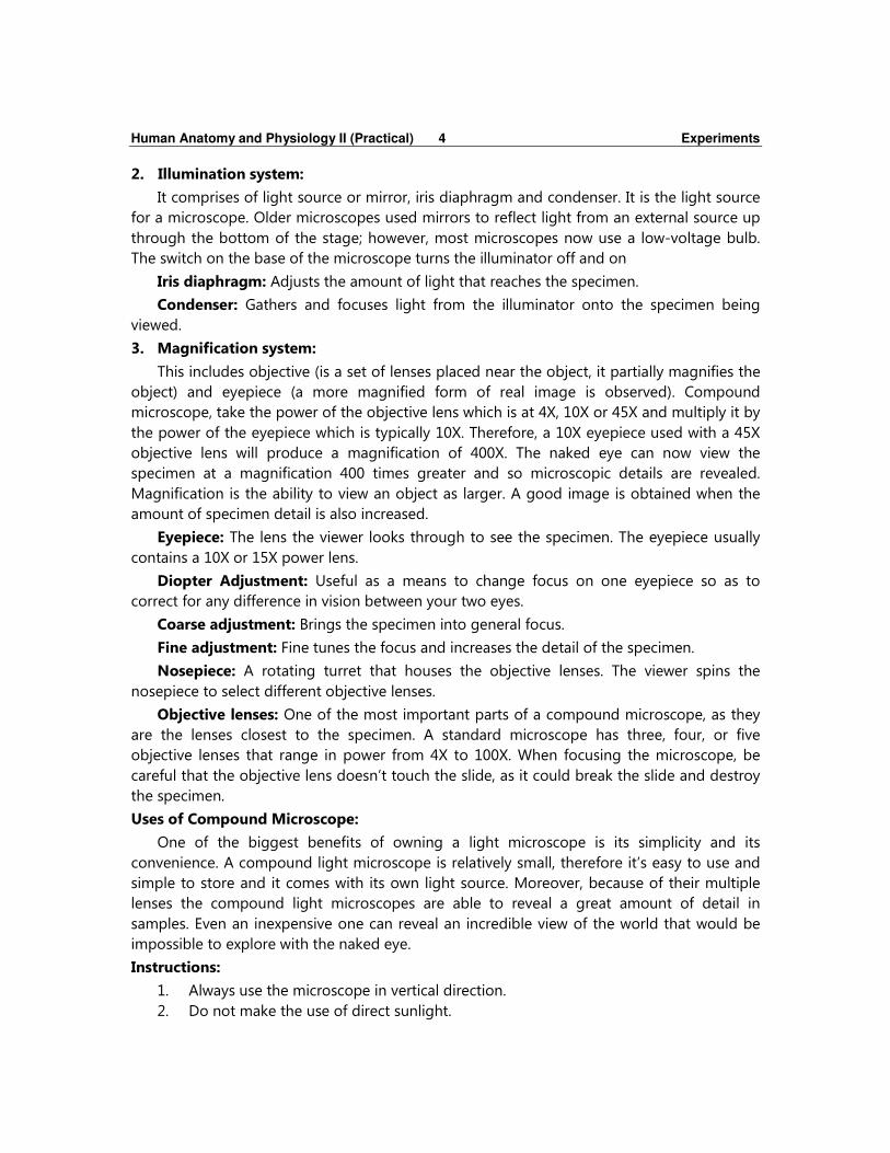
2. Illumination system:
It comprises of light source or mirror, iris diaphragm and condenser. It is the light source
for a microscope. Older microscopes used mirrors to reflect light from an external source up
through the bottom of the stage; however, most microscopes now use a low-voltage bulb.
The switch on the base of the microscope turns the illuminator off and on
Iris diaphragm: Adjusts the amount of light that reaches the specimen.
Condenser: Gathers and focuses light from the illuminator onto the specimen being
viewed.
3. Magnification system:
This includes objective (is a set of lenses placed near the object, it partially magnifies the
object) and eyepiece (a more magnified form of real image is observed). Compound
microscope, take the power of the objective lens which is at 4X, 10X or 45X and multiply it by
the power of the eyepiece which is typically 10X. Therefore, a 10X eyepiece used with a 45X
objective lens will produce a magnification of 400X. The naked eye can now view the
specimen at a magnification 400 times greater and so microscopic details are revealed.
Magnification is the ability to view an object as larger. A good image is obtained when the
amount of specimen detail is also increased.
Eyepiece: The lens the viewer looks through to see the specimen. The eyepiece usually
contains a 10X or 15X power lens.
Diopter Adjustment: Useful as a means to change focus on one eyepiece so as to
correct for any difference in vision between your two eyes.
Coarse adjustment: Brings the specimen into general focus.
Fine adjustment: Fine tunes the focus and increases the detail of the specimen.
Nosepiece: A rotating turret that houses the objective lenses. The viewer spins the
nosepiece to select different objective lenses.
Objective lenses: One of the most important parts of a compound microscope, as they
are the lenses closest to the specimen. A standard microscope has three, four, or five
objective lenses that range in power from 4X to 100X. When focusing the microscope, be
careful that the objective lens doesn’t touch the slide, as it could break the slide and destroy
the specimen.
Uses of Compound Microscope:
One of the biggest benefits of owning a light microscope is its simplicity and its
convenience. A compound light microscope is relatively small, therefore it’s easy to use and
simple to store and it comes with its own light source. Moreover, because of their multiple
lenses the compound light microscopes are able to reveal a great amount of detail in
samples. Even an inexpensive one can reveal an incredible view of the world that would be
impossible to explore with the naked eye.
Instructions:
1. Always use the microscope in vertical direction.
2. Do not make the use of direct sunlight.

Human Anatomy and Physiology II (Practical) 5 Experiments
3. The object to be examined should be mounted in mounting medium & should be
covered with cover slip before observation.
Precautions:
1. Do not keep the microscope near the edge of the table.
2. Always keep the stage clean and dry.
3. Do not use excess mounting medium.
4. Microscope when not in use should be covered properly.
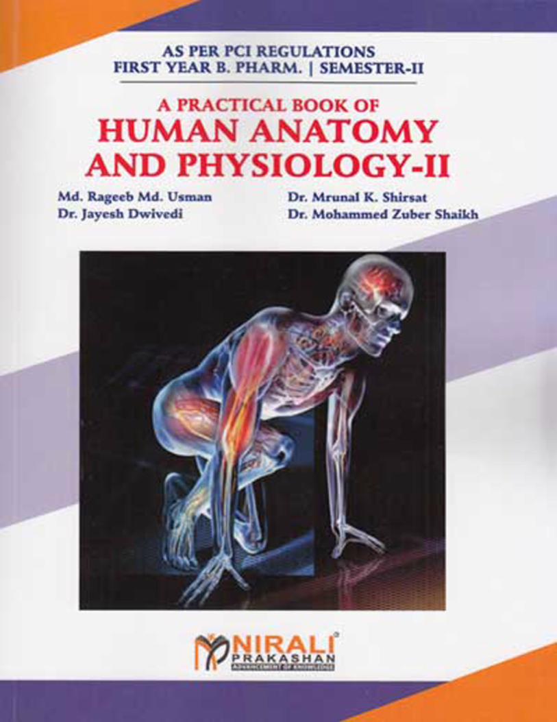
3. Electronic Microscope:
An EM is a microscope that focuses beams of energetic electrons to examine objects up
to nano-scales. They utilize the same principles behind an optical microscope, but rather than
photons or particles of light, concentrate electrons, charged particles located on the outside
of atoms, onto an object. Additional differences include preparation of specimens before
being placed in the vacuum chamber, the use of coiled electromagnets instead of glass
lenses, the use of a thermionic gun as an electron source and the image or electron
micrograph is viewed on a screen rather than an eyepiece. All EMs use electromagnetic or
electrostatic lenses, which consist of a coil of wire wrapped around the outside of a tube,
commonly referred to as a solenoid.
In addition, EMs use digital displays, computer interfaces, software for image analysis and
a low vacuum or variable pressure chamber, which upholds the pressure differential between
the high vacuum levels essential to the gun and column area and the low pressure required
in the chamber.
Fig. 1.3: Electronic Microscope
All electron microscopy samples must be prepared before placed in the microscope
vacuum. Techniques, which vary based on type of specimen and analysis, include:
• Cryofixation
• Fixation
• Dehydration

Human Anatomy and Physiology II (Practical) 6 Experiments
• Embedding
• Sectioning
• Staining
• Freeze-fracture and Freeze-etch
• Sputter Coating
Most of these techniques require specialized training and, due to sample manipulation,
can result in artifacts or inadvertent changes to the structure of the specimen.
The following gives you a description of two types of EMs, the Transmission (TEM) and
Scanning Electron Microscope (SEM).
Transmission Electron Microscope:
The transmission electron microscope (TEM), the first type of EM, has many
commonalities with the optical microscope and is a powerful microscope, capable of
producing images 1 nanometer in size. They require high voltages to increase the
acceleration speed of electrons, which, once they pass through the sample (transmission),
increase the image resolution. The 2-d, black and white images produced by TEMs can be
seen on a screen or printed onto a photographic plate. Although recent innovations in
software help to minimize, TEM resolution is hampered by spherical and chromatic
aberrations. The TEM is a popular choice for nanotechnology as well as semiconductor
analysis and production.
Fig. 1.4: Transmission Electron Microscope

A Practical Book Of Human AnatomyAnd Physiology - II
Publisher : Nirali Prakashan ISBN : 9789386943507
Author : Prof. Md. RageebMd. Usman, Dr. Mrunal K.Shirsat, Dr. JayeshDwivedi, Dr. MohammedZuber Shaikh
Type the URL : http://www.kopykitab.com/product/19712
Get this eBook
60%OFF