Embed Size (px)
Citation preview
A Primer on Mechanical Ventilation
David J Pierson MD Although mechanical ventilation is a key component of intensive care, unfamiliar jargon and technical detail render it confusing and formidably difficult for many clinicians. The rapidity and complexity of change in this area of respiratory medicine in recent years adds to the problem. Most of the current literature and nearly all the controversy in mechanical ventilation apply to only a small fraction of the patients who are intubated and ventilated in acute care hospitals. This small fraction consists of those with severe respiratory failure due to acute diffuse lung injury—acute lung injury (ALI) or the acute respiratory distress syndrome (ARDS)—and also those with severe obstructive lung disease (COPD or asthma). For the other 80 or 90 percent of ventilated patients the issues are much less difficult. At best, mechanical ventilation can support gas exchange and lung inflation in a reasonably normal fashion. It is supportive, replacement therapy only; there is no disease that intubating and ventilating a patient will cure. When done as well as we know how, mechanical ventilation can come close to replacing the normal functions of the lungs and chest bellows. Considering the many adverse consequences of intubation and positive-pressure ventilation, the clinician should always be striving to make the period of ventilatory support as short as possible. This primer summarizes the main issues in mechanical ventilation, explains many of the modes, settings, and terms involved, and reviews aspects of monitoring and complications with which the clinician should be familiar. It focuses on invasive mechanical ventilation (that is, on ventilating intubated patients), and on the care of adults rather than children or infants. Parts of it are adapted from the chapter on invasive mechanical ventilation in Clinical Respiratory Medicine, by Albert, Spiro, and Jett.1 Table 1 lists a number of factors that are key to developing a rational approach to mechanical ventilation in acute illness. It is also important that the fundamental goals and objectives of this therapy (Table 2) be understood, both in general and relative to the individual patient. 1 Pierson DJ. Invasive mechanical ventilation. In Albert RK, Spiro SG, Jett JR, eds. Clinical respiratory medicine. London/Philadelphia, Saunders, 2nd edition, 2004:189-209
1

INDICATIONS FOR INVASIVE MECHANICAL VENTILATION In the absence of a contraindication, mechanical ventilation is indicated whenever any of the circumstances listed under "clinical objectives" in Table 2 exists to a degree that threatens the life of the patient. Apart from apnea, however, few individual symptoms, signs, or laboratory findings by themselves always mandate the initiation of ventilatory support. Rather, this therapy becomes necessary in the presence of the right combination of clinical setting, severity of abnormality, and rapidity of development or worsening of physiologic abnormality. A summary of the main categories of indications for invasive ventilatory support is provided in Table 3. Apnea and Impending Respiratory Arrest No one would argue with providing ventilatory support for a patient who has stopped breathing altogether. On the other hand, "impending respiratory arrest" is difficult to define prospectively, and attempts to study it as an indication for intubation and mechanical ventilation have so far proved unsuccessful. These things are commonly carried out because the clinician judges the patient to be in severe respiratory distress, "tiring," or "about to arrest," but inter-observer variation and the extent to which these subjective impressions predict an unfavorable outcome if intubation is not performed have not been investigated. Exacerbation of Chronic Obstructive Pulmonary Disease The substantial number of studies of noninvasive positive-pressure ventilation (NPPV) in severe exacerbations of COPD has not directly addressed the validity of the criteria used for intubation. The writing committee for the Global Initiative for Obstructive Lung Disease (www.goldcopd.org), using best available evidence, recommends invasive mechanical ventilation when patients with acute exacerbations of COPD have cardiovascular instability, somnolence or other altered mental state, uncooperativeness, a high risk of aspiration, copious or very viscous respiratory tract secretions, any craniofacial condition (such as recent trauma or surgery) potentially rendering NPPV difficult, or extreme obesity. Very severe or progressive respiratory acidosis is also accepted as an indication, but agreement is lacking as to whether a pH of 7.25, a PaCO2 of 60 mm Hg, or some other thresholds should be used. Acute Severe Asthma Retrospective studies have shown that relatively few patients with acute severe asthma require invasive mechanical ventilation, but no clinical trials to define the specific indications have been reported. These indications may be similar to those for acute COPD exacerbations, although the potential for more rapid physiologic improvement in asthma, and the fact that patients with asthma are typically younger and healthier than those with severe COPD raise doubt about this assumption. Unlike the situation with COPD exacerbations, the benefit of NPPV in acute severe asthma has not yet been clearly established. Neuromuscular Disease
2

In acute respiratory insufficiency complicating neuromuscular disorders such as the Guillain-Barré syndrome and myasthenia gravis, there is agreement among experienced clinicians that invasive mechanical ventilation is best initiated before the patient develops frank respiratory acidosis. Although vital capacity and the maximum inspiratory pressure generated against an occluded airway have been used in assessing the need for intubation in such patients, the thresholds shown in Table 3 have not been established through prospective studies. Acute Hypoxemic Respiratory Failure Severe hypoxemia by itself is seldom an indication for invasive mechanical ventilation. For example, isolated hypoxemia in patients who have diffuse pneumonia or pulmonary edema can often be managed with high-flow oxygen by mask, with or without continuous positive airway pressure (CPAP). Typically, patients who demonstrate severe hypoxemia in the setting of severe acute illness have other indications for ventilatory support, such as evidence for excessive work of breathing or diminished ventilatory drive. There is no evidence to indicate what threshold of PaO2/FIO2 or other measure of oxygenation failure should be used as an independent indication for intubation and mechanical ventilation, or in fact whether such a threshold exists. Several studies of NPPV in various forms of acute hypoxemic respiratory failure have yielded inconclusive results, although available data suggest that it may be possible to avoid intubation in some immunocompromised patients with the use of this modality. The presence of cardiovascular instability, altered mental status, or evidence of the inability to adequately protect the lower airway are clear indications for intubation in acute hypoxemic respiratory failure; in the absence of these findings it may be reasonable to attempt NPPV if there is single-organ failure in an otherwise healthy patient and reason to believe that the patient's condition will improve rapidly. Heart Failure and Cardiogenic Shock Available evidence on the management of cardiac pulmonary edema indicates that CPAP or NPPV may improve gas exchange--possibly with a reduced need for intubation--but that rates of clinical recovery and other outcomes may not be different compared to management without these interventions. Cardiogenic shock, however, may represent a separate indication for invasive mechanical ventilation, to decrease the oxygen cost of breathing at a time of severely impaired cardiac function. While this has not been subjected to a prospective clinical trial, retrospective studies of patients managed with intra-aortic balloon pumps have found higher rates of weaning from the pump and improved hospital mortality in patients who were intubated and ventilated. Acute Brain Injury Short-term hyperventilation can rapidly decrease intracranial pressure in patients with traumatic brain injury by constricting cerebral blood vessels and decreasing both cerebral blood flow and cerebral blood volume. However, available evidence indicates that routine hyperventilation in such patients does not improve survival or neurologic outcome, and may in fact worsen the latter. Although brief periods of hyperventilation are used acutely to reduce sudden increases in intracranial pressure while more
3

definitive measures are undertaken, the presence of acute brain injury is not by itself an indication for hyperventilation.
CONTRAINDICATIONS TO INVASIVE MECHANICAL VENTILATION In general, intubation and mechanical ventilations should not be used in the following circumstances:
• No indication for ventilatory support exists (see Table 3) • Noninvasive ventilation is indicated in preference to invasive mechanical
ventilation • Intubation and mechanical ventilation are contrary to the patient’s expressed
wishes • Life-support interventions, including mechanical ventilation, would constitute
medically futile therapy
TYPES AND MODES OF MECHANICAL VENTILATION A confusing assortment of possible ways to mechanically ventilate a patient’s lungs is available to the clinician, and these ways are distinguished by numerous variables. Phase variables are used to initiate one of the three phases (trigger, limit, and cycle) of the ventilatory cycle. The trigger variable, which causes inspiration to begin, can be a preset pressure variation (pressure triggering), a preset volume (volume triggering), a designated flow change (flow triggering), or an elapsed time (time triggering). The limit variable is the pressure, volume, or flow target that cannot be exceeded during inspiration. An inspiration may thus be limited when a preset peak airway pressure is reached (pressure limiting), when a preset volume is delivered (volume limiting), or when a preset peak flow is attained (flow limiting). Cycling refers to the factors that terminate inspiration. A breath may be pressure, volume, or time cycled when a preset pressure, volume, or flow as time interval has been reached, respectively. Three different types of breath can be provided during mechanical ventilation, depending upon whether the ventilator or the patient does the work and whether the ventilator or the patient initiates (triggers) the breath. These types are mandatory, assisted, and spontaneous breaths. Mandatory breaths are machine cycled, and are triggered, limited, and cycled by the ventilator. The patient is entirely passive, and the ventilator performs the work of breathing. Assisted breaths are like mandatory breaths in that they are limited and cycled by the ventilator, but are triggered by the patient. Breathing work is thus partly provided by the ventilator and partly by the patient. Spontaneous breaths are triggered, limited, and cycled by the patient, who performs all the work of breathing.
4

The relationship between the various possible types of breath and the inspiratory phase variables just discussed is called a mode of ventilation. Table 4 lists and describes the modes currently available for positive-pressure ventilatory support. The distinction between volume-targeted and pressure-targeted ventilation is clinically important. Volume-targeted modes deliver a fixed tidal volume (Vt) with each breath. This means that airway pressure during a given breath can vary depending on the resistance to airflow during inspiration and on the patient’s lung and chest wall compliance.
Figure 1: Pressure profile of a volume-targeted breath in which flow is interrupted at end-inspiration, allowing measurement of the quasi-static properties of the respiratory system. PEEP, positive end-expiratory pressure; PIP, peak inspiratory pressure; Pplat, end-inspiratory plateau pressure; auto-PEEP, pressure above the set level of PEEP that is generated by dynamic hyperinflation due to incomplete exhalation in the presence of obstructive lung disease. As shown in Figure 1, the pressure profile of a volume-targeted breath has several components. Once the breath has been delivered, an end-inspiratory hold maneuver can be performed to measure the static or plateau pressure (Pplat). The latter is a reflection of lung distention and is used to calculate the static compliance of the respiratory system (equals ΔV/ΔP). During inspiration, airway pressure reflects the resistance to flow as well as the compliance of the system. Thus, an increase in inspiratory flow, bronchospasm, or airway secretions increases PIP, but does not directly affect Pplat.
5
Volume-Targeted Modes The most commonly used volume-targeted ventilator modes are assist/control (A/C, or AMV) and synchronized intermittent mandatory ventilation (SIMV). Figures 2-4 illustrate these schematically along with controlled mechanical ventilation (CMV), conceptually the “default mode” of volume-targeted ventilation. With CMV, the patient receives a preset number of fixed-volume breaths and cannot increase minute ventilation by triggering more machine breaths or by breathing spontaneously between them. The difference between A/C ventilation and CMV is that the patient may trigger additional fixed-volume machine breaths if desired. With intermittent mandatory ventilation (IMV), a fixed number of preset-volume breaths is delivered by the ventilator and, if desired (and capable), the patient can also breathe spontaneously from the ventilator circuit. Conceptually, IMV is therefore somewhat like being on both CMV and a T-piece at the same time. The theoretic risk of ‘stacking’ a mandatory breath on top of a large spontaneous breath, which might produce barotrauma, means that most ventilators deliver IMV in such a way that mandatory breaths can only be delivered after expiration is sensed, called SIMV.
6

Controlled Mechanical Ventilation (CMV)
Figure 2: Schematic depiction of changes in pressure at the airway opening, lung volume, and flow during controlled mechanical ventilation (CMV). With CMV, all breaths are machine-triggered mandatory breaths, and the patient is passive throughout the cycle.
7

Assist/Control Ventilation (A/C, or AMV)
Figure 3: Schematic depiction of changes in pressure at the airway opening, lung volume, and flow during assist/control mechanical ventilation (A/C, or AMV). Assist/control is essentially the same as CMV except that the patient may, if desired, trigger the set-volume machine breaths at a more rapid rate. Note on the pressure tracing that in this example each breath is patient-triggered, as shown by the slight negative deflection of the airway pressure tracing as it is initiated.
8

Synchronized Intermittent Mandatory Ventilation (SIMV)
Figure 4: Schematic depiction of changes in pressure at the airway opening, lung volume, and flow during synchronized intermittent mandatory ventilation (SIMV). With SIMV, a set number of machine-triggered, mandatory breaths are delivered, as with CMV. However, in SIMV the patient can also breathe spontaneously between mandatory breaths if desired. When the mandatory rate in SIMV is sufficient to provide all the ventilation the patient needs, this mode is effectively the same as CMV. Full ventilatory support is provided by CMV, which means that all work performed on the respiratory system during ventilation is provided by the ventilator (the patient is passive). In A/C ventilation, full ventilatory support is provided when the patient is not triggering, but partial ventilatory support when the patient breathes at a rate greater than the fixed backup rate. Studies have shown that patient work can be substantial in A/C, continuing throughout the inspiratory phase, particularly if the patient is air-hungry and the inspiratory flows provided by the ventilator are low. In SIMV, full ventilatory support is provided when the patient is not attempting to breathe above the mandatory rate, and partial ventilatory support when any spontaneous ventilation is present.
9

Pressure-Targeted Modes A number of modes are available that preset the maximum inflation pressure rather than a fixed Vt. Most widely used among these are pressure support ventilation (PSV) and pressure control ventilation (PCV), illustrated in Figures 5 and 6. With PSV, the patient breathes spontaneously and is assisted with every breath to a preset inspiratory pressure target. Although the technical aspects of its delivery are different, this is conceptually the same as intermittent positive-pressure breathing (IPPB), used in the past as a means of ventilatory support but now primarily an adjunct to lung-expansion maneuvers for preventing or treating atelectasis. Pressure support can be combined with SIMV, so that when the patient takes a spontaneous breath, over and above the set frequency of mandatory volume-targeted breaths, inspiration is assisted to the set pressure support level. Similar to CMV, in PCV the rate is fixed and cannot be increased by patient effort, but a difference is that it is the peak inflation pressure rather than the Vt that is set. Technically, the term for this mode is pressure-controlled continuous mandatory ventilation. On some ventilators it is also possible to deliver pressure-controlled A/C ventilation and pressure-controlled SIMV. Pressure-controlled inverse ratio ventilation (PCIRV) is not a separate mode, but rather PCV with the inspiratory phase longer than expiration. This variant of PCV has been used in patients who have severe hypoxemic respiratory failure in an attempt to improve oxygenation, but its popularity has waned because of the high incidence of hemodynamic compromise and barotrauma.
10

Pressure Support Ventilation (PSV)
Figure 5: Schematic depiction of changes in pressure at the airway opening, lung volume, and flow during pressure support ventilation (PSV). Pressure support is essentially spontaneous breathing with a preset positive pressure boosting each inspiration. Patients on PSV receive no ventilation if apnea occurs.
11

Pressure Control Ventilation (PCV)
Figure 6: Schematic depiction of changes in pressure at the airway opening, lung volume, and flow during pressure control ventilation (PCV). Pressure control ventilation is analogous to controlled mechanical ventilation (CMV, Figure 2), in that the rate is fixed and the patient cannot trigger additional breaths or breathe spontaneously between mandatory breaths; it differs from CMV in that maximum inspiratory pressure rather than tidal volume is fixed, and both tidal volume and minute ventilation can vary if the patient’s lung-thorax compliance or airway resistance changes. A key distinction between volume- and pressure-targeted modes relates to what happens when the mechanics of the patient-ventilator system change. When a patient who receives volume-targeted ventilation develops a pneumothorax or partial airway obstruction by inspissated secretions, the same Vt is delivered as before, but at higher peak and static airway pressures. However, with pressure-targeted ventilation, maximal airway pressure is preset and cannot increase under these circumstances. Instead, with obstruction in the airway or a decrease in compliance, the pressure stays the same and the delivered Vt decreases. Thus, the clinician needs to be aware that complications may be manifested differently in the different modes. When managing a patient whose pulmonary process may improve rapidly, as in acute asthma or pulmonary edema, the clinician needs to be prepared to make frequent ventilator adjustments when pressure ventilation is used.
12

Combined Modes Most critical care ventilators of recent manufacture offer hybrid "modes" combining features of volume-targeted and pressure-targeted ventilation, in an attempt to avoid both the high peak airway pressures of volume ventilation and also the varying Vts that may occur with pressure ventilation. Manufacturers have attempted to make these combinations unique to their own machines in a competitive market, and as a result several apparently new modes have appeared on the scene. Essentially, these combinations consist either of volume ventilation with high inspiratory flow and a limitation on peak pressure, or of pressure ventilation regulated to provide a preset minute ventilation. For example, recent Siemens/Maquet ventilator models offer pressure-regulated volume control (PRVC), in which all breaths are mandatory, the rate is fixed, and the inspiratory pressure is varied to maintain a preset Vt. These same ventilators also offer volume support, a combined mode that consists of PSV with a preset target Vt or minute volume, which the ventilator achieves by adding mandatory breaths, the inspiratory pressures of which are varied as necessary to achieve the set volume goal. Auto-mode, a third variation provided on recent Siemens/Maquet critical care ventilators, essentially combines PRVC and volume support. Auto-mode combines dual-control breath-to-breath time-cycled breaths with dual-control breath-to-breath flow-cycled breaths, patient effort or the lack thereof determining how individual breaths are cycled. Each manufacturer features its own slightly-different blending of volume- and pressure-targeted modes. Other examples include auto-flow (Dräger), adaptive pressure regulation (Hamilton), and volume-assured pressure support (Bird). Automatic tube compensation (ATC), a feature of recent Dräger ventilators, is not a mode per se but rather an attempt to overcome resistive work of breathing added by the endotracheal tube. The operator enters the endotracheal tube diameter, and the ventilator applies positive pressure to overcome the desired proportion of calculated tube resistance via closed-loop control. All these so-called combined modes are intended to give market advantage to the ventilator models that feature them. None has been demonstrated to have a detectable effect on clinical outcomes such as survival, complications, or weaning time. In Table 5, a comparison is given of the most commonly used volume- and pressure-targeted ventilator modes in terms of their relative advantages and disadvantages.
POSITIVE END-EXPIRATORY PRESSURE (PEEP) Manipulation of inspiration by means of the phase variables and modes just discussed is one of the two main processes involved in mechanical ventilation. The other is manipulation of end-expiratory pressure, which may be kept equal to that of the
13

atmosphere or deliberately raised to produce positive end-expiratory pressure (PEEP). The application of PEEP has two primary purposes:
• To increase lung volume in patients who have acute lung restriction that produces hypoxemia; and,
• To reduce the effort required for patients to trigger the ventilator or breathe spontaneously in the presence of dynamic hyperinflation and auto-PEEP (see below).
When PEEP is applied to the breathing circuit connected to the closed respiratory system of an intubated patient, all breaths start and end at a pressure above ambient. Continuous pressurization of the system from which a patient breathes spontaneously is referred to as CPAP, a term applicable only during spontaneous breathing. Whenever positive pressure above the end-expiratory level is applied during inspiration, the term PEEP is used. Thus, a patient can be on A/C + PEEP, or on SIMV + CPAP, or on SIMV + PSV + PEEP. As end-expiratory, end-inspiratory, and mean airway pressures are all increased in the presence of PEEP, the potential exists for a fall in cardiac output because of diminished venous return to the right side of the heart. Regional or generalized lung overdistention can also stretch pulmonary vessels, which reduces their caliber and increases pulmonary vascular resistance. In the presence of a reduced cardiac output secondary to either or both of these mechanisms, any gain in arterial oxygenation may be offset, and tissue oxygen delivery may actually fall. In addition, the application of PEEP may increase end-inspiratory lung volume to the point at which individual lung units become overdistended and rupture alveolar membranes, which leads to clinical barotrauma. Unless PEEP is being adjusted according to the ARDS Network PEEP-FIO2 ladder according to institutional protocol, whenever feasible a systematic, incremental ‘PEEP trial’ should be performed in as controlled a manner as possible (Table 6). Ideally, only one variable—the amount of PEEP—is altered during the trial, with Vt, fraction of inspired oxygen (FIO2), body position, and other factors that might affect oxygenation unchanged. An assessment for both favorable and adverse PEEP effects is made at each level as PEEP increased. As the condition of the patient may change over time, to determine the effects of PEEP most clearly the intervals at each level must be kept short. As PEEP is increased, the occurrence cardiac impairment becomes progressively more likely. Direct measurement of cardiac output during the trial should be considered if any of the following circumstances exist:
• Levels of PEEP are to be used that are likely to impair cardiac function (e.g. 15 cm H2O or more);
• Unexplained tachycardia or other manifestations of possible hypovolemia are present; or
• The patient has underlying cardiac disease.
14

Changes in cardiac output and lung compliance are likely to occur rapidly following an increase in PEEP, and should be sought within the first 3-5 minutes at each level. As PEEP is increased, PaO2 is measured sequentially as the primary index of a favorable response. If a substantial increase in PaO2 occurs with no evidence of either cardiac impairment or alveolar overdistention (as assessed using static compliance), that PEEP level can be maintained and the FIO2 titrated downward to maintain the target PaO2. Improvement in PaO2 tends to occur more slowly than changes in cardiac function or compliance as PEEP is increased, and arterial blood gas specimens should be drawn 10 to 20 minutes after each change. If PEEP is reduced prematurely, some alveoli may remain sufficiently unstable to collapse, which worsens oxygenation. If this happens, PEEP higher than the previous baseline level may be required to reopen the collapsed alveoli and, conceivably, the patient’s requirement for mechanical ventilation may be unnecessarily prolonged. It is thus important to be able to predict when patients are ‘ready’ for PEEP weaning. The protocol given in Table 7 is designed to facilitate PEEP withdrawal while protecting the patient from possible worsening of hypoxemia. For patients whose arterial oxygenation has previously been shown to be ‘PEEP responsive’, the following criteria should be met before PEEP is reduced:
• Hemodynamic stability with no changes in PEEP during the previous 6–12 hours; • No signs of sepsis present; and • Adequate arterial oxygenation, as shown by a PaO2/FIO2 ratio of 200 mm Hg or
more. Using the protocol in Table 7, maximum protection from unintended hypoxemia caused by premature PEEP weaning can be achieved if a 3-minute trial of PEEP reduction is carried out. Advances in understanding of ventilator-induced lung injury, along with results from major clinical trials, have led to a new overall approach to the ventilator management of acute lung injury (ALI) and the acute respiratory distress syndrome (ARDS), as discussed below. The lung-protective ventilation protocol summarized in Figure 11.14 and the "FIO2-PEEP Ladder" shown in Figure 11.15 provide overall guidance for application of both PEEP trials and PEEP weaning as presented here.
GUIDELINES FOR VENTILATOR SETTINGS IN DIFFERENT CLINICAL CIRCUMSTANCES
Table 8 summarizes the recommended approach to setting the ventilator depending on the patient’s acute respiratory problem and the clinical setting.
15

Routine ventilatory support Most patients who require a period of invasive mechanical ventilation have relatively normal underlying lung function. What may be referred to as “routine ventilatory support” is encountered most frequently in the postoperative period or in the setting of short-term loss of spontaneous ventilation, such as with a drug overdose. In such settings, a volume-targeted mode such as A/C ventilation is simplest and most reliable, and usually requires fewer adjustments than does pressure-targeted ventilation. Some clinicians prefer to add 5 cm H2O of PEEP routinely in order to counteract the modest drop in functional residual capacity (FRC) that has been shown to occur with endotracheal intubation, although this is probably unnecessary in most patients. Healthy individuals normally breathe with a Vt of 5-7mL/kg. However, diffuse microatelectasis and an increased difference between alveolar and arterial oxygen tensions [P(A-a)O2] soon develops because of decreased surfactant function if these individuals do not more fully expand their lungs several times per hour by sighing. The same problem exists with intubated patients who have normal lungs and who are ventilated with ‘normal’ Vts in the absence of sigh breaths. The need for sighs, which in the presence of pulmonary disease might overdistend and rupture alveoli, can be obviated if a larger Vt (10-12 mL/kg) is used. The cycling rate and hence minute ventilation are adjusted to provide normal arterial pH and PaCO2 values (e.g. 7.40 ± 0.05 units and 40 ± 5 mmHg, respectively). Enough supplemental oxygen is used to prevent hypoxemia, although maintaining PaO2 > 100 mm Hg is unnecessary. Obstructive lung disease Compared to individuals who do not have chronic lung disease, patients who have severe COPD or asthma are at increased risk for circulatory impairment and barotrauma when subjected to invasive mechanical ventilation, and a number of modifications of the routine approach are required to avoid these. The most common problem is pulmonary hyperinflation, which patients who suffer obstructive lung disease typically have at baseline, and which worsens during times of acute exacerbation. The three main goals of invasive mechanical ventilation in patients who have acutely exacerbated COPD or acute severe asthma are to:
• rest the ventilatory muscles; • avoid further dynamic hyperinflation; • avoid overventilation and acute alkalemia.
Resting the ventilatory muscles may be achieved either by providing full ventilatory support using volume-targeted ventilation (with either A/C ventilation or SIMV), so that the patient makes no respiratory effort, or by providing partial ventilatory support using PSV, such that tachypnea and respiratory distress are relieved. If the former strategy is
16

used, Vts of 5-7 mL/kg and a rapid inspiratory flow (e.g. 80–100 L/min) to maximize expiratory time and avoid air trapping (see subsequent section on auto-PEEP) are recommended. This is one of the two clinical settings in which permissive hypercapnia is appropriate, the other being acute lung injury as discussed below. When the degree of airflow limitation is severe, it may not be possible to provide a sufficient minute ventilation (i.e. respiratory rate x Vt) to reduce the PaCO2 enough to produce a normal pH without a resultant worsening in hyperinflation. In obstructive lung disease, PEEP serves a different function from that in acute lung injury. Its purpose here is not to increase lung volume (which is already excessive), but to decrease the muscular effort required to trigger the ventilator or breathe spontaneously in the presence of dynamic hyperinflation and auto-PEEP. Acute Lung Injury and the Acute Respiratory Distress Syndrome The goals of mechanical ventilation in acute lung injury (ALI) and the acute respiratory distress syndrome (ARDS) are to:
• support oxygenation; • avoid circulatory compromise; and • avoid ventilator-induced lung injury.
The first of these goals is accomplished through manipulations of FIO2 and PEEP, the aim of which is to balance the risks of pulmonary oxygen toxicity with those of raised intrathoracic pressures and lung volumes. Attempts to avoid ventilator-induced lung injury include a ‘lung-protective’ ventilation strategy that involves low Vt, limited alveolar pressure, and permissive hypercapnia. Either volume- or pressure-targeted ventilation may be used to manage ALI-ARDS. The use of PCV has several potential advantages, although whether these lead to improved outcomes or fewer complications remains to be determined. Whatever mode is used, it has been demonstrated both in animal models and in patients that the larger-than-physiologic Vts used for routine ventilatory support contribute to lung injury. Current best evidence indicates that a "lung-protective" ventilation strategy that keeps VT to a maximum of 6 mL/kg predicted body weight and avoids end-inspiratory plateau (static) pressures above 30 cm H2O minimizes ventilator-induced lung injury and reduces mortality. Because of the increasing prevalence of obesity in the developed world, and the risk of unintended lung overdistention in overweight patients if admission weight rather than predicted body weight is used to determine VT, the following formulas are used to determine initial settings: Males:
17

Predicted body weight (kg) = 50 + 2.3 [(height in cm - 152) divided by 2.54] = 50 + 2.3 (height in inches - 60) Females: Predicted body weight (kg) = 45.5 + 2.3 [(height in cm - 152) divided by 2.54] = 45.5 + 2.3 (height in inches - 60) The rate must be adjusted so that auto-PEEP does not develop, a limitation that results in hypercapnia in many patients. Most patients tolerate arterial pH values in the range 7.20-7.30 without difficulty, and bicarbonate infusion is not used by most authorities unless the value falls well below this range. In managing patients who have severe ARDS, the clinician may need to accept ‘permissive hypoxemia’ as well as permissive hypercapnia. Although the goal is to maintain PaO2 in the normal range, this may not be attainable in some patients without the use of potentially injurious levels of PEEP. A PaO2 of 50-60 mm Hg [oxygen saturation as measured by pulse oximetry (Spo2) 80–90%] is usually well tolerated if hemoglobin concentration and cardiac function are adequate. As both hypercapnia and hypoxemia distress patients, appropriate sedation is required when pursuing the ‘lung protective’ ventilatory strategy in ARDS. The "PEEP-FIO2 Ladder" (Table 9) titrates these two ventilator settings according to the original ARDS Network protocol. A second study comparing this "ladder" for setting PEEP and FIO2 to a similar titration employing higher PEEP levels and correspondingly lower FIO2s found no differences in patient outcomes. Unilateral or Markedly Asymmetric Lung Disease Patients who have lobar pneumonia, lobar or whole-lung atelectasis, and other markedly asymmetric pulmonary involvement present a special problem in mechanical ventilation, particularly in the presence of severe hypoxemia. Such patients illustrate why PEEP should not automatically be applied as treatment for hypoxemic respiratory failure. Respiratory system compliance is much higher in relatively normal areas of lung than in areas of consolidation or collapse. As a result, application of PEEP may preferentially expand these more normal areas and not produce the desired effect in the involved lung. Distention of normal lung tissue stretches and narrows pulmonary vessels, which can raise pulmonary vascular resistance sufficiently to divert blood to the abnormal areas. Accordingly, applying PEEP can worsen rather than improve arterial oxygenation in such instances. Some patients who have hypoxemic respiratory failure and apparently asymmetric lung involvement respond favorably to PEEP, however, which emphasizes the need to perform PEEP trials as routine. Neuromuscular disease Patients who have acute neuromuscular disease or cervical spinal cord injury and whose lung function is relatively normal may benefit from ventilator management at
18

higher than usual Vts and flows. Such patients may experience dyspnea at Vts of 10-12 mL/kg, which improves when larger volumes (12-16 mL/kg) are used, although this is controversial. Similarly, these patients typically prefer faster inspiratory flows (e.g. 80–100 L/min). Such settings often result in a mild-to-moderate respiratory alkalosis, which is usually well tolerated and soon accompanied by a compensatory metabolic acidosis. Alternatively, low levels of PEEP may accomplish the same relief of dyspnea as the attendant hyperventilation. Many patients who have traumatic quadriplegia or other acute neuromuscular disorder experience recurrent atelectasis, which can cause more severe hypoxemia than is usually seen with lobar collapse in other clinical settings. Ventilation with larger than usual Vts, with or without the addition of low-level PEEP, is important in such patients to prevent recurrence. Frequent changes in posture may also be beneficial. Acute brain injury Patients who have closed head injury or other acute brain insult may lose the normal autoregulation of cerebral perfusion pressure (CPP). In such patients anything that decreases mean arterial pressure or raises central venous pressure must be avoided. Thus, PEEP is used cautiously, if at all, in patients who have acute brain injury, as the raised intrathoracic pressure is transmitted via the vertebral veins to the central nervous system. Maneuvers that induce coughing and may raise intracranial pressure, such as tracheal suctioning, are avoided whenever possible in these patients. For many years, deliberate hyperventilation to arterial PCO2 levels of 25-30 mmHg was an integral part of ventilator management in patients who had acute brain injury. Results of recent studies call this practice into question, however, and hyperventilation is no longer used in many institutions, except as a temporary emergency measure while other treatments for intracranial hypertension are initiated. Flail chest Several studies demonstrate that the clinical course and outcome of flail chest injury are determined mainly by the underlying pulmonary injury rather than the flail segment per se. Patients who sustain multiple rib fractures without associated lung contusion or pneumonia generally recover uneventfully, while flail chest in the setting of acute lung injury typically follows the course of that illness, with little separate contribution from the chest wall instability. Ventilator management of patients who have a flail chest injury is thus essentially that used to manage the underlying pulmonary condition. Attention must be given to pain control, however, particularly when ventilator modes providing only partial ventilatory support are used, as these force the patient to use the intercostal muscles associated with the flail. Many clinicians prefer to use full ventilatory support until the patient is ready for weaning. Intercostal nerve blocks or administration of epidural narcotics can greatly aid in pain control and ventilator weaning in such patients.
19

WEANING AND EXTUBATION The clinician faces multiplicity of options when reducing or discontinuing ventilatory support, which has resulted in considerable controversy regarding the ‘best’ approach. In the last decade there has been a fundamental change in the approach to discontinuing invasive mechanical ventilation--from "predicting" to "checking." For the last 30 years of the Twentieth Century clinicians used a variety of measures of ventilatory mechanics in attempting to identify the point at which a patient was ready to switch from full ventilatory support to fully spontaneous breathing. These predictive measures or "weaning parameters" included spontaneous respiratory rate, often divided by the patient's average spontaneous tidal volume during a brief disconnection from the ventilator (f/VT, or the "rapid shallow breathing index"), inspiratory vital capacity, minute ventilation, and the maximum inspiratory pressure the patient could generate during end-expiratory airway occlusion. Most often, patients remained fully supported until threshold criteria were reached in one or more of these "weaning parameters," and then a trial of spontaneous breathing or progressively reduced inspiratory support was undertaken. A systematic review of existing evidence for the efficacy of the substantial number of weaning predictors, singly and in combination, was undertaken in conjunction with the development of international practice guidelines for weaning.2 Two important findings of this process were that no existing measurement, index, or combination of measurements could predict the ability to discontinue ventilatory support with sufficient accuracy, and that many patients considered by the clinicians managing them to be "unweanable" or in need of a prolonged weaning regimen could, in fact, breathe fine without assistance if simply taken off ventilatory support. This led to the recommendation that, rather than attempting to predict when patients were ready for weaning, clinicians should simply "check," both early and repeatedly, to determine whether they were, in fact, ready. Assessment for Readiness to Wean According to the new evidence-based guidelines, patients should be checked for readiness to wean when the following conditions are present:
• Evidence for some reversal of the underlying cause for acute respiratory failure; • Adequate arterial oxygenation (for example, arterial PO2 at least 60 mm Hg on
40% oxygen with positive end-expiratory pressure 5 cm H2O or less); • Acceptable acid-base balance (for example, arterial pH 7.25 or higher); • Hemodynamic stability (absence of active myocardial ischemia, and blood
pressure supportable without requirement for significant vasopressor support); and,
• Sufficient ventilatory drive and neuromuscular function to initiate a spontaneous inspiratory effort.
20

2 MacIntyre NR, Cook DJ, Guyatt GH, eds. Evidence-based guidelines for weaning and discontinuing ventilatory support. American College of Chest Physicians, American Association for Respiratory Care, and American College of Critical Care Medicine. Chest. 2001 Dec;120(6 Suppl):375S-484S The Spontaneous Breathing Trial Patients meeting the above criteria should have a spontaneous breathing trial. Although the time-honored T-piece method remains the favorite of many clinicians, this requires disconnecting the patient from the ventilator and interposing a different breathing circuit. A simpler alternative is to leave the patient connected to the ventilator circuit and switch to the CPAP mode, so that no mandatory or assisted breaths are provided. Low-level CPAP (eg, 5 cm H2O) can be provided during the trial at the discretion of the clinician, although adding more than 5 cm H2O of inspiratory pressure support tends to defeat the purpose of the spontaneous breathing trial and should be avoided. Increasing the FIO2 by 0.10 during the trial is recommended to avoid hypoxemia, as lung volumes and ventilation-perfusion matching may change during spontaneous breathing. Patients should be observed closely during the trial for evidence of increasing respiratory distress (such as progressively increasing respiratory rate over several minutes to 35 breaths/min or more), or for increasing tachycardia or systemic arterial hypertension, indicating that ventilatory support should be reinstituted. If spontaneous breathing is clinically tolerated for 30 minutes, an arterial blood specimen should be obtained for analysis to confirm that arterial oxygenation and acid-base status have not deteriorated unacceptably. The trial may be extended for up to 120 minutes if it is not initially clear whether it will be successful. However, there is little to gain by lengthening the period of spontaneous breathing beyond this point,. Prompt extubation should be considered for patients who tolerate the trial clinically and maintain acceptable arterial blood gas values, providing no separate indication for an endotracheal tube is present (see below). Evaluation of the Patient Who Fails a Spontaneous Breathing Trial An unsuccessful spontaneous breathing trial in a patient who fulfills the criteria for initiating it should be regarded as a diagnostic and therapeutic problem requiring deliberate attention. Table 10 presents an algorithm for sorting out the reason or reasons for a failed trial. According to best available evidence, patients should undergo a spontaneous breathing trial once each day. Between trials, a level of support should be provided (such as assist-control ventilation, or pressure support sufficient to prevent tachypnia and maintain patient comfort) such that the patient is able to rest without development of ventilatory muscle fatigue. For difficult cases, as with prolonged mechanical ventilation in acute respiratory failure or in patients who have a serious underlying illness in other organ systems, it is helpful
21

to approach weaning in a systematic fashion, considering possible impediments to success in approximate order of their likelihood, as summarized in Table 9. In most cases of initial weaning failure, the reason becomes apparent on proceeding through the algorithm in a stepwise fashion. Indeed, perhaps the most common reason for inability to wean from mechanical ventilation among patients who have been critically ill for a week or more is that the primary illness has not improved sufficiently for weaning to be achievable. When patients are unable to be weaned because of a high minute ventilation requirement, as is commonly seen in ARDS, the clinician may be helped by an analysis of the physiologic mechanism responsible. Only three basic mechanisms can account for a higher than normal minute ventilation: hyperventilation (i.e. respiratory alkalosis), increased carbon dioxide production, and increased dead space ventilation (VD/VT). Hyperventilation is readily identified by the presence of hypocapnia. Carbon dioxide production and VD/VT can be assessed either by collecting expired gas in a bag or using a metabolic cart (as used for determining nutritional requirements), in conjunction with an arterial blood sample. Making these simple measurements can aid in identifying the reason for a high minute ventilation:
• If increased carbon dioxide production is the cause, the clinical problem is a systemic rather than pulmonary one – perhaps the patient is receiving excessive nutritional support;
• If increased VD/VT is responsible, increased ventilation requirement results from inefficient ventilation, as seen with ARDS, dynamic hyperinflation, or pulmonary thromboembolism.
Weaning technique The main techniques currently in use for gradual weaning from ventilatory support are the T-piece method and PSV. The time-honored T-piece method consists of repetitive periods of spontaneous ventilation to the patient’s tolerance, interspersed with periods of rest with full ventilatory support. When PSV is used in weaning, the patient is switched from full ventilatory support to PSV at an inspiratory pressure sufficient to provide the same VT as before; inspiratory pressure is then gradually reduced, using the patient’s respiratory rate (usually maintained below 30 breaths/min) as a guide to the adequacy of support. Weaning with SIMV, which consists of gradually reducing the mandatory rate and thus progressively decreasing the ventilator’s contribution to the required minute ventilation, using the total respiratory rate (mandatory plus spontaneous breaths) and other clinical signs as indicators of progress, is no longer recommended because multicenter clinical trials have shown it to prolong the process when compared to T-piece trials and PSV. Extubation Discontinuation of ventilatory support and extubation are not the same thing. In most instances, both can be carried out together – as after general anesthetic. However,
22

failure to recognize the important exceptions can lead to serious problems. In addition to the need for mechanical ventilation, indications for translaryngeal intubation include upper airway problems, inability to protect the lower airways from aspiration of oropharyngeal or gastric contents, and inability to clear secretions without suctioning or other measures. Unfortunately, no specific tests or measurements are currently available to assess patients’ ability to protect their airway from aspiration and their ability to clear secretions. A trial of extubation is often the best way to determine whether the patient needs continued airway protection (with a tracheostomy, for example). COMPLICATIONS ASSOCIATED WITH INVASIVE MECHANICAL VENTILATION Although frequently life saving, invasive mechanical ventilation is also associated with numerous complications, some of which can be life threatening in themselves (Table 11). The expression ‘associated with’ is important conceptually, as many adverse effects that occur while a patient is being mechanically ventilated cannot be shown conclusively to arise directly from the ventilator or its operation. Nosocomial pneumonia in the ventilated patient, for example, is more likely a consequence of critical illness and the breakdown of normal host defenses than of the ventilator per se. Since they are related temporally and clinically to intubation and mechanical ventilation, however, it is logical to consider all such complications together here. Physiologic effects of positive-pressure ventilation Initiating positive-pressure mechanical ventilation in any patient alters pulmonary mechanics and respiratory function. Several of these changes are more ‘effects’ than ‘complications’ in that they are direct, predictable results of changes in lung volume and intrathoracic pressure. In several instances, although these effects have been well known for years, the precise mechanisms remain uncertain. Impaired cardiac function. The effects of positive-pressure ventilation on cardiac function are more common with the application of PEEP, but may also occur when mechanical ventilation is used without PEEP, especially in volume-depleted patients and those who have pre-existing cardiac disease. Several mechanisms for decreased cardiac function have been postulated, and which is most important remains unclear. However, most investigators agree that two mechanisms predominate:
• reduced right ventricular preload as a result of raised mean intrathoracic pressure (which is an absolute effect of positive pressure ventilation); and
• increased right ventricular afterload because of increased lung volume (which need not occur if gas trapping does not occur and if tidal residual volumes are physiologic).
23

Compared with spontaneous breathing, positive-pressure ventilation increases both peak and mean intrathoracic pressure, and thus increases the pressure gradient that must be overcome by venous blood as it returns to the right atrium. Mean intrathoracic pressure is further increased by application of PEEP, and in the presence of relative hypovolemia, even small amounts of PEEP can compromise cardiac function. Patients who have pre-existing right ventricular dysfunction are at increased risk for cardiac impairment on addition of PEEP. Some investigators have suggested that the reduction in venous returns may be the result of an increase in inferior vena caval resistance because of the increase in lung volume rather than the decrease in driving pressure grouping venous return. Marked increases in lung volume cause an increase in pulmonary vascular resistance. This increases right ventricular afterload, which may compromise the forward output of that chamber. The increase in right ventricular volume associated with raised pulmonary vascular resistance may in turn impair left ventricular function, since both ventricles share the interventricular septum, pericardial sac, and certain circumferential muscle fibers. Positive-pressure ventilation does not always impair cardiac function, and in certain circumstances it may even improve it. Although the work of spontaneous breathing normally accounts for a small proportion of overall oxygen consumption, this may increase drastically during acute respiratory failure. When cardiac function is severely impaired, as in cardiogenic shock, the heart may not be able to meet the demands imposed by this excessive work of breathing. In addition, the increase in juxtacardiac pressure that occurs as a result of the increase in intrathoracic pressure induced by positive pressure causes a functional reduction in left ventricular afterload. Increased intracranial pressure. Positive pressure ventilation can increase jugular venous pressure, which in turn can impede venous return from the brain and raise increased intracranial pressure (ICP). Normally, over a wide range of CPP (i.e. mean arterial pressure minus ICP), cerebral blood flow is kept constant through autoregulation, and changes in ICP or cardiac output have little effect. When ICP is elevated, however, as after head injury, autoregulation is lost, the relationship of cerebral blood flow to CPP becomes linear, and either an increase in ICP or a drop in cardiac output can reduce cerebral blood flow. Fortunately, conditions such as ARDS that require high levels of PEEP to support oxygenation also markedly reduce lung compliance, so that less pressure is transmitted to the jugular venous system. Gastric distention. Gastric and intestinal distention with air may occur when manual ventilation with bag-and-mask raises mouth pressure above lower esophageal sphincter pressure. During mechanical ventilation via a cuffed endotracheal tube, it is also not uncommon for patients who have low respiratory system compliance to develop gastric distention, presumably because tracheal pressure exceeds both cuff pressure and lower
24
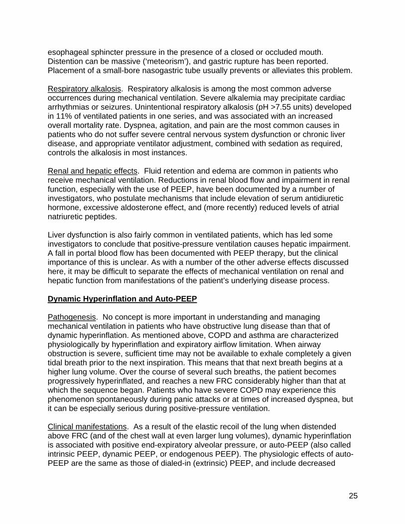
esophageal sphincter pressure in the presence of a closed or occluded mouth. Distention can be massive (‘meteorism’), and gastric rupture has been reported. Placement of a small-bore nasogastric tube usually prevents or alleviates this problem. Respiratory alkalosis. Respiratory alkalosis is among the most common adverse occurrences during mechanical ventilation. Severe alkalemia may precipitate cardiac arrhythmias or seizures. Unintentional respiratory alkalosis (pH >7.55 units) developed in 11% of ventilated patients in one series, and was associated with an increased overall mortality rate. Dyspnea, agitation, and pain are the most common causes in patients who do not suffer severe central nervous system dysfunction or chronic liver disease, and appropriate ventilator adjustment, combined with sedation as required, controls the alkalosis in most instances. Renal and hepatic effects. Fluid retention and edema are common in patients who receive mechanical ventilation. Reductions in renal blood flow and impairment in renal function, especially with the use of PEEP, have been documented by a number of investigators, who postulate mechanisms that include elevation of serum antidiuretic hormone, excessive aldosterone effect, and (more recently) reduced levels of atrial natriuretic peptides. Liver dysfunction is also fairly common in ventilated patients, which has led some investigators to conclude that positive-pressure ventilation causes hepatic impairment. A fall in portal blood flow has been documented with PEEP therapy, but the clinical importance of this is unclear. As with a number of the other adverse effects discussed here, it may be difficult to separate the effects of mechanical ventilation on renal and hepatic function from manifestations of the patient’s underlying disease process. Dynamic Hyperinflation and Auto-PEEP Pathogenesis. No concept is more important in understanding and managing mechanical ventilation in patients who have obstructive lung disease than that of dynamic hyperinflation. As mentioned above, COPD and asthma are characterized physiologically by hyperinflation and expiratory airflow limitation. When airway obstruction is severe, sufficient time may not be available to exhale completely a given tidal breath prior to the next inspiration. This means that that next breath begins at a higher lung volume. Over the course of several such breaths, the patient becomes progressively hyperinflated, and reaches a new FRC considerably higher than that at which the sequence began. Patients who have severe COPD may experience this phenomenon spontaneously during panic attacks or at times of increased dyspnea, but it can be especially serious during positive-pressure ventilation. Clinical manifestations. As a result of the elastic recoil of the lung when distended above FRC (and of the chest wall at even larger lung volumes), dynamic hyperinflation is associated with positive end-expiratory alveolar pressure, or auto-PEEP (also called intrinsic PEEP, dynamic PEEP, or endogenous PEEP). The physiologic effects of auto-PEEP are the same as those of dialed-in (extrinsic) PEEP, and include decreased
25
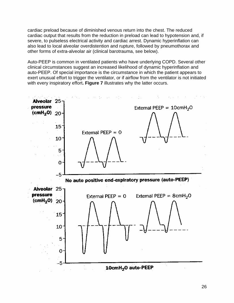
cardiac preload because of diminished venous return into the chest. The reduced cardiac output that results from the reduction in preload can lead to hypotension and, if severe, to pulseless electrical activity and cardiac arrest. Dynamic hyperinflation can also lead to local alveolar overdistention and rupture, followed by pneumothorax and other forms of extra-alveolar air (clinical barotrauma, see below). Auto-PEEP is common in ventilated patients who have underlying COPD. Several other clinical circumstances suggest an increased likelihood of dynamic hyperinflation and auto-PEEP. Of special importance is the circumstance in which the patient appears to exert unusual effort to trigger the ventilator, or if airflow from the ventilator is not initiated with every inspiratory effort. Figure 7 illustrates why the latter occurs.
26

Figure 7: Impact of dynamic hyperinflation and auto-PEEP on a patient’s ability to trigger the ventilator, and the effect of adding external PEEP. Upper diagram: Triggering the ventilator in the absence of auto-PEEP. The addition of external PEEP does not affect the relative ease of initiating breaths from the ventilator. Lower diagram: Triggering the ventilator in the presence of 10 cm H2O of auto-PEEP. In the absence of external PEEP (left portion of each figure), in order to initiate inspiratory flow from the ventilator, the patient must generate enough ventilatory muscle contraction to overcome the 10 cm H2O of auto-PEEP in order to reduce tracheal pressure below zero. One occasion (lower left, 3rd breath attempt), insufficient force is generated to trigger the ventilator. When 8 cm H2O of external PEEP is added, the patient can trigger the ventilator much more easily, since much less ventilatory muscle contraction is needed to bring tracheal pressure down to the external PEEP level in order to initiate inspiratory flow. Detection and measurement. Unlike externally applied PEEP, which is indicated on the ventilator’s pressure manometer, auto-PEEP cannot be detected unless a special maneuver is performed to allow pressures to equilibrate from the patient’s alveoli to the exhalation valve of the ventilator circuit at end expiration. At the author’s institution, respiratory practitioners who care for ventilated patients routinely check for the presence and magnitude of auto-PEEP (at least once each shift) in every patient who does not make spontaneous ventilatory efforts. When patients breathe spontaneously or attempt to trigger the ventilator, the end-expiratory occlusion technique cannot be used, and a different method such as stepwise addition of external PEEP must be used. Table 12 lists the different techniques available for detecting and quantitating auto-PEEP. Prevention and management. The key to prevent the development of dynamic hyperinflation, and to reduce its severity when present, is to increase expiratory time. Dynamic hyperinflation occurs because insufficient time is available to complete lung emptying following each positive-pressure breath. Therefore, prevention and management hinge on measures to reduce airway obstruction, and to decrease the time spent on inspiration during each minute. Table 13 lists practical steps that can be taken to prevent auto-PEEP in ventilated patients, and to minimize it when present. If the patient makes active inspiratory efforts, the addition of external PEEP decreases the muscular effort needed to trigger breaths from the ventilator, and thus decreases work of breathing and increases patient comfort. The added external PEEP should be at about 80% of the measured or estimated auto-PEEP level, as the latter is an average of various actual auto-PEEP levels in different lung regions, some of which are lower than the measured auto-PEEP. The level of auto-PEEP is checked frequently as the patient’s condition changes, with adjustments in external PEEP made accordingly. Barotrauma
27

Pneumothorax, pneumomediastinum, subcutaneous emphysema, and other forms extra-alveolar air detected during mechanical ventilation are collectively termed ‘barotrauma.’ Although this term implies that excessive pressure is the cause, it is more likely that alveolar disruption results from overdistention (i.e. excessive peak inflating volume) rather than high pressure per se. Extra-alveolar air that first becomes evident during ventilatory support may also be unrelated to the ventilator. Depending upon how much air enters the pulmonary interstitium and where the path of least resistance takes it, a variety of clinical manifestations can ensue. Pneumomediastinum, pneumoperitoneum, and subcutaneous emphysema are rarely of physiologic significance and do not require specific treatment. However, pneumothorax is different in that the air collects in a space (the pleural cavity) from which it cannot naturally escape as pressure increases. Pneumothorax in a patient who receives positive-pressure ventilation can quickly progress to tension pneumothorax, which may rapidly cause cardiovascular collapse and death; for this reason, it must always be relieved promptly if ventilatory support cannot immediately be discontinued. Table 14 outlines steps to minimize the risk of alveolar rupture and barotrauma during mechanical ventilation. These steps are especially important in high-risk patients, such as those who have obstructive or unevenly-distributed lung disease, pulmonary infection, and late-phase ARDS (e.g. beyond 1–2 weeks). Ventilator-induced parenchymal lung injury, apart from clinically apparent barotrauma as discussed here, has been shown to affect both lung dysfunction and clinical outcomes in acute lung injury. Repetitive opening and closing of collapsed alveoli subjects them to high local pressures and can lead to activation of inflammatory mediators (so-called "biotrauma"). Similarly, overdistention of alveoli or lung regions can injure them without producing extra-alveolar air. Current evidence indicates that the prevention or reduction of ventilator-induced lung injury is an important reason for improved survival and other outcomes when patients with acute lung injury or ARDS are managed by the lung-protective ventilatory strategy discussed earlier. Other complications Several important complications associated with invasive mechanical ventilation are not discussed in this chapter. However, three important topics are briefly discussed here that are not generally included in reviews of ventilator complications. ‘Fighting the ventilator’ Agitation and respiratory distress that develop in a patient on a mechanical ventilator who has previously appeared comfortable represents an important clinical circumstance for which the clinician needs an organized approach. The patient should not automatically be sedated, but must instead be evaluated for several potentially life-threatening developments that can present in this fashion. The ventilator is first disconnected from the patient, and the patient ventilated manually using 100% oxygen and a self-inflating bag. If this procedure relieves the patient’s distress, the problem lies proximal to the endotracheal tube, and the ventilator and its
28

29
circuitry must be inspected or replaced. If agitation and respiratory distress persist, the airway are suctioned and a brief physical examination performed, noting recent trends in vital signs and any other new developments in the hours that preceded the onset of distress. A more detailed examination is required if the likely cause is not apparent, which must include a new set of measurements of arterial blood gases and a bedside chest radiograph. On rare occasions, if the patient appears in extremis and tension pneumothorax cannot be excluded at the bedside, empiric measures to drain the chest may be warranted. Deteriorating oxygenation in the ventilated patient Deteriorating arterial oxygenation during mechanical ventilation, a common reason for ‘fighting the ventilator’, should initiate a systematic search for specific mechanisms and therapy rather than simply an increase in inspired oxygen fraction or level of PEEP. Possible causes for worsening oxygenation fall into several categories. The problem could be with the ventilator and its circuitry. The patient’s primary disease process (e.g. pneumonia, ARDS) could be worsening, or a new medical problem may have appeared. Examples of the last include pneumothorax, acute lobar atelectasis, pulmonary edema from fluid overload, nosocomial pneumonia or sepsis, aspiration of gastric contents, retained secretions, and bronchospasm. A fall in cardiac output can also cause worsening oxygenation in a patient who has significant pulmonary venous admixture. Interventions and procedures can also lead to a decline in oxygenation. Examples include the effects of airway suctioning, chest physical therapy, or even changes in body position, especially in patients affected by heterogeneously distributed pulmonary involvement. Bronchoscopy, thoracentesis, and hemodialysis can also lead to a decline in oxygenation. Finally, a number of drugs administered to patients undergoing mechanical ventilation can interfere with arterial oxygenation. Among these are vasodilators (which can decrease hypoxic vasoconstriction), b-blockers (which can depress cardiac output and induce bronchospasm), and bronchodilators (which can alter ventilation/perfusion ratios). Effects of suboptimal ventilator management Some adverse effects of mechanical ventilation are iatrogenic. With increasing complexity of the ventilators and their modes, it becomes more likely that the physician, nurse, or respiratory therapist who adjusts the ventilator may not fully understand the consequences of a given adjustment. Table 15 lists several such iatrogenic problems, the clinical circumstances in which they typically occur, and steps to prevent or correct them.