Embed Size (px)
Citation preview

A PROSPECTIVE STUDY ON THE OUTCOME OF EARLY
POSTOPERATIVE ACTIVE MOBILISATION FOLLOWING
TENDON TRANSFER PROCEDURES FOR CLAW HAND
CORRECTION
Dissertation submitted for
M.S. Degree Examination
Branch II – ORTHOPAEDIC SURGERY
APRIL-2013
THE TAMILNADU DR. M.G.R. MEDICAL UNIVERSITY
CHENNAI, TAMIL NADU
CERTIFICATE
This is to certify that this dissertation entitled “A PROSPECTIVE
STUDY ON THE OUTCOME OF EARLY POSTOPERATIVE
ACTIVE MOBILISATION FOLLOWING TENDON TRANSFER
PROCEDURES FOR CLAW HAND CORRECTION” is the bonafide
work done by Dr.P.MURUGESHKUMAR, under my supervision in the
Department of Orthopaedic Surgery, Madurai Medical College, Madurai-20.
Prof. Dr. P.V.PUGALENTHI, M.S Ortho., D. Ortho
Professor and Head,
Department of Orthopaedics & Traumatology
Madurai Medical College,
Madurai.
CERTIFICATE
This is to certify that this dissertation entitled “A PROSPECTIVE
STUDY ON THE OUTCOME OF EARLY POSTOPERATIVE
ACTIVE MOBILISATION FOLLOWING TENDON TRANSFER
PROCEDURES FOR CLAW HAND CORRECTION” is the bonafide
work done by Dr.P.MURUGESHKUMAR, under my direct guidance and
supervision in the Department of Orthopaedic Surgery, Madurai Medical
College, Madurai-20.
Prof. Dr. T.CHANDRA PRAKASAM M.S Ortho., D. Ortho
Professor of Hand surgery
Department of Orthopaedics & Traumatology
Madurai Medical College,
Madurai.
DECLARATION
I Dr.P.MURUGESHKUMAR , solemnly declare that the dissertation
titled “A PROSPECTIVE STUDY ON THE OUTCOME OF EARLY
POSTOPERATIVE ACTIVE MOBILISATION FOLLOWING TENDON
TRANSFER PROCEDURES FOR CLAW HAND CORRECTION ” has
been prepared by me. This is being submitted to “The Tamil nadu Dr. M.G.R.
Medical University, Chennai in partial fulfilment of the regulations for the
award of M S degree branch II orthopaedics.
Place : Madurai Dr.P.MURUGESHKUMAR
Date :

ACKNOWLEDGEMENT
At the very outset I would like to thank Dr.MOHAN M.S, the Dean,
Dr.SWAMINATHAN MS Medical Superintendent , Madurai Medical College
and Govt Rajaji Hospital, Madurai for permitting me to carry out this study in
this hospital.
I am greatly indebted to my beloved chief Prof.Dr P.V.PUGALENTHI ,
MS Ortho., D Ortho, Professor and Head, Department of Orthopaedic Surgery
and Traumatology, Madurai Medical College for his invaluable help to me in
preparing this dissertation
I am greatly indebted to my beloved chief ,
Prof.Dr.T.CHANDRAPRAKASAM , MS Ortho., D Ortho., Professor of
hand surgery , Department of Orthopaedic Surgery and Traumatology, Madurai
Medical College for his invaluable help, encouragement and guidance rendered
to me in preparing this dissertation.
I am most indebted and take immense pleasure in expressing my deep
sense of gratitude to my beloved chief Prof.Dr.S.Shanmuganathan, MS
Ortho.,D.Ortho., Prof.Dr.L.D.Thulasiram MS Ortho., D.Ortho,
Prof.Dr.R.Sivakumar MS Ortho., D Ortho., for their easy accessibility and
timely suggestion, enabled me to bring out this dissertation.

Sincre thanks I do extend to Dr.Aa.Rajamani, MS Ortho.,D Ortho, for
providing valuable guidance and consistent support as a part of my
dissertation.
I also take this opportunity to thank Dr.K.Ravichandran,,
Dr.S.Ramanathan., Dr.M.N.karthi., Dr.N.Thanappan.
Dr.P.V.Thirumalaimurugan.,Dr.K.P.Saravanakumar.,Dr.T.C.Premkumar.
,Dr.T.Saravanamuthu, Dr.Pathiarasakumar., Dr.J.Maheswaran.,
Dr.V.A.Prabhu, Dr.Gnaprakasam Assistant Professors, Department of
Orthopaedics, Madurai Medical College for their timely help and guidance
given to me during all stages of the study.
I also thank all Postgraduates, Tutors, Staff, and other members of the
Department of Orthopaedics of Madurai Medical College for their help.
Lastly, my sincere thanks to all my beloved patients & their parents, who,
with their excellent cooperation became the backbone of this dissertation.
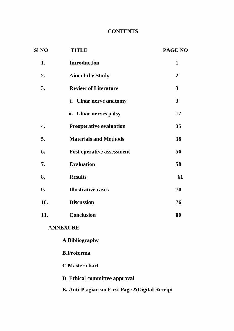
CONTENTS
Sl NO TITLE PAGE NO
1. Introduction 1
2. Aim of the Study 2
3. Review of Literature 3
i. Ulnar nerve anatomy 3
ii. Ulnar nerves palsy 17
4. Preoperative evaluation 35
5. Materials and Methods 38
6. Post operative assessment 56
7. Evaluation 58
8. Results 61
9. Illustrative cases 70
10. Discussion 76
11. Conclusion 80
ANNEXURE
A.Bibliography
B.Proforma
C.Master chart
D. Ethical committee approval
E, Anti-Plagiarism First Page &Digital Receipt

INTRODUCTION
Post operative immobilization of the hand in a cast is the conventional
practice following the tendon transfer for the claw hand deformity correction.
The wrist and the Metacarpophalangeal joints (MCP) are immobilized for a
period of 4 weeks following the Zancolli's lasso procedure for claw deformity
correction. After cast removal, 4 weeks is required for the tendon transfer re-
education before the patient is allowed to use the hand for daily living
activities. The period of morbidity with the post-operative immobilization of
the hand extends up to 7-8 weeks. Post immobilization stiffness may increase
the rehabilitation time and further delay in return to activities.
The concept of immediate active mobilization of tendon transfer was
reported recently by Rath. His immediate post operative active mobilisation
trial shows the benefit of 40% reduction in rehabilitation time .
To test this concept on tendon transfer for claw deformity correction ,
we did a prospective study of an Analysis of results of Early Postoperative
Active Mobilization following FDS middle finger 4 tail pulley insertions
(4TP) for claw hand correction ( Lasso procedure). The outcomes of this
procedure were compared with conventional immobilization in a cast for claw
hand deformity correction.

AIM OF THE STUDY
To Test the safety, efficacy and reliability of early postoperative active
mobilisation after tendon transfer surgery and to compare the outcomes
with those after conventional post operative immobilisation in the claw
hand deformity correction
REVIEW OF LITERATURE
ULNAR NERVE ANATOMY
Orgin of the ulnar nerve
The ulnar nerve arises from the medial cord of brachial plexus and is
composed of fibers from the anterior rami of C8 and T1.
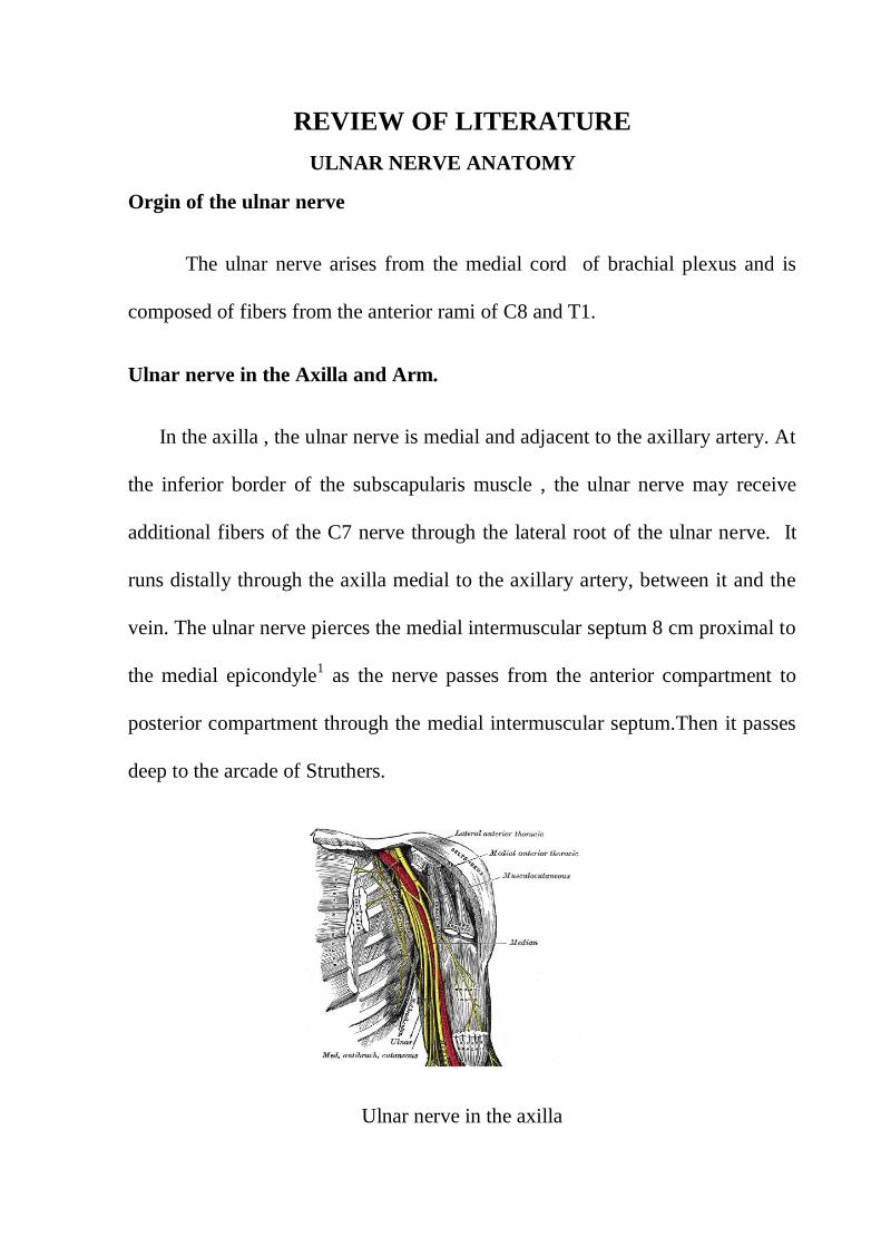
Ulnar nerve in the Axilla and Arm.
In the axilla , the ulnar nerve is medial and adjacent to the axillary artery. At
the inferior border of the subscapularis muscle , the ulnar nerve may receive
additional fibers of the C7 nerve through the lateral root of the ulnar nerve. It
runs distally through the axilla medial to the axillary artery, between it and the
vein. The ulnar nerve pierces the medial intermuscular septum 8 cm proximal to
the medial epicondyle1 as the nerve passes from the anterior compartment to
posterior compartment through the medial intermuscular septum.Then it passes
deep to the arcade of Struthers.
Ulnar nerve in the axilla

The ulnar nerve remains on the medial aspect of the superior ulnar
collateral artery. Both nerve and artery continue distally and medially on the
anterior surface of the medial head of the triceps muscle. Then the ulnar nerve
enters the interval between the medial epicondyle of the humerus and the
olecranon, and passes onto the ulnar groove on the dorsal aspect of medial
epicondyle. The ulnar nerve does not normally innervate any muscles in the
arm, although the muscular branch to the flexor carpi ulnaris may branch from
the ulnar nerve 1 cm proximal to the medial epicondyle.2
Fig 2. Ulnar nerve course in the arm and elbow

ANOMALOUS VARIATION OF ULNAR NERVE IN THE AXILLA AND
ARM.
The ulnar nerve normally originates from the medial cord of brachial
plexus. It may receive fibers from several other sources, including lateral cord ,
the middle trunk, and the anterior division of the middle trunk. These neural
elements are collectively referred to as the lateral root of the ulnar nerve.
ARCADE OF STRUTHERS
As the ulnar nerve passes from the anterior to posterior compartment of the
arm, it may encounter a myofibrous or faciomyofibrous band called the arcade
of Struthers . This common structure was first described by Struthers in 18543.
It is a fibrous or fascial sheet located in the distal third of the medial aspect of
the humerus. It is formed by a thickening of the deep investing facia of the
distal part of the arm , and by superficial muscular fibers of the medial head of
the triceps 4. The anterior border of the arcade of Struthers is the medial
intermuscular septum. The lateral border of the arcade is formed by the medial
aspect of the humerus covered by deep muscular fibers of the medial head of the
triceps. The arcade of Struthers may be a potential area of compression. If
compression is present , the fascial sheet of the arcade of the struthers should be
incised.

The arcade of struthers
THE FIRST BRANCH OF THE ULNAR NERVE.
The articular branch , normally the first branch of the ulnar nerve , exits
from the main trunk in the ulnar groove and passes horizontally into the joint.
The first muscular branch usually to the flexor carpi ulnaris exits immediately
distal to the articular branch.
ULNAR NERVE IN THE ELBOW AND FOREARM.
Ulnar nerve in the cubital Tunnel
The cubital tunnel at the elbow is a fibro osseous tunnel2. The lateral border
consists of the humerus, ulna, and elbow joint. The medial and inferior border
consists of a fascial sheath confluent with the brachial and antebrachial fascia of
the adjacent muscles. The distal medial border consists of the aponeurosis or

fascia between the two heads of flexor carpi ulnaris 4, 8,9
. As noted by Siegel and
Gelberman the tunnel can be divided geographically into three parts . The first
part , usually provides one branch or several small articular branches to the
elbow.
The second and middle part of the tunnel consists of a fascial arcade. The
nerve usually gives off two branches to innervate the flexor carpi ulnaris. One
branch usually supplies the humeral head and one supplies the ulnar head
In the second portion of the cubital tunnel, the distance between the
medial humeral epicondyle and the olecranon is the shortest with elbow
extension. This distance increases with elbow flexion. The roof of the cubital
tunnel is formed by the fascial arcade, which becomes taut with elbow flexion.
The third and most distal part of the tunnel consists of the muscle bellies
of the flexor carpi ulnaris.The nerve then continues distally in the forearm
between the flexor digitorum profundus , located dorsally and laterally to the
nerve, and the flexor carpi ulnaris located anteriorly and medially. The nerve
runs a straight course through the forearm. In the distal third of the forearm ,the
ulnar nerve courses more superficially , lying just radial and deep to the flexor
carpi ulnaris2.

Motor Branches Of The Ulnar Nerve In The Forearm.
In the forearm, and distal to the exit of the motor branches to the flexor
carpi ulnaris, the ulnar nerve usually has three additional main branches. These
are the motor branch to the flexor digitorum profundus( to the ring and small
fingers), the palmar cutaneous portion of the ulnar nerve and the dorsal branch
of the ulnar nerve2,7
.
Palmar cutaneous branch of the ulnar nerve
It arises a vriable levels from the ulnar nerve in the distal forearm. It
innervates the skin in the hypothenar eminence, ulnar artery and occasionally ,
the Palmaris brevis muscle.
In 80% of upper limbs a single branch from the ulnar nerve supplies the
flexor digitorum profundus. In appropriately 20% two or more branches supply
the muscle.
Dorsal cutaneous branch of the ulnar nerve
It arises from the medial aspect of main ulnar nerve trunk in the distal
forearm and curves dorsally to supply cutaneous innervation to the dorsal aspect
of the small finger and ring finger7,8,9

Anomalous connection between the ulnar and median nerve
In the distal forearm, a crossing of nerve fibers from the ulnar nerve to the
median nerve can occur, although with less frequency than the more common
crossing of fibers in the opposite direction from median nerve or anterior
interosseus nerve to the ulnar nerve ( Martin - Gruber anastomosis).
The ulnar nerve in the forearm
Variations in innervation of the Flexor Digitorum Profundus muscle
The ulnar nerve is thought to innervate the flexor digitorum profundus to the
ring and little fingers, and the median nerve innervates the index and long
fingers. However this pattern was found in only 50% of upper limbs. In several

specimens, the median nerve was found to innervate the ring and little fingers
and ulnar nerve was found to supply the long finger. It is more common for the
median nerve to innervate muscles traditionally supplied by the ulnar nerve than
for the ulnar nerve to innervate muscles usually supplied by the median
nerve18,19,20
.
Many of the variations in branching occur in the muscle belly of the
flexor digitorum profundus, and therefore are difficult to identify by superifical
visualization and examination of the muscle. The flexor digitorum profundus to
the index finger, however does seem to be innervated most consistently by the
median nerve.
Ulnar nerve compression by Anamolous Anconeus Epitrochlearis
The ulnar nerve may be compressed at the elbow by an anomalous
muscle, the anconeus epitrochlearis. The anconeus epitrochlearis originates
from the medial border of the olecranon and adjacent triceps tendon and inserts
into the medial epicondyle of the elbow.
CUBITAL TUNNEL SYNDROME.
Chronic compression of the ulnar nerve at the cubital tunnel may occur as a
result of ischemia or mechanical compression by repeated elbow flexion, post-
traumatic scarring, anomalous musculature, or direct compression, although the
exact cause may be difficult to identify. Acute trauma to the ulnar nerve from
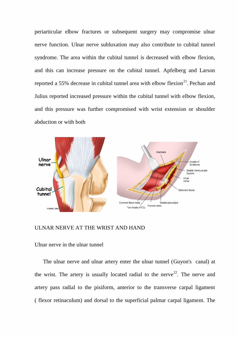
periarticular elbow fractures or subsequent surgery may compromise ulnar
nerve function. Ulnar nerve subluxation may also contribute to cubital tunnel
syndrome. The area within the cubital tunnel is decreased with elbow flexion,
and this can increase pressure on the cubital tunnel. Apfelberg and Larson
reported a 55% decrease in cubital tunnel area with elbow flexion21
. Pechan and
Julius reported increased pressure within the cubital tunnel with elbow flexion,
and this pressure was further compromised with wrist extension or shoulder
abduction or with both
ULNAR NERVE AT THE WRIST AND HAND
Ulnar nerve in the ulnar tunnel
The ulnar nerve and ulnar artery enter the ulnar tunnel (Guyon's canal) at
the wrist. The artery is usually located radial to the nerve22
. The nerve and
artery pass radial to the pisiform, anterior to the transverse carpal ligament
( flexor retinaculum) and dorsal to the superficial palmar carpal ligament. The
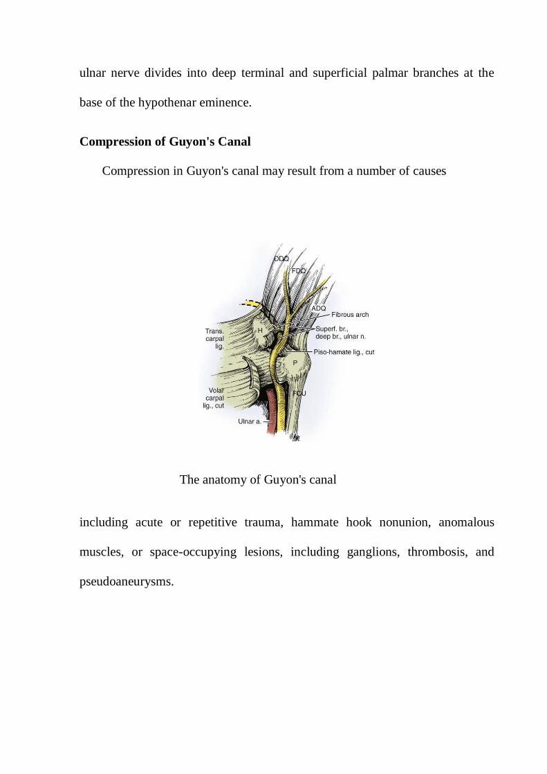
ulnar nerve divides into deep terminal and superficial palmar branches at the
base of the hypothenar eminence.
Compression of Guyon's Canal
Compression in Guyon's canal may result from a number of causes
The anatomy of Guyon's canal
including acute or repetitive trauma, hammate hook nonunion, anomalous
muscles, or space-occupying lesions, including ganglions, thrombosis, and
pseudoaneurysms.

Guyon's canal
The unique anatomy of Guyon's canal will influence the symptoms. The nerve
can be compressed proximal to its bifurcation (zone I), thereby yielding a mix
of both motor and sensory deficits; along the course of the deep motor branch
(zone II), characterized by pure motor loss; or along its superficial sensory
branch (zone III), which is associated with pure sensory changes23
.
Figure shows the two types of traumatic aneurysms of ulnar artery in hand.
Saccular “false” aneurysm arising from ulnar artery.

Superficial palmar branch of the ulnar nerve
The superficial palmar branch exits the distal ulnar tunnel with the superficial
terminal branch of the ulnar artery. The nerve then provides several small twigs
to innervate the skin on the medial side of the hand. The motor branches to the
Palmaris brevis may leave the nerve at this point . The nerve continues distally
and radially and divides into the proper digital nerve to the ulnar side of the
little finger and the common palmar digital nerve to the fourth web space. At
the level of metacarpal shafts the common digital nerves divides into two proper
digital nerves, one each to supply adjacent aspects of the fourth web space
between the small and ring fingers. In the palm, the nerves lies dorsal to the
superficial palmar arch and palmar to the flexor tendons. Immediately after
division, in the region of the metacarpal necks, the proper digital nerves course
anteriorly to lie palmar to the digital arteries. The neurovascular bundles are
stabilized in the digits by the retaining skin ligaments, cleland‟s ligaments
located dorsal to the neurovascular bundle, and grayson‟s ligaments located
palmarly.
Deep terminal branch of the ulnar nerve
It exits from the zone II of the ulnar tunnel dorsoulnar to the deep terminal
branch of the ulnar artery23,24
. The nerve passes medial to the hook of hamate,
deep to the fibrous arch of the hypothenar muscle origin

The ulnar nerve in the course in the hand
The proper digital nerves supply the palmar skin of the digits, and the skin distal
to the distal interphalangeal joints on the dorsal surface.
. The nerve continues between the abductor digiti minimi and flexor digiti
minimi muscles supplying motor branches to each. The nerve then pierces and
innervates the opponens digiti minimi26
. The deep branch then crosses the palm
with the ulnar artery. Along its course, the nerve is deep to the extrinsic flexor
tendons and deep to the mid palmar and thenar fascial clefts, but palmar to the
interossei. At the level of the third metacarpal, the deep branch of the ulnar
nerve cross between the oblique and transverse heads of the adductor pollicis.

Along its deep course, the nerve innervates each of the seven interossei15
, the
third and fourth lumbricals , the adductor pollicis, the flexor pollicis brevis, and
the hypothenar muscles. The deep terminal branch provides sensory afferent to
nerves to the ulno carpal , intercarpal, and carpo metacarpal joints.
Riche – cannieu communication
The Riche-Cannieu communication consists of a communication between
the deep terminal branch of the ulnar nerve and the motor branch of the median
nerve27
.

ULNAR NERVE PALSY
Ulnar nerve palsy is common with leprosy in areas where this disease is
endemic, but it most commonly occurs after a traumatic injury to the ulnar
nerve . Traumatic ulnar nerve palsy may either due to the open or the closed
injuries. The open injuries can occur with the sharp weapon and the closed
injuries can occur with posterior dislocation of the elbow and Medial
epicondyle fractures. The Tardy ulnar neve palsy is due to cubitus valgus
deformity or severe compression of the ulnar nerve in the cubital tunnel called
as cubital tunnel syndrome . Compression may be either due to osteoarthritis ,
an enlarging synovial cyst , or anomalous accessory muscle , fibrous bands , or
ligaments in the cubital tunnel. Less frequently, the ulnar nerve may be
compressed in Guyon's canal at the wrist, which may result in loss of intrinsic
muscle function without loss of sensation. Recurrent subluxation of the ulnar
nerve can also cause the ulnar nerve palsy . Injury to the medial cord of the
brachial plexus, compression of the T1 nerve root and neurologic diseases such
as syringomyelia, hereditary motor and sensory neuropathy (Charcot-Marie-
Tooth disease), poliomyelitis, and motor neuron disease may mimic ulnar nerve
palsy
Ulnar nerve injuries are classified as high or low. Low injuries occur distal
to the origins of the motor branches to the FCU and ring and little finger flexor
digitorum profundus (FDP) muscles. Strength of the extrinsic hand muscles is
unaffected, but sensation is lost on the ulnar border of the hand and in the ring
and little fingers, and the ulnar-innervated intrinsic muscles are paralyzed. This
results in weakness of thumb pinch, claw deformity, loss of the normal pattern
of finger flexion, and significant loss of hand dexterity and strength28,29
. High
injuries occur above the origins of the motor branches to the FCU and ring and
little finger FDP muscles. In this situation, loss of active ring and little DIP joint
flexion and wrist flexion compound the aforementioned findings, although
paradoxically, the claw deformity tends to be less severe
Ulnar nerve palsy in leprosy
In 1948 Dr.paul brand pioneered reconstructive surgery on patients afflicted
with leprosy and performed the first correction of claw hand at the Christian
Medical College hospital , vellore, India. He evolved a system of detailed
clinical evaluation and introduced modalities of preoperative approach for
correction of deformity and new procedures for claw hands. Some of these have
remained the „ work horse
‟ for surgeons treating leprosy and are used as the
standard by which all other procedures are evaluated. The other Indian authors
who are pioneers of reconstruction surgery of hands affected with leprosy are
Palande , H.Srinivasan , Antia .
In leprosy there is profound loss of sensation along the Ulnar nerve
distribution. Patients may present with blisters or ulceration in the little and ring
fingers, which in some instances may be the actual reason for them to seek

medical attention rather than for nerve pain, weakness or hypopigmented
patches on the skin. This is primarily due to the insidious nature of clinical
manifestations of leprosy while the patient unknowingly delays seeking medical
attention.
Clinical features of ulnar nerve paralysis
Clinical examination should preferably begin with observing the
outstretched hand. The obvious characteristic is a claw deformity also referred
to as the “benediction” hand because of the conspicuous flexion deformities in
the little and ring fingers with a lesser degree of involvement of the middle and
index fingers. The palmar aspect of the hand is next viewed by placing it palm
tip on the table to note the flattened palm and wasting of the hypothenar region
as well as a shallow mid palmar space distal to the thenar and hypothenar
eminences . Supple and delicate hands show longitudinal palmar furrows
between prominent long flexors beneath the palmar skin that indicate wasting of
lumbricals.
The dorsum of the hand shows pronounced wasting with shallow
concavities between the intermetacarpal space of the interosseus muscles and
particularly of the thumb web. The hand proper takes on the shape of an
isosceles triangle with its base distally rather than the normal rectangular
contour. This change of configuration is mainly due to the absence of the
hypothenar muscle bulge medial to the fifth metacarpal and the combined
adductor pollicis and first dorsal interosseus muscle . Manual workers or those
engaged in heavy hands on rural farming demonstrate the dorsal wasting and
contour changes in the ulnar palsied hand remarkably well.
DISABILITY IN THE CLAW HAND
Loss of intrinsic muscle power may cause hyperextension of the
metacarpophalangeal joints in a mobile hand; however, this deformity usually is
not the primary or most disabling aspect of this paralysis. It has been shown that
with intrinsic paralysis, grasp is diminished 50% or more because of the lack of
power of flexion at the metacarpophalangeal joints. In addition, there is
asynchronous movement in flexion of the fingers themselves . The roll-up
maneuver of the fingers in the intrinsically paralyzed hand shows this
characteristic .
The interphalangeal joints must flex first, followed next by the
metacarpophalangeal joints and ultimately by full flexion of the fingers. The
flexion of the metacarpophalangeal joints is lost with the loss of intrinsic muscle
power; the hand is unable to grasp a large object. it also lacks power of grasp
because metacarpophalangeal flexion depends entirely on the long flexors in the
absence of intrinsics.

Figure shows the Flexion of metacarpophalangeal joint occurs only after
interphalangeal joints are fully flexed. Fingers curl into hand and push away any
large object they wish to grasp
Power of pinch also is diminished in addition to the effects of paralysis of
the thenar muscles because the collateral ligaments of the metacarpophalangeal
joints of the fingers are lax in extension, and the stabilizing intrinsic
musculature that would ordinarily give lateral stability is paralyzed. Divergence
of the fingers is automatic with extension produced by the long extensor
tendons, and as a result of the alignment of the finger flexors, convergence of
the tips on grasping is automatic. To stabilize the fingers in extension at the
metacarpophalangeal joint, especially for the resistance of the index finger to
the pinch pressure of the thumb, the intrinsics are essential.
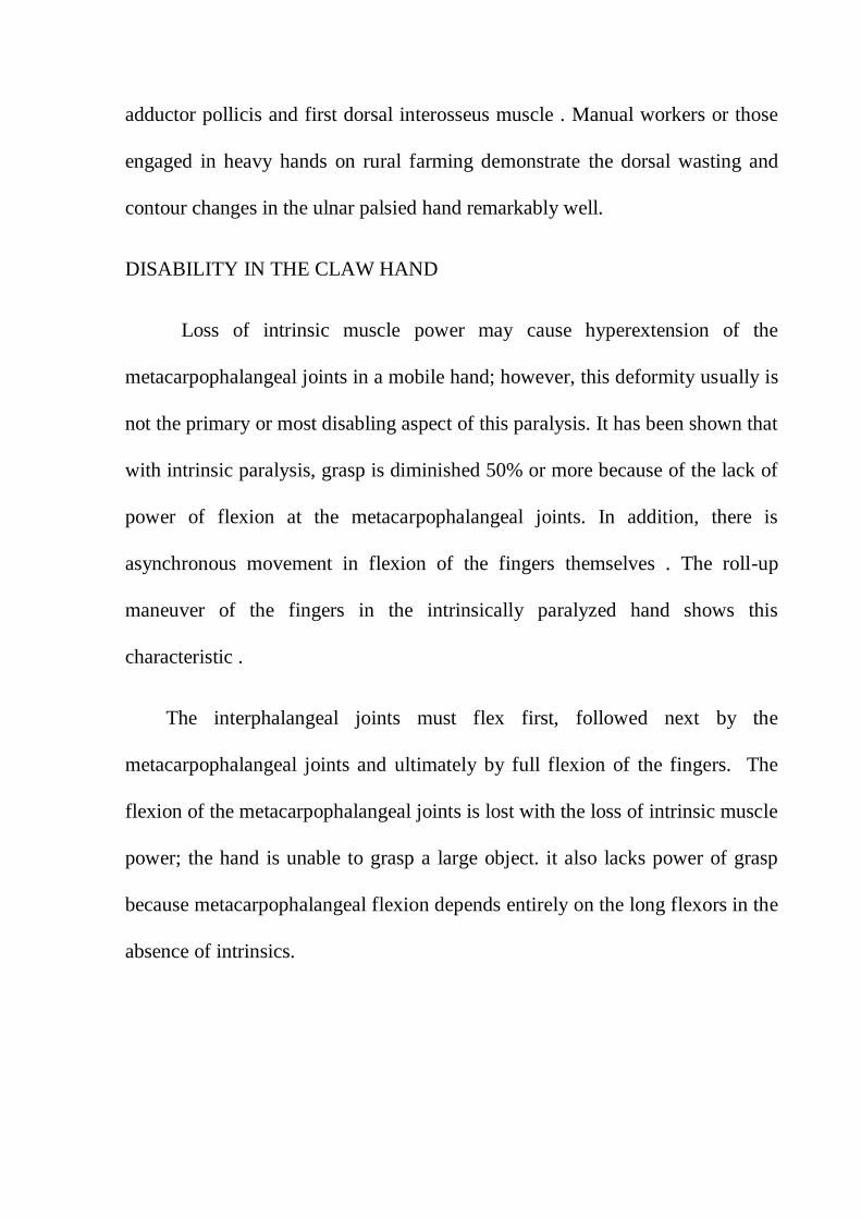
MUSCLE TESTING
The muscles which are to be tested in case of ulnar nerve palsy are Abductor
digiti minimi, First dorsal interossei, FCU, FDP to little and ring fingers
Muscle testing for abductor digiti minimi
Specific signs and tests of Motor dysfunction30
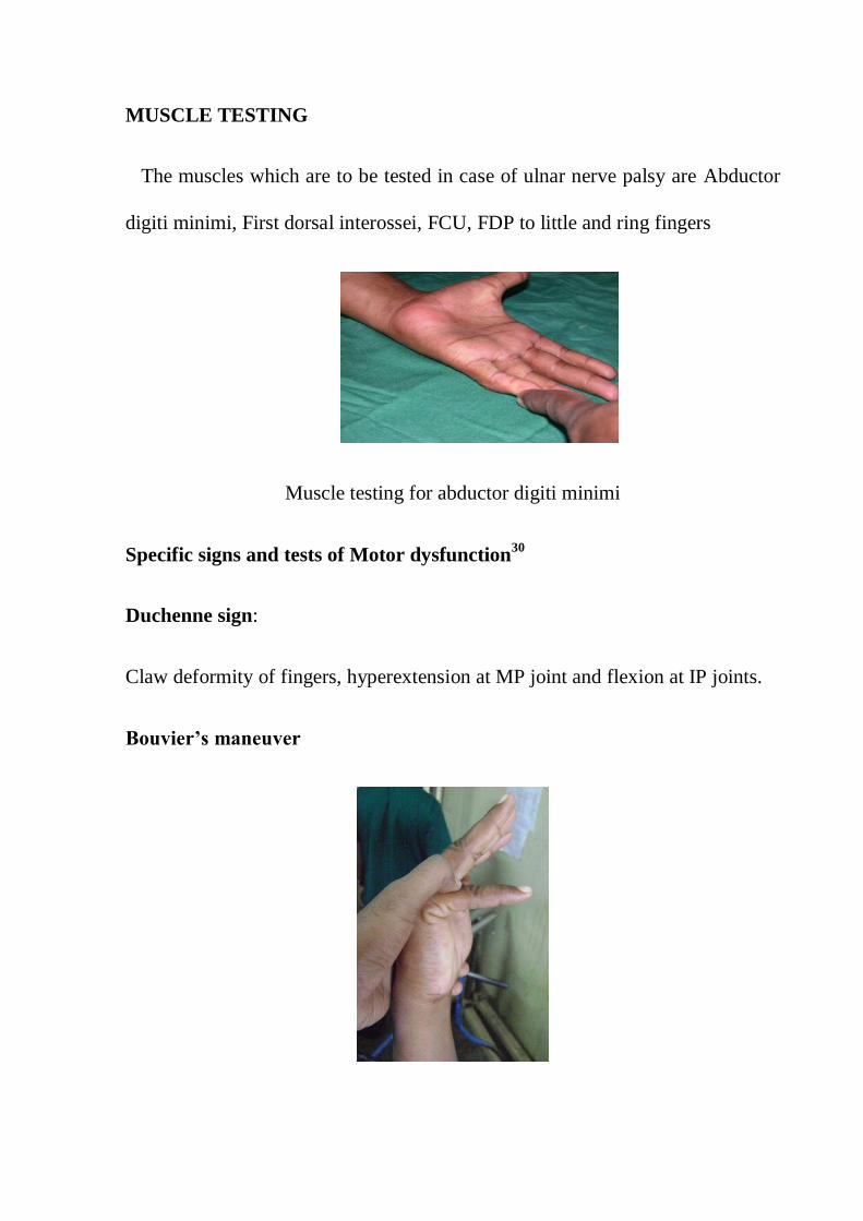
Duchenne sign:
Claw deformity of fingers, hyperextension at MP joint and flexion at IP joints.
Bouvier’s maneuver

When correcting the hyperextension at the MP joints results in full extension
of the IP joints
Andre Thomas sign :
Increase in claw deformity when patient makes an effort to extend the fingers
by flexing the wrist attempting to tenodese the extensor tendons
Pitres – Testut sign :
Inability to abduct the extended middle finger to the radial and ulnar side
when the hand is placed on a flat surface a test for second and third dorsal
interosseous muscles
Earle, valstou sign :
Inability to cross middle finger dorsally over the index finger or the index
finger over the middle finger the cross your fingers test ( a test of the first volar
interosseus and second dorsal interosseous muscles)
Flatt sign
Loss of integration of MP and IP flexion Mp joint does not flex until IP joint
flexion has been completed due to paralysis of the lumbrical muscles of the
ring and little fingers
Brand
The fingers curl or roll into the palm and objects are pushed away instead of
being grasped
Jeanne’s sign
Hyperextension of the MP joint of thumb during key pinch or gross grip due to
paralysis of the adductor pollicis muscle, which acts as first metacarpal
adductor, flexor of the thumb MP joint and an extensor of the thumb IP joint.
Masse’s sign :
Flattened metacarpal arch due to paralysis of the opponens digiti quinti and
decreased range of flexion of the little finger MP joint
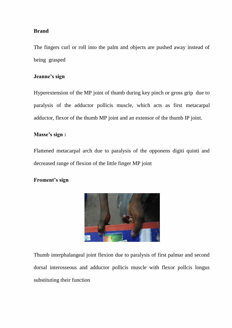
Froment’s sign
Thumb interphalangeal joint flexion due to paralysis of first palmar and second
dorsal interosseous and adductor pollicis muscle with flexor pollcis longus
substituting their function
Bunnell’s O sign:
Combined hyperextension at MCP joint and hyper flexion at IP joint noticed
when patient makes a pulp to pulp pinch with thumb and index finger
Smith sign:
Loss of active lateral mobility with the fingers in extension due to paralysis of
interosseous and hypothenar muscles.
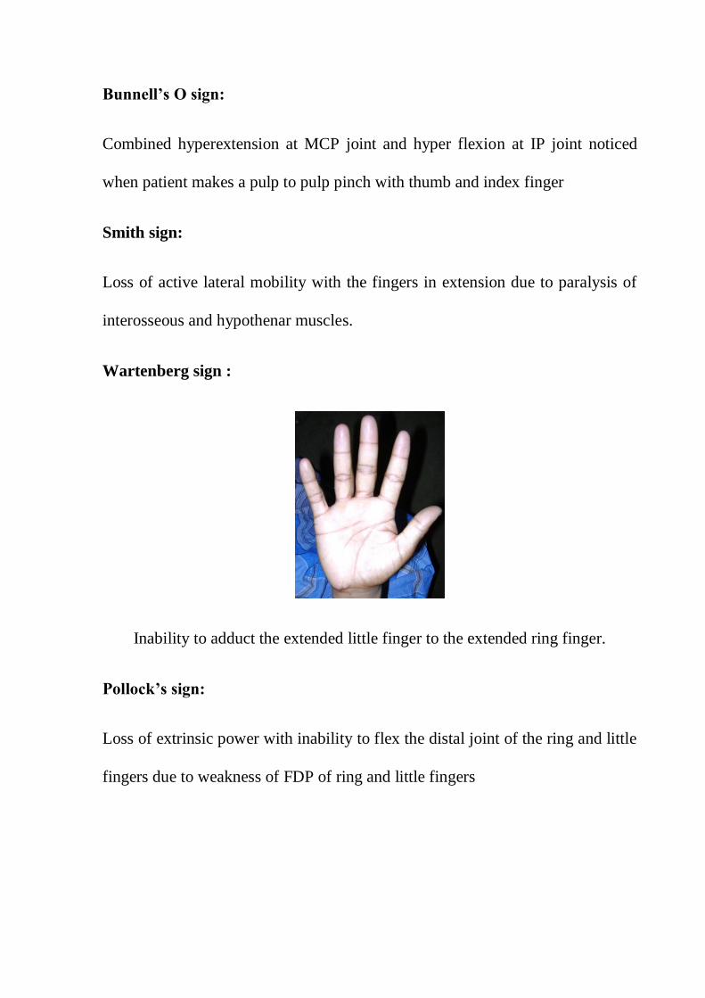
Wartenberg sign :
Inability to adduct the extended little finger to the extended ring finger.
Pollock’s sign:
Loss of extrinsic power with inability to flex the distal joint of the ring and little
fingers due to weakness of FDP of ring and little fingers

Bowden and Napier :
Partial loss of wrist flexion with inability to perform power grip with wrist in
neutral with paralysis of flexor carpi ulnaris
INDICATION FOR SURGERY
The ulnar Nerve paralysis of more than one year duration and Completion
of multi-drug therapy for treatment of Hansen's disease. All MCP and PIP
joints should be supple for tendon transfer
PRINCIPLE OF SURGERY
The interossei and lumbrical muscles flex the MCP and extend the IP joints
of the fingers, but the long finger extensors are capable of extending the IP
joints if the MCP joints are stabilized and cannot hyperextend . This principle is
the basis for many of the surgical procedures
GOAL OF THE SURGERY8
The restoration of grasping power of hand and to improve the thumb pinch,
Correction of finger clawing is more important . Restore the normal pattern of
finger flexion.

In claw hand all four fingers show the disability of the intrinsic muscle
paralysis and also paralysis of the index and middle fingers. All the introssei
muscle palsy make the fingers weak . So all the four fingers should be
operated.
SURGICAL PROCEDURES
Surgical procedures for claw correction are grouped into two categeries. It is
divided into the Static procedures and the Dynamic procedures. The Static
procedures commonly used in the Bouvier's positive hand ( correcting the
hyperextension at the MP joints results in full extension of the IP joints ) . A
tendon transfer should be done if the PIP joints remain flexed when the MP
joint hyperextension is corrected . . If there are fixed flexion deformities of the
IP joints, these need to be corrected before surgery
STATIC PROCEDURES
These prevent hyperextension of the finger MP joints by either shortening the
palmar capsules or by creating “check rein” ligaments or tenodeses. Although
bone blocks to prevent MP joint hyperextension31
, proximal flexor sheath
release to cause bowstringing of the extrinsic finger flexors32
, and excision of an
ellipse of skin and fascia from the palm of the hand to shorten the length of the
palm (fasciodermadesis) have been described, these procedures are now rarely
used.
PALMAR CAPSULODESIS OF METACARPOPHALANGEAL JOINT
ZANCOLLI'S TECHNIQUE33
Zancolli described a procedure using transverse incisions over the A1 pulley
of each affected finger. The A1 pulley of each finger is completely divided , and
the flexor tendons are retracted to one side to expose the palmar plate of the MP
joint. Two parallel longitudinal incisions are made in the palmar plate, and its
insertion onto the metacarpal neck is released, thus creating a capsular flap with
its base attached to the base of the proximal phalanx. The MP joint is
hyperextended to improve the exposure, and a small transverse bone tunnel is
created in the metacarpal neck with a fine awl or Kirschner wire. The
metacarpal neck is roughened with a curette or bur to create a bleeding bed for
adherence of the capsular imbrication. Zancolli then passed a monofilament
wire through the tunnel and the flap of the palmar plate with sufficient tension
to maintain the MP joint in 20 degrees of flexion, after which he firmly twisted
the ends of the wire and cut them to lie flat on the metacarpal. However, a small
bone anchor with an attached 3-0 Vicryl suture is also ideal for attaching the
capsular flaps onto metacarpal necks. The capsuloplasties are performed
sequentially from the index to the little MP joint.
Omer34
modified Zancolli's technique by cutting away a triangular portion of
the deep transverse metacarpal ligament on each side of the palmar plate flap ,
but this is probably unnecessary. He also recommended excision of

a 1.5-cm-wide ellipse of palmar skin to prevent stretching of the volar plate and
immobilized the hand with the MP joints flexed for 6 weeks
Omers34
modification of zancollis capsulodesis
DYNAMIC TENDON TRANSFERS
Many transfers have been described to correct finger clawing and these use a
variety of muscles as motors. Commonly used tendons are the superficialis
tendon transfers , the ECRL transfers and the EIP & EDM transfers
SUPERFICIALIS TENDON TRANSFER TECHNIQUES
Commonly used procedure are the Bunnel Procedue and the Lasso Procedure

SUPERFICIALIS INSERTION SITES
Burkhalter35,36
inserted the superficialis slips onto the proximal phalanx in
patients with traumatic ulnar nerve palsy to avoid the risk of PIP joint
hyperextension, which may occur when the transfer is attached to the lateral
band of the extensor apparatus . For the same reason, Riordan37
recommended
attaching the tendon slips to the A1 pulley if the PIP joints are lax and
hyperextended . Zancolli38
usually looped the tendon slips beneath the entire A1
pulley (lasso technique). Omer34
preferred the A2 pulley over the A1 pulley as
the insertion site to increase the force of MP flexion. Anderson and Oberlin
performed “extended pulley insertions,” looping a slip of the superficialis
tendon around both the A1 and the proximal portion of the A2 (A2a) pulleys in
each finger .
STILES AND FORRESTER-BROWN PROCEDURE 41
Transfer of one slip of each superficialis tendon into the corresponding
extensor digitorum tendon over the proximal phalanx
STILES- BUNNEL PROCEDURE
Bunnell42
modified the above procedure by rerouting both slips of all the
superficialis tendons through the lumbrical canals and anchoring them into both
the radial and ulnar lateral bands of the extensor mechanism . Bunnell's aim was
to correct the claw deformity and restore MP abduction and adduction . It often
resulted in the development of overcorrection and intrinsic-plus deformity
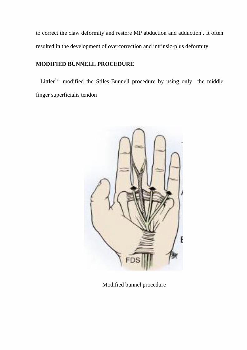
MODIFIED BUNNELL PROCEDURE
Littler43
modified the Stiles-Bunnell procedure by using only the middle
finger superficialis tendon
Modified bunnel procedure
ZANCOLLI
LOOPED THE TENDON SLIPS BENEATH THE ENTIRE
A1 PULLEY (LASSO TECHNIQUE)
Blocking the hyperextension by producing flexion of the
metacarpophalangeal (MCP) joints can allow the extrinsic extensors to extend
the interphalangeal (IP) joints. In the lasso procedure, the FDS tail is inserted
into the A-1 or A-2 pulley of the flexor tendon sheath . A transverse incision is
made at the level of distal palmar crease, then A1 pulleys of the flexor sheath
exposed. The FDS tendon of the middle finger is exposed & withdrawn into
the palm & divided into 4 slips . FDS tendon each tail is inserted into the A-1
or A-2 pulley . The limb is immobilised in POP cast with wrist in neutral,
transverse metacarpal arch well formed and MCP in 70° flexion for 4 weeks
ECRL TRANSFERS
This can be divided into Dorsal route and Volar route transfers.
Dorsal Route Transfer of the ECRL44
( BRAND- 1)
This dorsal route transfer was described by Brand. The ECRL or ECRB tendon
is exposed at the site of their insertion through a short transverse incision . They
are withdrawn into a second transverse incision 8 cm proximally in the distal
forearm. The plantaris tendon is harvested and attached to the distal end of the
motor's tendon. Brand advised a “wrap around” technique of anastomosis that
resulted in its surface being covered with epitenon with no cut end of either the
graft or the extensor tendon exposed to reduce adhesion formation .
The plantaris tendon graft is split into four tails . Dorso radial incisions are
made at the base of the middle, ring, and little fingers and the radial lateral
band of each of finger and the ulnar lateral band of the index finger are
identified. Tendon-tunneling forceps are passed from the finger incisions,
through the lumbrical canals and intermetacarpal spaces, volar to the deep
transverse metacarpal ligaments to emerge in the wrist wound. The tendon
grafts for each of the fingers are passed and stitched in equal tension, suturing
the index finger first, then the little finger and finally the middle and ring
fingers .
Flexor Route Transfer of the ECRL44
- BRAND -2 (EF4T)
The ECRL extended with graft is Passed through the carpal tunnel and
routed through the lumbrical canal and attached to the radial lateral bands of
the extensor expansions of middle , ring finger and ulnar lateral band of index
finger .

ECRL transfer , Brand- 1procedure
Flexor Route Transfer of the ECRL - BRAND -2

PRE OPERATIVE EVALUATION
The following angles are measured and recorded.
1.CLAW DEFORMITY ANGLE59
It is measured by precalibrated triangular discs & goniometer
( srinivasan 1979). . In a open hand position assess the hyperextension of MCP
joint and flexion of PIPjoints angles were measured and recorded.
The deformity angle measurements
2.UNASSISTED EXTENSION ANGLE59
It is measured In the intrinsic plus position , flex the fingers fully at MCP
joins and then extend the fingers fully at PIP joints . Measure the PIP joint
flexion angle, In normal hand it is 0 degree

Unassisted angle measurements
3.ASSISTED EXTENSION ANGLE 59
Press the flexed proximal phalanx down with the goniometer , ask the patient
to straighten the finger at the PIP joint and measure the PIP joint angle . In
normal hand this angle is 0o ,indicating extensor apparatus is intact and normal .
Assisted extension angle up to 20o is accepted
.
Assisted angle measurements
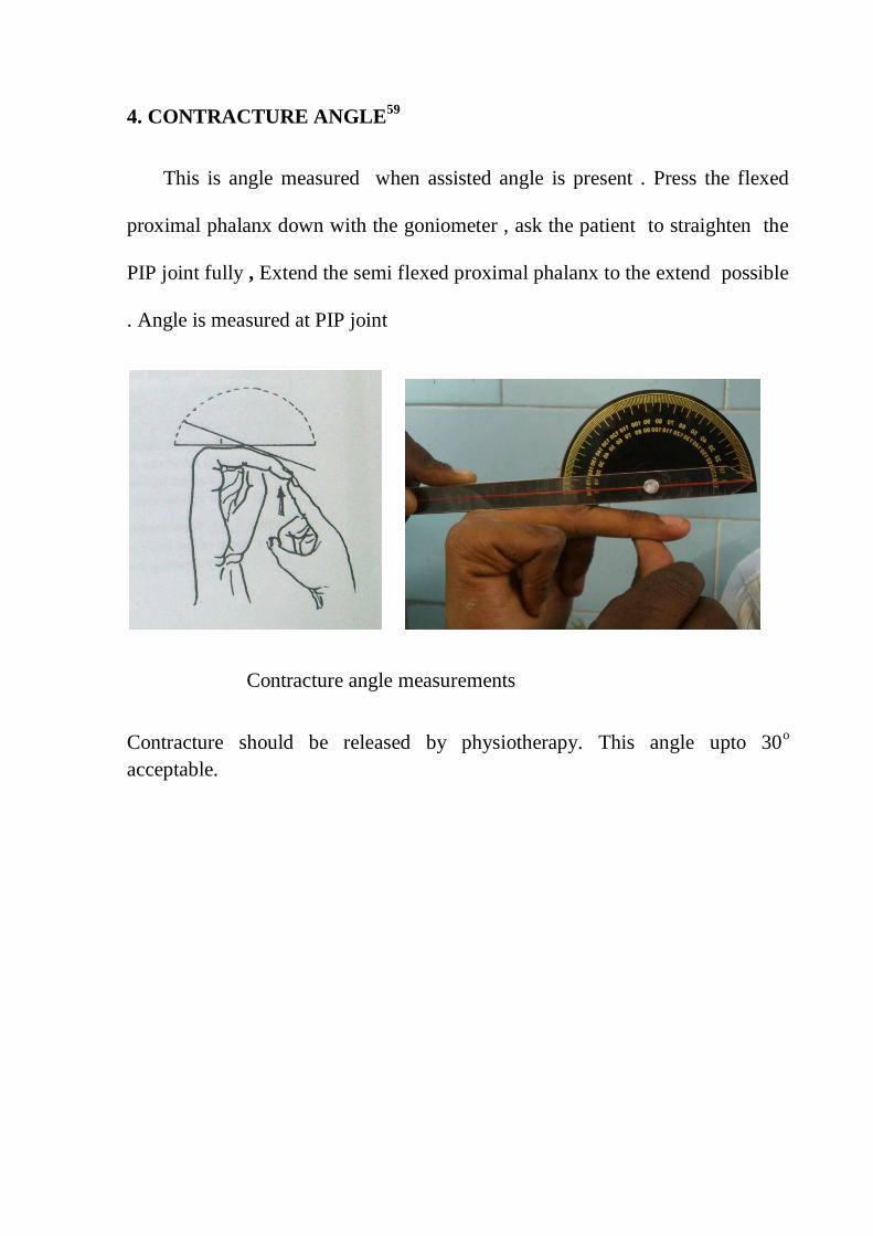
4. CONTRACTURE ANGLE59
This is angle measured when assisted angle is present . Press the flexed
proximal phalanx down with the goniometer , ask the patient to straighten the
PIP joint fully , Extend the semi flexed proximal phalanx to the extend possible
. Angle is measured at PIP joint
Contracture angle measurements
Contracture should be released by physiotherapy. This angle upto 30o
acceptable.

MATERIALS AND METHODS
Patients with the mobile claw hand deformities due to the complete ulnar nerve
paralysis are taken up for our study.
TOTAL NO OF THE PATIENTS – 12 cases
1.AGE DISTRIBUTION
Mean Age – 32 yrs( range 18 -50 yrs)
TABLE: 1.AGE DISTRIBUTION
AGE
11-20
21-30
31-40
41-50
NO OF
PATIENTS
1(8 %)
4(33%)
5(42%)
2 (17%)
0
0.5
1
1.5
2
2.5
3
3.5
4
4.5
5
11-20 YRS 21-30 YRS 31-40 YRS 41-50
AGE
AGE

2.SEX RATIO
TABLE ;2. SEX RATIO
SEX
MALE
FEMALE
NO O F PATIENTS
9
3
75%
25%
PATIENTSMALES FEMALES
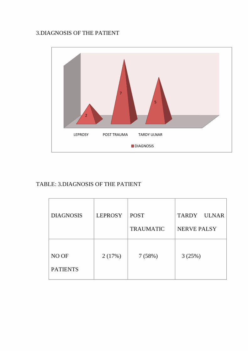
3.DIAGNOSIS OF THE PATIENT
TABLE: 3.DIAGNOSIS OF THE PATIENT
DIAGNOSIS
LEPROSY
POST
TRAUMATIC
TARDY ULNAR
NERVE PALSY
NO OF
PATIENTS
2 (17%)
7 (58%)
3 (25%)
LEPROSY POST TRAUMA TARDY ULNAR
2
7
5
DIAGNOSIS
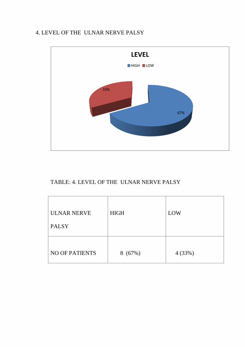
4. LEVEL OF THE ULNAR NERVE PALSY
TABLE: 4. LEVEL OF THE ULNAR NERVE PALSY
ULNAR NERVE
PALSY
HIGH
LOW
NO OF PATIENTS
8 (67%)
4 (33%)
67%
33%
LEVEL
HIGH LOW

5. SIDE OF THE LESION
TABLE : 5. SIDE OF THE LESION
LESION
RIGHT
LEFT
NO OF PATIENTS
7 (58%)
5(42%)
SIDE0
2
4
6
8
RIGHT
EFT
SIDE
SIDE
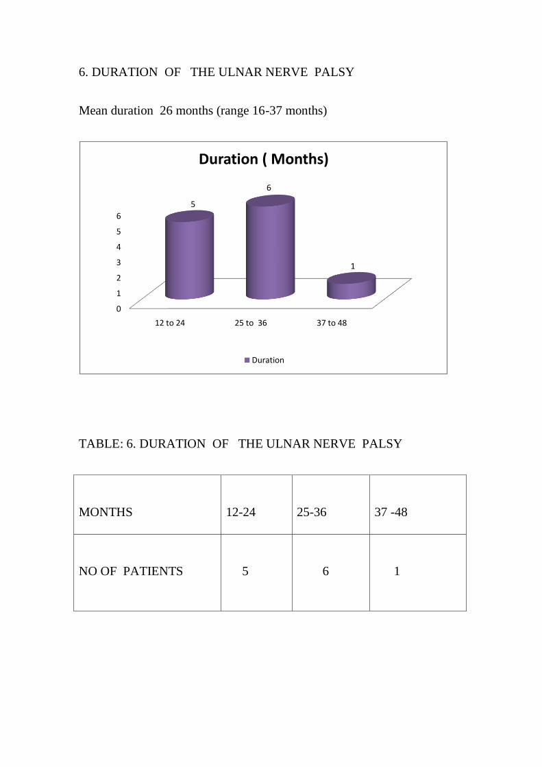
6. DURATION OF THE ULNAR NERVE PALSY
Mean duration 26 months (range 16-37 months)
TABLE: 6. DURATION OF THE ULNAR NERVE PALSY
MONTHS
12-24
25-36
37 -48
NO OF PATIENTS
5
6
1
0
1
2
3
4
5
6
12 to 24 25 to 36 37 to 48
5
6
1
Duration ( Months)
Duration
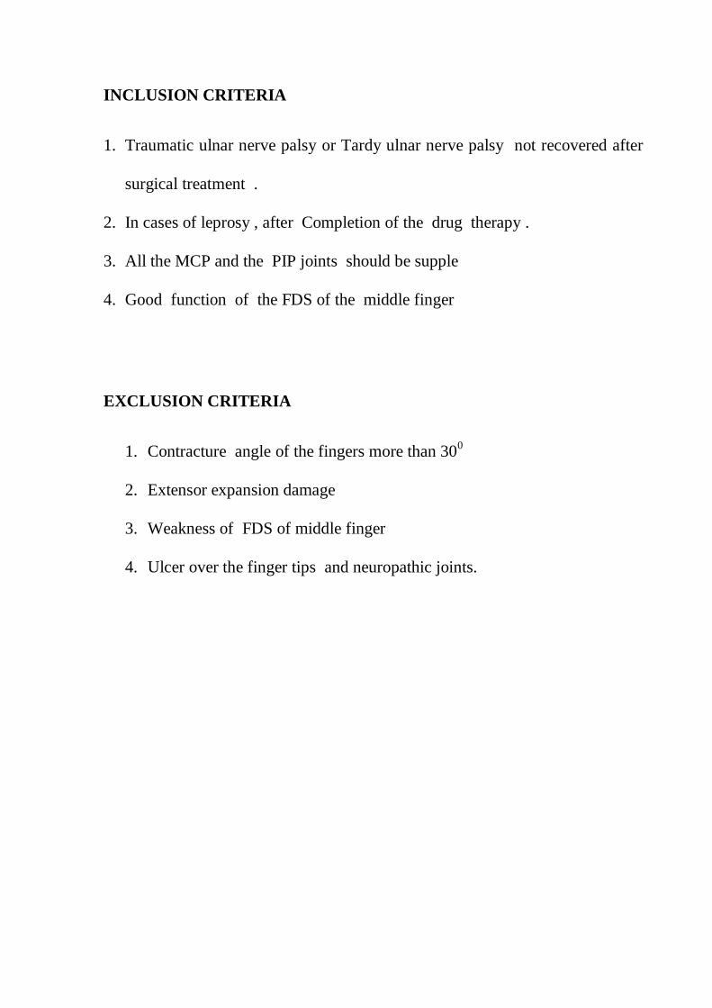
INCLUSION CRITERIA
1. Traumatic ulnar nerve palsy or Tardy ulnar nerve palsy not recovered after
surgical treatment .
2. In cases of leprosy , after Completion of the drug therapy .
3. All the MCP and the PIP joints should be supple
4. Good function of the FDS of the middle finger
EXCLUSION CRITERIA
1. Contracture angle of the fingers more than 300
2. Extensor expansion damage
3. Weakness of FDS of middle finger
4. Ulcer over the finger tips and neuropathic joints.
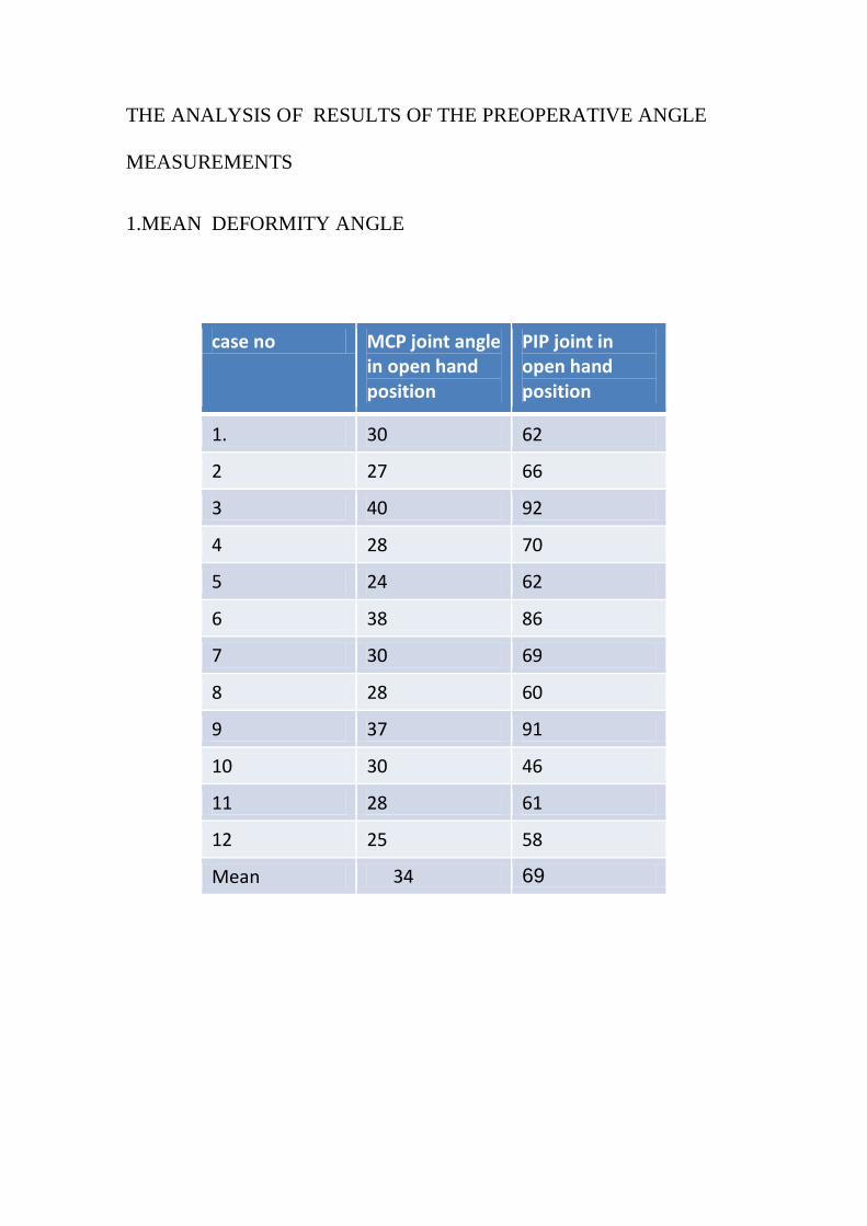
THE ANALYSIS OF RESULTS OF THE PREOPERATIVE ANGLE
MEASUREMENTS
1.MEAN DEFORMITY ANGLE
case no MCP joint angle in open hand position
PIP joint in open hand position
1. 30 62
2 27 66
3 40 92
4 28 70
5 24 62
6 38 86
7 30 69
8 28 60
9 37 91
10 30 46
11 28 61
12 25 58
Mean 34 69

2. ANALYSIS OF RESULTS OF THE UNASSISTED ANGLE , ASSISTED
ANGLE , AND CONTRACTURE ANGLE AT PREOPERATIVELY
Case no
Unassisted angle
Assisted angle
Contracture angle
1. 12 0 0
2 16 0 0
3. 31 12 6
4. 20 0 0
5. 18 0 0
6. 26 21 16
7. 8 0 0
8 10 0 0
9 32 12 12
10 6 0 0
11. 10 0 0
12. 12 0 0
Mean 17 18 11
The mean unassisted angle is 17 0,the mean assisted angle is 18
0 and the mean
contracture angle is 110 ( in three patients)
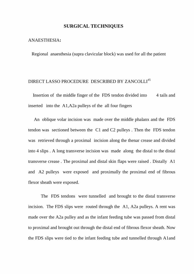
SURGICAL TECHNIQUES
ANAESTHESIA:
Regional anaesthesia (supra clavicular block) was used for all the patient
DIRECT LASSO PROCEDURE DESCRIBED BY ZANCOLLI45
Insertion of the middle finger of the FDS tendon divided into 4 tails and
inserted into the A1,A2a pulleys of the all four fingers
An oblique volar incision was made over the middle phalanx and the FDS
tendon was sectioned between the C1 and C2 pulleys . Then the FDS tendon
was retrieved through a proximal incision along the thenar crease and divided
into 4 slips . A long transverse incision was made along the distal to the distal
transverse crease . The proximal and distal skin flaps were raised . Distally A1
and A2 pulleys were exposed and proximally the proximal end of fibrous
flexor sheath were exposed.
The FDS tendons were tunnelled and brought to the distal transverse
incision. The FDS slips were routed through the A1, A2a pulleys. A rent was
made over the A2a pulley and as the infant feeding tube was passed from distal
to proximal and brought out through the distal end of fibrous flexor sheath. Now
the FDS slips were tied to the infant feeding tube and tunnelled through A1and

A2 pulley and brought out. The tendon slips were folded back and attached to
themselves by a pulvertafts weave46
thechnique with 4-0 prolene.
With the wrist in neutral position the tendon slips are tensioned ideally
to produce MCP joint flexion angles of 50° to 70° with higher flexion in the
ulnar digits. Wound closure was done . A dorsal POP was applied with wrist
in neutral position and MCP joint in 900
flexion and IP joints in extension.
Oblique volar incision & Incision of the FDS tendon
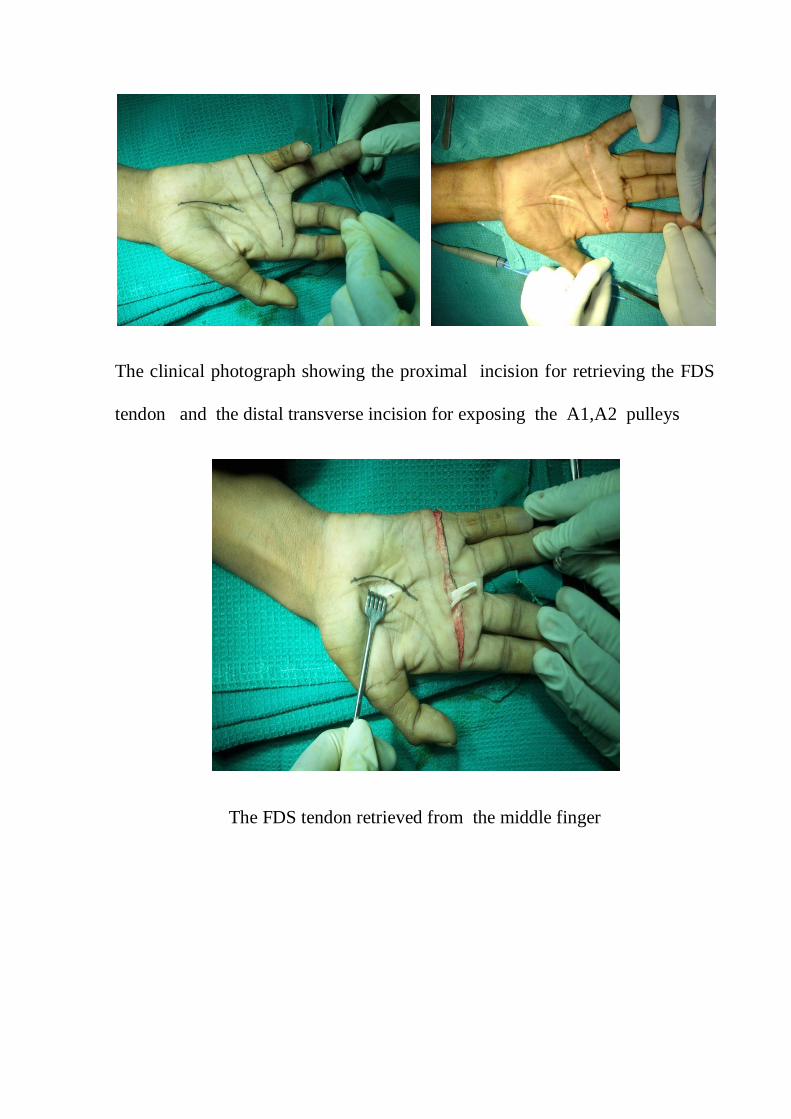
The clinical photograph showing the proximal incision for retrieving the FDS
tendon and the distal transverse incision for exposing the A1,A2 pulleys
The FDS tendon retrieved from the middle finger

Figure shows the FDS tendon of two slips
Figure shows the FDS tendon of each slips divided into 2 slips

The FDS tendon divided into 4 slips
The FDS tendons were tunnelled and brought to the distal transverse incision
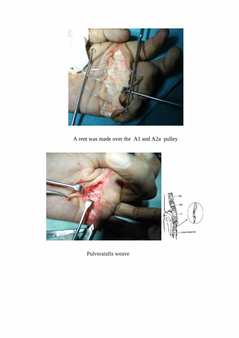
A rent was made over the A1 and A2a pulley
Pulveratafts weave
.
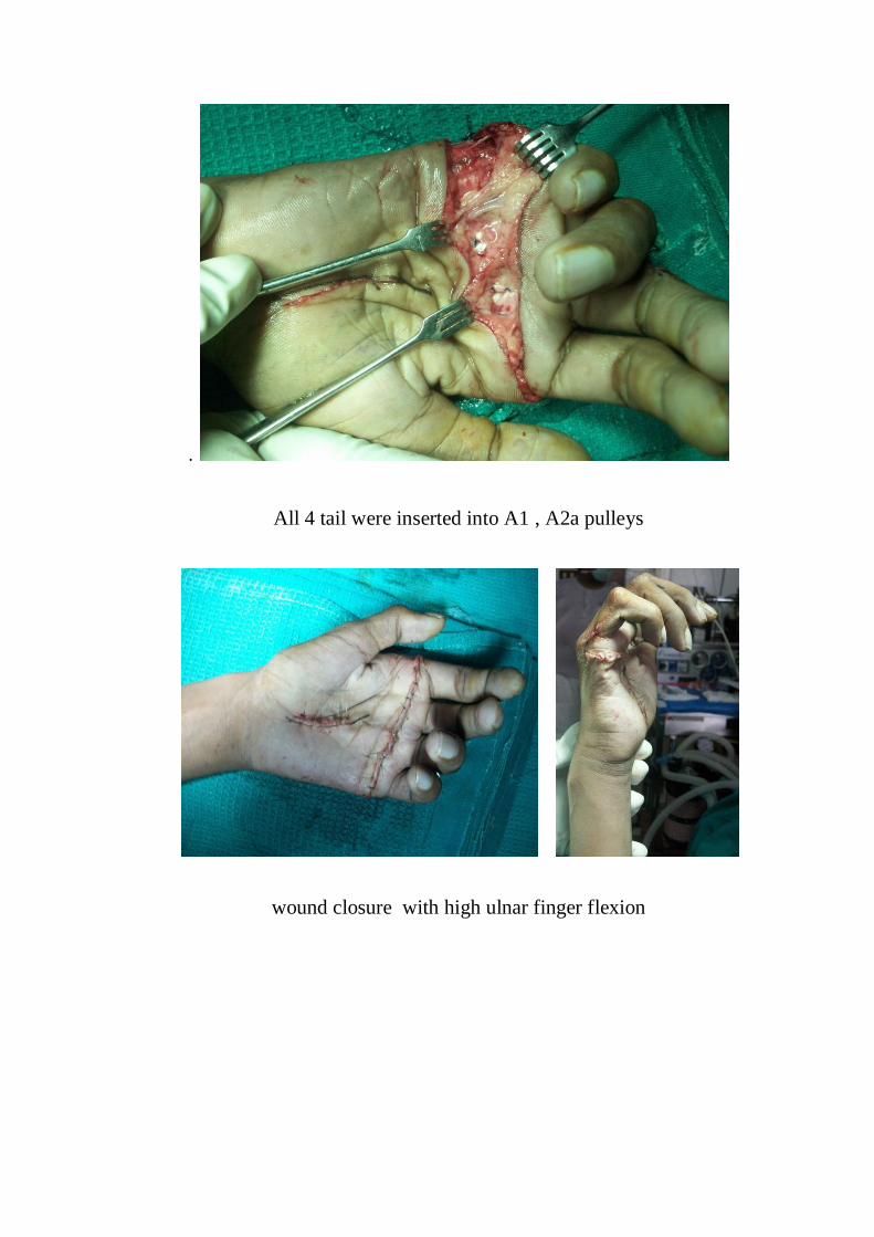
All 4 tail were inserted into A1 , A2a pulleys
wound closure with high ulnar finger flexion
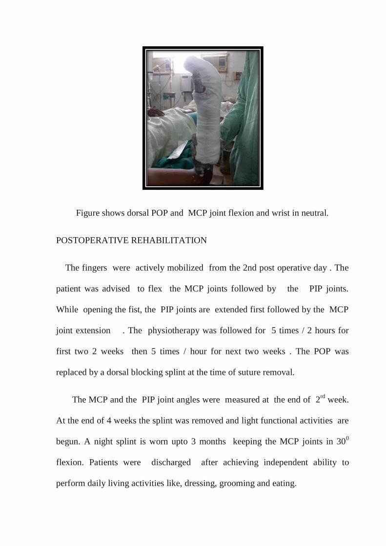
Figure shows dorsal POP and MCP joint flexion and wrist in neutral.
POSTOPERATIVE REHABILITATION
The fingers were actively mobilized from the 2nd post operative day . The
patient was advised to flex the MCP joints followed by the PIP joints.
While opening the fist, the PIP joints are extended first followed by the MCP
joint extension . The physiotherapy was followed for 5 times / 2 hours for
first two 2 weeks then 5 times / hour for next two weeks . The POP was
replaced by a dorsal blocking splint at the time of suture removal.
The MCP and the PIP joint angles were measured at the end of 2rd
week.
At the end of 4 weeks the splint was removed and light functional activities are
begun. A night splint is worn upto 3 months keeping the MCP joints in 300
flexion. Patients were discharged after achieving independent ability to
perform daily living activities like, dressing, grooming and eating.
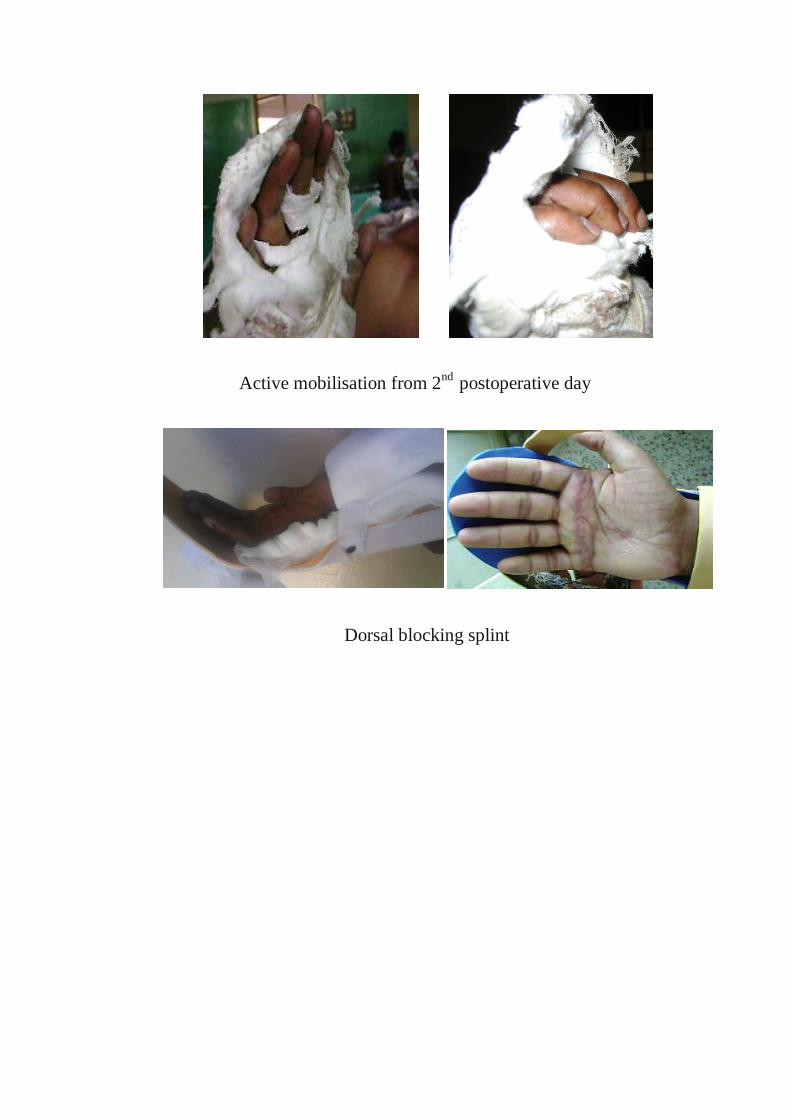
Active mobilisation from 2nd
postoperative day
Dorsal blocking splint

POSTOPERATIVE ASSESSMENT
The MCP joint and the PIP joint angles in the open hand position
provides an objective assessment of the deformity correction and intrinsic plus
position provides an assessment of the tendon transfer integration. The angles
were measured using a goniometer over the dorsum.
1.OPEN HAND POSITION FOR DEFORMITY CORRECTION
The MCP joint and PIP joint angles were measured with patients actively
extending the MCP joints to the maximum possible extent .
Figure shows the Measurement of the MCP and PIP joint angles in the
open hand position
2.INTRINSIC PLUS POSITION :
Patients were advised to actively attempt to flex the MCP joint and keeping
the PIP joint in extension. The MCP and PIP joint angles were measured

Figure shows The .MCP, and the PIP joint angle measurement in intrinsic plus
position
3.FIST CLOSURE.
The ability of the finger tips to reach the distal palmar crease or proximal
palmar crease or inability to touch the palm with active fist closure is noted.
FOLLOW UP
Angles were recorded biweekly for 2 months and then every month for 6
months
EVALUATION OF RESULTS
While evaluating the results of surgical procedures , the following factors were
taken into consideration.
1.TENDON RUPTURE DURING ACTIVE MOBILIZATION
Any sudden increase in MCP joint extension with loss of active MCP joint
flexion indicates the transferred tendon ruptures
2. MORBIDITY
The time required from the day of surgery to the patient able to perform
daily living activities like dressing and eating
3.DEFORMITY CORRECTION
At 6th month follow up the MCP & the PIP Joint angles were measured
and compared .The results of MCP and PIP joint angles in open hand position
were categorized for each hand into good, fair, poor as per the criteria of
Palande47
In our study all the 12 operated patients were followed and angles were
recorded for each digit.
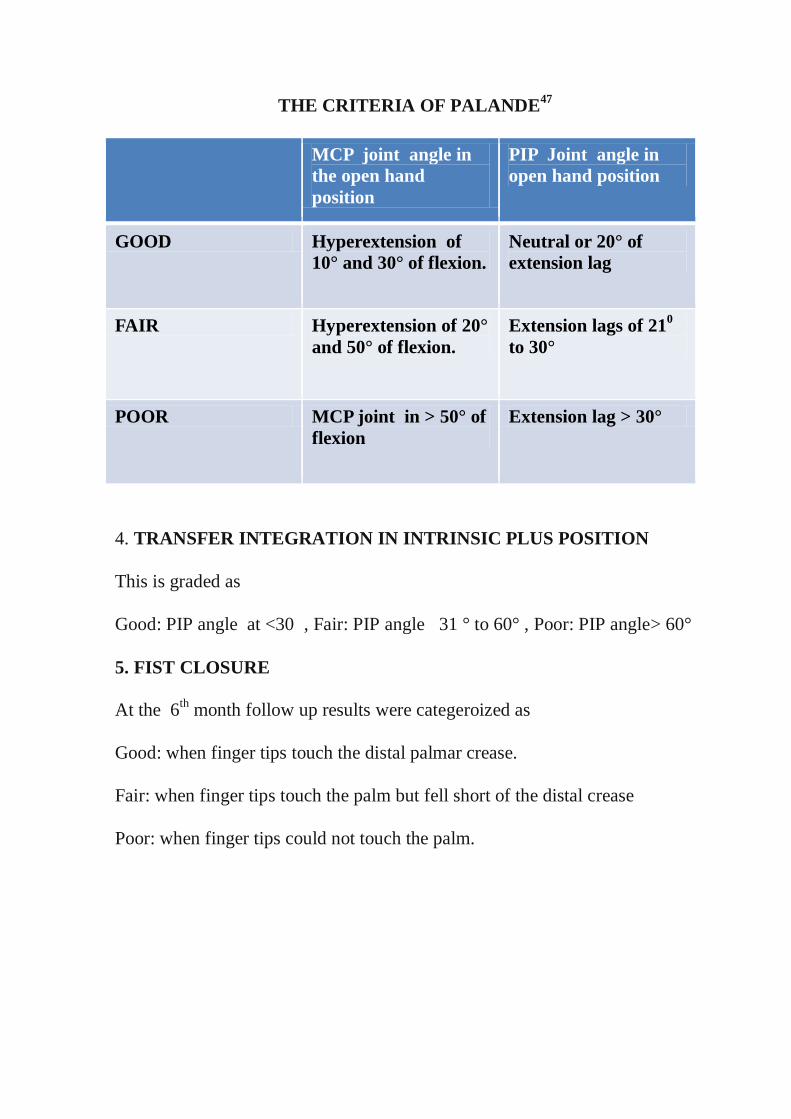
THE CRITERIA OF PALANDE47
MCP joint angle in
the open hand
position
PIP Joint angle in
open hand position
GOOD Hyperextension of
10° and 30° of flexion. Neutral or 20° of
extension lag
FAIR Hyperextension of 20°
and 50° of flexion. Extension lags of 21
0
to 30°
POOR MCP joint in > 50° of
flexion Extension lag > 30°
4. TRANSFER INTEGRATION IN INTRINSIC PLUS POSITION
This is graded as
Good: PIP angle at <30 , Fair: PIP angle 31 ° to 60° , Poor: PIP angle> 60°
5. FIST CLOSURE
At the 6th month follow up results were categeroized as
Good: when finger tips touch the distal palmar crease.
Fair: when finger tips touch the palm but fell short of the distal crease
Poor: when finger tips could not touch the palm.

(i). BASED ON THE ABOVE FACTORS OUR RESULTS OF EARLY
ACTIVE MOBILISATION WERE COMPARED WITH PUBLISHED
REPORTS OF THE CONVENTIONAL IMMOBILISTION OF
TENDON TRANSFER FOR CLAW HAND DEFORMITY
CORRECTION
(ii). WE ALSO COMPARED OUR RESULTS OF THE EARLY
ACTIVE MOBILISATION WITH PUBLISHED REPORT OF THE
EARLY ACTIVE MOBILISATION OF TENDON TRANSFER FOR
CLAW HAND DEFORMITY CORRECTION BY OTHER AUTHORS

RESULTS
A prospective study was conducted in our institution on 12 patients with claw
hand deformity from May 2010 to November 2012. All the patients were
treated with lasso procedures and early active mobilisation protocol was
followed.
The following observations are made in this study :
1.The affected male and female ratio is 3:1
2.The most common age group affected is 4th decade (43 %)
3.The most common cause of the ulnar nerve palsy is post traumatic
etiology ( 58%)
4.The most common level of the ulnar nerve palsy is high ulnar nerve
palsy (67%)
5. The right side (67%) is more commonly affected .
6 .The mean duration of the ulnar nerve palsy is 26 months
7. Three patients had the mean contracture angle of 11 degree ( range 6 -16o ),
which was managed with physiotherapy and splinting preoperatively.

The following are the results of the study:
1.There was no incidence of the transferred tendon rupture during early active
Mobilisation
2. The mean morbidity was 34 days ( range 29 -38 days)
3.The morbidity was reduced by 20 days (37%) with early active mobilisation
compared with Rath immobilisation group
4. The analysis of results of the deformity correction in the open hand position
( MCP joint and PIP joint angles) as per the criteria of Palande shows 82%
of the patients had good deformity correction , 10% had fair results and 8%
had poor results.
5. The analysis of results of the tendon transfer integration in intrinsic plus
position shows good results in 91% of the patients and fair in 6% and poor in
3% of the patients
6. The analysis of the results of fist closure at the follow up of 6 th month
shows 74% of the patients had good results , 20% of the patients had fair
results and 6% in poor results

7. Comparision of the early active mobilisation results with published reports
of immobilisation of tendon transfer for claw hand deformity correction shows
better outcome and also added benefit of reduced morbidity.

Table 7. The analysis of results of the morbidity of early active mobilisation
Morbidity
26 -30 days
31 -35 days
36-40 days
No of the pts
3
5
4
The mean morbidity is 34 days ( range 29-38) days
26-30 31-35 36-40
3
5
4
MORBIDITY(DAYS)
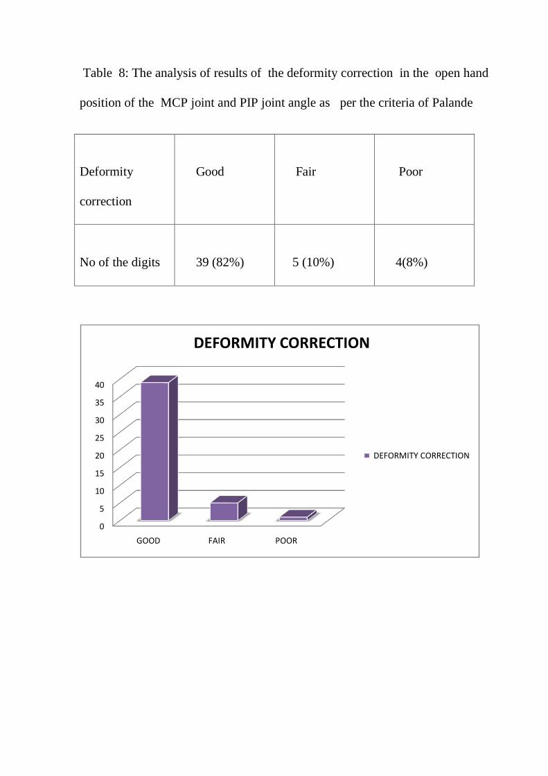
Table 8: The analysis of results of the deformity correction in the open hand
position of the MCP joint and PIP joint angle as per the criteria of Palande
Deformity
correction
Good
Fair
Poor
No of the digits
39 (82%)
5 (10%)
4(8%)
0
5
10
15
20
25
30
35
40
GOOD FAIR POOR
DEFORMITY CORRECTION
DEFORMITY CORRECTION
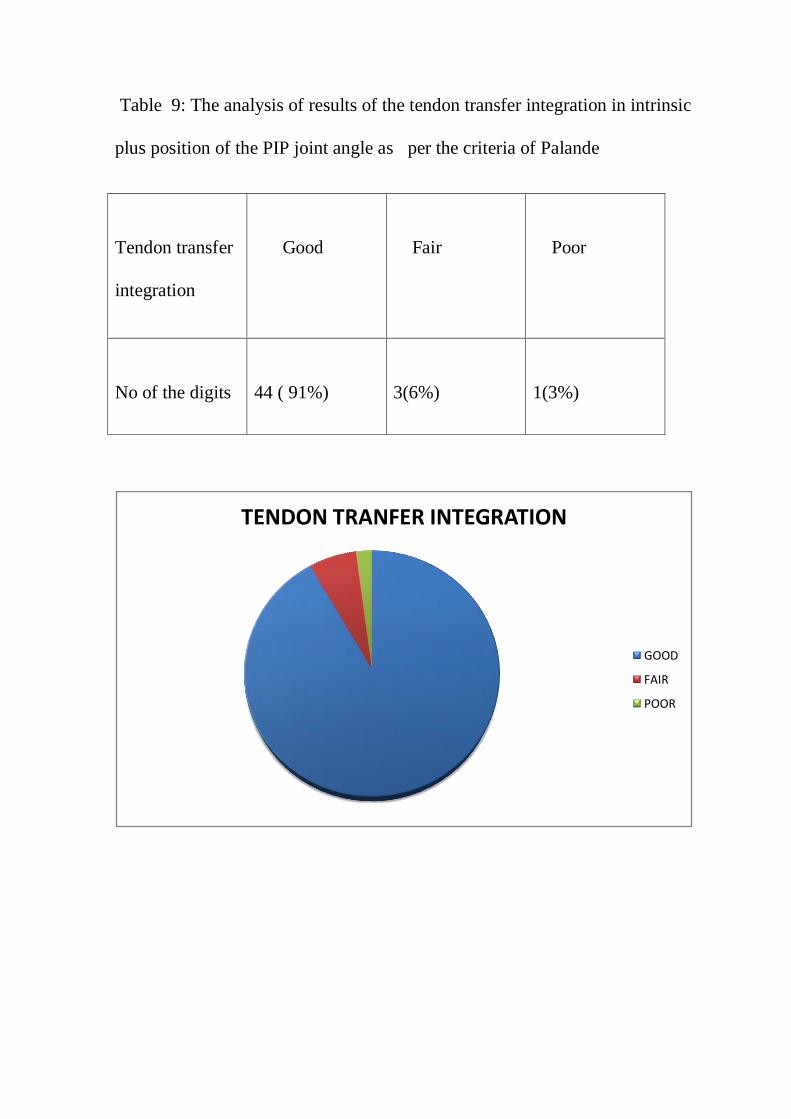
Table 9: The analysis of results of the tendon transfer integration in intrinsic
plus position of the PIP joint angle as per the criteria of Palande
Tendon transfer
integration
Good
Fair
Poor
No of the digits
44 ( 91%)
3(6%)
1(3%)
TENDON TRANFER INTEGRATION
GOOD
FAIR
POOR
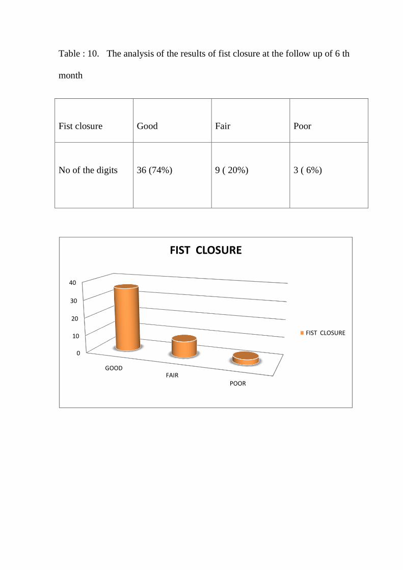
Table : 10. The analysis of the results of fist closure at the follow up of 6 th
month
Fist closure
Good
Fair
Poor
No of the digits
36 (74%)
9 ( 20%)
3 ( 6%)
0
10
20
30
40
GOODFAIR
POOR
FIST CLOSURE
FIST CLOSURE

TABLE : 11 .COMPARISION OF THE EARLY ACTIVE
MOBILISATION RESULTS WITH PUBLISHED REPORTS OF THE
CONVENTIONAL IMMOBILISTION OF TENDON TRANSFER FOR
CLAW HAND CORRECTION
deformity
correction
Patond4
8
99 H
n
=322D
Hasting &
Mccollam49
12 Hands
Anderson50
96 H ands
Rath 51
Immobil
isation
32 H
n =127
D
Our study of
early active
mo
bilisation 12
H
n=48D
Good n =262
81%
n = 19
83 %
86% n =81
63%
n=39
82%
Fair n = 23
18%
n =5
10%
Satisfactory
(good+Fair)
81% 83% 86% 81% 92%
poor n = 4
17%
n =23 D
19%
n = 4
8%
H =Hands , N = No of digits , D= digits
The satisfactory results of deformity correction with early active mobilisation
are better than those in the published reports of conventional immobilisation.
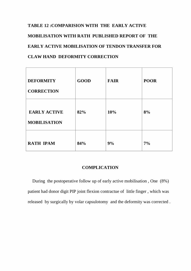
TABLE 12 :COMPARISION WITH THE EARLY ACTIVE
MOBILISATION WITH RATH PUBLISHED REPORT OF THE
EARLY ACTIVE MOBILISATION OF TENDON TRANSFER FOR
CLAW HAND DEFORMITY CORRECTION
DEFORMITY
CORRECTION
GOOD
FAIR
POOR
EARLY ACTIVE
MOBILISATION
82%
10%
8%
RATH IPAM
84%
9%
7%
COMPLICATION
During the postoperative follow up of early active mobilisation , One (8%)
patient had donor digit PIP joint flexion contractue of little finger , which was
released by surgically by volar capsulotomy and the deformity was corrected .

ILLUSTRATIVE CASES
CASE :1.
A case of tardy ulnar nerve palsy, in which the ulnar nerve transposition
was done earliar.
Pre operative photograph
Postoperative photograph

CASE :2
A case of post traumatic low ulnar nerve palsy
Preoperative
Post operative

CASE :3
A case of posttraumatic high ulnar nerve palsy
Pre operative
Postoperative
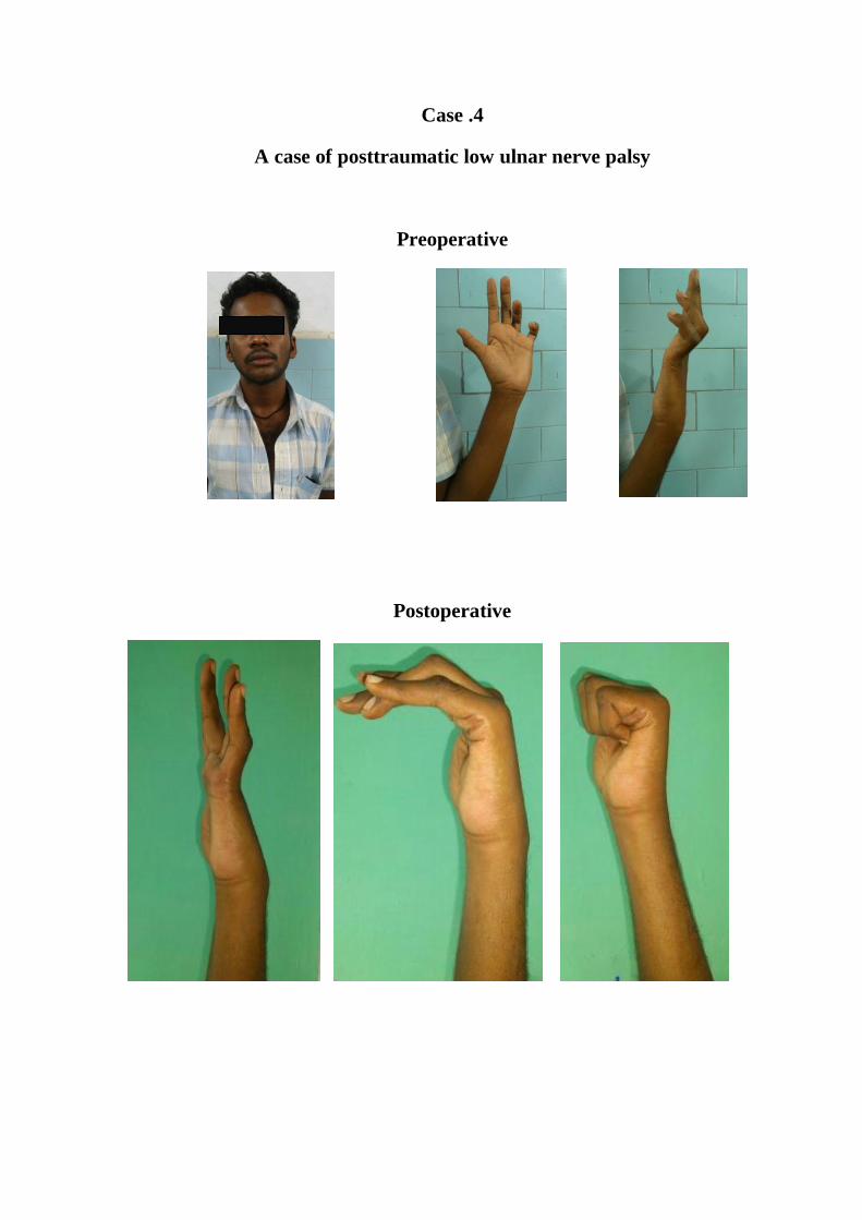
Case .4
A case of posttraumatic low ulnar nerve palsy
Preoperative
Postoperative
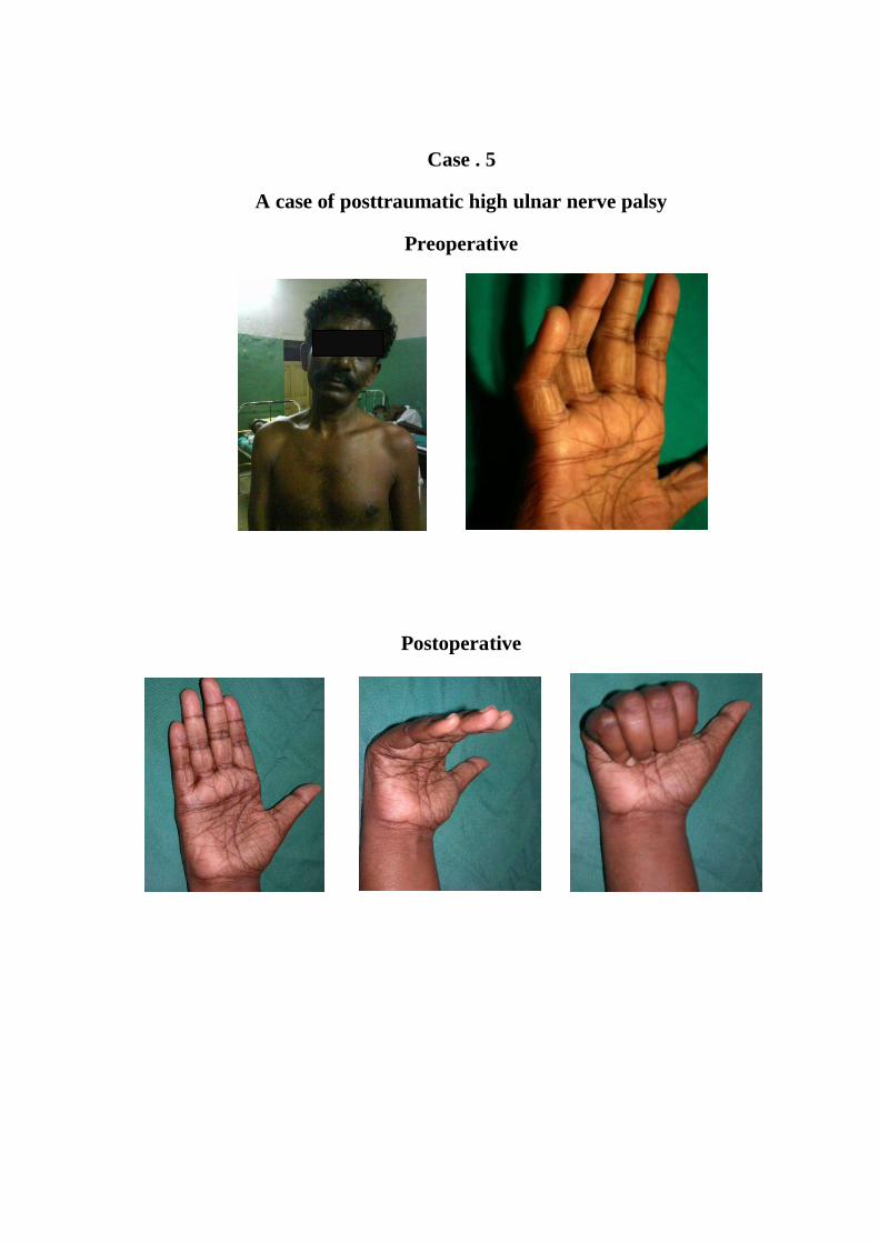
Case . 5
A case of posttraumatic high ulnar nerve palsy
Preoperative
Postoperative

Case 6.
A Case of leprosy with ulnar nerve palsy
Preoperative
Postoperative

COMPLICATION – PIP JOINT CONTRACTURE
Case ; 7
Preoperative
Postoperative
DISCUSSION
The evoluation of the tendon transfer techniques and the principles
were developed over 200 years ago . The earliest tendon transfer was described
by Velapeau in 1839 and Malgaigne in 1845 for reconstruction of severed
tendons . In 1882 , Nicoladoni applied the principle of the transfer of a an
intact muscle to paralytic muscles of lower limb. Bunnell described the
principles of tendon transfer for hand . Brand performed first surgical
correction of claw hand of leprosy patient in Christian Medical College ,
Vellore ,India in 194852
.
It is the usual practice after tendon transfer surgery to immobilize the hand
with pop cast for 3 – 4 weeks to allow union of the transferred tendon53
.
Subsequent to removal of the cast , post operative therapy is needed for 4 to 6
weeks . The average rehabilitation time in the conventional techniques is
usually 2 to3months . The period of postoperative immobilisation contributes
to 40-50% of the rehabilitation time in the claw hand deformity correction.
Early active mobilisation means starting the postoperative therapy for the
transferred tendon in the first week of surgery . The principle of early active
mobilisation has been proven to be safe and found to be significantly improve
the outcome. The conventional immobilisation is associated with adhesion
formation31
and a poor tendon gliding.
Laboratory studies demonstrated that the early mobilized tendon healed
faster , gained tensile strength quicker, and had better excursion . Because of
the less adhesion formation than unstressed repair54,55
early mobilized tendon
healed by an intrinsic mechanism and steadily gained strength following
repair56
.
The principles behind early mobilisation techniques involve protected
mobilisation in a restricted range with a dorsal blocking splint.
The early mobilisation has the benefit of reduced morbidity which will
considerably reduce the loss of income for self-employed patients for
undergoing surgery and loss of work compensation payment by the state.
Comparing the results of early active mobilization following FDS 4TP
with published reports62,63
of pulley insertion and immobilization has limitations
as the outcomes measures in these reports are not defined26
or uniform between
the studies· The factors taken into consideration for assessment of results in the
published reports i.e. deformity correction, integration of transfer and digit
flexion have similarities to the method of assessments as per Palande's criteria
used in the present study and can therefore be compared . The satisfactory i.e.
combined good and fair results of deformity correction with early active
mobilisation are better than those in the published reports of conventional
immobilisation.
Anderson57
allowed use of the hand with a knuckle bender splint 7 weeks
after surgery. Klein58
recommended a period of 12 weeks after surgery to allow

splint free hand activities. In our study the early active mobilisation patients
used hands for activities of daily living on an average of 5 weeks (34 days)
after surgery.
It is postulated that pre-operative isolation of FDS middle finger improves
the 'individuation index' of the little, ring and index finger by establishing new
neuronal networks in the brain. Early post operative mobilization activates the
new neuronal networks in the brain as described by Bezuhly60
whereas
immobilization may temporarily erase these neuronal networks. In addition
immobilization of the transfer will temporarily erase the cortical representation
of FDS middle finger is as demonstrated by de Jong et all61
with dynamic
immobilization of flexor tendons.
The study of de Jong et al41
demonstrated the impact of a relatively short
period of immobilization of digits on the functional organization of the brain. A
6 week period of immobilization induced temporary loss of efficient cerebral
control of hand movement. Functional restoration of hand movement occurs in
6-8 weeks time with use of the hand..
The major limitation of this study is comparing the results of a
prospective trial (early active mobilisation ) with retrospective historical data
CONCLUSION
The early active mobilisation is safe and has the better outcomes of
deformity correction compared to immobilisation with a selected donor
with a strong insertion.
Increased strength of tendon transfer insertion and protection of the
insertion during early active mobilisation are the two essential
requirements for this new tendon transfer rehabilitation protocol.
Wider application of this principle to other tendon transfer can
be investigated with incorporating the above two principles.
Future randomized controlled trials will provide insight into the
differences in post operative behavior of the tendon transfer with early
mobilization versus immobilization.
The neurophysiologic basis of the isolation of donor, integration of
tendon transfer and movement restoration can now be tracked in the
immediate postoperative period with functional MRI or PET scans.

BIBLIOGRAPHY
1. Hollingshead WJH.Anatomy for surgeons : the back and limbs. Vol3.
Philadelphia : JB Lippincott,1982 :341-529
2. Siegel DB,Gelberman RH.. ulnar nerve : applied anatomy and operative
exposure. In: Gelbermann RH, ed.operative nerve repair and
reconstruction.Philadelphia : JB Lippincott,1991:413-424.
3. Struthers J. On some points in the abnormal anatomy of the arm. Br
Foreign Med Chir Rev 14:170-179,1854
4. Spinner M. Injuries to the major branches of peripheral nerves of the
forearm,2nd
ed. Philadelphia : WB Saunders,1978.
5. Osborne G. compression neuritis of the ulnar nerve at the elbow. Hand
2:10- 13.1970
6. Uriburu IJF,Morchio FJ,Marin JC.compression syndrome of the deep
branch of ulnar nerve (piso- hamate hiatus syndrome). J Bone Joint surg
Am 58:145-147,1976.
7. Botte MJ, Cohen MS, Lavernia CJ et al. The dorsal branch of the ulnar
nerve : an anatomic study. J Hand Surg [Am] 15:603-607.1990
8. Fischer L, Neidhardt JH,Comlet JJ. Premilinary note on the topography
of the dorsal cutaneous branch of ulnar nerve : its value in surgical
approach of the inferior quarter of the ulna. Lyon Med 224:897-899,1970.

9. Fischer JL,Neidhardt comlet JJ.et.al Surgical anatomy of the dorsal
cutaneous branch of ulnar nerve. C R Assoc Anat 147:226-270,1970.
10. Anderson JE.Grant‟s atlas of anatomy. Baltimore : Williams & Wilkins,
1983
11. Clemente CD. Anatomy: a regional atlas of the human body. Philadelphia
Lea & Febiger,1975.
12. Williams PW.Gray‟s anatomy : the anatomical basis of medicine and
surgery,38 ed New York : Churchil Livingstone,1995 : 425-736.
13. Basmajian JV, Slonecker CE. Grant‟s metod of anatomy : a clinical
problem solving approach. Philadelphia : Williams & wilkins, 1989.
14. Mayfield FH.Compression syndromes of the shoulder girdle and arms. In:
Vinken PJ,Bruyn GW,eds . Handbook of clinical neurology, vol 7 :
Disease of nerves. New York : American Elsevier,1970:441-444.
15. Clemente CD. Gray‟s anatomy, 13th ed. Baltimore : Williams &
Wilkins,1985.
16. Hollingshead WJH.Anatomy for surgeons : the back and limbs. Vol3.
Philadelphia : JB Lippincott,1982 :341-529
17. Grant JCB. An atlas of anatomy,6th ed. Baltimore: Williams &
Wilkins,1972:1-104.
18. Sunderland S.Ran LJ.Metrical and non- metrical features of the muscular
branches of the median nerve.J Comp Neurol 85:113-120,1946.
19. Sunderland S. The intraneural Topography of the radial, median and
ulnar nerves.Brain 68:243-299,1945
20. Sunderland S. The innervation of the flexor digitorum profundos and
lumbrical muscles.Anat Rec 93:317-321,1945.
21. Campells text book of orthopaedics 11 th edition 4125-4173.
22. Guyon E : Note sur une disposition anatomique proper a la face
anteriecure de la region du poignet et non encore de crite par le docteur.
Bull Soc Anat paris 6: 184,1861.
23. Campells text book of orthopaedics 11 th edition 4125-4173
24. Bonnel R, Vila RM. Anatomical study of the ulnar nerve in the hand. J
Hand Surg [Br] 10:165-168,1985.
25. Bergfield TG, Aulicino PL. Variation of the deep motor branch of ulnar
nerve at the wrist.J Hand Surg [ Am]13:368-369,1982.
26. Rowntree T. Anomalous innervation of the hand muscles. J Bone Joint
surg Br 31:505-510,1949.
27. Kaplan EB, Spinner M. Normal and anomalous innervation patterns in
the upper extremity.In : Omer GE, Spinner M,eds. Management of
peripheral nerve problems.Philadelphia : WB Saunders,1980:75-99
28. Bowden R, Napier J: The assessment of hand functions after peripheral
nerve injuries. J Bone Joint Surg Br 1961; 43:481-492
29. Latimer J, Shah M, Kay S: Abductor digiti minimi transfer for the
restoration of opposition in children. J Hand Surg [Br] 1994; 19:653-658

30. greens hand surgery 5th edition
31. Mikhail IK: Bone block operation for clawhand. Surg Gynecol Obstet
1964; 118:1077-1079
32. Bunnell S: Surgery of the intrinsic muscles of the hand other than those
producing opposition of the thumb. J Bone Joint Surg Am 1942; 24:1-3
33. Zancolli EA: Claw-hand caused by paralysis of the intrinsic muscles: a
simple surgical procedure for its correction. J Bone Joint Surg Am 1957;
39:1076-1080
34. Omer GE: Ulnar nerve palsy. In: Green D, Hotchkiss R, Pederson W, ed.
Green's Operative Hand Surgery, New York: Churchill Livingstone;
1999:1526-1541
35. Burkhalter WE: Early tendon transfer in upper extremity peripheral nerve
injury. Clin Orthop 1974; 104:68-79
36. Burkhalter WE, Strait JL: Metacarpophalangeal flexor replacement for
intrinsic-muscle paralysis. J Bone Joint Surg Am 1973; 55:1667-1676
37. Riordan D: Tendon transfers for median, ulnar or radial palsy. Hand
1969; 1:42-46
38. Riordan DC: Tendon transplantations in median-nerve and ulnar-nerve
paralysis. J Bone Joint Surg Am 1953; 35:312-320
39. Anderson GA: Ulnar nerve palsy. In: Green D, et al ed. Green's
Operative Hand Surgery, Philadelphia: Elsevier; 2005:1161-1196.
40. Oberlin C: Zancolli's “lasso” operation in intrinsic palsy of leprous
origin: a study of twenty-six cases. Ann Chir Main 1985; 4:22-30
41. Stiles H, Forrester-Brown M: Treatment of Injuries of Peripheral Spinal
Nerves. London, Frowde & Hodder & Stoughton, 1922
42. Bunnell S: Surgery of the intrinsic muscles of the hand other than those
producing opposition of the thumb. J Bone Joint Surg Am 1942; 24:1-3
43. Littler JW: Tendon transfers and arthrodeses in combined median and
ulnar nerve paralysis. J Bone Joint Surg Am 1949; 31:225-234
44. Brand P: Tendon grafting: illustrated by a new operation for intrinsic
paralysis of the fingers. J Bone Joint Surg Br 1961; 43:444-453
45. Zancolli E. Intrinsic paralysis of the Ulnar Nerve - Physiopathology of
the Claw hand. InZancolli E. ed. Structural and Dynamic Bases of Hand
Surgery. 2nd eds. Philadelphia: J.B Lippincott Company, 1979 : 159-206.
46. De Jong BM, Coert JH, Stenekes Mw, Leenders KL, Paans AlYf],
Nicolai JPA. Cerebral reorganization of human hand movement
following Dynamic immobilization. NeuroReport2003; 14: 1693--1696.
47. Palande D. Correction of Intrinsic- Minus Hand Associated with Reversal
of the Transverse Metacarpal Arch: J Bone Joint Surg [American] 1983;
65-A: 514-21.
48. Patond KR, Betal BD, Kumar A. Surgical Correction Of Claw Fingers In
Leprosy Using Flexor Superficialis Direct Lasso Procedure. Indian
Journal of Leprosy 1997; 69:25-32.

49. Hastings H 2nd, McCollam SM. Flexor digitorum superficialis lasso
tendon transfer in isolated ulnar nerve palsy: a functional evaluation. J
Hand Surgery [British] 1994; 19: 275-80.
50. Anderson GA. The surgical management of the deformities of the hand in
Leprosy. J Bone Joint Surg. [British] 2006; 88-B: 290-94.
51. Santhosh rath . Journal of Hand Surgery, 2008 [American]; 33A : 232-
240
52. Anerson GA. The surgical management of defprmities of the hand in
leprosy
53. Srinivasn H, .Mukarjee SM and Subramanian RA . two tailed transfers of
tibialis posterior correction of foot drop in leprosy.jbjs 1968;50-B, 623-
628
54. Kleinert , H, E; Primary epair of lacerated flexor tendons on in no mans
hand , JBJS (America) 1987;49 A ;557
55. Kleinert , H E,kuntz,je A primary repair of flexor tendon injury.
OCNA,1973;4;865-876
56. Hichcock T,F, The effect of immediate constrained digital motion on the
strength of the flexor tendon repair in chickens. JHS 1987,12A;590-595
57. Lipscomb PR and Sanchez J). Anterior Transplantation of the Posterior
Tibial Tendon for Persistent Palsy of the Common Peroneal Nerve. J
Bone Joint Surg. [America] 1961;43-A (1): 60-66.
58. Brandsma Jw, Ebenezer M. Pre- and Postoperative Therapy Following
Tendon Transfer Surgery. In: Schwarz R, Brandsma Jw, ed. Surgical
Reconstruction and Rehabilitation in Leprosy and Other Neuropathies.
Ekta Books, Kathmandu, Nepal, 2004: 303-15.
59. Rath S. Early Results of a Randomized Controlled Trial of Immediate
Postoperative Active Mobilization versus Immobilization for Zancolli's
'LASSO' Procedure (abstract).
60. Rath S, Nagesh P. Is Immobilization Necessary Following Tendon
Transfers? A Trial of immediate Active Mobilization for Common
Tendon Transfers to the Hand (abstract).10th International Federation of
Societies of Surgery of the Hand Congress, 2007
61. Silfverskiold KL May EJ. Early active mobilization after tendon transfers
using mesh reinforced suture techniques. J Hand Surgery [British] 1995;
20(3): 291-300.
62. Strickland J W The Scientific Basis for Advances in Flexor Tendon
Surgery. J Hand Therapy 2005; 18:94-110.
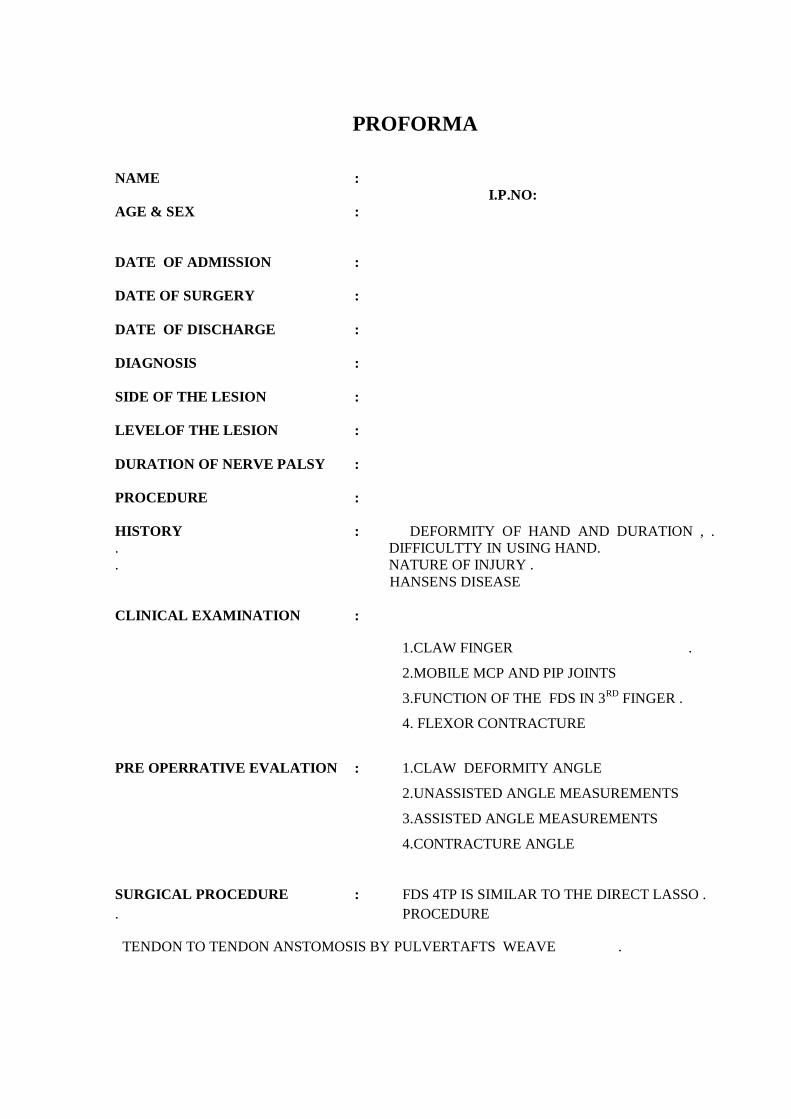
PROFORMA
NAME :
I.P.NO:
AGE & SEX :
DATE OF ADMISSION :
DATE OF SURGERY :
DATE OF DISCHARGE :
DIAGNOSIS :
SIDE OF THE LESION :
LEVELOF THE LESION :
DURATION OF NERVE PALSY :
PROCEDURE :
HISTORY : DEFORMITY OF HAND AND DURATION , .
. DIFFICULTTY IN USING HAND.
. NATURE OF INJURY .
HANSENS DISEASE
CLINICAL EXAMINATION :
1.CLAW FINGER .
2.MOBILE MCP AND PIP JOINTS
3.FUNCTION OF THE FDS IN 3RD
FINGER .
4. FLEXOR CONTRACTURE
PRE OPERRATIVE EVALATION : 1.CLAW DEFORMITY ANGLE
2.UNASSISTED ANGLE MEASUREMENTS
3.ASSISTED ANGLE MEASUREMENTS
4.CONTRACTURE ANGLE
SURGICAL PROCEDURE : FDS 4TP IS SIMILAR TO THE DIRECT LASSO .
. PROCEDURE
TENDON TO TENDON ANSTOMOSIS BY PULVERTAFTS WEAVE .

POSTOPERATIVE REHABLITATION
THERAPY PROTOCOL
ASSESSMENT
ANGLES ARE RECORDED IN BIWEEKLY
ANGLES MCP JOINTS PIP JONTS
1.OPEN HAND POSITION
2. INTRINSIC PLUS POSITION
MEAN
FOLLOW UP
EVERY MONTH FOR 6 MONTHS
EVALUATION
1.TENDON RUPTURE DURING ACTIVE MOBILISATION
2.MORBIDITY
3.FIST CLOSURE
4.COMPARISION OF EARY ACTIVE MOBILISATION WITH IMMOBILISATION
RESULTS
COMPLICATION


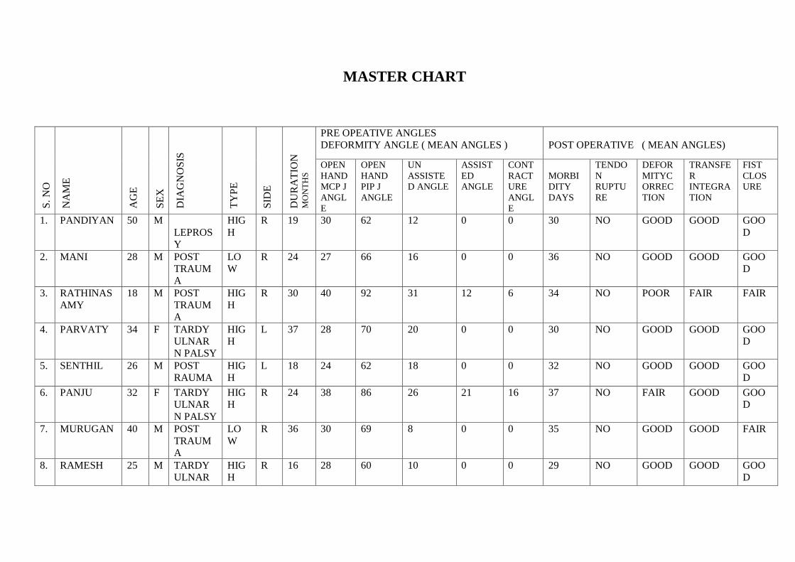
MASTER CHART
S.
NO
NA
ME
AG
E
SE
X
DIA
GN
OS
IS
TY
PE
SID
E
DU
RA
TIO
N
MO
NT
HS
PRE OPEATIVE ANGLES
DEFORMITY ANGLE ( MEAN ANGLES )
POST OPERATIVE ( MEAN ANGLES)
OPEN
HAND
MCP J
ANGL
E
OPEN
HAND
PIP J
ANGLE
UN
ASSISTE
D ANGLE
ASSIST
ED
ANGLE
CONT
RACT
URE
ANGL
E
MORBI
DITY
DAYS
TENDO
N
RUPTU
RE
DEFOR
MITYC
ORREC
TION
TRANSFE
R
INTEGRA
TION
FIST
CLOS
URE
1. PANDIYAN 50 M
LEPROS
Y
HIG
H
R 19 30 62 12 0 0 30 NO GOOD GOOD GOO
D
2. MANI 28 M POST
TRAUM
A
LO
W
R 24 27 66 16 0 0 36 NO GOOD GOOD GOO
D
3. RATHINAS
AMY
18 M POST
TRAUM
A
HIG
H
R 30 40 92 31 12 6 34 NO POOR FAIR FAIR
4. PARVATY 34 F TARDY
ULNAR
N PALSY
HIG
H
L 37 28 70 20 0 0 30 NO GOOD GOOD GOO
D
5. SENTHIL 26 M POST
RAUMA
HIG
H
L 18 24 62 18 0 0 32 NO GOOD GOOD GOO
D
6. PANJU 32 F TARDY
ULNAR
N PALSY
HIG
H
R 24 38 86 26 21 16 37 NO FAIR GOOD GOO
D
7. MURUGAN 40 M POST
TRAUM
A
LO
W
R 36 30 69 8 0 0 35 NO GOOD GOOD FAIR
8. RAMESH 25 M TARDY
ULNAR
HIG
H
R 16 28 60 10 0 0 29 NO GOOD GOOD GOO
D
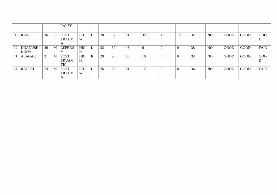
PALSY
9 RANI 36 F POST
TRAUM
A
LO
W
L 28 37 91 32 16 12 32 NO GOOD GOOD GOO
D
10 DNANUSH
KODY
46 M LEPROS
Y
HIG
H
L 32 30 46 6 0 0 38 NO GOOD GOOD FAIR
11 ALAGAR 31 M POST
TRUAM
TIC
HIG
H
R 29 28 58 10 0 0 33 NO GOOD GOOD GOO
D
12 RAJESH 23 M POST
TRAUM
A
LO
W
L 26 25 61 12 0 0 36 NO GOOD GOOD FAIR

Your digital receiptThis receipt acknowledges that Turnitin received your paper. Below you will find the receipt informationregarding your submission.
Paper ID 290490095
Paper title“A PROSPECTIVE STUDY ON THE OUTCOME OF EARLYPOSTOPERATIVE ACTIVE MOBILISATION FOLLOWING TENDONTRANSFER PROCEDURES FOR CLAW HAND CORRECTION
Assignmenttitle Medical
Author Murugesh Kumar 22101602 M.S. Orthopaedic SurgeryE-mail [email protected]
Submissiontime 24-Dec-2012 09:09PM
Total words 10303
First 100 words of your submission
“A PROSPECTIVE STUDY ON THE OUTCOME OF EARLY POSTOPERATIVE ACTIVEMOBILISATION FOLLOWING TENDON TRANSFER PROCEDURES FOR CLAW HANDCORRECTION ” Dissertation submitted for M.S. Degree Examination Branch II – ORTHOPAEDICSURGERY APRIL-2013 THE TAMILNADU DR. M.G.R. MEDICAL UNIVERSITY CHENNAI, TAMILNADU 1 CERTIFICATE This is to certify that this dissertation entitled “A PROSPECTIVE STUDY ONTHE OUTCOME OF EARLY POSTOPERATIVE ACTIVE MOBILISATION FOLLOWING TENDONTRANSFER PROCEDURES FOR CLAW HAND CORRECTION” is the bonafide work done byDr.P.MURUGESHKUMAR, under my supervision in the Department of Orthopaedic Surgery, MaduraiMedical College, Madurai-20. Prof. Dr. P.V.PUGALENTHI, M.S Ortho., D....
Copyright 2012 Turnitin. All rights reserved.