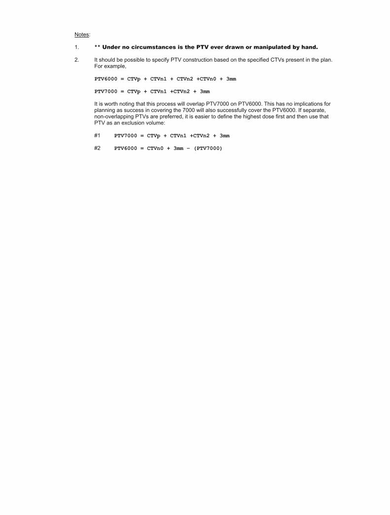
Embed Size (px)
Citation preview
Journal of Radiation Oncology Informatics
A Rational Informatics-enabled approach to the
Standardised Naming of Contours and Volumes in
Radiation Oncology Planning
Research Article
Alexis A. Miller 1,2∗
1 Centre for Oncology Informatics, Faculty of Engineering & Information Science, University of Wollongong, WollongongNSW, Australia
2 Department of Radiation Oncology, Illawarra Cancer Care Centre, Wollongong NSW, Australia
Abstract: The standardising of nomenclature in the radiotherapy planning process has deep implications for the ability of
the profession to examine the adequacy of construction of radiotherapy plans in outcomes research, particularlyin relation to disease control and toxicity generation. After surveying the literature for similar attempts at
Standardised Nomenclature, this paper proposes a logical standardised nomenclature which can be used by
any individual or institution as a template for a mappable local standard.The nomenclature is systematically constructed using the Foundational Model of Anatomy, ICRU Report 50
and ICRU report 62. The system foreshadows a XML metadata structure to detail the method of construction
of volumes. Treatment Planning System vendors should build their software with the ability to use thissystematic construction technique so that contours and volumes in a radiotherapy plan can be annotated.
This metadata will allow the investigation of how a radiation plan’s construction can affect the therapy
outcome.A Standardized Nomenclature is provided as an Appendix.
J Radiat Oncol Inform 2014;6:1:53-70
doi: 10.5166/jroi-6-1-22
Author has no conflict to disclose.
Keywords: radiotherapy • contour • volume • oncology • standardized nomenclature • Foundational Model of Anatomy
c© Journal of Radiation Oncology Informatics
1. Introduction
Nomenclatures are lists of words associated with meaning for use in particular circumstances. Standard nomen-
clatures are derived from local or regional efforts to achieve consistency of terms for application within that
jurisdiction. More than this, a standardised nomenclature should have an logical and reproducible basis which is
not subject to the whims of a local or regional group.
∗ E-mail: [email protected], m: +61 406654239
53
A Rational Informatics-enabled approach to the Standardised Naming of Contours and Volumes in Radiation Oncology Planning
1.1. What makes a good nomenclature?
Nomenclatures are built for a reason, which shapes their construction and ultimately determines their useful-
ness [1]. Determining the reasons for expending the effort should precede and adequately justify the effort.
Furthermore, since construction requires effort, adequate solutions should not be superseded.
From an Informatics perspective, a nomenclature will be most useful if it corresponds to an ontology that reflects
clinical care, supports information exchange with internal and external systems, reveals clinical decision making
and accumulates structured information (such as occurs in a database) to assist in quality assurance measures
and to aid research. A nomenclature is a device to benefit a profession, rather than the individual professional,
but it does not substitute a well-formed ontology since nomenclatures do not exist outside the structure of the
expert domain’s knowledge base. Rather, it provides a list of instances of acceptable terms and definitions for
pre-existing knowledge structures, e.g., Gross Tumour Volume (GTV) is the term given to the visible tumour
defined by a ROI on a clinical image-set.
A comprehensive nomenclature would specify terms used by the radiation oncologist pertinent to the patient’s
characteristics and their assessment of the patient, as well as diagnosis, stage, the plan of care, therapies applied,
the care actually delivered, the patient’s responses to care, and the actual patient outcomes. In Radiation
Oncology to date, the only standard nomenclatures reported have related to the names for contours and volumes
assigned by the oncologist during the Simulation and Planning Processes. This report confines itself to the same
nomenclature subset.
A standard nomenclature will permit synonymy, i.e., the expression the same concept in different ways depending
on local preference. The point is that a local preference should be able to be mapped to the standard
nomenclature [2]. The answer to lack of use of standards is not a Lord Of The Rings response (”one ring to
rule them all”) with a single defined nomenclature (such as SNOMED is purported to be), but rather local
nomenclatures constructed to be interoperable with the standardised nomenclature, i.e., deliberately devised to fit
well-constructed knowledge structures which have been formalised into an ontology. This deliberate construction
would allow subsequent transfer of local names into a standardised common identifier. For radiation oncologists,
it does not matter if the spherical anatomical item with which you see this page is called an eye, eyeball, globe
or any other non-English term (ojo, oculus, occhio, oogappel, etc), so long as a colleague elsewhere using the
term ‘right eyeball’ can translate this name into a standardised anatomical nomenclature (e.g., FMAID 12514).
And when I use the term ‘EYE R’ locally, that this too will translate into the same standardised anatomical
nomenclature (i.e., FMAID 12514). However, the use of the term ’ball’ to describe either an eye or a testis in a
single department is problematic, as is the lack of a departmental conversion table that specifies that local use of
‘ball’ corresponds to the anatomical term ‘testis’. Given that classifications enlarge over time, a translation table
54
A. A. Miller
will cater for these changes [3]
When used in a clinical process, a nomenclature should have sufficient granularity to describe differences in
clinical care. Discussions between oncologists about recommended treatment can result in widely variable targets
being defined, e.g., a recommendation to “treat the necks nodes” might result in ipsilateral or bilateral volumes,
inclusion or exclusion of Levels 1A, 1B or 5 depending on the recipient’s understanding of the recommendation.
Similarly, when treating a Merkel Cell Carcinoma of the skin with radiotherapy, the recommendation to treat the
primary site “with a wide margin” could result in margins of between 2 and 10cm. Since margin expansions are an
important determinant of geographical miss probability, a standard nomenclature should seek to systematically
quantify margins to allow analysis of variation and consequent outcomes.
The process of making a nomenclature useful requires synonymy which is clinical relevant, but also coding
in a consistent manner that makes retrieved nomenclature consistent in structure and therefore able to be
manipulated by the computer. The difference between ‘EYE R PRV 3’ and ‘PRV R EYE 3’ is non-trivial if
they occur in a single institution.
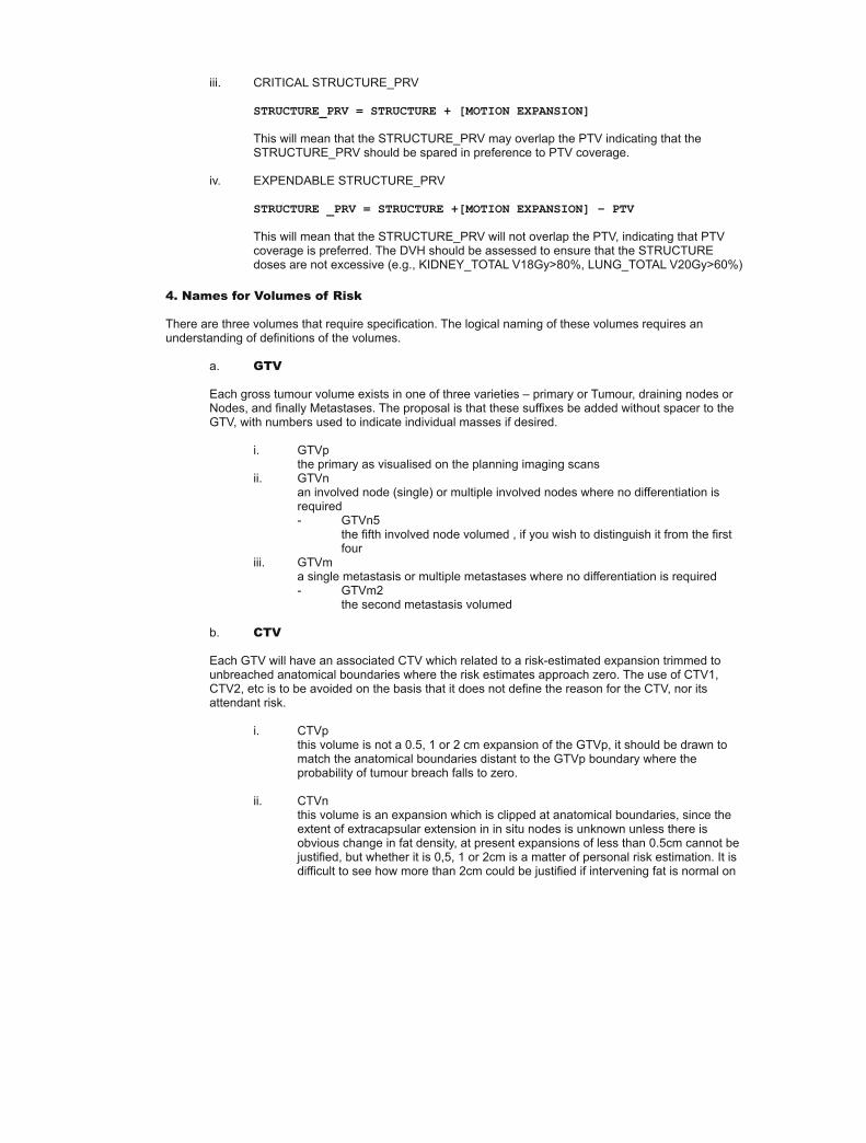
2. Post-Nomenclature Radiotherapy Planning
Non-ambiguity in terms is ensured by clear, precise definition. However the tendency to impose a structure on
the nomenclature to describe all processes used in normal clinical processes, even when precise and relevant,
is counter-productive. Firstly the nomenclature becomes unwieldy when more than 2-3 additional processes
are coded. Secondly, the names of volumes can become lengthy and highly codified. One schema could have
a target volume titled ‘SPINALCORD PRV 3’ where SPINALCORD is a defined anatomic structure, PRV
corresponds to an expansion of the previous specified anatomic structure, and 3 meaning that a symmetric
expansion of 3mm has occurred. While this example combines terms in a way that describes the clinical process,
the problem of asymmetric expansion is not adequately addressed.
The concatenation of additional information into the nomenclature increases its expressiveness but also reduces
its simplicity and legibility. The expanded nomenclature that includes domain specific knowledge is therefore a
temporary solution.
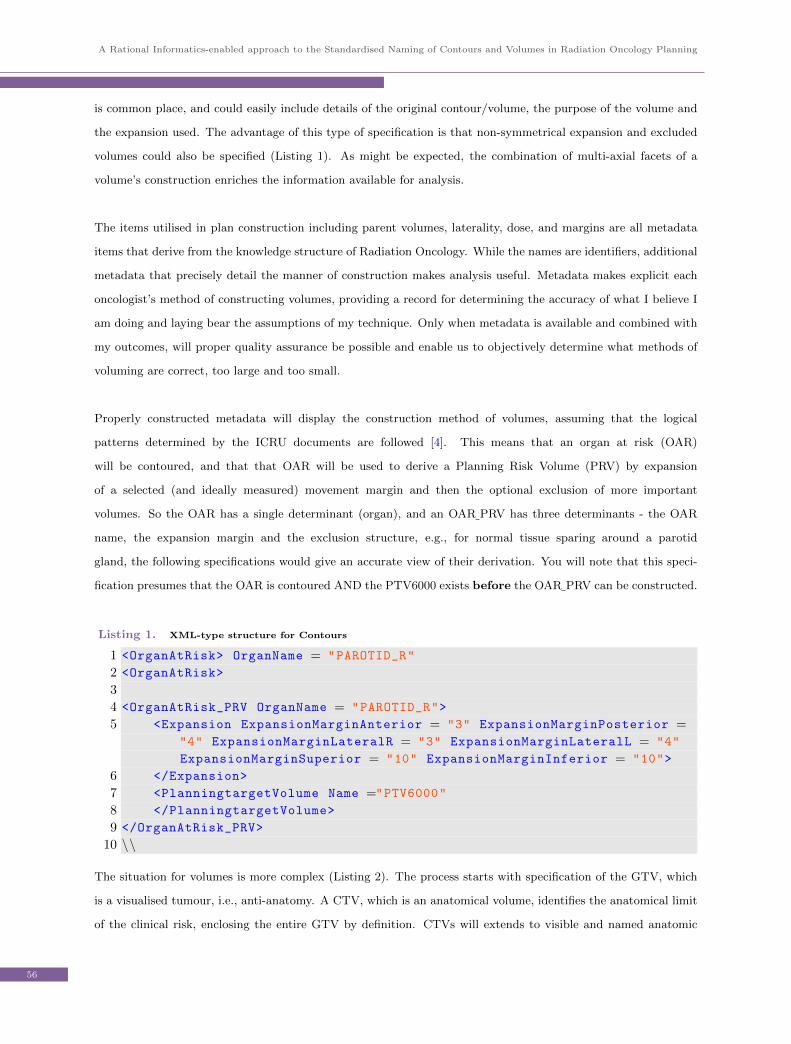
Improving the semantic richness of a volume name to describe its construction requires a method to capture
the details of the construction process as metadata. The capture of this information could be automated in
current treatment planning software. The specification of metadata in a hierarchical markup language like XML
55
A Rational Informatics-enabled approach to the Standardised Naming of Contours and Volumes in Radiation Oncology Planning
is common place, and could easily include details of the original contour/volume, the purpose of the volume and
the expansion used. The advantage of this type of specification is that non-symmetrical expansion and excluded
volumes could also be specified (Listing 1). As might be expected, the combination of multi-axial facets of a
volume’s construction enriches the information available for analysis.
The items utilised in plan construction including parent volumes, laterality, dose, and margins are all metadata
items that derive from the knowledge structure of Radiation Oncology. While the names are identifiers, additional
metadata that precisely detail the manner of construction makes analysis useful. Metadata makes explicit each
oncologist’s method of constructing volumes, providing a record for determining the accuracy of what I believe I
am doing and laying bear the assumptions of my technique. Only when metadata is available and combined with
my outcomes, will proper quality assurance be possible and enable us to objectively determine what methods of
voluming are correct, too large and too small.
Properly constructed metadata will display the construction method of volumes, assuming that the logical
patterns determined by the ICRU documents are followed [4]. This means that an organ at risk (OAR)
will be contoured, and that that OAR will be used to derive a Planning Risk Volume (PRV) by expansion
of a selected (and ideally measured) movement margin and then the optional exclusion of more important
volumes. So the OAR has a single determinant (organ), and an OAR PRV has three determinants - the OAR
name, the expansion margin and the exclusion structure, e.g., for normal tissue sparing around a parotid
gland, the following specifications would give an accurate view of their derivation. You will note that this speci-
fication presumes that the OAR is contoured AND the PTV6000 exists before the OAR PRV can be constructed.
Listing 1. XML-type structure for Contours
1 <OrganAtRisk> OrganName = "PAROTID_R"
2 <OrganAtRisk>
3
4 <OrganAtRisk_PRV OrganName = "PAROTID_R">
5 <Expansion ExpansionMarginAnterior = "3" ExpansionMarginPosterior =
"4" ExpansionMarginLateralR = "3" ExpansionMarginLateralL = "4"
ExpansionMarginSuperior = "10" ExpansionMarginInferior = "10">
6 </Expansion>
7 <PlanningtargetVolume Name ="PTV6000"
8 </PlanningtargetVolume>
9 </OrganAtRisk_PRV>
10 \\
The situation for volumes is more complex (Listing 2). The process starts with specification of the GTV, which
is a visualised tumour, i.e., anti-anatomy. A CTV, which is an anatomical volume, identifies the anatomical limit
of the clinical risk, enclosing the entire GTV by definition. CTVs will extends to visible and named anatomic
56
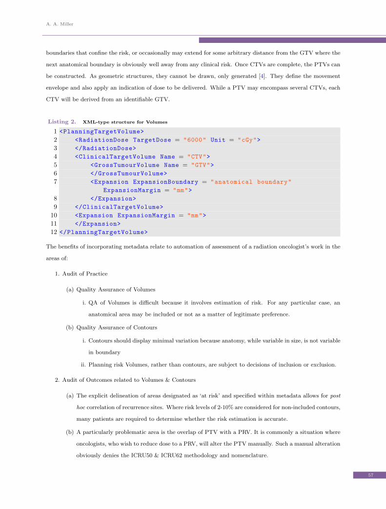
A. A. Miller
boundaries that confine the risk, or occasionally may extend for some arbitrary distance from the GTV where the
next anatomical boundary is obviously well away from any clinical risk. Once CTVs are complete, the PTVs can
be constructed. As geometric structures, they cannot be drawn, only generated [4]. They define the movement
envelope and also apply an indication of dose to be delivered. While a PTV may encompass several CTVs, each
CTV will be derived from an identifiable GTV.
Listing 2. XML-type structure for Volumes
1 <PlanningTargetVolume>
2 <RadiationDose TargetDose = "6000" Unit = "cGy">
3 </RadiationDose>
4 <ClinicalTargetVolume Name = "CTV">
5 <GrossTumourVolume Name = "GTV">
6 </GrossTumourVolume>
7 <Expansion ExpansionBoundary = "anatomical boundary"
ExpansionMargin = "mm">
8 </Expansion>
9 </ClinicalTargetVolume>
10 <Expansion ExpansionMargin = "mm">
11 </Expansion>
12 </PlanningTargetVolume>
The benefits of incorporating metadata relate to automation of assessment of a radiation oncologist’s work in the
areas of:
1. Audit of Practice
(a) Quality Assurance of Volumes
i. QA of Volumes is difficult because it involves estimation of risk. For any particular case, an
anatomical area may be included or not as a matter of legitimate preference.
(b) Quality Assurance of Contours
i. Contours should display minimal variation because anatomy, while variable in size, is not variable
in boundary
ii. Planning risk Volumes, rather than contours, are subject to decisions of inclusion or exclusion.
2. Audit of Outcomes related to Volumes & Contours
(a) The explicit delineation of areas designated as ‘at risk’ and specified within metadata allows for post
hoc correlation of recurrence sites. Where risk levels of 2-10% are considered for non-included contours,
many patients are required to determine whether the risk estimation is accurate.
(b) A particularly problematic area is the overlap of PTV with a PRV. It is commonly a situation where
oncologists, who wish to reduce dose to a PRV, will alter the PTV manually. Such a manual alteration
obviously denies the ICRU50 & ICRU62 methodology and nomenclature.
57
A Rational Informatics-enabled approach to the Standardised Naming of Contours and Volumes in Radiation Oncology Planning
(c) The issue of radiation plan quality has been highlighted as responsible for significant deficits in out-
come [5]. However the boundaries for declaring a significant deviation were very loose 1. One would
hope that these criteria were rarely broached in the IMRT era, but automated assessment of this type
is not possible without metadata specification. It is therefore not surprising that the authors have
not closed the loop by voluming and contouring to define the exact deficits in the HEADSTART plan
generation.
2.1. What makes something a standard?
Physicians have always been “knowledge workers”, collaborating with other health care actors to provide patient
care by applying published data to direct patient management. The process of evidence production, which is
largely clinic-based, is now challenged by the paradigm of Informatics in the guise of Information Technology
and electronic records. We have moved on from a time of independent components designed for specific but
separate functions with data stored in proprietary formats in non-interacting silos. Despite a high degree of
semantic homogeneity, data heterogeneity was imposed by the lack of domain standards which caused confusion.
Standards were poorly implemented, training was insufficient and relied on sales people, and there was poor
understanding and inadequate use of accumulated information because new paradigms of innovative data use
could not be demonstrated. It may come as a surprise that this description comes from the recent paper by
Gibaud [6] describing the problems of standards in Radiology!
Radiation Oncology has the same challenges, but unfortunately has a much smaller commercial base making
the improvement of software more difficult. Given that most treatment planning systems can export to the
DICOM-RT format, the issue of nomenclature has an elevated importance. As the DICOM-RT format is a
standard, research by electronically accessing multiple files is supported. However looking inside the files will
reveal many naming conventions and examples which will prevent collation. The Radiation Oncology Data
Alliance (RODA), based on MOSAIQ use [7], found this problem of name proliferation to the detriment of
analysis [8].
Making a nomenclature into a standard is a corporate task requiring recognition by a professional or governmental
group. At present, none of the professional organisations in this region (TROG, RANZCR) have an approved
1 Table 1. Protocol-Specified Criteria for Significant Deviations (pp.2997)Tumor Dose at 200cGy/fraction delivered to target volumes*.All gross disease (except nodes <2 cm) must receive at least 6650cGyNo more than 10% of the planning target volume (PTV) enclosing gross disease must receive <6650cGy (<5700cGyfor small nodes) or >7500cGy, excluding volumes within the gross tumor volume or air cavities.No more than 10% of PTV defining electively treated areas must receive <4000cGy
58
A. A. Miller
standardised nomenclature. The result is that the evolution of biomedical knowledge, which requires a necessary
informatics articulation between care delivery and biomedical research [6] is stunted. In essence, the lack of
standardised nomenclature results in a lack of openness, and the use of quasi-proprietary protocols (I own my
naming system, you own yours, etc.) and so poor performance [6] in interoperability and collaboration which are
the necessary infrastructure for research.
However, it is inevitable that nomenclatures managed by corporate agencies will be unwieldy and slow to change.
It is possible to overcome this tendency by adopting an Open Source approach to nomenclature maintenance
where any professional can commit an alteration, but a maintainer oversees and approves changes to maintain
consistency of purpose. To aid in this task, the re-use of pre-existing nomenclatures and ontologies makes sense.
In terms of anatomical names, radiation oncologists do not have any particular expertise. We only use the names.
So an anatomy ontology or nomenclature should be used to derive names, if it is available and maintained. Such
ontologies are available.
The development of standards and focus of interoperability have seen real benefits for the business of Radiation
Oncology (e.g., IHE-RO [9]) so that we have the some ability to re-use data to perform different tasks [6].
However this interoperability does not translate into the clinical area since questions of re-use of data from tasks,
letters, reports and literature to support outcomes research is not enabled. Interoperability requires the shared
knowledge model behind the data [6] and the standard nomenclature to prevent data heterogeneity in the face
of semantic homogeneity, thereby leading to ignorance while swimming in a sea of electronic data. This shared
knowledge model is domain-specific and must represent an achievable standard for Radiation Oncology.
With the emergence of ’omics and the push for personalised medicine, radiation oncologists need to have and use
their clinical data to overcome the present personalisation gap [10, 11]. The use of standardised protocols [8] in
patient planning has benefits for the patient [5], it also has benefits for process efficiency [12].
As stated previously, the standardised nomenclature should only be seen as a stop gap measure. Nomenclatures
are limited to simple concatenated terms with the same function as metadata, and should be specified within
a formal description of the domain’s specialist knowledge in the form of ontologies [13, 14] which include the
vocabulary of terms defined in a formal language with the attributes of first-order logic to support reasoning. A
common format is OWL and its variant, OWL-DL [15–17].
Not all standardised nomenclatures are useful in Radiation Oncology. The Systematised Nomenclature of
Medicine (SNOMED) is not a true ontology having started life as a collection of terms. There is no formal
analysis of its usefulness in Radiation Oncology, but the fact that it does not include the term ‘radiation
59
A Rational Informatics-enabled approach to the Standardised Naming of Contours and Volumes in Radiation Oncology Planning
oncologist’, and does not list ‘medical oncologist’ among ‘medical specialist’ indicates some shortcomings.
2.2. What should be embedded in a nomenclature?
A nomenclature for use by radiation oncologists in Simulation and Planning should include several abilities which
are listed below:
CONTOURS
Contours are ROIs drawn to define anatomical structures.
Organ - the name of the organ being contoured must be unambiguous and consistent, and should be
derived from or locally correlated with an external anatomical source. The anatomical ontology is to
be preferred over another nomenclature because of the added functionality of an ontology.
Laterality - the differentiation of right, left and combined total organs is required.
VOLUMES
Volumes are oncological ROIs based on the probability assessment that a moving area seen on an image set
is or contains cancer, and therefore which need to be included in the daily radiation target.
Gross Tumour Volume - the GTV delineates all tumour visible on an image set, which is to say that its
definition is ‘anti-anatomical’. Differentiation of GTVs that exist in the primary site, multiple nodal
deposits and/or multiple metastatic deposits is required.
Clinical Target Volume - the CTV defines the risk volume on the image set and is an anatomically
defined volume, with each CTV being derived from one GTV by defining the surrounding risk area.
It should be clear from the nomenclature which GTV resulted in a particular CTV.
Intermediate Target Volume - the ITV defines the physiological motion of an anatomically defined risk
volume, i.e., a volume encompassing a moving CTV. It should be clear from the ITV nomenclature
which CTVs have been combined to produce a particular ITV.
Planning Treatment Volume - the PTV is a volume is firstly defined by the total expected motion of
the CTV while being treated. Secondly it is also defined by the dose which is expected to be delivered
to the volume. The prescribed dose should be clear from the PTV nomenclature.
Planning Risk Volume - the PRV is a volume which defines a moving organ that is to be spared when
constructing a radiation plan. This PRV will look like some portion of an expanded organ, and may
encroach into a PTV. Overlap of PTV and PRV indicates an area of clinical decision making balancing
tumour coverage and critical organ dosing. It should be clear from the nomenclature which organ
forms the basis of the PRV, and what expansion has been used in its construction.
60
A. A. Miller
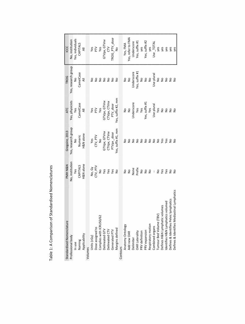
3. What Standardised Nomenclatures are available?
There are examples of departments and collaborative groups developing guidelines for consistent naming. However
the term ”standardised” could be a synonym for ‘locally consistent’ rather that being deliberately constructed or
achieving the status of a standard. After the discussion below, Table 1 provides a comparison of the features of
the nomenclatures.
• Princess Margaret Hospital Head & Neck Nomenclature
In 2007, the Princess Margaret Hospital (PMH) in Toronto reported on a standardized Head & Neck
voluming nomenclature that they had implemented in 2004 [18] to facilitate planning, quality assurance
(QA) and future outcome audits of IMRT. It was designed to conform to ICRU 50 and 62. They used
case-sensitive terminology for normal structures, gross disease and target volumes are which they described
as unique and descriptive e.g. CORD (spinal cord), R facial (right facial node). Guidelines were developed
to handle separate, multiple or combined targets.
Physicians contour the primary disease with a GTV. For patients who have undergone surgery where there
may be no gross tumour objects, the site(s) of preoperative tumour were considered high risk (CTV), as
currently there is no ICRU terminology for this principle. Physicians contour all nodal gross disease and
localized its anatomic position to specific surgical levels in the neck. A laterality prefix ‘R’ or ‘L’ is used
for gross or elective neck nodal targets with the addition of nodal levels, e.g. R2A (right level IIa node).
Dose was applied to CTVs as a suffix. The term CTV without prefix refers to a volume surrounding
the GTV. In combination, their nomenclature specified RCTVs and LCTVs (right and left CTV) with
corresponding PTVs, LPTVs and RPTVs. Increased specification produced these patterns - CTV50 (50
Gy CTV surrounding GTV), R2ACTV70 (70 Gy CTV encompassing a right level IIa node). All PTVs
and PRVs are generated by planners [18].
The authors felt that the voluming nomenclature had served the purpose of facilitating multidisciplinary
communication, quality assurance review of H&N planning and had enabled the safe automation of complex
programming tasks [18].
There are some attractive principles espoused in this nomenclature, including the unambiguous use provided
by capitalised names, a clear specification of laterality and specific surgically defined neck lymph nodes sites.
However, the source of the anatomical names is not described, and is limited to H&N cases by original
intent [1]. The positioning of the laterality as a prefix without separation from the volume’s primary name
makes reading difficult, and the lack of delimiter use affects legibility of more complex names, for example
the name required for planning (CTV) may be disguised (R2ACTV70). While not specifically mandated
in the ICRU reports, the use of dose in Gray is problematic for fractional doses (e.g., 50.4Gy). The use of an
integer with centiGray (cGy), is preferred as it avoids the use of period in the name. Although supposedly
based on ICRU 50/62, the nomenclature contravenes these standards by not identifying involved lymph
61
A Rational Informatics-enabled approach to the Standardised Naming of Contours and Volumes in Radiation Oncology Planning
nodes as gross tumour volume ( [4], Section 2.3.1 pg 6). In terms of aesthetics, one can compare R2ACTV70
with a rearranged and separated version, CTV R 2A 7000, to decide on the legibility issue. While the
PMH H&N nomenclature wraps dose with the CTV, in neither ICRU 50 [4] nor ICRU 63 [19] is there a CTV
described with a juxtaposed dose. These reports only use dose when describing a PTV, and when specifying
coverage of a PTV. Finally, in terms of workload, the nodal levels in the neck are defined separately while
in fact the nodal groups are in continuity and might be defined differently in the future [3].
• Advanced Technology Consortium Nomenclature
The second example of a nomenclature is one developed by a consortium, predominantly of physicists in
USA and European cancer centres (Advanced Technology Consortium, ATC) [20]. This work indicates that
nomenclatures are also useful to professions downstream from radiation oncologists who have responsibilities
in differing facets of radiotherapy.
Unfortunately radiation physicists are not the purveyors of knowledge structure of the medical domain
of Radiation Oncology, only of the physics portion of the domain. The limited engagement of radiation
oncologists has the potential to skew the nomenclature away from its general applicability in radiation
oncology [1]. Failure to make the nomenclature relevant to radiation oncologists will result in under-use of
the nomenclature.
The group correctly identify that a nomenclature with consistent language and terminology is a key com-
ponent of any effective process improvement and work flow management infrastructure [20]. The intra-
departmental variability of free-text structure names prevents the reliable mapping of names by automated
heuristic methods. The only solution is a great deal of manual quality assurance [20]. The pervasive nature
of the Internet is moving attention onto inter-institutional data sharing and analyses. When this occurs,
lack of standardisation will prevent sharing. Interoperability for analysis requires the profession to iden-
tify, adopt, and maintain a list of standardized structure names [20]. This nomenclature purports to be
successfully implemented and in use [21].
The ATC schema [20] is more complex and comprehensive than the PMH H&N nomenclature by attempting
to address all circumstances, not just H&N, and divides structure names into target volumes (TV) and
contours, which could be either Organs At Risk or Planning organ at Risk Volumes (PRV) derived from
the OARs. The tenets of the schema are state that all radiation dose levels are specified in units of cGy
with a maximum of five characters, and that margins are specified in units of millimeters with a maximum
of two characters. The volume name is constructed using a capitalised TV base name, e.g., GTV, CTV,
PTV, followed by a suffix associated with the target (p=primary, n=node) and target multiplicity (1, 2,
. . . ) separated by an underscore ( ) delimiter, but does not specify laterality. The inclusion of dose into
CTV is not specified by any other standard [4, 19] and does not follow clinical reasoning. The CTV is an
expression of risk, not dose. The oncologist defines the risk areas initially but only then specifies the dose
62
A. A. Miller
to the corresponding PTV [4, 19] based on the level of risk. This specification should be discussed with
oncologists, the relevant users.
The order of suffixes may not reflect clinical importance. The volume GTVn1 when expanded by 15mm
becomes CTVn1 15 5000. Irrespective of the inclusion of dose with CTV, a discussion should occur to see
whether CTVn1 15 5000 or CTVn1 5000 15 is the preferred format.
They introduce the Tumour Bed Volume (TBV) and quantify respiratory motion in the respiratory cycle
(EE (end expiration) or EI (end inspiration)) to allow the generation of an ITV. The source of the anatomic
names used is not defined, nor how to add additional names. Base OAR names are constructed with more
ambiguous and error prone CamelCase truncated at 16 characters. For repeating organs (ribs, cranial
nerves, vertebral bodies) a laterality and suffix number is used inconsistently, e.g., (CN I R, CN II L,
Rib1 L, Rib2 R, VB C1 R (C=cervical, T=thoracic,L=lumber, S=sacral). Vasculature was named as
A name (artery) or V name (vein), and combined bilateral organs were named simply as a plural or-
gan (Lung R + Lung L = Lungs; Kidney R + Kidney L = Kidneys). Planning Risk Volumes as
derived from the base OAR names with an underscore ( ) delimiter for uniform expansion margins in
millimetres (2 numerals maximum) (e.g., SpinalCord 05). Asymmetric PRV expansion uses the suf-
fix PRV (e.g., SpinalCord PRV), but this could cause confusion (e.g., SpinalCord, SpinalCord 05,
SpinalCord PRV)
• TransTasman Radiation Oncology Group
The TransTasman Radiation Oncology Group (TROG) has recently published their Standardised Nomen-
clature on their website2 as the output of an XLS file without instructions on use. The document (from
14 May 2014) is a list of names with no instructions to describe use. Unconventional and general names
are not provided with any definition to permit accurate delineation. The contour BaseOfTongue has no
specification to assist an oncologist draw the contour. While the FMA does recognise the concept of regions,
the lack of distinct boundaries prevents definition and so should be avoided in Planning Nomenclatures.
The specification also contains some lateralised organs (Breast L, Breast R) but not others (CN VII,
Atrium). It also duplicates names without indication of how they differ (LargeBowel/Colon,
Constrictors/PharynxConst, Eye/Globe, MainBronchus/BronchialTree/LobarBronchus L).
Some names are unusual (MassMuscle, GreatVessel, Vessels, A Hypophyseal), while others are
inconsistent (FemHead/FemoralJoint, Lung/BilatLung, A subclavicular/V SubClav).
The lack of extensibility, duplications, undefined contours and inconsistencies indicate that this standard
is not mature.
2 The document can be downloaded here - http: // trog. com. au/ SiteFiles/ trogcomau/ TROG_ standardised_structure_ names. pdf
63
A Rational Informatics-enabled approach to the Standardised Naming of Contours and Volumes in Radiation Oncology Planning
• Gregoire Research Consortium Nomenclature
A recent publication [3] provides consensus guidelines from several research groups, specifically DAHANCA,
EORTC, HKNPCSG, NCIC CTG, NCRI, RTOG and TROG, on the nomenclature for neck lymphatic
regions. The report focuses on this anatomical area alone, and as such provides no assistance in how to
incorporate these names into a system to identify what has been contoured, or how to construct volumes
in a way that describes their antecedents. Furthermore, it controversially suggests that the entire surgical
nomenclature should be altered. It may be that such a proposal fails to gain widespread use outside of
radiation oncology trials in H&N.
• Illawarra Cancer Care Centre Standardised Nomenclature
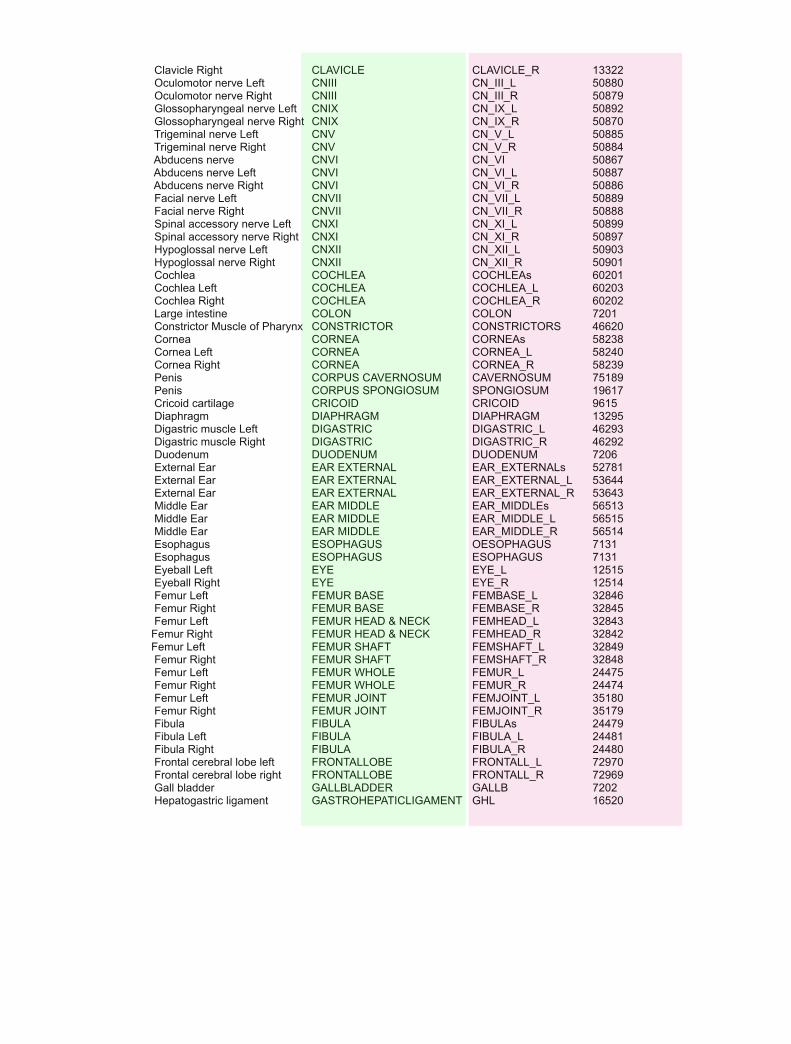
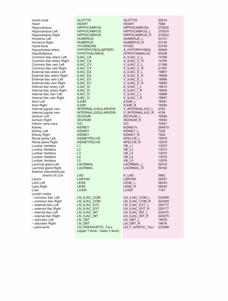
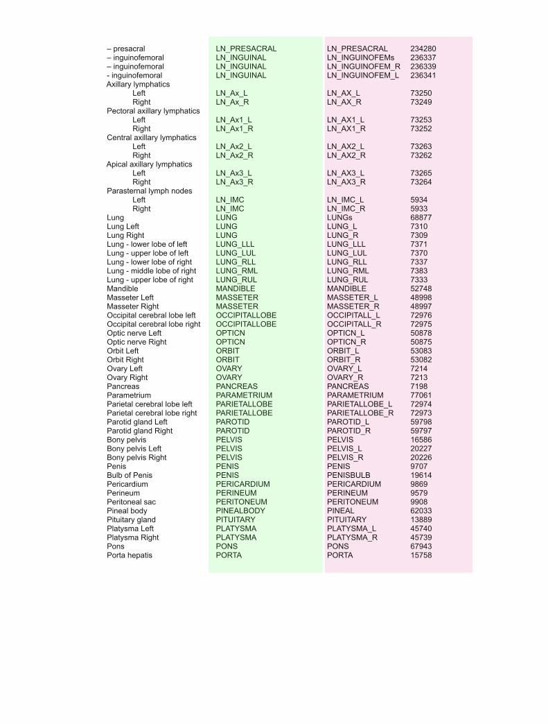
This specification is provided in Appendix 1, and is an adaptable nomenclature based on the Foundational
Model of Anatomy for OAR names, and a similar, though independently derived, TV construction to the
ATC nomenclature. Being developed by an oncologist, it addresses the definition of lymphatic levels that
are to be assigned to different risk levels, the ability to include new anatomy names, and the clinical use of
the ICRU reports in generating TVs.
The specification enshrines the logic of the ICRU process in moving from OARs to GTVs then through to
PTVs. Using the GTVs, the definition of most CTVs occurs by combination of GTV expansion and OAR
confinement. The generation of PTVs uses the aggregation of CTVs. This standardised nomenclature is
also compared in Table 1. While the system does not use a meta-data definition for its construction method,
as the next section demonstrates, the logic embedded in the metadata definition is identical to the logic
described in this schema.
The system also stipulates that PRVs and PTVs are not to be manually shaped, so that there is the
possibility of assessing the outcome of assigning constraint priority to PRVs or PTVs can be detected.
64
Tab
le 1
: A C
om
par
iso
n o
f St
and
ard
ised
No
men
clat
ure
s
Stan
dar
dis
ed N
om
encl
atu
re
PM
H N
&N
G
rego
ire,
20
13
A
TC
TRO
G
ICC
C
Pro
fess
ion
al b
od
y N
o, i
nst
itu
tio
n
Yes,
res
ear
ch g
rou
p
Yes,
ph
ysic
ists
Ye
s, r
ese
arch
gro
up
N
o, i
nst
itu
tio
n
In u
se
Yes
N
o
Yes
N
o
Yes,
ind
ivid
ual
s N
amin
g C
AP
ITA
LS
Nu
mer
ic
Cam
elC
ase
Cam
elC
ase
CA
PIT
ALS
A
pp
licab
ility
H
&N
alo
ne
H&
N a
lon
e A
ll
All
All
Vo
lum
es
U
nit
s (c
Gy)
N
o, G
y Ye
s
Yes
N
o
Yes
Do
se a
ssig
ned
to
C
TV, P
TV
CTV
, PTV
P
TV
No
P
TV
Co
mp
lies
wit
h IC
RU
50/
62
N
o
No
Ye
s
No
Ye
s D
elin
eate
d G
TV
Yes
G
TVp
x, G
TVn
x G
TVp
x, G
TVn
x N
o
GTV
px,
GTV
nx
Del
inea
ted
CTV
Ye
s
CTV
px,
CTV
nx
CTV
px,
CTV
nx
No
C
TV
Gen
erat
ed P
TV
Yes
P
TVp
x _d
ose
P
TV_d
ose
N
o
TRO
G_P
TV_
do
se
Mar
gin
s d
efin
ed
No
Ye
s, s
uff
ix #
1, m
m
Yes,
su
ffix
#2
, mm
N
o
No
C
on
tou
rs
A
nat
om
y O
nto
logy
N
o
No
N
o
No
Ye
s, F
MA
A
dd
new
OA
R
No
N
o
No
N
o
Yes,
ref
er
to F
MA
D
elim
iter
N
on
e N
o
Un
der
sco
re
Un
der
sco
re
Un
der
sco
re
OA
R L
ate
ralit
y P
refi
x
No
Ye
s
Yes,
su
ffix
#1
Ye
s, s
uff
ix #
1
PR
V d
efin
itio
n
No
N
o
Yes
N
o
yes
PR
V e
xpan
sio
n
No
N
o
Yes,
su
ffix
#1
No
Ye
s, s
uff
ix #
2
Res
pir
ato
ry m
oti
on
N
o
No
Ye
s
No
ye
s C
om
bin
ed o
rgan
s --
N
o
Use
plu
ral
Use
plu
ral
Use
_TO
TAL
Tum
ou
r B
ed V
olu
me
(TB
V)
No
N
o
Yes
N
o
yes
Def
ines
H&
N L
ymp
hat
ic v
olu
mes
Ye
s
Yes
N
o
No
ye
s Id
enti
fies
H&
N L
ymp
hat
ics
volu
med
Ye
s
Yes
N
o
No
ye
s D
efin
es &
Iden
tifi
es P
elvi
c ly
mp
hat
ics
No
N
o
No
N
o
yes
Def
ines
& Id
enti
fies
Med
iast
inal
Lym
ph
atic
s N
o
No
N
o
No
ye
s
A Rational Informatics-enabled approach to the Standardised Naming of Contours and Volumes in Radiation Oncology Planning
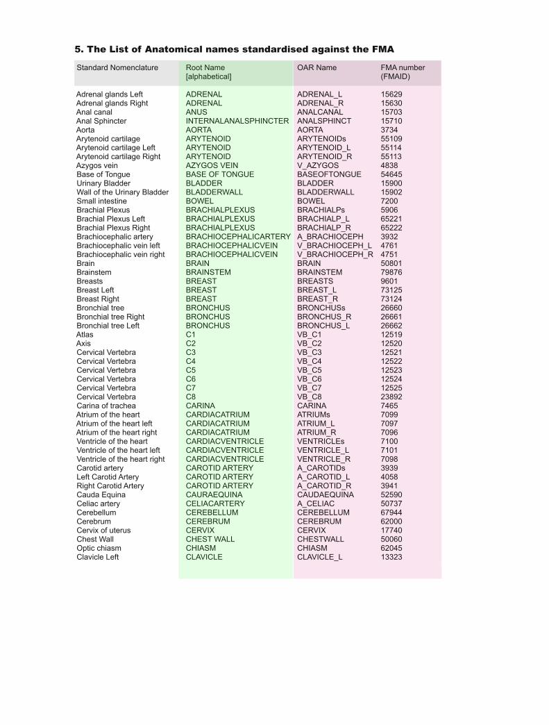
4. Standardised Anatomy Nomenclature
The use of a standardised framework is useful whenever trying to define and use expert domain knowledge.
Anatomy is not the sole province of oncology and as its own expert domain has been subject to attempts at
definition already.
The Foundational Model of Anatomy (FMA) was built to accommodate the knowledge of anatomy to be defined
in a way that allows for machine argument. The ontology is built with terms (more than 110,000 [22]) that have
a relationship (more than 170 types [22]). Examples of relationships that are well understood in the Radiation
Oncology domain include:
• Tip of Tongue drains to Level IA lymphatics.
• Level IA lymphatics are bounded anteriorly by Mandible.
• Level IA lymphatics drains to Level IB lymphatics.
• Level IB lymphatics drains to Level II lymphatics.
• Level II lymphatics drains to Level III lymphatics.
• Level III lymphatics drains to Level IV lymphatics.
• Facial Artery is anterior to Masseter Muscle.
Once connected by one of the many relationships possible, logical arguments and searches can be built to answer
questions. It is possible to ask the question, “What is the lymphatic drainage of the Tip of Tongue?” and
establish that there is an anatomic link between the Tip of Tongue and the Level 4 lymphatics, which is
what we teach our trainees to consider when drawing contours and volumes in the neck for radiotherapy. The
benefit of the description logic in the FMA is seen where nodal drainage in the neck is described from the FMA
and its relationships (‘afferent’, ‘efferent’) [23]. There is data demonstrating the use of an anatomy ontology
in radiotherapy planning [24–26].
The use of a knowledge structure, such as the FMA, will permit easier outcomes research. It will allow faster
and more accurate audit of practice, if practice definition allows the anatomical sites included to be defined
retrospectively. Organ contouring can be compared between sites, even to the point of looking at CT numbers
around the region of interest (ROI) boundary and adjudicating on conformality. It is likely that many ROIs will
not be controversial (e.g., KIDNEY), but others such as CTVs and PTVs will be very variable [27].
66
A. A. Miller
5. What benefits accrue?
What can we do together if we use a standardised nomenclature? A standardised nomenclature for planning
allows radiation oncologists to speak the same language, a language of intent and process relating to the con-
struction of a plan. A logically constructed nomenclature allows for a limited description of actions undertaken
in producing volumes from contours and GTVs.
The purpose of implementing a standardised nomenclature is to carry forward the original intent of the ICRU
expressed in Reports 50 and 62 so that the construction of the radiation plan becomes logical and reveals the
intent of the oncologist.
While there are international efforts to accumulate radiotherapy planning data [28], the lack of a nomenclature
limits the effort by having mismatched ROI names, and by being unable to compare the actual method of
construction of the ROIs. In fact, a later publication makes no mention at all of these nomenclature issues [29].
The comparison of variably named ROIs without any hint of the intent in construction cannot lead to useful
conclusions to inform radiation oncologists.
6. Conclusion
The definition of contours and volumes in Radiation Oncology requires the use of names. These names have
a use that lasts long after the development of an acceptable plan and can be used to define, or obscure, the
voluming procedures and their consequence when developing new knowledge. At the heart of this ability is the
need to combine data from many departments to discover explicit decisions and their consequences.
The realisation of this functionality is the problem of interoperability which Informatics addresses. It does
this by applying knowledge modelling techniques to the development of standardised nomenclatures which
embed components of knowledge and work flow. In its final state the components will be expressed in the meta-
data attached to a specific volume. The XML schema provides an excellent way of realising this level of metadata.
However while the application of Informatics to Radiation Oncology problems requires and assists in the
production of nomenclatures for use, the realities of language and local use dictate that local instances of these
nomenclatures must be permitted, and that a mechanism for translating locally developed nomenclatures into an
ontologically sound and enabled schema should be in place.
This paper is followed by an appendix containing a clinically utilised specification where the correlation of organ
67
A Rational Informatics-enabled approach to the Standardised Naming of Contours and Volumes in Radiation Oncology Planning
name with the Foundational Model of Anatomy provides the planning process with an structured ontology
capable of machine manipulation. The appendix also displays that the logical development of target volume
names also provides a specification of the knowledge flow and critical decision making undertaken by the radiation
oncologist to assist with data mining of medical decisions. The nomenclature is superior to previous patterns, it is
not completely expressive, because the granularity of metadata required cannot be specified in a single short name.
The development of such a granular process requires that commercial vendors approach the problem is a way
that satisfies the Informatics needs of the expert domain.
References
[1] J. Ingenerf, “Taxonomic vocabularies in medicine: the intention of usage determines different established
structures,” MEDINFO, vol. 8 Pt 1, pp. 136–139, 1995. 00040 PMID: 8591138.
[2] W. A. Meissner, “What’s in a name?The standardization of tumor nomenclature,” International journal of
radiation oncology, biology, physics, vol. 1, pp. 1245–1246, Nov. 1976.
[3] V. Grgoire, K. Ang, W. Budach, C. Grau, M. Hamoir, J. A. Langendijk, A. Lee, Q.-T. Le, P. Maingon,
C. Nutting, B. OSullivan, S. V. Porceddu, and B. Lengele, “Delineation of the neck node levels for head
and neck tumors: A 2013 update. DAHANCA, EORTC, HKNPCSG, NCIC CTG, NCRI, RTOG, TROG
consensus guidelines,” Radiotherapy and Oncology. 00000.
[4] International Commission on Radiation Units and Measurements, Prescribing, recording, and reporting pho-
ton beam therapy. [ICRU 50]. Bethesda, MD: International Commission on Radiation Units and Measure-
ments, 1993. 00000.
[5] L. J. Peters, B. O’Sullivan, J. Giralt, T. J. Fitzgerald, A. Trotti, J. Bernier, J. Bourhis, K. Yuen, R. Fisher,
and D. Rischin, “Critical impact of radiotherapy protocol compliance and quality in the treatment of ad-
vanced head and neck cancer: results from TROG 02.02,” Journal of clinical oncology: official journal of the
American Society of Clinical Oncology, vol. 28, pp. 2996–3001, June 2010. 00132 PMID: 20479390.
[6] B. Gibaud, “The quest for standards in medical imaging,” European Journal of Radiology, vol. 78, pp. 190–
198, May 2011.
[7] S. Hopkins, L. Oakes, S. Upasani, and J. Goldwein, “Establishment of a common radiation oncology data
registry system: Report of a pilot program,” International Journal of Radiation Oncology * Biology * Physics,
vol. 75, pp. S139–S139, Nov. 2009. 00000.
[8] J. Christodouleas, M. van der Pas, and J. Goldwein, “The case for clinical pathways in radiation oncology,”
Frontiers in Oncology, vol. 3, Sept. 2013. 00000 PMID: 24046816 PMCID: PMC3763216.
[9] M. Abdel-Wahab, R. Rengan, B. Curran, S. Swerdloff, M. Miettinen, C. Field, S. Ranjitkar, J. Palta, and
68
A. A. Miller
P. Tripuraneni, “Integrating the healthcare enterprise in radiation oncology plug and PlayThe future of
radiation oncology?,” International Journal of Radiation Oncology*Biology*Physics, vol. 76, pp. 333–336,
Feb. 2010. 00013.
[10] X. Wang and L. Liotta, “Clinical bioinformatics: a new emerging science,” Journal of Clinical Bioinformatics,
vol. 1, no. 1, p. 1, 2011. 00037.
[11] I. S. Kohane, “Bioinformatics and clinical informatics the imperative to collaborate,” Journal of the American
Medical Informatics Association, vol. 7, pp. 512–516, Sept. 2000. 00068 PMID: 10984470.
[12] B. S. Chera, L. Mazur, M. Jackson, K. Taylor, P. Mosaly, S. Chang, K. Deschesne, D. LaChapelle, L. Hoyle,
P. Saponaro, J. Rockwell, R. Adams, and L. B. Marks, “Quantification of the impact of multifaceted initiatives
intended to improve operational efficiency and the safety culture: A case study from an academic medical
center radiation oncology department,” Practical Radiation Oncology, vol. 4, pp. e101–e108, Mar. 2014.
[13] T. Bittner and B. Smith, “Normalizing medical ontologies using basic formal ontology,” in Proceedings of
GMDS Innsbruck, pp. 199–201, 2004.
[14] J. J. Cimino and X. Zhu, “The practical impact of ontologies on biomedical informatics,” Yearbook of medical
informatics, pp. 124–135, 2006. PMID: 17051306.
[15] S. Zhang, O. Bodenreider, and C. Golbreich, “Experience in reasoning with the foundational model of
anatomy in OWL DL,” Pacific Symposium on Biocomputing. Pacific Symposium on Biocomputing, pp. 200–
211, 2006. PMID: 17094240.
[16] F. W. Hartel, R. Dionne, G. Fragoso, and J. Golbeck, “Modeling a description logic vocabulary for cancer
research,” Journal of Biomedical Informatics, vol. 38, pp. 114–129, 2005.
[17] C. Golbreich, J. Grosjean, and S. J. Darmoni, “The foundational model of anatomy in OWL 2 and its use,”
Artificial Intelligence in Medicine, vol. 57, pp. 119–132, Feb. 2013.
[18] J. Kim, S. Breen, J. Waldron, A. Bayley, B. Cummings, J. Ringash, L. Dawson, C. Liu, S. Huang, and
B. O’Sullivan, “A standardized nomenclature system for head and neck (H&N) IMRT contouring, planning
and quality assurance,” International Journal of Radiation Oncology Biology Physics, vol. 69, p. S473, Nov.
2007.
[19] International Commission on Radiation Units and Measurements, Prescribing, recording, and reporting pho-
ton beam therapy. [ICRU 62]. Bethesda, Md.: International Commission on Radiation Units and Measure-
ments, 1999. 00000.
[20] L. Santanam, C. Hurkmans, S. Mutic, C. van Vliet-Vroegindeweij, S. Brame, W. Straube, J. Galvin, P. Tripu-
raneni, J. Michalski, and W. Bosch, “Standardizing naming conventions in radiation oncology,” International
Journal of Radiation Oncology Biology Physics, vol. 83, pp. 1344–1349, July 2012.
[21] J. van der Leer, F. F. M. van Aarle, M. a. J. van Lieshout, M. J. van der Sangen, and C. W. Hurkmans, “Im-
plementation of the new standardised naming conventions in radiation therapy,” Radiotherapy and Oncology,
vol. 103, pp. S157–S157, May 2012.
69
A Rational Informatics-enabled approach to the Standardised Naming of Contours and Volumes in Radiation Oncology Planning
[22] C. Rosse and J. L. Mejino Jr., “A reference ontology for biomedical informatics: the foundational model of
anatomy,” Journal of Biomedical Informatics, vol. 36, pp. 478–500, Dec. 2003.
[23] I. J. Kalet, J. L. V. Mejino, V. Wang, M. Whipple, and J. F. Brinkley, “Content-specific auditing of a large
scale anatomy ontology,” Journal of Biomedical Informatics, vol. 42, pp. 540–549, June 2009. 00007.
[24] J. Barker, M. M. Austin-Seymour, I. J. Kalet, M. Whipple, C. Teng, and L. Shapiro, “Automated delineation
of regional target volumes for patients with head and neck cancer treated conformally,” International Journal
of Radiation Oncology Biology Physics, p. 15, 1999.
[25] C.-C. Teng, L. G. Shapiro, and I. J. Kalet, “Head and neck lymph node region delineation using a hybrid
image registration method,” in 3rd IEEE International Symposium on Biomedical Imaging, (Arlington, VA),
pp. 462–465, IEEE Comput. Soc, 2006.
[26] C.-c. Teng, M. M. Austin-Seymour, J. Barker, I. J. Kalet, L. G. Shapiro, and M. Whipple, “Head and
neck lymph node region delineation with 3-d CT image registration,” in Proceedings of the AMIA Annual
Symposium, pp. 767–771, 2002.
[27] T. Hong, W. Tome, R. Chappell, and P. Harari, “Variations in target delineation for head and neck imrt:
An international multi-institutional study,” International Journal of Radiation Oncology, Biology, Physics,
vol. 60, no. 1 Supplement, pp. S157–S158, 2004.
[28] E. Roelofs, L. Persoon, S. Qamhiyeh, F. Verhaegen, D. De Ruysscher, M. Scholz, G. Iancu, M. Engelsman,
C. Rasch, L. Zijp, G. D. Meerleer, M. Coghe, J. Langendijk, C. Schilstra, M. Pijls-Johannesma, and P. Lambin,
“Design of and technical challenges involved in a framework for multicentric radiotherapy treatment planning
studies,” Radiotherapy and oncology: journal of the European Society for Therapeutic Radiology and Oncology,
vol. 97, pp. 567–571, Dec. 2010. PMID: 20864198.
[29] E. Roelofs, A. Dekker, E. Meldolesi, R. G. van Stiphout, V. Valentini, and P. Lambin, “International data-
sharing for radiotherapy research: An open-source based infrastructure for multicentric clinical data mining,”
Radiotherapy and Oncology, 2013.
70


A. A. Miller
93


![MEDC 603 Fall 20071 Biological Discovery High Volume Screening Combinatorial Diversity Rational [Structure, Design, Informatics] Lead Series Biodisposition](https://img.pdfslide.net/doc/110x75/56649d9f5503460f94a898d3/medc-603-fall-20071-biological-discovery-high-volume-screening-combinatorial.jpg)