Embed Size (px)
Citation preview

A re-evaluation of the genus Myceliophthora (Sordariales, Ascomycota):its segregation into four genera and description
of Corynascus fumimontanus sp. nov.
Yasmina Marin-FelixAlberto M. Stchigel1
Mycology Unit, Medical School and IISPV, UniversitatRovira i Virgili, C/ Sant Llorenc 21, 43201 Reus,Tarragona, Spain
Andrew N. MillerIllinois Natural History Survey, University of Illinois,1816 S. Oak St., Champaign, Illinois 61820
Josep GuarroJose F. Cano-Lira
Mycology Unit, Medical School and IISPV, UniversitatRovira i Virgili, C/ Sant Llorenc 21, 43201 Reus,Tarragona, Spain
Abstract: Based on a number of isolates of Myce-liophthora (Chaetomiaceae, Sordariales, Ascomycota)recently isolated from soil samples collected in USA,the taxonomy of the genus was re-evaluated throughphylogenetic analyses of sequences from the nucrDNA internal transcribed spacer region and genesfor the second largest subunit of RNA polymerase IIand translation elongation factor 1a. Members ofMyceliophthora were split into four monophyleticclades strongly supported by molecular and pheno-typic data. Such clades correspond with Myce-liophthora, now restricted only to the type species ofthe genus Corynascus, which is re-established with fivespecies, the new monotypic genus Crassicarpon andalso the new genus Thermothelomyces (comprising fourspecies). Myceliophthora lutea is mesophilic anda permanently asexual morph compared to themembers of the other three mentioned genera, whichalso are able to sexually reproduce morphs withexperimentally proven links to their asexual morphs.The asexual morph of M. lutea is characterized bybroadly ellipsoidal, smooth-walled conidia with a wide,truncate base. Crassicarpon thermophilum is thermo-philic and heterothallic and produces spherical tocuneiform, smooth-walled conidia and cleistothecialascomata of smooth-walled, angular cells and ascos-pores with a germ pore at each end. Corynascus spp.are homothallic and mesophilic and produce spher-ical, mostly ornamented conidia and cleistothecialascomata with textura epidermoidea composed of
ornamented wall cells, and ascospores with one germpore at each end. Thermothelomyces spp. are thermo-philic, heterothallic and characterized by similarascomata and conidia as Corynascus spp., but itsascospores exhibit only a single germ pore. Adichotomous key to distinguish Myceliophthora fromthe other mentioned genera are provided, as well asdichotomous keys to identify the species of Corynascusand Thermothelomyces. A new species, namely Cory-nascus fumimontanus, characterized by verrucoseascomatal wall cells and irregularly shaped ascos-pores, is described and illustrated.
Key words: Chaetomiaceae, Crassicarpon, Pezizo-mycotina, soilborne fungi, Thermothelomyces
INTRODUCTION
Myceliophthora spp. (Chaetomiaceae, Sordariales)traditionally were characterized by the production ofone-celled, subhyaline to reddish brown, smooth-walled to verrucose, globose to pyriform blastoconi-dia, sessile or arising on swollen protrusions from thevegetative hyphae, solitary or in short chains, andshow a narrow basal scar due to their rhexolyticdehiscence (Oorschot 1980). Mycelia and conidia ofMyceliopthora spp. are mostly hyaline or nearly so,with the only exception of the conidia of Myce-liophthora hinnulea, which become dark brown withage, and the mycelium of Myceliopthora vellerea, whichis pale brown. The conidiogenesis in Myceliophthoraspp. is similar among them, producing holoblasticconidia, sessile (frequently named as aleuroconidia)or on micronematous to semimicronematous con-idiophores, mostly solitary but also grouped in shortchains of 2–4 conidia. Sessile holoblastic conidia arealso present in other members of the family Chaeto-miaceae, that is Thielavia arenaria, Thielavia micro-spora and Thielavia subthermophila (Mouchacca1973). Myceliophthora spp. are mostly found in soilbut they also have been reported on compost used forgrowing mushrooms (Costantin and Matruchot1894), some species being parasites of mushrooms(Costantine 1892) and rarely infecting human (Hooget al. 2000).
The genus Myceliophthora was erected by Costantin(1892) to accommodate the mycoparasitic fungusMyceliophthora lutea, characterized by pyriform toglobose conidia born terminally or laterally on aerial
Submitted 28 Aug 2014; accepted for publication 18 Jan 2015.1 Corresponding author. E-mail: [email protected]
Mycologia, 107(3), 2015, pp. 619–632. DOI: 10.3852/14-228# 2015 by The Mycological Society of America, Lawrence, KS 66044-8897
619
hyphae, sometimes with a basal short pedicel, andoccasionally producing an additional apical conidi-um. Later three new species were added to the genus,they are Myceliophthora sulphurea Goddard (Goddard1913), Myceliophthora fusca Doyer (Doyer 1927) andMyceliophthora inflata Burnside (Burnside 1928). vanOorschot (1977, 1980) revised the genus and trans-ferred three additional species, Myceliophthora fergusii(Klopotek) Oorschot and Myceliophthora thermophila(Apinis) Oorschot from Chrysosporium and Myce-liophthora vellerea (Sacc. & Speg.) Oorschot fromSporotrichum. The same author excluded M. fusca,M. inflata and M. sulphurea from the genus.Myceliophthora fusca was thought to be identical toPtychogaster rubescens Boud., the anamorph of thebasidiomycete Punctularia atropurpurascens (Berk. &Br.) Petch; M. inflata was synonymized with Taifan-glania inflata (Burnside) Z.Q. Liang, Y.F. Han & H.L.Chu; and M. sulphurea was found indistinguishablefrom Chrysosporium merdarium (Ehrenb.) J.W. Car-mich. More recently Myceliophthora hinnulea Awao &Udagawa (Awao and Udagawa 1983) was described.
The sexual morphs of Myceliophthora have beenincluded in several genera belonging to differentorders and even classes, Arthroderma (Arthrodermatuberculatum Kuehn; order Onygenales; class Euro-tiomycetes), Corynascus (Corynascus spp.; order Sor-dariales; class Sordariomycetes) and Ctenomyces (Cte-nomyces serratus Eidam; order Onygenales; classEurotiomycetes) (Oorschot 1980, Guarro et al. 1985,Stchigel et al. 2000). Some of these sexual morphsfrom heterothallic species have been obtained in vitroby crossing sexually compatible strains, as is the casefor Arthroderma tuberculatum, Myceliopthora thermo-phila/Corynascus heterothallicus and Myceliophthorafergusii/Corynascus thermophilus (Klopotek 1974,1976). On the other hand, crossings of isolates ofMyceliophthora gutulata, Myceliophthora hinnulea andMyceliophthora lutea have never been reported toproduce their sexual stage. The homothallic speciesof Corynascus produced ascomata in monosporecultures on several culture media (von Arx et al.1984; Oorschot 1980). Myceliophthora spp. linked withtheir sexual morph have never been treated asa Myceliophthora-like asexual stage.
The genus Corynascus was proposed by von Arx in1973 based on two species of Thielavia (i.e. Thielavianovoguineensis Udagawa & Y. Horie and Thielaviasepedonium C.W. Emmons) that possess ascosporeswith two germ pores, one at each end, as opposed tospecies of Thielavia that have only a single germ pore.Three additional species of Thielavia also weretransferred under the same criteria to Corynascus,(i.e. Thielavia heterothallica Klopotek, Thielavia setosaDade and Thielavia thermophila Fergus & Sinden
Klopotek (Klopotek 1974, von Arx 1975, von Arx et al.1984). In 1978 Corynascus setosus moved to Chaeto-midium (Lodha 1978). In the current century, threenew species have been included in Corynascus (i.e.Corynascus sexualis Stchigel, Cano & Guarro, Cor-ynascus similis Stchigel, Cano & Guarro and Corynas-cus verrucosus Stchigel, Cano & Guarro [Stchigel et al.2000]).
In a recent phylogenetic study (van den Brink et al.2012), Corynascus spp. grouped together with thetype species of Myceliophthora (M. lutea), and basedon the current fungal nomenclature (McNeill et al.2012), the name Myceliophthora was chosen whileCorynascus was considered a synonym. In the samestudy Myceliophthora vellerea was placed far fromM. lutea, clustering with C. serratus and A. tubercu-latum in a different and phylogenetically distant clade(family Arthrodermataceae), being therefore exclud-ed from Myceliophthora. Myceliophthora is restrictedcurrently to those species belonging to the familyChaetomiaceae (Sordariales), characterized by theproduction of cleistothecial ascomata with an asco-mata wall of textura epidermoidea, unitunicate asciand one-celled, ellipsoidal or broadly fusiform,brownish ascospores, usually with a distinct germpore at each end (Stchigel et al. 2000, Guarro et al.2012). Zhang et al. (2014) described the new speciesMyceliophthora guttulata Y. Zhang & L. Cai, from a soilsample in China. The following 11 species ofMyceliophthora currently are accepted (i.e. the alreadymentioned M. fergusii, M. guttulata, M. heterothallica,M. hinnulea, M. lutea, M. thermophila, in addition toM. novoguineensis [Udagawa & Y. Horie] van denBrink & Samson, M. sepedonium [C.W. Emmons] vanden Brink & Samson, M. sexualis [Stchigel, Cano &Guarro] van den Brink & Samson, M. similis[Stchigel, Cano & Guarro] van den Brink & Samson,and M. verrucosa [Stchigel, Cano & Guarro] van denBrink & Samson [van den Brink et al. 2012, Zhanget al. 2014]).
During a survey on soilborne ascomycetes fromGreat Smoky Mountains National Park (USA), severalfungi belonging to Myceliophthora were isolated.Becausee some of these isolates could not be properlyidentified, a phylogenetic and phenotypic study wasconducted to better define the boundaries betweenMyceliophthora and related genera, resulting in theproposal of two new genera and one new species.
MATERIALS AND METHODS
Soil sampling and isolation of fungi.—Soil samples werecollected in Aug 2008 in Great Smoky Mountains NationalPark (35.60, 283.52), USA, located in Tennessee and NorthCarolina and containing more than 2100 square kilometres.
620 MYCOLOGIA
This area is mainly composed of cove hardwood, hemlock,northern hardwood, pine-oak and spruce-fir forests (Whit-taker 1956) and includes more than 1570 species of vascularplants of which 130 are native trees (Sharkey 2001). To carryout the isolation of soilborne ascomycetes we followeda previously described protocol for activation of thedormant ascospores using acetic acid (Stchigel et al.2001). Fungal structures of those specimens that developedin the primary cultures were examined under the stereo-microscope and transferred with a sterile needle to Petridishes containing oatmeal agar (OA; oatmeal flakes, 30 g;agar-agar, 20 g; tap water, 1 L), and incubated at 15, 25 and35 C.
Phenotypic study.—Fungal isolates were grown on OA,potato-carrot agar (PCA; grated potatoes, 20 g; gratedcarrot, 20 g; agar-agar, 20 g; L-chloramphenicol, 100 mg; 1%
w/v dieldrinTM in dimethyl-ketone, 20 drops; tap water, 1 L)and potato dextrose agar (PDA; Pronadisa, Madrid, Spain)at 5, 15, 25, 30, 35, 40, 45 and 50 C. Color notations inparentheses in the species descriptions are from Kornerupand Wanscher (1984). Fertile fungal structures weremounted and measured in lactic acid. Photomicrographswere obtained with a Zeiss Axio Imager M1 light micro-scope. The scanning electron microscope (SEM) tech-niques used were described by Figueras and Guarro(1988). SEM micrographs were taken with a Jeol JSM 840at 15 keV.
Phylogenetic studies.—DNA of the isolates was extracted andpurified directly from fungal colonies according to the FastDNA Kit protocol (MP Biomedicals, Solon, Ohio). Theamplification of the internal transcribed spacer region(ITS) of the nuc rDNA (ITS1-5.8S-ITS2) and partialsegments of the translation elongation factor 1-a (EF1)and RNA polymerase II (RPB2) loci was performed for allisolates, according to Cano et al. (2004) (ITS) andHoubraken et al. (2007) (RPB2 and EF1). The sequencesof these amplicons were obtained with the protocol of theTaq Dye-Deoxy Terminator Cycle Sequencing Kit, and PCRproducts were purified and sequenced by Macrogen Europe(Amsterdam, the Netherlands) with a 3730XL DNAanalyzer (Applied Biosystems). Consensus sequences wereobtained with SeqMan (7.0.0; DNASTAR, Madison, Wiscon-sin), and the sequences were aligned with Clustal X 2.0(Larkin et al. 2007) followed by manual adjustments witha text editor. Sequences retrieved from GenBank andincluded in these analyses are provided (TABLE I). Thephylogenetic analyses was carried out with MEGA 5.21 ofthe combined dataset (ITS, RPB2, EF1) of our isolates, thetype and reference strains of the accepted species ofMyceliophthora, the type strain of Corynascella inaequalisand one strain of Thielavia terricola, Chaetomidium arxii andChaetomium globosum, using the type strain of Hypocreaaurantefussa and a strain of Nectria pseudotrichia as out-groups, (Tamura et al. 2011). The combined dataset wastested for incongruence with the partition homogeneity test(PHT) as implemented in PAUP* (Swofford 2002). Maxi-mum likelihood (ML) analysis was conducted on the datasetusing the Tamura-Nei model, with gamma distribution andthe pairwise deletion of gaps option. The robustness of
branches was assessed by bootstrap analysis with 1000replicates. Bayesian inference (BI) was carried out withMrBayes 3.1 following the parameters detailed in Alvarez etal. (2010). The sequences generated in this study aredeposited in GenBank, and the alignments used in thephylogenetic analyses are deposited in TreeBASE: (www.treebase.org, accessionURL: http://purl.org/phylo/treebase/phylows/study/TB2:S16736).
RESULTS
The individual alignments used in the combineddataset were 473 bp (ITS), 634 bp (EF1) and 499 bp(RPB2), and the final total alignment was 1606 bp,361 bp of which were parsimony informative. Becausethe result of the partition homogeneity test showedthat the dataset for the three loci were congruent(P 5 0.508), they were combined into a single dataset.ML analysis produced a single tree (FIG. 1). Three ofour recently collected American isolates (CBS 137294,CBS 135878, CBS 137791) grouped in a main clade(71% bs and less than 0.95 bayesian posteriorprobability [pp]) with the type strains of M. lutea,M. novoguineensis, M. sepedonium, M. sexualis and M.similis. This clade was divided into two sister clades.The first one included the type strain and otherstrains of M. lutea (100% bs/1 pp), which werecharacterized by holoblastic, pyriform to globose,thick- and smooth-walled hyaline conidia, broadlytruncate at the base, sometimes with a pedicel, borneterminally or laterally on aerial hyphae (FIG. 2).Myceliophthora lutea is mesophilic, with an optimalgrowth at 30–35 C. The second sister clade (99% bs/1 pp) grouped species that previously were includedin Corynascus, including the type species of thisgenus. For this reason we think Corynascus should bere-established. Within the Corynascus spp. sister cladeour isolate CBS 137294 formed a terminal branch,although at significant distance, together with M.sexualis (100% bs/1 pp). This isolate had bothasexual and sexual morphs, the latter being charac-terized by cleistothecial ascomata with an ascomatalwall of textura epidermoidea composed of verrucosecells, and irregularly-shaped ascospores with a germpore at each end. Its conidia were globose, yellowishand verrucose (FIG. 3). The optimal growth of thisfungus was at 35–40 C. This combination of featuresdoes not match any known species. Its most closelyrelated species, M. sexualis, can be differentiated bythe absence of an asexual morph and ascomatacomposed of verrucose cells and limoniform ascos-pores. The type strains of M. novoguineensis,M. sepedonium, M. sexualis, M. similis and M. verrucosa,all grouped in the same clade, being characterizedby their homothallism, in contrast with the membersof the other clades, and by the production of
YASMINA MARIN-FELIX ET AL.: SEGREGATION GENUS MYCELIOPHTHORA 621
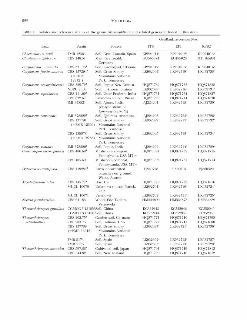
TABLE I. Isolates and reference strains of the genus Myceliophthora and related genera included in this study
GenBank accession Nos.
Taxa Strain Source ITS EF1 RPB2
Chaetomidium arxii FMR 12364 Soil, Gran Canaria, Spain KP204014a KP204012a KP204013a
Chaetomium globosum CBS 148.51 Man, Greifswald,Germany
GU563374 KC485028 NT_165981
Corynascella inaequalis CBS 331.75T Soil, Kirovograd, Ukraine KP204017a KP204015a KP204016a
Corynascus fumimontanus CBS 137294T
(5FMR12372T)
Soil, Great SmokyMountains NationalPark, Tennessee
LK932694a LK932719a LK932733a
Corynascus novoguineensis CBS 359.72T Soil, Papua New Guinea HQ871762 HQ871733 HQ871838NBRC 9556 Soil, unknown location LK932698a LK932716a LK932731a
Corynascus sepedonium CBS 111.69T Soil, Uttar Pradesh, India HQ871751 HQ871734 HQ871827CBS 632.67 Unknown source, Russia HQ871759 HQ871744 HQ871830IMI 378521 Soil, Ajmer, India
(ex-type strain ofCorynascus similis)
AJ224201 LK932715a LK932730a
Corynascus verrucosus IMI 378522T Soil, Quilmes, Argentina AJ224203 LK932723a LK932726a
CBS 137791(5FMR 12369)
Soil, Great SmokyMountains NationalPark, Tennessee
LK932699a LK932717a LK932732a
CBS 135878(5FMR 12783)
Soil, Great SmokyMountains NationalPark, Tennessee
LK932695a LK932718a LK932734a
Corynascus sexualis IMI 378520T Soil, Jaipur, India AJ224202 LK932714a LK932729a
Crassicarpon thermophilum CBS 406.69T Mushroom compost,Pennsylvania,USA;MT 2
HQ871794 HQ871732 HQ871715
CBS 405.69 Mushroom compost,Pennsylvania, USA; MT +
HQ871793 HQ871731 HQ871714
Hypocrea aurantefussa CBS 119284T Partly decorticatedbranches on ground,Weins, Austria
FJ860728 FJ860613 FJ860520
Myceliophthora lutea CBS 145.77T Hay, UK HQ871775 HQ871722 HQ871816MUCL 10070 Unknown source, Natick,
USALK932701a LK932710a LK932724a
MUCL 10071 Unknown LK932702a LK932711a LK932725a
Nectria pseudotrichia CBS 641.83 Wood, Edo Tachira,Venezuela
HM534899 HM534878 HM534889
Thermothelomyces guttulata CGMCC 3.15185TSoil, China KC352943 KC352946 KC352949CGMCC 3.15186 Soil, China KC352944 KC352947 KC352950
Thermothelomycesheterothallica
CBS 202.75T Garden soil, Germany HQ871771 HQ871710 HQ871798CBS 203.75 Soil, Indiana, USA HQ871772 HQ871711 HQ871800CBS 137789(5FMR 13215)
Soil, Great SmokyMountains NationalPark, Tennessee
LK932697a LK932721a LK932736a
FMR 5174 Soil, Spain LK932692a LK932712a LK932727a
FMR 5175 Soil, Spain LK932693a LK932713a LK932728a
Thermothelomyces hinnulea CBS 597.83T Cultivated soil, Japan HQ871791 HQ871719 HQ871813CBS 544.82 Soil, New Zealand HQ871790 HQ871718 HQ871812
622 MYCOLOGIA
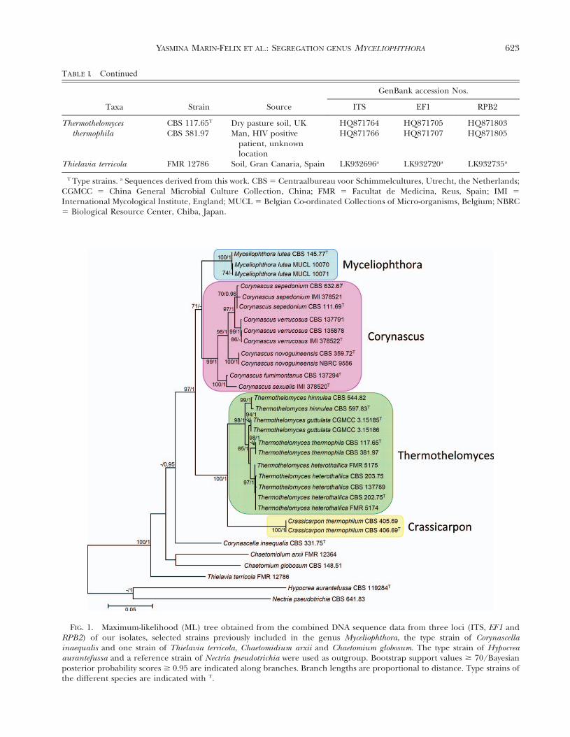
FIG. 1. Maximum-likelihood (ML) tree obtained from the combined DNA sequence data from three loci (ITS, EF1 andRPB2) of our isolates, selected strains previously included in the genus Myceliophthora, the type strain of Corynascellainaequalis and one strain of Thielavia terricola, Chaetomidium arxii and Chaetomium globosum. The type strain of Hypocreaaurantefussa and a reference strain of Nectria pseudotrichia were used as outgroup. Bootstrap support values $ 70/Bayesianposterior probability scores $ 0.95 are indicated along branches. Branch lengths are proportional to distance. Type strains ofthe different species are indicated with T.
TABLE . Continued
GenBank accession Nos.
Taxa Strain Source ITS EF1 RPB2
Thermothelomycesthermophila
CBS 117.65T Dry pasture soil, UK HQ871764 HQ871705 HQ871803CBS 381.97 Man, HIV positive
patient, unknownlocation
HQ871766 HQ871707 HQ871805
Thielavia terricola FMR 12786 Soil, Gran Canaria, Spain LK932696a LK932720a LK932735a
T Type strains. a Sequences derived from this work. CBS 5 Centraalbureau voor Schimmelcultures, Utrecht, the Netherlands;CGMCC 5 China General Microbial Culture Collection, China; FMR 5 Facultat de Medicina, Reus, Spain; IMI 5
International Mycological Institute, England; MUCL 5 Belgian Co-ordinated Collections of Micro-organisms, Belgium; NBRC5 Biological Resource Center, Chiba, Japan.
I
YASMINA MARIN-FELIX ET AL.: SEGREGATION GENUS MYCELIOPHTHORA 623
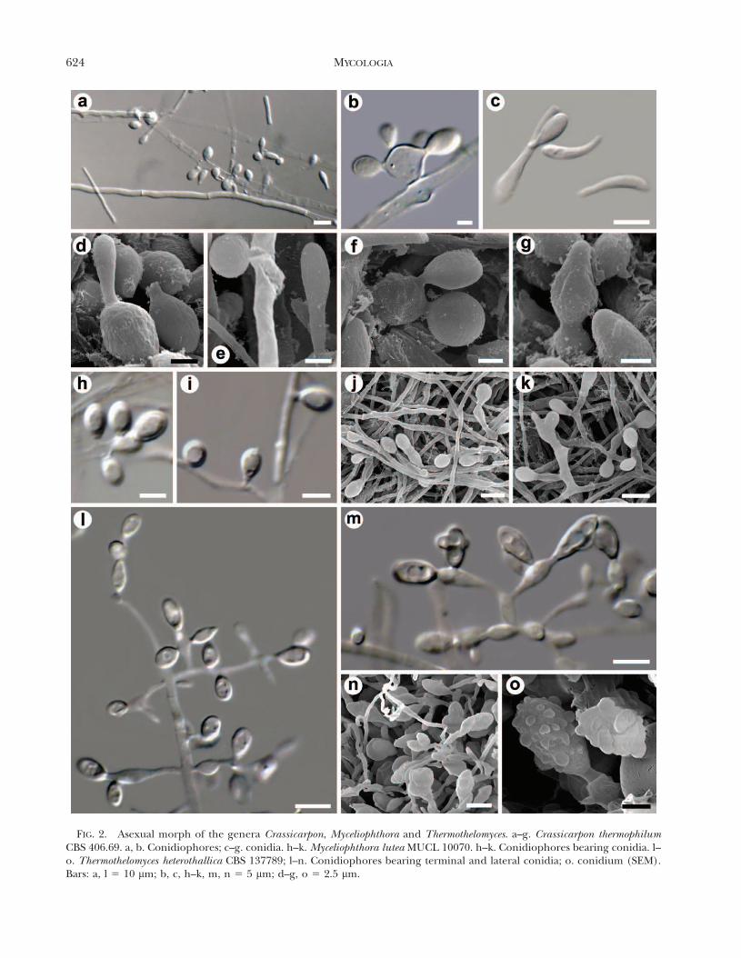
FIG. 2. Asexual morph of the genera Crassicarpon, Myceliophthora and Thermothelomyces. a–g. Crassicarpon thermophilumCBS 406.69. a, b. Conidiophores; c–g. conidia. h–k. Myceliophthora lutea MUCL 10070. h–k. Conidiophores bearing conidia. l–o. Thermothelomyces heterothallica CBS 137789; l–n. Conidiophores bearing terminal and lateral conidia; o. conidium (SEM).Bars: a, l 5 10 mm; b, c, h–k, m, n 5 5 mm; d–g, o 5 2.5 mm.
624 MYCOLOGIA

cleistothecial ascomata of textura epidermoidea andornamented (mostly reticulate) ascomata wall cells,and brown ascospores with a distinct germ pore at eachend (FIG. 4). The asexual morph was observed in allthese species with the exception of M. sexualis, as
was reported by Stchigel et al. (2000), and wascharacterized by holoblastic, spherical or nearly so,hyaline to pale yellow conidia with an ornamented cellwall, except for M. novoguineensis, which producedsmooth-walled conidia, sessile or on short protrusions,
FIG. 3. Corynascus fumimontanus CBS 137294. a. Ascomata. b. Irregular network of distorted hyphae on the ascomatal wall.c. Detail of the ascomatal wall. d. Asci. e, f. Ascospores. g. Sessile conidia. h. Conidia on short inflated protusion. i. Intercalaryconidia. j. Conidia (SEM). Bars: a 5 50 mm; b, c, f–j 5 5 mm; d 5 15 mm; e 5 10 mm.
YASMINA MARIN-FELIX ET AL.: SEGREGATION GENUS MYCELIOPHTHORA 625
sometimes also on swollen, sometimes catenate,conidiogenous cells (FIG. 5). These species weremesophilic with an optimal growth at 25–40 C. Thetype strains of M. sepedonium and M. similisgrouped together in the same clade with a nucleotideidentity over 99%. Morphologically both species weredistinguished only by the shape of the ascospores andthe position of the germ pores (i.e. irregularly shaped
ascospores with two subapical germ pores in M. similis)and broadly fusiform ascospores with two apical germpores in M. sepedonium.
The other species of Myceliophthora (i.e. M. fergusii,M. guttulata, M. heterothallica, M. hinnulea, M.thermophile) were located in two distinct, well-supported sister clades, each of them representinga new genus. The first contains the type strains of M.
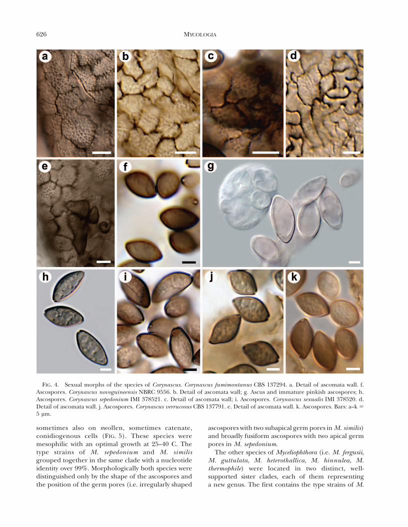
FIG. 4. Sexual morphs of the species of Corynascus. Corynascus fumimontanus CBS 137294. a. Detail of ascomata wall. f.Ascospores. Corynascus novoguineensis NBRC 9556. b. Detail of ascomata wall; g. Ascus and immature pinkish ascospores; h.Ascospores. Corynascus sepedonium IMI 378521. c. Detail of ascomata wall; i. Ascospores. Corynascus sexualis IMI 378520. d.Detail of ascomata wall. j. Ascospores. Corynascus verrucosus CBS 137791. e. Detail of ascomata wall. k. Ascospores. Bars: a–k 5
5 mm.
626 MYCOLOGIA

guttulata, M. heterothallica, M. hinnulea andM. thermophila (98% bs/1 pp), and the other includesM. fergusii (100% bs/1 pp). The members of both cladeswere thermophilic, with an optimal growth at 40–45 C.The species in the first clade produced holoblastic,subglobose or obovoid to ellipsoidal conidia truncate atthe base, brown, thick-walled and ornamented, with theexception of M. guttulata that produces hyaline,smooth-walled and guttulate conidia on terminally and
laterally on hyphae (sessile), or on ampulliform toclavate polyblastic conidiogenous cells, sometimes witha short or long basal pedicel (FIG. 2). Only M.heterothallica was capable of producing sexual morphsin culture after mating sexually compatible strains. Theywere dark cleistothecial ascomata with an ascomatal wallof textura epidermoidea, producing ellipsoid to ovoidascospores with a terminal germ pore. The strains of M.fergusii produced holoblastic, hyaline to yellow in mass,
FIG. 5. Asexual morphs of the species of Corynascus. a–d. Corynascus fumimontanus CBS 137294. a. Sessile conidium; b.Conidia on short inflated protrusion; c. sessile and intercalary conidia; d. conida (SEM). e–h. Corynascus novoguineensis NBRC9556. e. Sessile conidia; f. conidia on short inflated protrusion; g. conidiophores; h. conidium (SEM). i–l. Corynascus sepedoniumIMI 378521. i. Sessile conidia; j. conidia on short inflated protrusion; k. conidiophore bearing terminal conidium (SEM); l.conidia (SEM). m–p. Corynascus verrucosus FMR 12369 (5 CBS 137791). m. Sessile conidia; n. conidia on short inflatedprotrusion; o. conidiophores bearing terminal conidia; p. conida (SEM). Bars: a–c, e–g, i–k, m–o 5 5 mm; d, h, l, p 5 2.5 mm.
YASMINA MARIN-FELIX ET AL.: SEGREGATION GENUS MYCELIOPHTHORA 627
thick- and smooth-walled conidia, sessile or in swollenconidiogenous cells, arising singly or in chains of up tofive conidia (FIG. 2). This fungus was also heterothallic,producing cleistothecial ascomata with a thick-walledascomatal wall of textura angularis and ellipsoidalascospores, pinkish when young but becoming darkbrown, with a germ pore at each end.
The nucleotide identities in the combined datasetamong the type strains of M. fergusii, M. lutea, M.sepedonium and M. thermophila were # 93%.
TAXONOMY
Based on the molecular and phenotypic resultsmentioned above, we propose the revalidation ofCorynascus as a genus distinct from Myceliophthora,and the new genera Thermothelomyces and Crassicar-pon. To accommodate the isolate CBS 137294, wepropose the new species C. fumimontanus.
KEY TO THE GENERA CORYNASCUS, CRASSICARPON,
MYCELIOPHTHORA AND THERMOTHELOMYCES
1. No growth at 50 C . . . . . . . . . . . . . . . . . . . . . . . . 21. Growth at 50 C . . . . . . . . . . . . . . . . . . . . . . . . . . 32. Sexual morph present in culture . . . . . . Corynascus2. Sexual morph absent in culture. . . . . Myceliophthora3. Conidia hyaline, spherical to cuneiform, smooth-
walled . . . . . . . . . . . . . . . . . . . . . . . . . Crassicarpon3. Conidia brown, subglobose or obovoid to ellipsoidal;
ornamentated or, rarely, smooth . . . . . Thermothelomyces
Corynascus Arx, Proc. K. Ned. Akad. Wet., Ser. C, Biol.Med. Sci. 76: 295. 1973. FIGS. 3, 4, 5Type species: Corynascus sepedonium (C.W. Em-
mons) Arx, Proc. K. Ned. Akad. Wet., Ser. C, Biol.Med. Sci. 76: 292. 1973.
Notes. Corynascus is characterized by its mesophilichabit, having an ascomata wall of textura epidermoi-dea composed of reticulate or verrucose cells,ascospores with a germ pore at each end andyellowish conidia usually verrucose or echinulate totuberculate, rarely smooth.
MycoBank MB809486Typification. USA. TENNESSEE: Great Smoky
Mountains National Park, Cosby Creek trail, 35.78,283.22, from forest soil, 01-VIII-2008, A.N. Miller,M. Calduch, A.M. Stchigel. (holotype CBS H-21594).Isotypes FMR 12372, ILLS 71950. Ex-type culturesFMR 12372, CBS 137294.
Etymology: From the Latin fumi-, smoky, and-montanus, mountains, referring to the name of thenational park where the fungus was isolated.
Diagnosis: This species is characterized by verru-cose ascomata wall cells, mostly irregularly shapedascospores, greenish brown when young, and con-idia sessile, intercalary or on swollen conidiogenouscells.
Mycelium composed of hyaline to pale yellow,branched, anastomosing, septate, smooth-walled hy-phae of 1–2 mm diam. Colonies on PCA attaining72–75 mm diam in 14 d at 35 C, light yellow with olivepatches, olive gray at center, flattened, powdery togranular due to the production of conidia andascomata, margins fimbriate; reverse pale yellow tolight yellow, with olive patches. Ascomata superficial,globose, cleistothecial, brown to dark brown, 50–110mm diam, ascomata wall of textura epidermoidea,composed of 1–3 layers of irregularly shaped,verrucose, golden brown to brown cells, covered byhyphae anastomosing with the ascomata wall cells.Paraphyses absent. Asci eight-spored, subglobose tobroadly ellipsoidal, 24–31 3 15322 mm, thin-walled,short-stipitate, evanescent. Ascospores one-celled,broadly fusiform to irregularly shaped, 13–17 3 7–9mm, hyaline to greenish brown when young becomingbrown, thick- and smooth-walled, with a conspicuoussubterminal to terminal germ pore at each end.Conidiophores micronematous, 1–2.5 mm wide andup to 26 mm long, or semimacronematous, flask-shaped, 5–10 3 3–8 mm. Conidia holoblastic, globoseto subglobose, 6–10 mm diam, subhyaline to paleyellow, thick-walled, verrucose, sessile or on swollenconidiogenous cells, and holothallic when intercalary,morphologically similar to the holoblastic ones.
Colonies on PDA attaining 73–75 mm diam in 14d at 35 C, yellowish white to pale yellow, velvety topowdery, radially folded, umbilicate, lobulate, mar-gins regular; reverse pale yellow to light yellow.Ascomata absent. The minimum and maximumtemperature of growth are 15 and 45 C, respectively.Optimal temperature 35–40 C.
Corynascus novoguineensis (Udagawa & Y. Horie) Arx,Proc. K. Ned. Akad. Wet., Ser. C, Biol. Med. Sci. 76:295. 1973. FIGS. 4b, g, h; 5e--hBasionym: Thielavia novoguineensis Udagawa & Y. Horie,
Bull. natn. Sci. Mus., Tokyo 15: 191. 1972.; Myceliophthora novoguineensis (Udagawa & Y. Horie)
van de Brink & Samson, in Brink, Samson, Hagen,Boekhout & Vries, Fungal Divers. 52: 206. 2012.
Notes. Corynascus novoguineensis is characterized byslightly verrucose ascomata wall cells, pinkish ascos-pores when young, and smooth-walled conidia. In theoriginal description the immature ascospores weredescribed as greenish brown (Udagawa and Horie1972).
628 MYCOLOGIA
Corynascus sepedonium (C.W. Emmons) Arx, Proc. K.Ned. Akad. Wet., Ser. C, Biol. Med. Sci. 76: 292.1973. FIGS. 4c, i; 5i-lBasionym: Thielavia sepedonium C.W. Emmons, Bull.
Torrey bot. Club 59: 417. 1932; Chaetomidium sepedonium (C.W. Emmons) Lodha, in
Subramanian (Ed.), Taxonomy of Fungi (Proc. int.Symp. Madras, 1973), Pt 1: 248. 1978.
; Myceliophthora sepedonium (C.W. Emmons) van denBrink & Samson, in Brink, Samson, Hagen, Boekhout& Vries, Fungal Divers. 52: 206. 2012.
5 Thielavia sepedonium var. minor B.S. Mehrotra &Bhattacharjee, Antonie van Leeuwenhoek 32: 391.1966.
5 Myceliophthora similis (Stchigel, Cano & Guarro) vande Brink & Samson, in Brink, Samson, Hagen,Boekhout & Vries, Fungal Divers. 52: 206. 2012.
Ascomata superficial, globose, cleistothecial, brownto dark brown, 50–110 mm diam, glabrous, ascomatawall of textura epidermoidea, composed of 1–3 layersof irregularly shaped, reticulate, golden-brown tobrown cells. Paraphyses absent. Asci eight-spored,subglobose to broadly ellipsoidal, 26–40 3 20–31 mm,thin-walled, short-stipitate, evanescent. Ascosporesone-celled, ellipsoidal to broadly fusiform or navicu-lar in lateral view, 11–23 3 6.5–13 mm, hyalinebecoming brown when mature, thick- and smooth-walled, with a conspicuous subterminal to terminalgerm pore at each end. Conidiophores micronema-tous or semimacronematous. Conidia holoblastic,globose to subglobose, 6–12 mm diam, subhyaline topale yellow, thick-walled, finely echinulate to tuber-culate, sessile or on swollen conidiogenous cells.
Notes. Corynascus sepedonium is characterized byreticulate ascomata wall cells and echinulate totuberculate conidia. The description is from theprotolog with slight modifications based on the studyof the type strain of C. similis (IMI 378521).
Corynascus sexualis Stchigel, Cano & Guarro, inStchigel, Sagues, Cano & Guarro, Mycol. Res. 104:880. 2000. FIG. 4d, j; Myceliophthora sexualis (Stchigel, Cano & Guarro) van
de Brink & Samson, in Brink, Samson, Hagen,Boekhout & Vries, Fungal Divers. 52: 206. 2012.
Notes. Corynascus sexualis differs from the otherspecies of the genus by the lack of asexual morph andits lemon-shaped ascospores.
Corynascus verrucosus Stchigel, Cano & Guarro, inStchigel, Sagues, Cano & Guarro, Mycol. Res. 104:884. 2000. FIGS.; 4e, k; 5m–p; Myceliophthora verrucosa (Stchigel, Cano & Guarro)
van de Brink & Samson, in Brink, Samson, Hagen,Boekhout & Vries, Fungal Divers. 52: 206. 2012.
Notes. Corynascus verrucosus is characterized byverruciform dark brown projections from the asco-mata wall, and broadly fusiform ascospores witha subterminal germ pore at each end.
KEY TO THE SPECIES OF CORYNASCUS
1. Asexual morph absent; ascospores limoniform C. sexualis1. Asexual morph present; ascospores irregularly
shaped, ellipsoidal or fusiform . . . . . . . . . . 22. Conidia smooth-walled, or nearly so; ascospores
pinkish when young . . . . . . C. novoguineensis2. Conidia verrucose or tuberculate; ascospores
greenish or brownish when young. . . . . . . . 33. Ascomata wall cells with verrucose projections;
ascospores irregularly shaped C. fumimontanus3. Ascomata wall cells reticulated . . . . . . . . . . . . . . . 4
4. Ascomata glabrous; ascospores ellipsoidal tobroadly fusiform... . . . . . . . . . . . C. sepedonium
4. Ascomata with short, brown verruciform projec-tions on entire ascomata wall; ascosporesbroadly fusiform . . . . . . . . . . . . . C. verrucosus
Crassicarpon Y. Marın, Stchigel, Guarro & Cano, gen.nov. FIG. 2a–gType species: Crassicarpon thermophilum (Fergus &
Sinden) Y. Marın, Stchigel, Guarro & Cano.Etymology: From the Greek Crassum- and -karpos,
referring to the thick ascomatal wall.Diagnosis: Characterized by its thermophilic habit,
blackish ascomata with a thick wall of texturaangularis, broadly ellipsoidal ascospores with a germpore at each end, and hyaline, smooth-walled conidia,yellow in mass.
Ascomata superficial or immersed, globose, cleis-tothecial, dark brown to black, glabrous, ascomatalwall thick, of textura angularis, composed of an outerlayer of thick-walled swollen cells, and an inner layerof flattened cells. Asci 4–6-spored, broadly clavate,thin-walled, stalked, evanescent. Paraphyses absent.Ascospores one-celled, broadly ellipsoidal, first hya-line, becoming pink and finally dark brown, smooth-and thick-walled, with a germ pore at each end.Conidiophores micronematous or semimacronema-tous. Conidia holoblastic, hyaline to yellow in masswith the age, spherical to cuneiform, variable in size,thick- and smooth-walled, sessile or produced inswollen conidiogenous cells, sometimes with shortpedicels; secondary apical conidia may be produced.Heterothallic. Thermophilic.
Crassicarpon thermophilum (Fergus & Sinden) Y.Marın, Stchigel, Guarro & Cano, comb. nov.
FIG. 2a–gMycoBank MB809488
YASMINA MARIN-FELIX ET AL.: SEGREGATION GENUS MYCELIOPHTHORA 629
Basionym: Thielavia thermophila Fergus & Sinden,Can. J. Bot. 47: 1635. 1969.; Corynascus thermophilus (Fergus & Sinden)
Klopotek, Arch. Mikrobiol. 98: 366. 1974.; Chaetomidium thermophilum (Fergus & Sinden)
Lodha, in Subramanian (Ed.), Taxonomy ofFungi (Proc. int. Symp. Madras, 1973), Pt 1: 248.1978.
5 Myceliophthora fergusii (Klopotek) Oorschot,Persoonia 9: 406. 1977.
; Chrysosporium fergusii Klopotek, Arch. Mikro-biol. 98: 366. 1974.
Notes. We decided to use the epithet thermophiluminstead fergusii, which had been chosen by van denBrink et al. (2012) for this taxon because Thielaviathermophila was the first morph described.
Myceliophthora Costantin, C. r. hebd. Seanc. Acad.Sci., Paris 114: 849. 1892. FIG. 2h–k
Type species. Myceliophthora lutea Costantin, C. r.hebd. Seanc. Acad. Sci., Paris 114: 2. 1892.
FIG. 2h–kNotes. Myceliophthora is characterized by its meso-
philic habit, hyaline and smooth-walled conidia andthe lack of sexual morph.
Thermothelomyces Y. Marın, Stchigel, Guarro &Cano, gen. nov. FIGS. 2l–o
MycoBank MB809489Type species: Thermothelomyces thermophila (Apinis)
Y. Marın, Stchigel, Guarro & Cano.Etymology. From the Greek thermos-, hot, thelo-, love,
and -myces, fungi, referring to the thermophilic habitof the fungus.
Diagnosis: Characterized by its thermophilic habit,ascomata with a wall of textura epidermoidea, ellipsoi-dal ascospores with a single apical germ pore, andhyaline or pale brown conidia, mostly ornamented.
Ascomata immersed to sub-immersed, globose,cleistothecial, black, glabrous, ascomata wall thin,of textura epidermoidea. Asci eight-spored, ellipsoi-dal, thin-walled, stalked, evanescent. Paraphysesabsent. Ascospores ellipsoidal, occasionally irregu-larly shaped, first hyaline, dark brown to black, thick-and smooth-walled, with one germ pore. Conidio-phores micronematous or semimacronematous.Conidia holoblastic, hyaline or pale brown, subglo-bose, ellipsoidal or obovoid to pyriform, thick-walled, conspicuously verrucose-spinulose or tuber-culate, rarely smooth-walled and guttulate, produc-ing terminally or laterally on hyphae, sometimes withshort or long pedicels, or on swollen conidiogenouscells in a number of 1–4; occasionally a secondary
apical conidium is produced. Heterothallic.Thermophilic.
Thermothelomyces guttulata (Y. Zhang & L. Cai) Y.Marın, Stchigel, Guarro & Cano, comb. nov.
MycoBank MB 809490Basionym: Myceliophthora guttulata Y. Zhan & L. Cai,
Mycol Progress 13: 165. 2014.Notes. Thermothelomyces guttulata is distinguished
from the other species by its hyaline, smooth-walledand guttulate conidia.
Thermothelomyces heterothallica (von Klopotek) Y.Marın, Stchigel, Guarro & Cano, comb. nov.
FIGS. 2l–oMycoBank MB809491Basionym: Thielavia heterothallica von Klopotek, Arch.
Mikrobiol. 107: 223. 1976.; Corynascus heterothallicus (von Klopotek) von
Arx, Dreyfuss & Muller, Persoonia 12: 174. 1984.; Myceliophthora heterothallica (von Klopotek) van
den Brink & Samson, in Brink, Samson,Hagen, Boekhout & Vries, Fungal Divers. 52:206. 2012.
Notes. This species is characterized by pale orange-brown, long ellipsoidal, tuberculate conidia and theproduction of ascomata after mating. The conidiapreviously were described as hyaline (Klopotek 1974,1976; van Oorschot 1977).
Thermothelomyces hinnulea (Awao & Udagawa) Y.Marın, Stchigel, Guarro & Cano, comb. nov.
MycoBank MB809492Basionym: Myceliophthora hinnulea Awao & Udagawa,
Mycotaxon 16: 436. 1983.Notes. This species is characterized by yellowish
brown to brown, subglobose to ovate, conspicuouslyverrucose-spinulose conidia.
Thermothelomyces thermophila (Apinis) Y. Marın,Stchigel, Guarro & Cano, comb. nov.
MycoBank MB809493Basionym: Sporotrichum thermophilum Apinis, Nova
Hedwigia 5: 74. 1963.; Chrysosporium thermophilum (Apinis) Klopotek,
Arch. Mikrobiol. 98: 366. 1974.; Myceliophthora thermophila (Apinis) Oorschot,
Persoonia 9: 403. 1977.Notes. The asexual morph of this species is similar
to those of T. heterothallica but T. thermophila doesnot produce a sexual morph after mating.
630 MYCOLOGIA

KEY TO THE SPECIES OF THERMOTHELOMYCES
1. Conidia smooth-walled and guttulate, (3.8–)4.8–7.23 3–5 mm . . . . . . . . . . . . . . . . . . . . . . T. guttulata
1. Conidia with an ornamented surface. . . . . . . . . . . . 22. Conidia 7–12 3 5–10 mm . . . . . . . . . . T. hinnulea2. Conidia 4.5–11 3 3–4.5 mm . . . . . . . . . . . . . . . . . . . . .
. . . . . . . . . . . . . . . T. heterothallica/T. thermophila*
* Thermothelomyces heterothallica produces ascomataafter mating.
DISCUSSION
Based on recent molecular studies that demonstratedthat M. lutea, the type species of Myceliophthora,clustered with some members of the family Chaeto-miaceae, that genus was restricted to the species ofsuch family (van den Brink et al. 2012, Zhang et al.2014); consequently M. vellerea (now renamedCtenomyces vellereus [Sacc. & Speg.] P.M. Kirk) andMyceliopthora anamorph of Arthroderma tuberculatumwere transferred to the family Arthrodermataceae,where they were phylogenetically located (van denBrink et al. 2012, Kirk 2014).
In previous phylogenies (van den Brink et al. 2012,2013) the species of Myceliophthora spp. grouped witha confidence value of below 50%, being divided intotwo main clades (each one composed of one or twoterminal clades depending on the nuclear lociemployed in the phylogenetic inference) accordingto their mesophilic and thermophilic habit. However,in our study, in agreement with Zhang et al. (2014),Myceliophthora spp. formed a well-supported clade, butthe genetic distances among the terminal clades(below of 93% similarity) are such that they shouldbe treated as separate genera. Consequently weproposed to split Myceliophthora into four genera,revalidating Corynascus and erecting two new genera:Crassicarpon and Thermothelomyces. Our proposal isalso supported by phenotypic data (e.g. Crassicarponthermophilum presents dark, thick-walled ascomatawith a wall of textura angularis composed of non-ornamented ascomata wall cells, while the Corynascusspp. and Thermothelomyces spp. are characterized bythe production of many pale, thin-walled ascomatawith a wall of textura epidermoidea composed ofornamented ascomata wall cells. The type of ascomatawall previously had been used successfully in thedelimitation of genera in Lasiosphaeriaceae (Millerand Huhndorf 2004, 2005; Cai et al. 2005). Thenumber of germ pores in the ascospores is alsoa distinctive feature because Corynascus spp. andCrassicarpon thermophilum have one at each end,whereas Thermothelomyces heterothallica presents onlyone. The conidia are produced by similar ontogenetic
processes in all four genera but also show morpholog-ical differences: both Myceliophthora lutea and Crassi-carpon thermophilum produce hyaline, smooth-walledconidia, but while in the first taxon they are pyriformto globose and produced singly (or rarely in chains upto two conidia), in the second one they are spherical tocuneiform and in chains of up to five conidia.Corynascus spp. and Thermothelomyces spp. producemostly ornamented, yellowish conidia, even thoughThermothelomyces spp. present more complex conidio-phores, with ovoid to clavate conidia with a truncatebase, whereas in Corynascus spp. they are spherical.
Corynascus fumimontanus sp. nov. is easy todistinguish morphologically from the rest of thespecies of the genus Corynascus by its verrucoseascomata wall cells (reticulate in the other species)and its irregularly shaped ascospores. Finally thesynonymy of C. similis with C. sepedonium wasproposed due to the high nucleotide identity andthe minor morphological differences among them.
ACKNOWLEDGMENTS
This work was supported by the Spanish Ministerio deEconomıa y Competitividad, grant CGL2013-43789. Thenew species was discovered during fieldwork supported bya National Science Foundation (USA) grant (DEB-0515558)to ANM.
LITERATURE CITED
Alvarez E, Garcia-Hermoso D, Sutton DA, Cano JF, StchigelAM, Hoinard D, Fothergill AW, Rinaldi MG, Dromer F,Guarro J. 2010. Molecular phylogeny and proposal oftwo new species of the emerging pathogenic fungusSaksenaea. J Clin Microbiol 48:4410–4416, doi:10.1128/JCM.01646-10
Awao T, Udagawa S. 1983. A new thermophilic species ofMyceliophthora. Mycotaxon 16:436–440.
Burnside CE. 1928. Saprophytic fungi associated with thehoneybee. Mich Acad Sci 8:59–86.
Cai L, Jeewon R, Hyde KD. 2005. Phylogenetic evaluation andtaxonomic revision of Schizothecium based on ribosomalDNA and protein coding genes. Fungal Divers 19:1–21.
Cano J, Guarro J, Gene J. 2004. Molecular and morpholog-ical identification of Colletotrichum species of clinicalinterest. J Clin Microbiol 42:2450–2454.
Costantin J. 1892. Sur quelques maladies du blanc deChampignon. Comptes rendus hebd. seances acad. sci.Paris 114:849–851.
———, Matruchot L. 1894. Recherches sur le Vert de Gris,le Platre et le Chanel, maladies du Blanc de Champi-gnon. Rev Gen Bot 6:289–300.
de Hoog GS, Guarro J, Gene J, Figueras MJ. 2000. Atlas ofclinical fungi. 2nd ed. Utrecht/Reus: the Netherlands/Spain: Centraalbureau voor Schimmelcultures/Univer-sitat Rovira i Virgili.
YASMINA MARIN-FELIX ET AL.: SEGREGATION GENUS MYCELIOPHTHORA 631
Doyer CM. 1927. Meded. Phytopath. Lab. Willie CommelinScholten 10:32.
Figueras MJ, Guarro J. 1988. A scanning electron micro-scopic study of ascoma development in Chaetomiummalaysiense. Mycologia 80:298–306.
Goddard HN. 1913. Can fungi living in agricultural soilassimilate free nitrogen?. Bot Gaz (Crawfordsville) 56:249–305.
Guarro J, Gene J, Stchigel AM, Figueres MJ. 2012. Atlas ofsoil Ascomycetes. CBS Biodiversity Series 10. Utrecht,the Netherlands: CBS-KNAW Fungal Biodiversity Cen-tre. 486 p.
———, Punsola L, Cano J. 1985. Myceliophthora vellerea(Chrysosporium asperatum) anamorph of Ctenomycesserratus. Mycotaxon 23:419–427.
Houbraken J, Due M, Varga J, Meijer M, Frisvad JC, SamsonRA. 2007. Polyphasic taxonomy of Aspergillus sectionUsti. Stud Mycol 59:107–128, doi:10.3114/sim.2007.59.12
Kirk PM. 2014. Nomenclatural novelties. Index Fungorum120:1.
Kornerup A, Wanscher JH. 1984. Methuen handbook ofcolor. 3rd ed. London: Eyre Methuen. 252 p.
Larkin MA, Blackshields G, Brown NP, Chenna R, McGetti-gan PA, McWilliam H, Valentin F, Wallace IM, Wilm A,Lopez R, Thompson JD, Gibson TJ, Higgins DG. 2007.Clustal W and Clustal X 2.0. Bioinformatics 23:2947–2948.
Lodha BC. 1978. Generic concepts in some Ascomycetesoccurring on dung. In: Subramanian, ed. Taxonomy ofFungi. Proceedings of the International Symposium onTaxonomy of Fungi, University of Madras, 1973, 1.Madras, India: Univ. Madras, 241–257.
McNeill J, Barrie FR, Buck WR, Demoulin V, Greuter W,Hawksworth DL, Herendeen PS, Knapp K, Marhold K,Prado J, Prud’homme van Reine WF, Smith GF,Wiersema H, Turland NJ. 2012. International code ofnomenclature for algae, fungi and plants (MelbourneCode) adopted by the 18th International BotanicalCongress Melbourne, Australia, Jul 2011. RegnumVegetabile 154. Germany: Koeltz Scientific Books.240 p.
Miller AN, Huhndorf SM. 2004. A natural classification ofLasiosphaeria based on nuclear LSU rDNA sequences.Mycol Res 108:26–34, doi:10.1017/S0953756203008864
———, ———. 2005. Multigene phylogenies indicateascoma wall morphology is a better predictor ofphylogenetic relationships than ascospore morphologyin the Sordariales (Ascomycota, Fungi). Mol PhylogenetEvol 35:60–75, doi:10.1016/j.ympev.2005.01.007
Mouchacca J. 1973. Les Thielavia des sols arides: especesnouvelles et analyse generique. Bull Soc Mycol Fr 89:295–311.
Sharkey MJ. 2001. The all taxa biotic inventory of the GreatSmoky Mountains National Park. Fla Entomol 84:556–564.
Stchigel AM, Cano J, Mac Cormack WP, Guarro J. 2001.Antarctomyces psychrotrophicus gen. et sp. nov., a newascomycete from Antarctica. Mycol Res 105:377–382, ,doi:10.1017/S0953756201003379
———, Sagues M, Cano J, Guarro J. 2000. Three newthermotolerant species of Corynascus from soil, witha key to the known species. Mycol Res 104:879–887,doi:10.1017/S0953756299002245
Swofford DL. 2002. PAUP* 4.0b10: phylogenetic analysisusing parsimony (*and other methods), Sunderland:Massachusetts: Sinauer Associates.
Tamura K, Peterson D, Peterson N, Stecher G, Nei M,Kumar S. 2011. MEGA 5: molecular evolutionarygenetics analysis using maximum likelihood, evolution-ary distance and maximum parsimony methods. MolBiol Evol 28:2731–2739, doi:10.1093/molbev/msr121
Udagawa SI, Horie Y. 1972. A new species of Thielavia andits Chrysosporium conidial state. Bull Natl Sci MusTokyo 15:191–196.
van Oorschot CAN. 1977. The genus Myceliophthora.Persoonia 9:401–408.
———. 1980. A revision of Chrysosporium and allied genera.Stud Mycol 20:1–89.
van den Brink J, Samson RA, Hagen F, Boekhout T, de VriesRP. 2012. Phylogeny of the industrial relevant, ther-mophilic genera Myceliophthora and Corynascus. FungalDivers 52:197–207, doi:1007/s13225-011-0107-z
———, van Muiswinkel GCJ, Theelen B, Hinz SWA, de VriesRP. 2013. Efficient plant biomass degradation by thermo-philic fungus Myceliophthora heterothallica. Appl EnvironMicrobiol 79:1316–1324, doi:10.1128/AEM.02865-12
von Arx JA. 1973. Ostiolate and nonostiolate Pyrenomy-cetes. Proc K Ned Akad Wet Amsterd C 76:289–296.
———. 1975. On Thielavia and some similar genera ofAscomycetes. Stud Mycol 8:1–31.
———, Dreyfuss M, Muller E. 1984. A revaluation of Chae-tomium and the Chaetomiaceae. Persoonia 12:169–179.
von Klopotek A. 1974. Revision der thermophilen Sporo-trichum-Arten: Chrysosporium thermophilum (Apinis)comb. nov. und Chrysosporium fergusii spec. nov. 5 statusconidialis von Corynascus thermophilus (Fergus undSinden) comb. nov. Arch Microbiol 98:365–369.
———. 1976. Thielavia thermophila spec. nov., die perfekteForm von Chrysosporium thermophilum. Arch Microbiol107:223–224.
Whittaker RH. 1956. Vegetation of the Great SmokyMountains. Ecol Monograph 26:1180.
Zhang Y, Wu WP, Dian MH, Su YY, Cai L. 2014. A newthermophilic species of Myceliophthora from China. MycolProg 13:165–170, doi:10.1007/s11557-013-0904-8
632 MYCOLOGIA